Int. J. Electrochem. Sci., 8 (2013) 11711 - 11722
International Journal of
ELECTROCHEMICAL
SCIENCE
www.electrochemsci.org
Amperometric and Photometric Responses of in Situ Coupled
Glucose Oxidase-Poly (Propylene Imine) Dendrimer Based
Glucose Biosensor
Sudheesh K. Shukla, Ajay K. Mishra, Bhekie B. Mamba, Omotayo A. Arotiba*
Department of Applied Chemistry, Doornfontein Campus, University of Johannesburg P.O. Box 17011, Doornfontein 2028, Johannesburg, South Africa
*
E-mail: oarotiba@uj.ac.za;
Received: 14 June 2013 / Accepted: 8 August 2013 / Published: 10 September 2013
In this work, poly (propylene imine) dendrimer (PPI) was coupled to glucose oxidase enzyme (GOx) in situ and used as a bioreceptor in a glucose biosensor. The coupling was accomplished by the use of 1-ethyl-3-(dimethylaminopropyl) carbodiimide (EDC)/N-hydroxysuccinimide (NHS) linker. Ultraviolet visible spectroscopy, Fourier transform infra red spectroscopy and scanning electron microscopy were used to confirm the coupling reaction. A biosensor was then prepared by drop coating the GOx-PPI onto a glassy carbon electrode and then used for the amperometric detection of glucose. The bio-recognition of glucose by the GOx-PPI was observed with a detection limit of 0.1 mM. Photometric responses showed good enzyme stability and activity with a Michaelis-Menten constant of 0.127 mM.
Keywords: glucose oxidase, poly (propylene imine) dendrimer, biosensor, glucose amperometric, photometric
1. INTRODUCTION
need to develop cost-effective, simple, accurate, portable and rapid sensors that are a socially acceptable [4]. Electrochemical glucose biosensors are based on glucose oxidase (GOx) enzyme catalytic oxidation of glucose to gluconolactone. The quantification of glucose can be achieved by electrochemical detection of the enzymatic release of H2O2. Most often GOx is used as a catalyst to catalyse the oxidation of glucose by the O2 to produce gluconic acid and hydrogen peroxide (H2O2) [5, 6].
β-D-glucose + O
2+ H
2O
Glucose OxidaseD-Gluconic acid + H
2O
2H
2O
22H
++ 2e + O
2
In direct electrochemical detection, it is difficult to exchange the electrons between the solid surface of the electrodes and the enzyme. This is because of the inaccessibility of its redox centre at the electrode interface and loss of some degree of bioactivity of the enzyme due to the conformational changes caused by adsorption on the electrode surface. These challenges present the need for electrode surface modification as a way of creating a biocompatible environment for the enzyme [7]. Polymers are suitable candidates for electrode modification of biosensor platforms. Nowadays, polymers are being used in the field of biosensors due to their high surface-to-volume ratio, surface area, electrical conductivity and considerable mechanical strength.
Dendritic macromolecules are synthetic three-dimensional hyperbranched, globular shaped polymers that emanate from a central core, having a defined number of generations and functional end groups [8, 9]. Dendrimer have been used in catalysis [10, 11] and very recently as biosensors [12, 13]. Poly (propylene imine) (PPI) is one of the notable and fascinating dendrimers that have been found to be compatible with bioreceptors such as enzyme and DNA [14, 15]. PPI can also be used as hydrogen donors, because of the high density of -NH2 groups [16]. Due to the presence of an easily accessible multiple terminal or end functional groups, PPI is ideal for the construction of star shaped polymer-conjugation and can be applied in biosensors as a platform for the fabrication of DNA-, immuno-, and enzyme sensors [17, 18]. The interaction between PPI and enzyme can be used to aid enzyme immobilisation owing to the amine functional group of the dendrimer. Furthermore, the PPI may form a conjugate that can enhance GOx activity and probably prolong the shelve life by inhibiting degradation.
In this study, GOx was conjugated to PPI and used as a bioreceptor in the development of a glucose biosensor. Carboxylic acid groups of GOx were converted to reactive NHS-ester using EDC and NHS, and thereafter the PPI G2 was covalently coupled by the reaction with NHS-activated carboxylic group of GOx. The stability of the conjugate was also investigated.
2. EXPERIMENTAL
2.1 Reagents and Materials
dianisidine dye (Aldrich), 1-ethyl-3-(3-dimethylaminopropyl) carbodiimide (EDC), N-hydroxysuccinimide (NHS), KH2PO4, K2HPO4, KCl, K3[Fe(CN6)], K4[Fe(CN6)] were purchased from Sigma-Aldrich. Generation 2 Poly(propylene imine) (PPI) dendrimer was purchased from SyMO-Chem, Eindhoven, Netherlands. 5 mg ml-1 GOx in phosphate buffer solution (PBS 0.1 M, pH 7) was stored at 4 °C. Ultra pure Mili Q water (from Milipore) was used throughout the experiments. 0.05 M phosphate buffer solution (PBS) at pH 5.0 was used as the electrolyte. All solutions were purged with high-purity argon for at least 15 min to remove oxygen and a blanket of argon was then kept over the solutions during all the measurements.
2.2 Instrumentation
All electrochemical experiments were performed using an IVIUM CompactStat electrochemical potentiostate/galvanostate workstation (Ivium Technologies B.V., Netherland). A conventional three electrode system consisting of a working 3 mm diameter glassy carbon electrode, a Ag/AgCl reference electrode (3 M NaCl type) and an auxiliary platinum wire. Amperometric studies were carried out under stirred conditions and all experiments were done at room temperature. The surface morphology of in situ coupled GOx and PPI dendrimer were studied by scanning electron microscope TESCAN (Vega 3 XMU) and the samples were fixed on the holder by using carbon coating.
The UV-Vis spectroscopy experiments were performed with a GBC UV/Vis 920 spectrophotometer (GBC Scientific Instruments, Australia). FTIR spectra were obtained from a PerkinElmer spectrum 100 spectrophotometer. The IR spectra were recorded on an IR universal ATR sampling system accessory transmission mode, with an accumulation of 16 scans and a resolution of 4 cm-1 in the range of 4000 to 550 cm-1.
2.3 In situ coupling of Glucose Oxidase and Poly (propylene imine)
Covalent in situ coupling between GOx and the generation 2 Poly(propylene imine) dendrimer (PPI) was performed using EDC and NHS. Variation of crosslinking conditions was accomplished by altering the reaction time, reaction volume, pH and the molar ratio of GOx, PPI and EDC/NHS.
2.4 Determination of Glucose Oxidase Activity
2.5 GOx –PPI coupling
GOx was conjugated onto PPI dendrimer by an EDC/NHS activation procedure [20]. For enzyme activation, 5 mg ml-1 of GOx was mixed in a phosphate buffer solution (1M, pH 7.5) which containing 0.02M EDC and 0.02 M NHS solution for 3 h with gentle stirring at room temperature. For the immobilisation of GOx, the PPI dendrimer (10 mM) was mixed in GOx activated phosphate buffer solution for 4 h at room temperature with gentle stirring. The in situ coupled GOx with EDC/NHS-crosslinked PPI G2 was stored in a refrigerator under dry conditions at 4 °C.
N N NH2 NH2 N H2 N H2 -COOH -NH2
H2N- -COOH
+
N N NH2 NH2 N N H H -NH2
H2
N-=O
Amide bond
PPI G2 Dendrimer
GOx
In situ
coupled GOx-PPI G2 Dendrimer
=O
Sheme 1. Preparation scheme for in situ coupled PPI-GOx dendrimer based Glucose Biosensor
2.6 Preparation of the Glucose bioactive electrode
Prior to enzyme immobilisation, the glassy carbon electrode was mechanically polished with 1.0, 0.3 and 0.05 µm α-Al2O3 powder and sonicated successively in ethanol and water. The polished GCE was then electrochemically cleaned by cyclic voltammetry between -0.6 V and +0.6 V at 50 mV s-1 in 0.05M PBS (pH 7.0) until stable cyclic voltammograms were obtained.
2.7 Photometric analysis
The bioelectrode was dipped in reaction mixture containing 3 mL PBS solution, 25 μL of HRP (4.1 mM in PBS), 25 μL of o-dianisidine dye (1% in deionized water) and 100 μL of glucose solution (5.52 mM). The mixture of reaction mixture was kept for approximate 3 min and the absorbance was taken.
3. RESULT AND DISCUSSION
3.1 Characterisation of GOx-PPI
[image:5.596.81.506.367.674.2]The coupling between GOx and PPI was confirmed by UV and FTIR spectroscopy. The UV spectra of GOx, PPI dendrimer and GOx-PPI couple are given in Fig. 1. The pristine GOx absorbed at 278 nm and 221 nm. The 278 nm band of the GOx shifted to 260 nm in the GOx-PPI owing to the chemical interaction between the dendrimer and the GOx. The dendrimer showed no absorbance in the 206 nm to 280 nm range.
Figure 1. UV spectra of (A) PPI-GOx dendrimer, (B) GOx and (C) PPI dendrimer.
C), the several peak positions of 1640, 1530, 1076 cm-1 were attributed to the presence of –COOH groups and 3280 cm-1 was attributed to N-H stretching of NH2 groups. The presence of 1637 cm-1 peak corresponding to amide bands as seen in Fig. 2 (spectrum B) confirms the attachment of GOx to PPI via carbodiimide chemistry [21-24]. These amide band is absent in Fig. 2 (spectra A & C). Furthermore, the broadening of the absorption band within 2880 cm-1 - 3718 cm-1 has been attributed to C-H and N-H stretching vibrations peculiar to amide interaction [24].
Figure 2. FTIR spectra in KBr monitored in the region between 4000 cm-1 and 500 cm-1; (a) PPI (b) GOx-PPI dendrimer and (c) GOx.
[image:6.596.75.523.196.512.2]
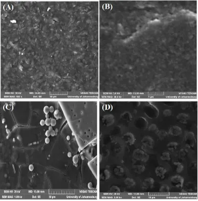
Figure 3. SEM image of (A) bare SPCE, (B) PPI dendrimer modified SPCE, (C) GOx modified SPCE (D) GOx-PPI dendrimer modified SPCE.
3.2 Electrochemical Studies of GOx-PPI
Fig. 4A shows the effect of the immobilised GOx-PPI in the presence of [Fe (CN)6]3-/4- ions. An increase in current was observed for the GOx-PPI modified electrode over the bare GCE owing to the nano-dimensional nature of PPI which increases the surface area of the electrode [26].
In the scan rate study (Fig. 4B), the peak current varies linearly with increased current suggesting that the kinetics of FC is diffusion controlled on the GOx-PPI modified electrode (GOx-PPI/GCE) as seen in Fig. 4B (inset) where a correlation coefficient 0.9937 was calculated. This diffusion controlled interfacial kinetics suggests that the GOx-PPI/GCE can be used for quantitative analytical purposes. Furthermore, the stability of the GOx-PPI film is depicted by shift in peak potential as scan rate increases. A large shift or irregular peak potential is expected when the film on the electrode is unstable or leaching into the solution, but this is not the case in Fig. 4B.
on the GOx-PPI modified GCE occurred between 450 mV and 600 mV as seen in Fig. 4C. This lowered oxidation potential reduces the risk of interferences. This oxidation is an evidence of successful immobilisation of the GOx-PPI on GCE. It also shows that the enzyme maintains its activity after coupling it to PPI.
A)
C)
Figure 4. A) Cyclic voltammetry studied of (i) bare GCE, (ii) PPI modified GCE; (iii) GOx-PPI modified GCE [Fe (CN)6]3-/4-. B) Cyclic voltammograms of GOx-PPI/GCE bioelectrode on increasing scan rate from 10 mV s-1 to 150 mV s-1. Inset is the corresponding plots of peak current versus the square root of scan rate. C) Cyclic voltammetry responses of a PPI-GOx dendrimer in a PBS solution (pH 7.5) in the absence (i) and presence (ii) of 30 mM glucose.
3.3 Chronomperometric determination of glucose
[image:9.596.130.467.70.314.2] [image:9.596.135.463.450.693.2]
Chronoamperometric experiments for the detection of glucose using the GOx-PPI modified GCE were investigated at +400 mV and +600 mV. The amperometric responses at these two potentials were similar and thus +400 mV was chosen as the optimum oxidation potential (Fig. 5). A response time of ca 5 sec was needed to obtain the glucose signal. A calibration plot of the biosensor response to glucose (Fig. 5 inset) gave a correlation of 0.99337 and a linear range of 0.4 mM to 14 mM. A detection limit of 0.1 mM was calculated.
3.4 Photometric response studies
Photometric studies were carried out to determine the GOx activity, stability and shelf life of GOx-PPI/GCE bioelectrode. Fig. 6 shows the variation of the difference between the initial and final absorbance value obtained at 500 nm after 3 min of incubation of the biosensor in glucose. It can be seen that the value of absorbance increases linearly with glucose concentration in the range of 0.552 mM to 22.1 mM. This experiment was carried out in triplicates with good reproducibility within 2% error.
Figure 6. Photometric response of in situ coupled GOx-PPI dendrimer modified SPCE as a function of glucose concentration (0.552 mM to 22.1 mM) in phosphate buffer (50 mM, pH 7.5) containing 0.9% NaCl.
[image:10.596.116.484.340.604.2]
3.5 Enzyme activity studies
The noticeable enzyme activity (Ucm-2) is defined as one unit of enzyme activity that results in the conversion of 1μmol of glucose into glucolactones per minute. It can be calculated by using the equation [30],
st enz appAV
Ucm
2
(2)
Where A is the difference in absorbance before and after incubation, V is the total volume (3.015 cm3), ε is the millimolar extinction coefficient (7.5 for o-dianisidine at 500 nm), t is the reaction time (3 min), and s is the surface area (cm2) of the GCE. The apparent enzyme activity was found to be ~0.032 (U cm-2) indicating that 0.032 units of enzyme per cm2 actively participate in the enzymatic reaction.
The shelf life of PPI-GOx biosensor was measured as a function of absorbance with respect to time with regular interval of 1 week. It was observed that GOx-PPI biosensor retained about 80% of glucose oxidase activity even after 9 weeks, when stored at 4 °C.
4. CONCLUSION
The successful coupling of poly (propylene imine) dendrimer (PPI) to glucose oxidase and its use as a biosensor for glucose is reported for the first time. The in situ coupling integrates the multi-functionality and nano-scopic properties of PPI into the enzyme moiety thereby enhancing immobilization, while retaining the enzyme activity on the electrode surface. The proposed biosensor displayed interesting amperometric response characteristics towards the efficient detection of glucose. These analytical parameters include low over potentials (+400 mV versus Ag/AgCl), very fast response time (~5 s), linear concentration range extending upto14 mM. This glucose biosensor was highly sensitive; to the extent that it was able to chronoamperometrically detect the target glucose concentration as low as 0.4 mM (R2 = 0.9933) in phosphate buffer with detection limit of 0.1 mM. Photometric results showed the good enzyme stability and activity with linear response in the range of 0.552 mM to 22.1 mM. The activity of the enzyme, as deduced from relatively low Michaelis-Menten constant (0.127 mM) indicates high enzyme affinity towards glucose, was found to be better than previous reports. In addition, the GOx-PPI G2 dendrimer glucose biosensor exhibits both good reproducibility and long term stability (9 weeks). Owing to the similarities in the functional groups of enzyme, there is a possibility of extending this simple carbodiimide coupling reaction to other types of enzymes. The result of which can lead to improved enzyme activity, ease of enzyme handling at room temperature and elongation of enzyme shelf life.
ACKNOWLEDGEMENT
Reference
1. C. Chen, Q. Xie, D. Yang, H. Xiao, Y. Fu, Y. Tan, S. Yao, RSC Adv., 3 (2013) 4473. 2. A. Heller, B. Feldmen, Chem. Rev., 108 (2008), 2482.
3. D. Odaci, B.N. Gacal, B. Gacal, S. Timur, Y. Yagci, Biomacromolecules, 10 (2009) 2928. 4. E. H. Yoo, S.Y. Lee, Sensor, 10 (2010) 4558.
5. M.C. Moreno-Bnd, O.S. Wolfbeis, M.J.P. Leiner, B.P.H. Schaffar, Anal. Chem., 62 (1990) 2377. 6. W. Trettnak, M.J.P. Leiner, O.S. Wolfbeis, Analyst, 113 (1988) 1519.
7. P. Noroizi, F. Faridbod, B. Larijani, M.R. Ganjali, Int. J. Electrochem. Sci., 5 (2010) 1213. 8. D.A Tomalia, A. M Naylor, W.A. Goddard II, Angew. Chem. Int. Ed., 102 (1990) 119. 9. J.M.J. Frechet, Science, 263 (1994) 1710.
10.D. Astruc, F. Chardac, Chem. Rev., 101 (2001) 2991.
11.R. Malgas, S.F. Mapolie, S.O. Ojwach, G.S. Smith, J. Darkwa, Catal. Commun., 9 (2008) 1612. 12.O.A. Arotiba, A. Ignaszak, R. Malgas, A. Al-Ahmed, P.G.L. Baker, S.F. Mapolie, E.I. Iwuoha,
Electrochim. Acta, 53 (2007) 1689.
13.O.A. Arotiba, P. G. Baker, B.B. Mamba, E.I. Iwuoha, Int. J. Electrochem. Sci., 6 (2011) 673. 14.C. Dufes, I.F. Uchegbu, A.G. Schatzlein, Adv. Drug Delivery Rev., 57 (2005) 2177.
15.Y, Gao, G. Gao, Y. He, T. Liu, R. Qi, Mini-Rev. Med. Chem., 8 (2008) 889.
16.A.P.H.J. Schenning, E. Peeters, E.W. Meijer, J. Am. Chem. Soc., 122 (2000) 4489. 17.J. Roovers, B. Comanita, Adv. Polym. Sci., 142 (1999) 179.
18.A. A. Baleg, N.M. Jahed, O.A. Arotiba, S.N. Mailu, N.R. Hendricks, P.G. baker, E.I. Iwuoha, J. Electroanal. Chem., 652 (2001) 18.
19.Sigma Chemical Co. (1983). Technical Bulletin No. 510. 20.A. Tiwari, S. Aryal. S. Pilla, S. Gong, Talanta, 78 (2009) 1401.
21.M.M. Reddy, D.J. Reddy, A. Moin, H.G. Shivakumar, Der Pharmacia Lettre, 3 (2011) 119. 22.A. Salimi, A. Noorbakhsh, Electrochim. Acta, 56 (2011) 6097.
23.S.K. Shukla, S.R. Deshpande, S.K. Shukla, A. Tiwari, Talanta, 99 (2012) 283.
24.P.R. Solanki, A. Kaushik, A.A. Ansari, A. Tiwari, B.D. Malhotra, Sens. Actuators, B, 137 (2009) 727.
25.O. Arotiba, J. Owino, E. Songa, N. Hendricks, T. Waryo, N. Jahed, P. Baker, E. Iwuoha, Sensors, 8 (2008) 6791.
26.O.A. Arotiba, E.A. Songa, P. G. Baker, E.I. Iwuoha, CHIM OGGI, 27 (2009) 55. 27.E.I. Iwuoha, A. Rock, M.R. Smyth, Electroanalysis, 11 (1999) 367.
28.P.R. Solanki, S.K. Arya, S.P. Singh, M.K. Pandey, B.D. Malhotra, Sens. Actuators, B, 123 (2007) 829.
29.A. Kaushik, R. Khan, P. R. Solanki, P. Pandey, J. Alam, S. Ahmad, B.D. Malhotra, Biosens. Bioelectron., 24 (2008) 676.
30.J. Singh, A. Roychoudhury, M. Srivastava, V. Chaudhary, R. Prasanna, D. W. Lee, S. H. Lee, B.D. Malhotra, J. Phys. Chem. C, 117 (2013) 8491.


![Figure 4C) . A) Cyclic voltammetry studied of (i) bare GCE, (ii) PPI modified GCE; (iii) GOx-PPI modified GCE [Fe (CN)6]3-/4-](https://thumb-us.123doks.com/thumbv2/123dok_us/1916590.150524/9.596.135.463.450.693/figure-cyclic-voltammetry-studied-bare-gce-modified-modified.webp)

