STUDIES ON THE STRUCTURE AND FUNCTION OF THE CRAB EYE
SALLY JANE STOWE
A Thesis submitted for the degree of Doctor of Philosophy of the Australian National University.
DECLARATION.
I declare all the original work presented in this thesis to be my own.
~
S. J. StoweChapters IV and V are taken from two published papers, with minor alterations to avoid repetition, and the addition of some extra photographs. These were:
Stowe, S., Ribi, W.A., and Sandeman, D.C. (1977). The organisation of the Lamina ganglionaris of the Crabs Scylla serrata and Leptograpsus variegatus. Cell.Tiss. Res. 178, 517-532
Stowe, S. (1977) The Retina-Lamina Projection in the crab Leptograpsus variegatus. Cell. Tiss. Res. 185, 515-525
A manuscript for publication based on Chapters II and III is in preparation.
One other publication was made during the tenure of my C.S.F.P. Scholarship:
ACKNOWLEDGEMENTS
I would like to thank Professor Horridge for the opportunity to work in a most stimulating environment, and for his encouragement during a crucial time.
It is a pleasure to thank my supervisors, who were, in order of appearance, Dr David Sandeman, Dr Willi Ribi, Dr Dave Dvorak, Dr David Blest, Prof. G. Adrian Horridge, and Dr Steve Shaw, for their support and willingness to offer help and advice. David Sandeman suggested an anatomical study of the crab optic lobe, and Willi Ribi gave me inv~luable advice on the art of Golgi staining.
Many other past and present members of the department have helped me enormously, especially Joachim Erber and Martin Wilson, who taught me something of electrophysiology, and Eldon Ball, who is always willing to give of his time and knowledge. Kevin Downing's ability to smooth out difficulties has always been an important support.
I have benefited greatly from discussions with many people, among them Yasuo Tsukahara, Pete Lillywhite, Barry O'Brien, and Simon Laughlin, to whom I am indebted for his comments on Chapter III, although he is not responsible for any errors remaining.
Bruce Ham braved all weathers to collect crabs, and was responsible for the design and maintenance of the crab tanks.
I would like to thank the staff of the EM Unit, Photography, and the
Workshop, who have always been extremely helpful.
All the colour prints are the work of Kerrie Ruth, and I am also grateful .to David Sandeman for drawing Fig. 1.1, and to Chris Snoek for Fig. 2.1.
David Blest has helped me with reduced silver staining, by
ABSTRACT
The eyes of crabs are of interest for a number of reasons. Some crabs, including Leptograpsus, are active over a wide range of light intensities, from night to bright sunlight, a change of seven or eight log units in brightness. They are known to be highly sensitive to very slow movements. Since their eyes are of the apposition rather than the superposition type they have much higher visual acuity than most other decapods.
The eyes of the rock crab, Leptograpsus variegatus, and the Queensland mud crab Scylla serrata, have been examined in a number of ways. In Scylla, the structural organisation of the first optic neuropile, the lamina, has been investigated using Golgi and reduced silver staining. It is very similar in structure to the lamina of decapods with superposition eyes, such as crayfish, and contains five types of monopolar neurons and at least three tYIJes of large tangential fibres. The seven retinula cells which make up the main part of the fused rhabdom terminate in two layers within the lamina. The eighth cell, which forms only the distal tip of the rhabdom, has an axon that passes through the lamina and ends in the second optic neuropile, the external medulla. By tracing Leptograpsus retinula cell axons through a series of 1 or 2 µm sections, it was found that all the retinula cell axons from one ommatidium go to the same lamina cartridge, and not to several, as has been recently proposed.
illumination conditions during the day and at night. The spectral transmission of the different types of screening pigment was measured by microspectrophotometry.
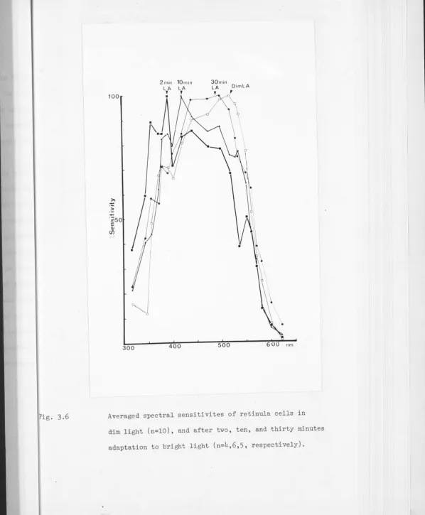
The spectral sensitivity of Leptograpsus retinula cells was found by intracellular recording from eyes in situ. The dark adapted spectral sensitivity peaks at c 485nm. Light adaptation causes various changes in spectral sensitivity which can largely be accounted for by movements of the screening pigments. The net effect is to produce a broader but fairly constant spectral sensitivity over a wide range of light levels.
The polarisation sensitivity is high, about 10:1. The angular sensitivity, which alters slightly in response to light and dark adaptation, ranges from about 2° in bright light, to .3-4° in the dark, in the forward-looking portion of the eye.
Chapter I Chapter II
Chapter III Chapter IV
Chapter V
Chapter VI
CONTENTS
GENERAL INTRODUCTION.
RETINAL PIGMENTS AND THEIR MOVEMENTS IN LEPTOGRAPSUS
RECEPTOR RESPONSES AND SPECTRAL SENSITIVITY. THE RETINA-LAMINA PROJECT IN THE CRAB
LEPTOGRAPSUS VARIEGATUS.
THE ORGANISATION OF THE LAMINA GANGLIONARIS OF THE CRABS SCYLLA SERRATA AND LEPTOGRAPSUS VARIEGATUS.
CONCLUSION.
1
5
22 53
64
Chapter I GENERAL INTRODUCTION
Even to the casual observer, crabs give the impression of being highly visual animals. The often brightly coloured crabs of mudflats have a large repertoire of display movements, the classic example being the threat and courtship signals of the various species of the fiddler crab Uca, reviewed by Crane (1975). Escape from predators is often visually mediated, as can be seen from the speed with which a slight movement will send a platoon of Mictyris many yards away on a flat beach scurrying off in the opposite direction, a foraging Leptograpsus disappearing down a crack in the rocks, or an Ozius caught investigating a shallow pool surreptiously gliding under a clump of weed.
Field ·and laboratory studies under more controlled conditions bear this out. Uca, for instance, finds the way back to the area of its burrow from some distance by a variety of clues which can include remembered land-marks and the polarisation pattern of the sky (Herrnkind 1972).
Recording from the optic nerve of Podophthalmus, Wiersma et al. (1964) demonstrated a varied array of movement detectors in crabs, in fact
2.
The ability to perceive very slow movements is one of the main reasons why the optic system of the crab is worth investigation. Another is the capacity of the retinula cells to analyse polarised light.
Crustaceans, because of the structure of their rhabdom, are potentially more sensitive to the polarisation plane of light than almost any other group. Yet apart from orientation responses to sky polarisation patterns, which are hardly unique to Crustacea, we do not know for what purpose, and to what extent, crabs use the polarisation information available to them.
The general structure of the decapod retina is well known, and is discussed in the next chapter. The rhabdom is formed by seven main cells with interdigitating orthogonally-oriented microvilli, and a distal eighth cell of varying importance. The properties of the eighth cell are
effectively unknown. Apart from the obvious.differences in the plane of polarisation sensitivity caused by the orientation of their microvilli, it is not known how differences in the structure of the main retinula cells (size of the rhabdomere, position in relation to the surrounding pigment cells) are reflected in their properties. The effect of the pigment screen has been well studied in crayfish, which have superposition eyes, but not in decapods with apposition eyes, such as crabs. The problem of spectral sensitivity in decapods is still obscure in spite of recent work. The manner in which retinula cells project to the lamina has been the subject of
speculation without conclusive evidence, and recently it has become obvious that in studying any visual system it. is necessary to determine whether there are any circadian changes in the rhabdom itself. I have tried to answer some of these questions.
An initial Golgi study of the lamina, the first optic ganglion, was performed using the portunid mud crab Scylla serrata. It was later
3.
much more reliable animal for behavioural and electrophysiological work. Since it is also available locally and has less well developed weaponry, it was used in later work. The basic construction of the eyes of the two crabs is very similar (Fig.
1
.
1).
As discussed in Chapter V, no large differences in _the structure of the lamina were revealed by the limited amount of work done on Leptograpsus, and considering the great similarities between the lamina of Scylla and that of other decapods (Hafner,1973,
"
Nassel,
1975, 1976),
it is unlikely that the lamina of another brachyuran crab will be very disparate. The same applies to the gross structure and arrangement of the retinula cells themselves. It is· another matter when considering the secondary pigment cells, which are notoriously variable throughout the decapods, and display quite different behaviours in the two crabs.The pigment systems of crustacean eyes move under a combination of circadian and environmental influences, in a way which must be determined for each species. Obviously, if any work is to be done more centrally in the nervous system of Leptograpsus, it is necessary to have some idea of what factors are causing its eye pigments to move, and their probable state in any experimental situation.
The projection pattern of axons from the retina to the lamina was traced because the "neural superposition" type of connection pattern that had been assumed (e.g. Waterman
1978)
seemed most unlikely. It was not well supported by the little anatomical evidence available, and no reasonable theoretical explanation for it had been advanced. A knowledge of theb
4.
Although some measurements were made of polarisation sensitivity
and angular sensitivity of retinula cells, the main focus of the
electrophysiological study described in Chapter III was spectral sensitivity,
since it was thought that it might be profitably studied in conjunction
5.
CHAPTER II
Retinal Pigments and their Movements in Leptograpsus
INTRODUCTION
Retinal screening pigments in Crustacea present a bewildering variety of systems, ranging from simple three-pigment eyes to combinations of five or six pigments, some stationary, some moving according to a
circadian rhythm, some because of a direct or hormonally-mediated effect of light. Although closely homologous pigment cell types can be recognised across many different species, one of the main lessons of work on pigment migration so far is that generalisation even between the species of one genus is often invalid.
It is necessary to determine the details of the retinal pigment system and its movements for any species whose visual physiology is to be investigated closely. To be most effective, such a study needs to be combined with knowledge of how the various pigments absorb light. This can be rather difficult to obtain, since many pigment cells contain several different types of pigment granules, and it may also be hard to entirely separate classes of pigment cells in fresh preparations. Luckily in Leptograpsus the relationship between the pigment cell types is such that an unfixed eye may be teased apart to yield small clumps containing either one sort of pigment cell, or two easily separable types.
6.
distal retinula cells in the day or light-adapted eye, restricting the amount of non-axial light that can reach the rhabdom. In the night or dark-adapted eye, the pigment moves distally and becomes concentrated within a smaller area, allowing much of the non-axial light to pass on to the rhabdoms. Dark granules of proximal pigment within the retinula cells themselves can move below the basement membrane to expose the rhabdom, or form a sheath around the rhabdom to limit the entrance of light from the side and perhaps attenuate axial light. Reflecting pigment, sometimes called accessory pigment, lies between the ommatidia, sometimes moving back and forth across the basement membrane. When it lies between the ommatidia it should stop light "leaking" from one column of retinula cells to another; when it lies fairly proximally, and is not screened by darker pigments, it acts as a tapetum, reflecting and scattering light back through the rhabdoms.
7,
In spite of a long history of research a coherent picture of the
hormonal control of pigment movements in Crustacea is only just beginning
to emerge (reviewed by Kleinholz,
1976)
.
Even now, much more is knownabout movements of two pigments, the distal pigment and the retinula cell
or proximal pigment, than any others in the eye. The dark distal pigment
(at any rate in the crayfish Procambarus) moves in response to hormones: the light-adapting or dark-adapting DRPH (Distal Retinal Pigment Hormones) .
Its movements take much longer to complete than those of the proximal
pigment, have a higher threshold, and do no occur in the isolated eye (reviewed by Arechiga,
1978)
.
Proximal pigment, as shown by Ludolph et al.(1973
)
in the crab Callinectes, can move independently in the differentretinula cells of one ommatidium. Olivo and Larsen
(1978)
showed thatmigration of proximal pigment initiated in an isolated eyestalk by a
brief exposure to light is continued in the dark.
This study was undertaken primarily to determine the position
of the retinal screening pigments of Leptograpsus under the conditions
used in the electrophysiological experiments, in order to gain some idea
of their effects on the responses of the retinula cells.
METHODS
Leptograpsus variegatus, the common grapsid rock crab, was
collected every three to five weeks near Bateman's Bay, on the New South
Wales coast. The crabs were kept in a fibre-glass tank of sea-water
containing rocks for shade and shelter. The sea-water was kept
circulating through several inches of gravel bed by air pumped in beneath the gravel. The tank was lit by fluorescent lights on a 12:12 light:dark
cycle in winter, and a 14:10 cycle in summer, when all the experiments
concerning pigment migration were carried out. The crabs were fed on
8.
their.fellows foolhardy enough to try to moult in a crowded tank. They were allowed .to acclimatise for two-three days at least before being used. Electron microscopy
For electron microscopy eyes were fixed in O.lM sodium cacodylate, 0.14M sucrose·, 2.5% gluteraldehyde and 2mM calcium chloride at pH 7.3.
The crabs used were of almost identical size, of carapace width between 2.8 and 3cm. The eyes of two crabs were fixed in the light adapted condition at midday, and the eyes of two more were fixed when dark-adapted at midnight, being dissected under a red light. They were fixed overnight at 4°c, washed in several changes of the cacodylate buffer (with · sucrose), osmicated in 1% osmium in cacodylate buffer for two hours, washed
in several changes of buffer·, dehydrated through an ethanol series, taken through propylene oxide and embedded in araldite. For examination at low magnifications, pale gold to silver sections were cut on glass knives, mounted on formvar-coated slot grids and stained in uranyl acetate (40 min) and Reynold's lead citrate (20 min). They were examined with a Jeol JEM_ lOOC electron-microscope. Measurements were made from photographs. Means and standard deviations were calculated from 40-50 measurements of each type of pigment granule, and 8-10 rhabdoms in each condition.
Pigment Movements
9.
completed in 70% alcohol after the eyes had been left overnight in cold
fixative. This was 2.27% gluteraldehyde in Millonig's buffer with 0.01%
calcium chloride and 15% glucose. "Day" crabs were fixed between 1. 30 and 3.30 pm in the following conditions: after 30 minutes at a window in
bright sunlight but on a cool substrate; after 60 minutes in "dim light" 2
(10-20 µW/cm ); after 30 and 60 minutes dark adaptation; and after 2, 5,
15 and 30 minutes under "bright" (8-lOmW/cm2) light from a 30 watt tungsten
microscope lamp. "Night" crabs were fixed between 10 and 12 pm when dark
adapted for two hours, 'dim' light adapted for one hour, and 'bright'
light adapted for 2, 5, 15 and 30 minutes. The dim and bright light regimes
were adopted in order to mimic conditions under which recordings from
retinula cells had been made.
After dehydration through an ethanol series (as fast as possible
in order to minimise loss of alcohol-soluble pigments) the eyes were taken
through propylene oxide to araldite. They were sectioned at 0.5 to 2µm
to provide different viewing conditions and mounted unstained under Permount.
Measurements were made as a proportion of cone length or retinula cell soma
length, to allow for varying crab sizes. The minimum diameter of the "iris"
-formed by the dark distal pigment around the cone tip was also measured.
An eyepiece graticule was used for all measurements of LM material. Mierospectrophotometry
The spectral absorption of the red basal pigment, the retinula
cell screening pigment, the dark distal pigment and the light distal pigment
was measured using a Zeiss UMSP 1. Fresh eyes were dissected in Carcinus
saline (Fantin, 1972), and the retina teased apart to yield pieces
10.
measuring diameter of 5 µmin the object plane, from 700 to 350 nm. A section of the slide containing no tissue was used to provide baseline measurements. The red basal pigment formed large oily droplets in the saline, and some of these easily covered the whole area being measured. Clusters of granules were measured for the dark distal and retinula cell screening pigments, and groups of small yellow-brown droplets for the light distal pigment. Quartz slides were used, but glass coverslips. Change in extinction was measured by comparing transmission through pigment and saline at ten or twenty nm intervals. Data for each run were normalised, taking the highest extinction value of each run as unity, then the normalised data were averaged and plotted.
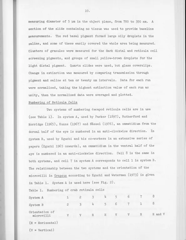
Numbering of Retinula Cells
Two systems of numbering decapod retinula cells are in use (see Table 1). In system A, used by Parker (1897), Rutherford and Horridge (1965), Kunze (1967) and ~assel (1976), an ommatidium from the dorsal half of the eye is numbered in an anti-clockwise direction. In System B, used by Eguchi and his co-workers in an extensive series of papers (Eguchi 196.5 onwards), an ommatidium in the ventral half of the eye is numbered in an anti-clockwise direction. Cell
8
is the same in both systems, and cell7
in system A corresponds to cell 1 in system B. The relationship between the two systems and the orientation of the microvilli in Grapsus according to Eguchi and Waterman (1973) is given in Table 1. System A is used here (see Fig. 2).Table l . Numbering of crab retinula cells
System A 1 2 3 4
5
6 78
System B 2 3 4
5
6 7 18
Orientation of
microvilli V V H H V V H H and V
[image:19.615.15.610.10.777.2]RESULTS
The cornea of Leptograpsus consists of any array of c.40 µm
hexagonal facets, with no external demarcation between them. One of the
axes of the array runs horizontally when the eye is in the normal position.
The corneal cuticle is flexible and thin (c.40 µm), and has no apparent
focusing effect when viewed through a hanging drop. Below the cornea of
each facet lie two cone producing cells and a crystalline cone 90-120 µm
long (Fig. 2.1). The cone tapers to a blunt point above the short rhabdom
of retinula cell eight. Four "cone cell roots" continue to the basement
membrane as fine threads between retinula cells 1 and 2, 3 and 4, 5, and 6,
and 7 and 1. The soma of R8 is divided into· four lobes (Fig. 2.2) one of
which sends a process proximally which enlarges to contain the nucleus and
then continues as an axon besides the soma of R7, eventually joining the
group of retinula cell axons that forms below the basement membrane. The
soma of R8 contains no pigment granules, and the cytoplasm is pale and full
of vacuoles during the day. The most distal parts of Rl-7 surround the
lobes of R8 (Fig. 2.3). Rl-7 form a fused rhabdom of the standard decapod
type. Along its 250-350 µm length, two sets of orthogonally oriented microvilli alternate R7, and 4 contribute horizontally oriented microvilli,
Rl, 2, 5 and 6 have microvilli running vertically when the crab holds its
eye in the normal position. The rhabdomere of each cell stretches halfway
across the rhabdom; the rhabdomere of R7 covers half the area of the
rhabom, the other cells all contribute one quarter each.
During the day, in the light-adapted state, there is little
variation in rhabdom diameter from the distal (1.67: 0.12 µm, Fig. 2.4)
to the proximal (Fig2.5) end of the rhabdom. The rhabdom is surrounded by
Fig. 2.1 Semi-schematic diagram of a Leptograpsus ommatidium
and associated cells, when light-adapted dur'ng the
Cone producing _ _ ,.--,, cell
l{," t t - - - - ~ ~- Light Distal pigment cell
- Reflecting pigment _ __...;~~,----.<.n
cell ~
R8 -~~~~~~
:"F:0'1i~M-li;:'J..__
i;~ke~is:~i'I _ _ _\~t
I
I
~R1-7~rc1·,
Rhabdom~---..r~
-.-.--.-=~-
Retinula cell ---:H"'.-'t=ilLight Adapted
[bright sunlight]
screening pigment
Basement
pigment cell
" ·
.
, "I,
,.'> 0 flt') 0 • •
• o" Q
0. 3
Dark Adapted
Fig. 2.2
Fig. 2.3
Fig
Cross-section of a light-adapted Leptograpsus eye at
the level of the eighth cell. Scale 5 µm.
Distal tip of the rhabdom at night, showing the four
lobes of RB lying between the pigment containing Rl-7.
Inset, day rhabdom at a level c. 10 µm proximal, and·
the same magnification. (4,900 X)
DD, dark distal pigment cell; R, rhabdom; P, palisade;
[image:23.614.10.574.16.709.2]at
our
Rl-7,
and
Fi Fig. 2.4 Distal part of the retina during the day, showing Rl-7
1-7
ent
Fig. 2.5 Proximal part of rhabdom in the day, showing clear spaces inside retinula cells, filled with microtubules and surrounded by mitochondria. (6,800 X)
12.
the rhabdom and the palisade is a thin rind of cytoplasm, approximately 0.3 µm thick, from which the microvilli sprout. Within the retinula cells are large clear areas, containing microtubules (Fig. 2.6). Above the basement membrane these areas cover most of the cross-section of the cell, and tend to become smaller as they continue distally as far as the nuclear region.
At night, the diameter of the distal part of the rhabdom increases dramatically to 5.22
!
0.52 µm (Fig. 2.7), an increase in cross-sectional area by a factor of about 10. The rhabdom tapers towards the basement+
membrane, where the diameter is 1.93 - 0.51 µm, a cross-sectional increase of c. 1.8. The "palisade" is 9.44
!
0.72 µmin outer diameter distally, but decreases towards the basement membrane, where it is hardly visible. The rind of cytoplasm around the rhabdom is not evident at night. The clear areas or microtubule fields are found just above the basement membrane and in the axons below it, and a proportion of them contain apparently non-membrane-bound yellow droplets of varying sizes, the largest filling up almost the whole cross-section of the cell (Fig. 2.8, 2 .12c).Between the crystalline cones lie thin sheets containing the yellow-brown light distal pigment in small, oily droplets 0.2 .:_ 0.1 µm
F Fig. 2.6 Halfway down retinula cell column, day eye. (6,900 X) . R, rind; P, palisade; M, microtubule-filled space;
N, nucleus of reflecting pigment cell; DD, extensions
x)
e;
Fig. 2.7
F
Fig. 2.8
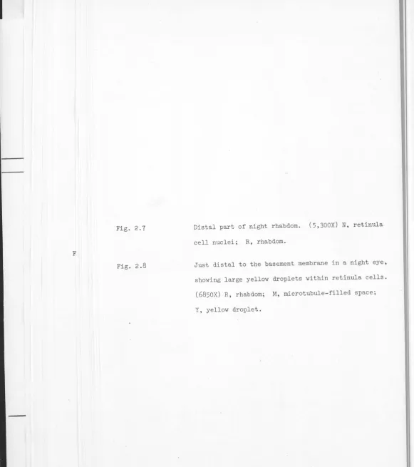
Distal part of night rhabdom. (5,300X) N, retinula
cell nuclei; R, rhabdom.
Just distal to the basement membrane in a night eye, showing large yellow droplets within retinula cells.
(6850X) R, rhabdom; M, microtubule-filled space;
[image:31.614.5.586.15.669.2]a
ye,
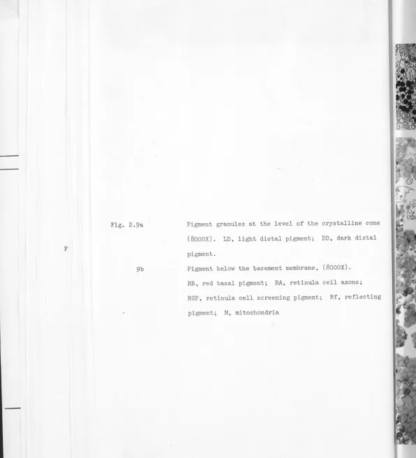
Fig. 2.9a
F
9b
Pigment granules at the level of the crystalline cone
(8000X). LD, light distal pigment; DD, dark distal
pigment.
Pigment below the basement membrane, (8000X).
RB, red basal pigment; RA, retinula cell axons;
RSP, retinula cell screening pigment; Rf, reflecting
[image:33.614.11.607.16.672.2]cone
stal
13.
Within the retinula cells themselves are brown screening pigment granules, of 0.3
±.
0.1 µm diameter. These are very mobile, and may beconcentrated around the rhabdom or scattered throughout the cell. The
retinula cell axons below the basement membrane have pigment granules
around their circumference, and in some conditions may be quite densely
filled with pigment.
The reflecting pigment is contained within large cells which are usually closely apposed to the retinula cell column over most of its length
from the basement membrane to the level of the distal tip of the rhabdom.
Processes of the cells extend below the basement membrane in some states
(Fig. 2.10). A thin strand of tissue containing reflecting pigment granules extends to the cornea. The nucleus lies about halfway down the retinula
cell column. The pigment granules, 0.3
±.
0.05 µmin diameter, are abrilliant white in reflected light, but pale brown in transmitted light.
The contents of the granules are usually lost in EM sections.
Beneath the basement membrane the retinula cell axons run through a layer of basal red pigment cells which contain large (from 0.3 to 0.7 µm diam.) oily red droplets, densely packed (Figs. 2.9b, 2.11). They form a thick barrier on the proximal side of the basement membrane, and sometimes send processes through it.
Mainly below this layer, but sometimes extending within it, are a small.number of irregularly arranged cells containing dense black pigment. These cells consist largely of widely separated processes wound around the retinula cell bundles from the sub-basement membrane area to the lamina, where they splay out over the distal lamina surface rather in the manner of exposed tree roots.
Fig. 2.10 Oblique section through the basement membrane in a
night eye. (4,900X) . RA, retinula cell axons, A',
axon passing through basement membrane in a
non-standard position. Y, yellow droplets; Rf,
Fig. 2.11 Longtitudinal section through the basement membrane (3,500X) L, sub-basement membrane lacunae; RSP, retinula cell
[image:38.594.22.578.16.763.2]14
.
· observed, green on one side only, that have become dislodged f'rom somewhere
around the crystalline cone. Nothing corresponding to them has been
observed in sectioned material in Leptograpsus. Schonenberger (1977)
sees similar structures in the eye of Sguilla, and suggests, as seems
likely also for Leptograpsus, that they are only related to the superficial
colour scheme of the animal.
pigment movement
The state of the five main pigments and the width of the 11iris11
formed by the dark distal pigment and/or the retinula cell screening
pigment is summarised in Table 1.
Allowing for individual variation and the range of crab sizes used,
all that can be said of the iris is that it is approximately twice as large
during the night as during the day, and that dark or light adaptation does
not cause a marked change within 30 or 60 mins.
The dark distal pigment is largely responsible for the size of
the iris, (Fig. 2.12d) but the bulk of the cell changes very little except
under very strong illumination, when pigment granules move into the fine
strand that extends to the basement membrane. However since this survey
has been done using light microscopy almost exclusively, it would not be
evident if any change had occurred in those parts of the cells that run in
to the rhabdom on either side of R7.
The light distal pigment covers all the cone that is not surrounded
by the more proximal darker screening pigments, except at night. When dark
adapted at night, the pigment retracts from the distal third or so of the
cone. Long exposure to moderate light levels at night causes the pigment
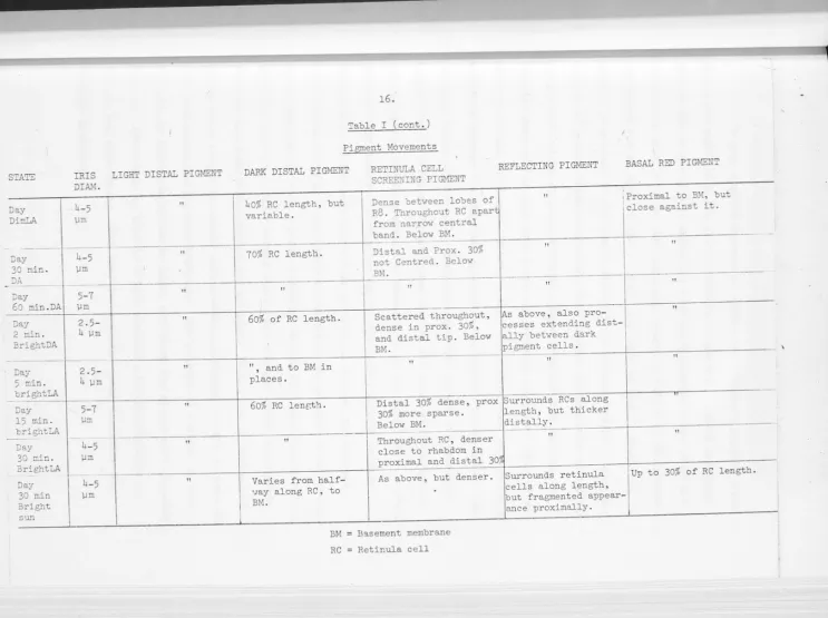
STATE Night DA Night DimLA Nig.1-it
2 min
BrightLA
Night
5 min.
BrightLA
Hight
15 min.
BrightLA
Night
30 min
IRIS
DIAM.
I
13~
I
I
.
10-13
µm
8-9.5
µm
8 µm
10-13
µm
5-7
µm
BrightLA
15.
Table I
Pigment Movements
LIGHT DISTAL PIGMENT DARK DISTAL PIGMENT
From l/3(distally) to Proximal 20% of cone
9/10 (proximally) of and distal 30% of
cone. RC.
Along cone. Along distal half of
RCs.
From 1/8 (distally) Along distal 75% of
to 9/10 (proximally) RCs.
of cone.
As above. Along distal half of
RCs. :
Proximal 95% of Along distal 60% of
cone. RC.
All along cone. As above, but up to
BM where red pigment
movement extensive.
RETINULA CELL I SCREENING PIGMENT
Below BM.
Sparse in the distal
30% of RC: and
below BM.
Light but centred
around rhabdom, in
prox. and dist. 10
-20% of RC. Below BM.
Centred in distal
20%, sparser and
scattered in proximal
30% of RC. Below BM.
Centred in distal 25%
and proximal 10%,
scattered throughout.
Below BM.
Light centred in
distal 25% scattered
centrally .• Thick and
centred in prox. 10%.
Below BM.
REFLECTING PIGMENT BASAL RED PIGMENT
Surrounds proximal Up to 50 µm below BM.
half of RCs, extends
C • 50 µm below BM.
Surrounds proximal Close below BM, some
half of RCs, fine small patches above, for
strands along distal c.15% RC length.
half.
As above, distal Close below BM.
strands slightly
thicker.
As above, thick Very slightly above BM
strands. patches.
More uniform, but Slightly above BM, up to
still denser proxim- 10% of RC length.
ally.
Uniformly surrounds Above BM in large patches
STA'E
Day Din:LA
-
-Day
30 min.
"JA
-
-Day
60 :nin.DA
"Jay
2 min.
BrightDA
Day
5 min.
brightLA
Day
15 min.
brightLA
-Day 30 min.
BrightLA
Day
30 min Bright
sun
IRIS
DIAM.
_ h-5 )Jm
-4-5 µm
-5-7
)Jm
2. 5-4 )Jm
--2 .5-4 µm 5-7 )Jm h-5 µm
4-5
µm
LIGHT DISTAL PIGMENT
II 11 I 11 11
I
II II II"
16.Table I (cont. )
Pigment Movements
DARK DISTAL PIGMENT
I
40% RC length, but
variable.
70% RC length.
RETINULA CELL
SCREENING PIGMENT
Dense between looes of R8. Throughout RC apart
from narrow central
band. Below BM.
----Distal and Prox. 30%
not Centred. Below
BM.
REFLECTING PIGMENT
II
11
·-· --
---
-11 II
II
60% of RC length. Scattered throughout, ~s above, also pro
-dense in prox. 30%, cesses extending dist
-and distal tip. Below ally between dark
BM. pigment
--
cells.-.
II and. to BM in 11
II
'
places.
60% RC lenr;t'1. Distal 30% dense, prox Surrounds RCs along
30% more sparse. length, but thicker
Below BM. distally.
---11 Throughout RC, denser
II
close to rhabdom in
proximal and distal 30,
Varies from half- As above, but denser. Surrounds retinula
cells along length,
BASAL RED PIGMENT
\Proximal to BM, but
close against it.
11
II
II
11
11
Up to 30% of RC length.
·.,ay along RC , to
BM. ~ut fragmented appear
-BM
RC
B':l.sement membrane
Retinula cell
ance proximally.
-I
[image:41.795.46.789.11.565.2]17.
In dark adapted animals at night, the screening pigment granules of the retinula cells are almost entirely below the basement membrane.
This is the only state in which the four non-pigmented lobes of R8 are
not clearly visible under the light microscope, outlined by the
pigment-containing distal tips of Rl-7. Under dim light at night, scattered
pigment is present in the distal third of the retinula cells. After two
minutes of bright light adaptation, this pigment concentrates around the
rhabdom and shifts slightly distally. Some granules move up into the
proximal part of the soma from below the basement membrane. Eyes fixed
after
5,
15 and 30 minutes of bright light adaptation show more granulesmoving up through the basement membrane to accumulate in the distal and
proximal thirds of the cell. Scattered granules appear in the central
region from 15 minutes on, but since they do not accumulate there they are presumably moving towards the distal part of the cell, which becomes
progressively darker. During the day in dim light, pigment extends
through most of the cell apart from the central region, but is more
concentrated distally. Dark adaptation causes the pigment to become less
dense, keeping the same general distribution but with less concentration
about the rhabdom. Under strong light, the granules first concentrate
distally and proximally, within two minutes. During the 30 minutes of
bright light adaptation used, more granules continued to move up into the
soma from below the basement membrane. The pattern is similar to that
produced by light adaptation at night but there are always more granules
present. After half an hour in bright sunlight, the distribution is
similar but even denser, with very few granules remaining below the
18.
At night, the proximal half of each retinula cell column is enveloped by reflecting pigment cells under all conditions examined.
However a progressive change takes place in the distal half. In the dark-adapted eye the only reflecting pigment in the distal half of the
eye is in the fine threads that run to the cornea, leaving a clear area between retinula cells. In dim light these threads are thickened. After 15 minutes of strong illumination the clear area is. no longer evident,
and after 30 minutes the reflecting pigment is equally distributed
between the proximal and distal halves of the retina. This is also the case during the day, under dim light or up to as much as 60 minutes of
dark adaptation. After two minutes of strong light adaptation, the
reflecting pigment begins to push up between the dark distal pigment cells. After 15 minutes, there is more reflecting pigment between the
retinula cells in the distal half of the retina than the proximal. At
thirty minutes, the extreme distal projections of reflecting pigment have been displaced by dark distal pigment. After thirty minutes in
bright sunlight (Fig. 2.12a) , the reflecting pigment cells extend over both proximal and distal halves of the retina. However, they do not completely envelope the retinula cell columns, but present a rather
tattered, fragmented appearance. The reflecting pigment cells also send
processes below the basement membrane, but when light adapted in the day,
these are masked by the basal red pigment.
When dark adapted at night, the basal red pigment is retracted to some extent (about 20-30 µm) from the basement membrane. Under dim light at night, and dim or bright artificial light during the day, the pigment is below the basement membrane but closely apposed to it (Fig. 2,12b). Under bright sunlight, or bright artificial light at night, the
Fig. 2.12a
12b
Oblique section through the retina of a crab exposed
to bright sunlight for 30 minutes. Osmicated,
otherwise unstained. The red basal pigment can be
seen lower left, dark distal pigment top right.
Retinula cell screening pigment is reddish-brown,
and the reflecting pigment light brown. (450X) .
Longtitudinal section of day eye, dim light adapted,
showing red basal pigment below the basement membrane.
ed
d,
ane.
Fig. 2.12c 7
12d
Oblique section of the n:1ght eye of Leptograpsus,
showing the increase in size of the rhabdom from
near the basement membrane (lower left) to the
distal retina (top right), and large yellow
droplets within the retinula cells. (llOOX)
Unstained longtitudinal section from crystalline
cone (top) to basement membrane (bottom) showing
position of the light distal pigment between the
cones, the aperture formed by the dark distal
pigment (arrow) and the distal concentration of
[image:46.614.13.583.12.719.2]19.
These processes were seen to extend up to one third of the way along the
retinula cells. Two of -the eyes adapted to dim light at night showed
small patches of red pigment extending up to 15% of the length of the
retinula cells. Since_ the red pigment in unosmicated eyes was largely
dissolved by the dehydrating alcohols, the amount of red pigment above
the basement membrane is probably under-estimated, although the basement
membrane did seem to offer some protection against the alcohol.
Microspectrophotometry·
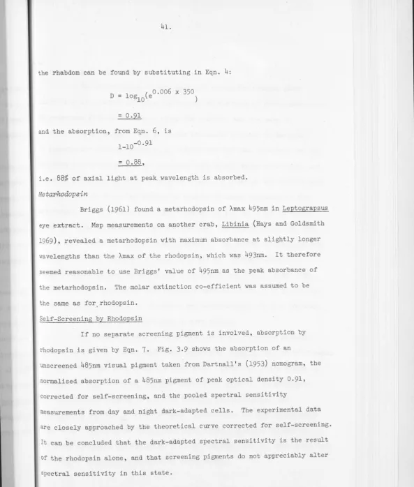
The retinula celi screening pigment (Fig. 13a) and the light
distal pigment (Fig. 13b) show a high absorbance in the ultra-violet,
increasing fairly smoothly towards longer wavelengths. The light distal
pigment has a slightly lower extinction at wavelengths longer than about
500 nm. The dark distal pigment also has its highest extinction in the
UV' but there is a rise in absorbance between 500 and 580 nm, peaking at
540 nm (F_ig. 13c). Extinction at the red end of the spectrum is markedly
higher than for the retinula cell screening pigment or the light distal
pigment. The absorbance of the red basal pigment -is low in both the
ultra-violet and the red, peaking in the green at about 500 nm (Fig. 13d).
The extinction curve of the reflecting pigment (not illustrated) shows a
similar shape to the retinula cell screening pigment but its reflectivity
characteristics were not measured.
DISCUSSION
Most of the screening pigments of the Leptograpsus eye appear
comparable to ·those described and sometimes chemically identified in
other Crustacea. The· exception is the light di-stal pigment, which does
not seem to occur commonly in Crustacea. The yellow pigment granules
described in the chromophores of Crangon, which have inclusions, (Elofsson
~ E
0·4
0-2
400 500 600 700
A
0·2
400 500 600 700
B
Fig. 2.13 Normalised differences in extinction between retinal screening pigments of Leptograpsus and a
sal."ne baseline.
A Light Distal Pigment (N=9)
10 '
-!.,;
0·8 '::-:::-::...._:-::-:-:--:-~-~":"" _
-
-
:"'_
06
~~~
-
~
0·4
0·2
400 500
400 500
C Dark Distal Pigment
(N=8)
D Red Basal Pigment
(N=ll)
600 700
C
600 700nm
20.
The granules of the dark distal pigment cells and the retinula cells are similar in size and structure to those containing ommochromes in other Crustacea (Elofsson and Hallberg,
1973)
.
The absorption spectrum of the dark distal pigment is very similar to that of onunin (Butenandt et al.1958,
quoted from Goldsmith,1964)
,
which is probably the main constituent of the granules. The retinula cell pigment granules, with a higher transmission at longer wavelengths, probably contain asignificant amount of the red and yellow xanthommatins.
The red basal pigment droplets are structurally similar to those found in the eye of Nebalia (Green,
1972
-
discussed by Elofsson and Hallberg,1973)
and in several mysids (Hallberg,1977)
.
These areconsidered to be formed of carotenoids, as are the red pigment-containing chromatophores of the body, and in fact Briggs
(1961)
found astaxanthin in extracts of Leptograpsus eyes.The white reflecting pigment of a number of brachyuran crabs has been examined by Zyznar and Nicol
(1971)
.
They found it to consist largely of pteridines, which are fluorescent in long wavelength ultra -violet, and a smaller amount of purines, which quench short wavelength UV. In all six crabs, isoxanthopterin was the most abundant pteridine.The retinula cell screening pigment, the reflecting pigment, the red basal pigment, and to some extent the dark distal pigment, are responsive to changes in the light level during both day and night. This might be
21.
The change in diameter of the rhabdom between night and day is of the same order of magnitude to that found.in some spiders (Blest,
1978)
and recently in Grapsus (N'assel and Waterman, in prep.) . In Grapsus the diameter increases substantially all down the rhabdorn, and it is not at all unlikely that the changes seen at 12 pm in Leptograpsus were not complete.22.
CHAPTER III
RECEPTOR RESPONSES AND SPECTRAL SENSITIVITY
INTRODUCTION
The general form of the receptor response has been described for several crabs (Shaw 1966, 1969), among them Leptograpsus (Erber and
Sandeman 1976). The response is typical of arthropod photoreceptors (reviewed by Fuertes and O'Bryan 1971), being a depolarisation which consists, at all but response levels below about 5mV, of a transient peak followed by a plateau. The polarisation sensitivity of cells Rl-7 is known to be high in decapods (Shaw 1966, 1969). However angular sensitivities have been little studied. Walcott (1974) found very wide angular sensitivities (24°) in the dark-adapted superposition eye of
Cherax, a crayfish. Leggett (1978), using Scylla with eyes in situ, studied changes in acceptance angle during dark adaptation, and found rather wide acceptance angles during the day (c. 4°) which became very broad (10-11°) when dark-adapted at night.
Crustacean spectral sensitivity, and in particular colour vision, is a somewhat confused area, with puzzling discrepancies in the evidence from various sources. Only one photopigment has been found by spectroscopic examination of eye extracts or isolated rhabdoms, but some electrophysiological and behavioural evidence implies there is more than one colour-type of
retinula cell. This evidence is now reviewed. ~Vidence from analysis of extracted pigments.
Measurement of the~ max (peak absorption) of photopigments that have been extracted from the rhabdom with digitonin often yields values up to 20nm shorter than are found by the examination of pigment in intact rhabdoms, or by recording the responses of retinula cells. A similar
23.
The cause is unknown. The size and inconsistency of the effect in crustaceans means that evidence from this source is of little use unless it is supported by observations using other methods.
In the majority of species studied, only one visual pigment has been found. The exceptions are the crayfish Procambarus and Orconectes, in which Wald (1967) found two pigments, one absorbing maximally in the yellow region and the other in the green. However neither of them can be identified with pigments found by other methods. Extracts of eye pigment
from brachyuran crabs have yielded green absorbing photopigments; 513nm
with a metarhodopsin of A max 495nm, in Leptograpsus and Hemigrapsus (Briggs 1961), 476nm in Callinectes (Fernande~ 1965) and 480nm in Uca (Bruno and Goldsmith 1974).
No trace of a photopigment maximally sensitive in the blue or violet range has been found in pigment extracts.
Evidence from microspectrophotometry
Microspectrophotometry allows measurement of visual pigment "in
situ" in the rhabdom, using a measuring beam diameter of as little as l .5µm.
Crab rhabdoms that have been measured with msp have all yielded a visual
pigment with a A max. of around 500nm; 493nm in Libinia (Hays and
Goldsmith 1969), 504nm in Callinectes (Goldsmith and Bruno 1973) and 502-506run in Carcinus (Bruno, Mote and Goldsmith 1973).
Only one visual pigment has been found in the rhabdoms of other crustaceans examined in this way, including crayfish, where Goldsmith (1978b)
found a photopigment with A max at 530nm.
Evidence from ERGs
Spectral sensitivity of a number of species has been tested Using the ERG response. Since the shape of the ERG depends on the number and ·structure of the responding elements, the surrounding tissue, and the
24.
form of the response of a single photosensitive cell, any conclusions drawn
from ERG data are necessarily rather tentative. The ERG of a large
proportion of tested species, including euphausiids (Boden et al. 1961)
and the hermit crab Eupagarus (Stieve 1960), offers evidence for only one visual pigment. Many crustacea have an ERG peaking around 500nm, very
similar to the curve that would be produced by a single unshielded pigment, and substantially unchanged by adaptation to red or blue light, as in the
lobster Homarus (Kennedy and Bruno 1961, Kampa et al.1963; Wald 1968,
A max 525nm), the crabs Callinectes (Goldsmith and Fernandez 1968, A max
505nm), Uca and Sesarma (Scott and Mote 1974, A max 508nm) an anomuran, Pleuroncodes (Fernandez 1973, A max 523nm) and the isopod Glyptonotis
(.Laughlin, pers .comm. 495-500nm) . In some species the shape of the spectral sensitivity curve is unchanged by light adaptation, but has a secondary peak at shorter wavelengths, as in Sguilla mantis (Schiff 1963), and
Porcellio (Goldsmith and Fernandez 1968).
In other animals, the shape of the spectral sensitivity curve of
a light-adapted eye is different from that of a dark-adapted eye. The
interpretation of these changes is unclear without additional evidence,
since. they could be caused by either changes in the distribution of
screening pigments, or by selective adaptation of one of several types of
visual pigment. The effect ~ay be independent of the adapting wavelength,
as in Libinia (Wald 1968), where the light-adapted sensitivity is almost
flat between 390 and 500nm, while the dark-adapted curve is symmetrical,
with a peak at 490nm.
In other cases, the effect depends on whether blue or red light is used to adapt the eye. In the crayfish Procambarus and Orconectes,
25.
red light suppressed the peak at 570nm and revealed a smaller peak at
425-450nm. Similarly, the shrimp Palaemonetes paludosus has a
dark-adapted spectral sensitivity peaking at 550nm with a shoulder at 380nm which
is selectively enhanced by red light adaptation (Goldsmith & Fernandez 1968).
The difference in dark-adapted spectral sensitivity peak of
crayfish ERG 570nm) and the A max of the visual pigment measured by msp
(530nm) has been explained as the effect of the red-brown screening pigments
by Kong and Goldsmith (1978), who examined a white-eyed mutant which lacked
them. Goldsmith (1978) extended the argument to cover other decapods with
superposition eyes and similar screening pigment. The most drastic shifts
so far seen as a result of selective adaptation, occur in the crab Carcinus
(Wald 1968). The dark-adapted curve was rather broad, peaking at 500-520nm.
Adaptation to red light caused a shift to 425nm. While adaptation to blue
light caused a shift in the other direction, to 565nm. However, both
light-adapted curves were very broad, maintaining a high sensitivity over most
of their range. This work was repeated by Bruno, Mote and Goldsmith (1973),
who found the dark-adapted curve was narrower, with a maximum of 493nm
making a reasonable fit with the absorbance of the rhodopsin as measured
by msp, but the results of their selective adaptation studies were
equivocal.
Intracellular recordings from retinula cells
Most published determinations of spectral sensitivity in crustaceans
using intracellular recording have been made using excised eyes, as have
some of the ERGs, and are therefore not necessarily totally reliable.
Studies on dark adapted crab eyes have shown peak spectral sensitivities very close to those found with ERGs (Carcinus, Bruno, Mote and Goldsmith
1973; Callinectes, Scott and Mote 1974). Leggett (1978) using the mud
crab Scylla, with eyes in situ, showed that there are sometimes considerable
26
.
a dark-adapted peak close to 500nm, (Laughlin, pers. comm.).
The crayfish Procambarus has been relatively closely studied.
Nosaki, in 1969, reported two classes of retinula cell, one maximally
sensitive to violet light, the other, more common type maximally sensitive
to yellow-orange light. · This study was repeated on the same equipment by
Waterman and Fernandez (1970). The "V" cells were grouped around 440nm,
the "Y-0" type averaged 594nm, ranging from 538 to 634nm peak sensitivity.
Micro-anatomical evidence from selective adaptation of retinula cells
Eguchi, Waterman and Akiyama (1973) exposed the eye of Procambarus
to long (6 h) periods of yellow or violet light, and counted the number of
"lysosome related bodies", supposedly associated with light adaptation in
Libinia (Eguchi and Waterman 1967), in cross-sections of the main retinula
cells Rl-7. Although some bodies were found in all cells under both light
regimes, there were ·more in Rl and R7 in yellow light, and in R4 and R5 in
violet light (Parker's numbering system) . The remaining cells showed no
clear effect. The distribution of retinula cell screening pigment, which
has been shown to move independently in the ~ells of one ommatidium (Ludolph
~ . (1973) was not mentioned. Msp measurements comparing the difference
spectra of points within rhabdoms of crayfish (Goldsmith 1978b) demonstrated
that pigment composition within the rhabdom of Rl-7 is spatially uniform.
It is therefore unlikely that the results of Eguchi et al. were caused by
a division of retinula cells into those with violet-sensitive or yellow
-sensitive pigment.
Evidence from interneuron recordings
27.
spectral sensitivity curves with maxima between 570-575nm,narrower than the receptor spectral sensitivity. Red light adaptation exposed an input with a maximum at 445nm. Near the threshold, the set of spikes produced by a single flash could be resolved into two bursts. The shorter latency burst (30-40 msec) was more sensitive in the range 575-650nm, while the sensitivity of the longer latency (120-130msec) burst peaked at 445nm. Woodcock and Goldsmith, in a similar experiment, found that 90% of the
units they recorded had peak sensitivity in the yellow-green, between 560-580nm, but about 10% had the peak in the blue, near 460nm. The shape of the curves suggested that each sustaining unit was receiving at least some input from both the yellow-green and the blue systems. During dark adaptation, the shifting of the peak response to shorter wavelengths paralleled the retraction of the screening pigment, and could be reversed by injecting the animal with eyestalk extract, which caused the screening pigment to extend to the light-adapted state.
Evidence from behaviour for colour vision
Colour discrimination is notoriously hard to demonstrate (e.g. Menzel, 1979 in press), and the behavioural evidence from decapods is sparse. While crabs in particular are often brilliantly coloured and apparently make use of this in visual display it is hard to rule out other factors in the behaviour that might be significant. The prime example of this is the display behaviour of Uca where close study has shown that the various species not only have different coloured chelae, but also wave
them in different patterns and make different sounds with them (Crane, 1975). Many laboratory studies involving the response to stripes, either in a
28.
the animals to distinguish between shades of grey. Experiments involving
coloured lights are less open to question. Many planktonic crustacea are,
under certain circumstances, phototactically repelled by short wavelengths
and attracted to longer wavelength light (Daphnia, von Frisch and
Kupelwieser 1913; Bosmina, Ceriodaphnia, several copepods, stomatopod
larvae, Baylor and Smith 1957), Hyatt (1975) succeeded in demonstrating
a discrimination in phototactic behaviour among some combinations of blue,
red, white, and UV light over a considerable range of intensities in Uca. With this background in mind, the following experiments were
undertaken in order to determine the spectral sensitivity of the retinula
cells of Leptograpsus over a wide range of background intensities. In view
of the variety of shielding-pigment states demonstrated in Chapter II,
and since none of the pigments, taken in isolation, approaches a neutral
density some changes in spectral sensitivity with light adaptation are to
be expected. Leggett (1978), working with Scylla, found a variety of
spectral classes which he interpreted as being due to a combination of
adaptation effects and differences in the properties of the screening pigments
from between omrnatidia. These differences (for which no other evidence was
given) supposedly make colour vision possible with only one photopigment.
The theoretical screening effects of the pigments present in Leptograpsus
on spectral sensitivity were calculated in order to see if those pigments
known to be common to all cells (at least Rl-7) could produce the range of
spectral sensitivities that were measured.
The polarisation sensitivity and angular sensitivity of some
cells was measured to demonstrate the reliability of results with intact
29
.
METHODS
Preparation and recording
Crabs were made to autotomise their legs, and the back of the carapace
fixed with quick-setting cyano-acrylate glue to a Perspex holder mounted on
a magnetic stand. The eyes were positioned as they would be in an alert
animal, secured with Plasticene, and immobilised by filling the eye-cup with
"Vertex" dental cement.
The crab was positioned in the recording set-up at the centre of a
Cardan arm perimeter device, and the eyes wiped with damp tissue to remove
any salt deposit left by drying sea-water. A chip of razor blade was used to
cut a triangular hole with sides about 5 facets or 200 µm long in the dorsal
cornea, and the electrode was quickly introduced vertically with the aid of
a Leitz joystick micromanipulator. Haemolymph drying around the electrode
provided some stability, but even so movements of the eye produced by the
heart-beat were large enough to prevent stable recording in about 50% of
the preparations.
The recording electrodes of resistance 150-200
Mn
,
were pulledfrom fibre-filled glass and filled with 3M potassium acetate. The
indifferent electrode was a silver wire pushed into the rear leg stump.
The signal was recorded through a Grass Pl6 amplifier and displayed on an
oscilloscope and a chart recorder. All measurements, apart from some
resting potentials, were made from the chart recorder. Crabs generally
remained in good condition, if they were kept moist, for about a day ..
Recordings were made from June 1978 to January 1979.
Stimulation
The light source was a 150 watt Xenon arc lamp. The stimulating
beam passed through a heat filter, a collimating lens, neutral density
30.
The beam was focused onto a 4mm diam quartz light guide attached to the Cardan arm. The quartz neutral density filters (Balzers) allowed attenuation over 5.6 log units in 0.2 log unit steps. The twenty interference filters
(Schott and Corion) ranged from 317 to 621 nm peak transmission. The light
guide subtended 1.3° at the crab eye. The set-up was calibrated about once
a month using a Hewlett Packard type 8330A radiant flux meter. Apart from
the first few weeks after the installation of a new bulb, the measured
transmission through the filters varied little. The quantal transmission
through the filters from 373 to 621 nm inclusive was adjusted to within
13% of 1.8 x 10¹³ Q/cm2/sec., but transmission through the UV filters 317,
345, and 358nm was 20-40% less, and was not adjusted. The correction
factors applied to the measured responses, then, were not more than 0.06
log units for filters above 373nm, but up to 0.39 log units for the three
shortest-wavelength filters.
During all the experiments described here, a 20 msec flash was
given at 10 second intervals, and where possible the N.D. filters were
adjusted to keep the response below about 25mV. Brighter or more frequent
flashes caused appreciable adaptation, so that the responses to consecutive
stimuli were not independent.
When a cell with a stable resting potential of 50mV or over was
found, the light guide was centred on the optical axis, using flashes
delivered once every second. The experimental run was started after the response to a flash of the same intensity, delivered once every 10 seconds,
had stopped increasing. Since cells could not be stimulated to produce their
maximum response (50-80mV) until they had been completely dark-adapted for
31.
maximum response was generally not determined. V/log I curves were measured by· starting near threshold and increasing the intensity in steps of 0.2 log units until the response of the cell had reached about 30mV. White light intensity series were interspersed between experimental runs
in all experiments. Monochromatic intensity series were sometimes taken,
and found to have the same slope as white light series, but they were not
. generally used as the light intensity available was often insufficient to
produce large responses. Polarisation sensitivity or angular sensitivity
was determined for some cells, but most were used only for spectral
sensitivity measurements. All cells used were in the central, anteriorly
directed part of the retina.
Polarisation sensitivity
Sensitivity to the plane of polarisation of white light was
measured by rotating a piece of polarising film (Polaroid type HN38) in
front of the light guide through 180° in 10° steps. The maximum and
minimum responses during the polarisation run were compared with the V/log I
curve to find the PS. Polarisation runs were considered valid if all
responses fell within the linear part of the V/log I curve (where measurement
error and intrinsic variation in response have least effect), and if the
size of the responses at O and 180° rotation of the polaroid were equal,
implying that the sensitivity of the cell had not changed during the run.
All PS measurements were made on dark-adapted cells.
Angular sensitivity
White light was used for angular sensitivity measurements. To
provide a point source, the light guide was covered with a metal mask with
an aperture that subtended 20' at the eye. After the point had been