COMPOUND FIXATIVES IN MICROWAVE
ASSISTED TISSUE PROCESSING
–
A STEP
TOWARDS FORMALIN FREE LABORATORY
PRACTICE
DISSERTATION
SUBMITTED TO TAMILNADU DR.M.G.R. MEDICAL UNIVERSITY, CHENNAI
in partial fulfilment of the requirements for the degree of
M.D. (PATHOLOGY)
BRANCH - III
TIRUNELVELI MEDICAL COLLEGE HOSPITAL,
TIRUNELVELI- 627011
CERTIFICATE
I hereby certify that this dissertation entitled “COMPOUND FIXATIVES IN
MICROWAVE ASSISTED TISSUE PROCESSING – A STEP TOWARDS
FORMALIN FREE LABORATORY PRACTICE” is a record of work done by
Dr.M.LAUVANYA, in the Department of Pathology, Tirunelveli Medical College,
Tirunelveli, during her postgraduate degree course period from 2015- 2018. This
work has not formed the basis for previous award of any degree.
Prof.Dr. K. SITHYATHIYAMUNAVARAH M.D.,
DEAN
CERTIFICATE
I hereby certify that this dissertation entitled “COMPOUND FIXATIVES IN
MICROWAVE ASSISTED TISSUE PROCESSING – A STEP TOWARDS
FORMALIN FREE LABORATORY PRACTICE” is a record of work done by
Dr.M.LAUVANYA, in the Department of Pathology, Tirunelveli Medical College,
Tirunelveli, during her postgraduate degree course period from 2015- 2018,under my
guidance and supervision, in the Department of Pathology Tirunelveli Medical
College & Hospital, Tirunelveli, in partial fulfilment of the requirement for M.D.,
(Branch III) in Pathology examination of the Tamilnadu Dr. M.G.R Medical
University will be held in MAY 2018. This work has not formed the basis for
previous award of any degree.
.
Prof. Dr. J. SURESH DURAI,M.D.,
Department of pathology, Tirunelveli Medical College,
Prof.Dr.K. SHANTARAMAN.MD.,
Professor and Head, Department of Pathology, Tirunelveli Medical College Tirunelveli- 627011.
ABBREVIATION
1. ˚C DEGREE CELCIUS
2. AMeX ACETONE-METHYL BENZOATE-XYLENE
3. cm CENTIMETRE
4. DNA
DEOXY RIBONUCLEIC ACID
5. GHz GIGAHERTZ
6. MHz MEGAHERTZ
7. mm MILIMETRE
8. NBF NEUTRAL BUFFERED FORMALIN
9. RNA RIBONUCLEIC ACID
10. RT-PCR REAL TIME POLYMERASE CHAIN REACTION
11. UMFIX UNIVERSAL MOLECULAR FIXATIVE
12. VS VERSUS
DECLARATION
I solemnly declare that this dissertation titled“COMPOUND FIXATIVES IN
MICROWAVE ASSISTED TISSUE PROCESSING – A STEP TOWARDS
FORMALIN FREE LABORATORY PRACTICE”submitted by me for the degree
of M.D, is the record work carried out by me during the period of 2015-2017 under
the guidance of Prof.Dr. J. SURESH DURAI, M.D, Professor of Pathology,
Department of Pathology, Tirunelveli Medical College, Tirunelveli. The dissertation is
submitted to The Tamilnadu Dr. M.G.R. Medical University, Chennai, towards the
partial fulfilment of requirements for the award of M.D. Degree (Branch III)
Pathology examination to be held in May 2018.
Place: Tirunelveli Dr.M.LAUVANYA,
Date: Department of Pathology,
Tirunelveli Medical College,
ACKNOWLEDGEMENT
This dissertation has come to your hands as a result of the combined effort of
lot of people. I am thankful to all these people for helping me in bringing out this
work.
I thank the DEAN Prof.Dr. SITHY ATHIYA MUNAVARAH, M.D for
permitting me to conduct this study and to avail the resources of the hospital.
I am also thankful to my Professor and Head, Department of Pathology
Prof.DR.K.SHANTARAMAN M.D, for his support and guidance in doing this
dissertation.
I express my heartfelt gratitude to my guide Prof.DR. J. SURESH DURAI
M.D, Professor, Department of Pathology, who has guided me through all the steps in
my study right from the beginning till the end.
I am extremely thankful to the respected Professors of my department
Prof.DR.SWAMINATHAN.K M.D, Prof.DR.ARASI RAJESH M.D,
Prof.DR.VASUKI MUTHURAMAN M.D, and Associate Professor;
DR.V.BAGIYALAKSHMI M.D Assistant Professors; DR.HIDAYA FATHIMA
M.D., DR.JOHNSY MERLA M.D., DR.MAHALAKSHMI M.D., DR.
SINDHUJA M.D., and Dr.DINA MARY M.D., for their concern, valuable
suggestions, and support during the study.
I also thank the lab technicians MRS.VEERALAKSHMI,
MR.SANKARANARAYANAN, MR.BALAMURUGAN, MRS.AMUTHA,
MRS.MAHALAKSHMI and MR.BALACHANDER for their support and
postgraduates and my friends without whom this study would not have been possible.
A special word of thanks to my family who have helped me in this journey.
I thank the almighty, for blessing me not only in this study, but in all
CONTENTS
S.NO TITLE PAGE.NO
1.
2.
3.
4.
5.
6.
7.
INTRODUCTION
AIM AND OBJECTIVES
REVIEW OF LITERATURE
MATERIALS AND METHODS
OBSERVATION AND RESULTS
DISCUSSION
SUMMARY & CONCLUSION
BIBILIOGRAPHY
MASTER CHART
1
3
4
47
56
71
CERTIFICATE - II
This is certify that this dissertation work title “COMPOUND FIXATIVES IN
MICROWAVE ASSISTED TISSUE PROCESSING – A STEP TOWARDS
FORMALIN FREE LABORATORY PRACTICE” of the candidate
Dr.M.LAUVANYA with registration Number 201513305 for the award of M.D.
Degree in the branch of PATHOLOGY (III). I personally verified the urkund.com
website for the purpose of plagiarism check. I found that the uploaded thesis file
contains from introduction to conclusion page and result shows 1 percentage of
plagiarism in the dissertation.
INTRODUCTION
Time is a vital parameter in relation to diagnosis, initiation of
therapy, prognosis and the overall patient care. Hence it has become
imperative to diminish any delay in histopathological diagnostic
procedures by cutting short the turnaround time of specimens. Quick and
precise histological diagnosis is the need of the hour especially in
neoplastic diseases so as to improve diagnostic-therapeutic pathways.
Formalin fixed paraffin embedded sections have been the bastion for
histopathological diagnosis for decades,at the cost of exposure to its
toxicity. The lethal side effects and its crosslinking property resulting in
antigen masking and disruption of immunohistochemical analysis have led
to the search for alternate but equally effective substitutes. Minimal
formalin containing compound fixatives are fast acquiring recognition in
the fixation of histopathological specimens.
Various studies have highlighted the fixation characteristics of
minimal formalin containing compound fixatives. Yet its application in
microwave fixation is still being widely researched.
Microwave technology is employed to even out the specimens and
make them firm to enhance thin section dissection, fix a variety of
specimens regardless of their size, decalcify bony tissues, quickly process
and step up staining. These can be achieved with little or no effect on the
histomorphological features.
Microwave energy accounts for most of its expediency. Microwave
processing is a technique employing internal heating of tissues by
excitation of the polar molecules that influences the diffusion of fluids into
and out of the cells. The end result is swift and homogenous heating of
tissues, relatively shorter processing time, and comparable sections,
sometimes even superior to conventional tissue processing methods.
Thus microwave processing using minimal formalin containing
compound fixative can be of great help in obtaining a healthy and
AIMS AND OBJECTIVES
AIM:
To study about compound fixatives in microwave assisted tissue
processing and a step towards formalin free laboratory practice.
OBJECTIVES:
To evaluate the fixation characteristics of a new (minimal formalin
containing alcoholic) compound fixative using microwave.
To diminish formalin exposure in those handling the histopathological
specimens.
REVIEW OF LITERATURE
Conventional tissue fixation and processing are time proven methods
which are considered the norms against which all newer technologies need
to be equated. Turnaround time has been a significant concern for many
years and has turned out to be exceedingly important.
Microwaves, a form of electromagnetic wave, when utilized in
histotechnology, constantly yields histologic material of similar or superior
quality to that provided by traditional processing methods, making it more
sought after in the recent times.
FIXATION:
The living cell is in a fluid or a semifluid state.1The pathologist tries
to stabilize the apparent microanatomy of tissue by the process of
fixation.Fixation is the foremost step in tissue preservation for pathological
diagnosis and its purpose is to maintain tissues enduringly in as life-like a
state as possible.2Fixation of tissues is fundamental for successful
dissection, processing and microscopic examination of histopathology
PRINCIPLE OF FIXATION :
Fixation leads to denaturation and coagulation of proteins in tissues. The
fixatives form cross links between proteins, thereby producing a gel,
keeping everything in their in vivo relation to each other.
AIM OF FIXATION:
1. To preserve the tissue as if like life as possible.
2. Hardening : The hardening action of fixatives permits easy treatment of soft tissue like brain, intestines etc.
3. Solidification: Changes the semifluid consistency of cells to an irreversible semisolid consistency.
4. Optimum Optical differentiation2:
Fixation alters the refractive indices of the different components of
cells and tissues so that components that are not stained are more easily
visualized than when unfixed.
5. Effects on staining:
Provides a mordanting effect, thus facilitating successive staining
of tissues.
Improves cell avidity for special stains.
6. To avert postmortem changes like autolysis and putrefaction.
CHARACTERISTICS OF A GOOD FIXATIVE:
Have quick penetration.
Be cheap, stable and safe
Segregation of proteins, RNA and DNA with not much noteworthy
biochemical alterations ought to be possible.3
Create only a minimal physical and chemical alteration of the tissues.
Must cater to assorted variety of specimens.
Must sustain histochemical as well as immunohistochemical studies and
other specialized procedures.
Be attuned with modern automated tissue processors.
Subsist for long term, easily disposable or recyclable and also provide
brilliant microtomy of embedded blocks.
CONDITIONS INFLUENCING FIXATION:
1. VOLUME:
The volume of fixative is important. It should be at least 10 times the
volume of the specimen.4 Changing the fixative at intervals helps to evade
exhaustion of the fixative. Agitation of the specimen in the fixative will
2. CONCENTRATION OF FIXATIVE:
The appropriate concentration of fixatives is influenced by the
effectiveness and solubility of the fixative. Formalin is best at 10%;
Glutaraldehyde is generally made up at 0.25% to 4%. Too high a
concentration may adversely affect the tissues and produce artifact similar
to excessive heat. Ethanol below 70% does not eliminate free water from
tissues efficiently.
3. pH
Fixation is best carried out close to neutral pH, in the range of 6-8.
Hypoxia of tissues lowers the pH, so there must be buffering capacity in
the fixative to prevent excessive acidity. Common buffers include
Phosphate, Bicarbonate, Cacodylate, Tris, and Acetate.3
4. TEMPERATURE:
Increasing the temperature will augment the speed of fixation. Hot
formalin fixes tissues faster. The diffusion of molecules becomes greater
with rising temperature due to their more brisk movement and vibration.
Hence microwaves are used now to speed up fixation and tissue
5. PENETRATION:
Penetration of tissues depends upon the diffusability of each
individual fixative. Formalin and Alcohol penetrate the best, and
Glutaraldehyde the worst. Mercurials and others are anywhere in between.
Sectioning the tissues thinly (2 to 3 mm) reduces this problem. Penetration
into a thin section will occur more rapidly than for a thick section. To
allow proper penetration of fixatives from all directions, the bottom of the
container should be wadded by fixative-soaked cotton or cloth and the
specimens placed over that. Bloody gross specimens ought to be washed
prior to putting into fixative.
6. OSMOLALITY:
Hypertonic and hypotonic solutions lead to cell shrinkage and
swelling respectively. The better morphological details are obtained with
solutions that are slightly hypertonic (400- 450 mOsm). The ionic
compositions of solutions must be as isotonic as possible to the tissues.
7. TIME INTERVAL FROM REMOVAL OF TISSUES TO FIXATION:
Ideally, tissues should be fixed immediately and completely from the
living state. Prefixation time refers to the time lapse from the surgical
excision of the specimen to the fixative.1Major biochemical alterations
Hence, the prefixation time should be kept slightest to minimize
RNA and protein degradation. The faster the tissue is obtained and fixed,
the better. If tissue is left outside, artifacts will be instigated by drying.
Hence they should be kept moist with saline. The longer the wait, larger is
the loss of cellular organelles. More nuclear shrinkage and artefactual
clumping also transpires.
FIXATION METHODS:
PHYSICAL :
Heat fixation
Freeze drying and freeze substitution.
Microwave fixation
CHEMICAL :
Cross linking fixatives
Coagulant fixatives
PHYSICAL METHODS:
HEAT FIXATION:
It is the simplest form of fixation. Boiling of tissues in normal saline
can produce adequate morphology of the tissue. In recent times, heat is
mainly used to speed up other forms of fixation and used in the tissue
processing steps.
FREEZE DRYING AND FREEZE SUBSTITUTION:
In freeze drying, thin sections are prepared and engrossed in liquid
nitrogen. The water molecules are then extracted in a vacuum chamber at
-40˚c.4It is valuable for analysing small and soluble particles.
In freeze substitution, at – 40˚c, tissues are immersed in fixatives
that lead to slow elimination of water molecules by dissolution of ice
crystals. Continuing raise in temperature to 4˚ c concludes the fixation
process.4
MICROWAVE FIXATION:
Microwave is an electromagnetic non-ionizing wave with a
frequency (300 MHz to 300 GHz)6 and wavelength that can be found
intermediate between a radio wave and visible light in the electromagnetic
spectrum.7Microwave heating accelerates fixation. It can lessen fixation
CHEMICAL FIXATION:
Organic and inorganic compounds are used for good morphological
preservation.
CROSS LINKING FIXATIVES: Formaldehyde
Glutaraldehyde
Other aldehydes: Chloral hydrate and Glyoxal
Salts of metals such as Mercuric and zinc chloride.
Metallic compounds such as osmium tetroxide.
FORMALDEHYDE:
Formaldehyde is a naturally occurring gaseous compound. The
liquid form branded as formalin is made up of 37-40% of formaldehyde
and 60-63% of water.8
The fixative action of formaldehyde is most likely to be wholly due
to its interactions with proteins. The aldehyde group merges with nitrogen
and other atoms of proteins producing a cross-link -CH2- known as a
methylene bridge.8
BENEFITS OF FORMALIN FIXATION:
Cheap.
Reasonably stable.
Frozen sections can be produced from formalin fixed material.
Fat staining can be effortlessly carried out on tissues fixed in formalin.
Penetration is good and does not produce excessive hardening.
Re-establishment of natural tissue colors through formalin fixation.
DRAWBACKS: Irritant.
Leads to Allergic contact dermatitis.
Unbuffered formalin causes dark brown artifact pigment.
Inappropriate for electron microscopy
Slow fixation (16–24 hours) prevents intra operative decision making.31
Sluggish quenching of enzymatic activity results in RNA degradation.31
ADVERSE EFFECTS:
International Agency for Research on Cancer has reported a
powerful alliance between formaldehyde contact and human
nasopharyngeal carcinoma.57 A connection between formaldehyde contact
and myeloid leukemia was recognized in 2009.52
Formaldehyde was initially quoted in the Second Annual Report on
and was then listed as “known to be a human carcinogen” in the 12th
edition National Toxicology program(2011).10
The frequent noxious effects of formalin are Headache, Throat
discomfort, and Breathing difficulties.
COAGULANT FIXATIVES:
Frequently used coagulant fixatives are Alcohols like Ethanol and
Methanol.
MECHANISM OF ACTION:
Coagulant fixatives operate by denaturing and precipitating proteins.
This is done by elimination of free water molecules and interruption of the
hydrogen bonds.
Free water molecules regularly surround hydrophobic areas of
proteins and by repulsion, the water molecules drive hydrophobic areas
closer and thereby stabilize hydrophobic bonding. When alcohol removes
water, this hydrophobic bonding weakens. Water molecules also take part
in hydrogen bonding in hydrophilic areas of proteins. So water removal in
alcoholic fixation weakens this hydrogen bonding. All these changes upset
the tertiary structure of proteins.
Coagulant fixation begins at a concentration of 50–60% for Ethanol
EFFECT ON TISSUES:
Various studies have shown that noncross linking alcoholic fixatives
constantly provided better-quality results for nucleic acid fixation than
aldehydes26,27Ethanol and methanol conserve nucleic acids better for the
reason that they lead to petite chemical changes. Physical and chemical
measurements have revealed that DNA is largely warped in Ethanol (65%
v/v) and Methanol.1When the denatured DNA is rehydrated there is
reversion to the original structure.27
Hundred percent ethanol and methanol are acknowledged to be
supreme fixatives for preserving both high-molecular weight DNA and
RNA. The low molecular weight and swift tissue penetration of alcohol is
thought-out to be accountable for the uniform tissue fixation and
insignificant loss of tissue components.26
OTHER COAGULANT FIXATIVES:
PICRIC ACID AND TRICHLOROACETIC ACID
Both Picric acid and Trichloroacetic acid may introduce a lipophilic
anion into a hydrophilic region and thereby upset the tertiary structure of
COMPOUND FIXATIVES:
Amalgamation of coagulant fixatives with non-coagulant cross
linking fixatives is known as compound fixatives.
As no fixative is efficient in conserving all the tissue constituents,
combination of fixatives are used in compound fixatives. This helps in
rectifying the inadequacies of one fixative by other. The addition of
formaldehyde to dehydrating agent like ethanol results in lesser amount of
shrinkage and hardening compared to pure dehydrating agents and it is
proficient in preserving molecules such as glycogen. Fixation of tissues in
alcoholic formalin might be of assistance to discriminate lymph nodes
entrenched in fat.
AMeXfixative:
Content: Acetone
Methyl benzoate
Xylene
DNA and RNA removal was good and similar to fresh frozen
specimens.64
BOUINS FLUID :
Glacial acetic acid
40% Formaldehyde
Advantages:
Homogeneous and speedy permeation with no shrinkage.
Most dependable for testicular biopsies.
CARNOY’S FIXATIVE:
Content: Ethanol
Chloroform
Glacial acetic acid
Advantage:
Conservation of nucleic acid in specimens is good.
Disadvantage:
Shrinkage and hardening of tissues
CLARKE’S SOLUTION:
Content: Ethanol
Glacial acetic acid
FINEFIX:
Content: Ethanol
Glycerol
Monomeric carbohydrates.
Advantages:
Also called as Soft-Fix, a formalin-free fixative prepared in 70%
ethanol. Produces shorter duration of fixation. Therefore histological
artifacts produced by alcohol based fixatives are absent.DNA and RNA
integrity is conserved well. Used for microwave and conventional fixation.
MODIFIED METHACARN:
Contents: Methanol
Chloroform
Glacial acetic acid.
RNA Conservation was good with this fixative.38
OMNIFIX :
Content:
Ethanol
Ethylene glycol
Acetic acid
Sodium chloride
Fixation is brought about by water removal, precipitation of nucleic
acid and stabilization of protein configuration.
PAGA:
Content:
Polyethylene glycol
Acetic acid
Ethanol
Glycerol.
Nuclear characteristics are better conserved.
RCL 2:
Content: Ethanol
Acetic acid
Complex carbohydrate.
Efficiency and conservation of morphological features are similar to
formalin fixation.
Universal molecular fixative:
Content:
Methanol
Polyethylene glycol.
Generally clubbed with microwave assisted tissue processing. It is
ZENKER’S FLUID:
Content:
Mercuric chloride
Potassium-di-chromate
Sodium sulphate
Glacial acetic acid.
Advantages:
Uniform permeation and swift fixation
Disadvantages:
The tissue must be washed in running water after fixation to remove
excess dichromate. Mercury pigment must be removed with Lugol’s
iodine.
ZINC BASED FIXATIVES:
Content: Zinc acetate
Zinc chloride
Calcium chloride in Tris buffer.
Advantage:
DNA and protein evaluation is better in variety of tissues.
Disadvantage:
Shrinkage of tissue
Histology might be modified
Suresh Durai et al have used a minimal formalin containing
compound fixative known as the new compound fixative.9
Composition of new compound fixative9:
Ethanol -20ml
Formalin-7ml
Glycerin-5 ml
Methylene blue-0.05g
Buffer - 4g of Sodium dihydrogen phosphate monohydrate
6g of anhydrous disodium hydrogen phosphate
0.7% hypotonic saline was used to make up the volume to100 ml.
The PH adjusted between 7. 2 - 7.4.
Cell shrinkage may be caused by Ethanol. This can be avoided by
reconstituting in hypotonic saline. Evaporation was curtailed by addition of
Glycerin. The fixative was light blue in color due to usage of Methylene
blue. This helped to scrutinize the color of fixatives and to shun the
100 unfixed tissue slices were fixed in new compound fixative as
well as in 10% NBF using microwave. Stained slides were scrutinized
using light microscope.
Architecture, nuclear features, cytoplasmic particulars, fixation
artifacts and staining characteristics, were observed. Scoring was given for
the nuclear, cytoplasmic and architectural features from 0- 9.
Nuclear parameters such as preservation of nucleus and nucleolus,
size of the nucleus, nuclear membrane regularity and mitotic figures were
considered. Score 3 was assigned to specimens fixed in new compound
fixative with nuclear characteristics comparable to specimens fixed in 10%
NBF. Score 2 was assigned to sections having 1 to 2 less distinct nuclear
features. Score 1 was assigned to sections having more than 2 less distinct
nuclear parameters. Score 0 was assigned to sections having poor
conservation of parameters and was not suitable for diagnosis.
Cytoplasmic characteristics like color, volume, nuclear cytoplasmic
contrast and erythrocyte integrity were scrutinised. Score 3 was assigned to
specimens fixed in new compound fixative with cytoplasmic characteristics
analogous to specimens fixed in 10% NBF. Score 2 was assigned to slices
with cytoplasmic shrinkage and less evident cytoplasmic granules. Score 1
was assigned to slices with more than 2 less definite cytoplasmic features.
Architectural variables such as shrinkage artifacts, distortion,
cracking and formalin pigments were evaluated. Score 3 was assigned to
specimens fixed in fixatives with architectural features parallel to tissues
fixed in 10% NBF. Score 2 was assigned to sections with 1 to 2 less
distinct architectural variables. Score 1 was assigned to sections with more
than 2 less distinct architectural variables. Score 0 was assigned to
sections with poor preservation of details which was unsuitable for
diagnosis.
MICROWAVE FIXATION:
Electromagnetic waves are classified based on their frequencies:
Radio waves
Television signals
Radar beams
Infrared waves
Visible and Ultraviolet light
X-rays and Gamma rays.
Electromagnetic waves having a frequency between 300 MHz and 300
GHz are known as Microwaves. Both these frequencies equate to
HISTORY OF MICROWAVES IN HISTO PROCESSING:
The phrase microwave has been found in text in the first issue of
Alta Frequenza.11. The Magnetron was designed at the GE Research
Laboratory in 1916. The microwave oven was discovered in 1945 and US
patent award was obtained in 1950. Microwave irradiation was first
engaged as a fixation method in the laboratory in 1970.12 Login was the
earliest to get satisfactory outcome in microwave fixation of surgical and
autopsy slices.12Microwavetechnique was put to use in tissue processing
in 1985 by Kok and Boon from Netherlands and Anthony Leong from
Australia.12 The first microwave histoprocessor was released to the world
by Milestone technology in 1990s.13
Microwave technology emerged from the expansion of radar (Radio
Detection and Ranging). Microwave pulses can be used for distance and
time measurement, as they are very short. The basic variety of radar
calculates the time for an echo to come back from a specific direction.
Microwaves pierce fog and clouds, pass through in straight lines, and offer
distinctive shadows and reflections.
Microwaves are able to pass all the way through substances with tiny or
no effect, or they are reflected or absorbed. Substances such as plastics and
glass, are considered “microwave transparent” since they remain unaltered
soon as substances take up microwave energy, they become animated and
create internal heat. Tissues absorb the Microwave energy and transform it
into kinetic and chemical energy.
Based on this principle, substances are classified as:16
1. Microwave transparent, e.g.: Plastic, Glass, Paraffin pellets
2. Microwave reflectant, e.g.: Metals
3. Microwave absorbent e.g.: Tissues, Proteins
PRINCIPLE OF MICROWAVE:
Microwaves speedens fixation and processing of tissues.
The effect of microwave energy on the tissues and the mechanism by
which they act is not entirely elucidated.
Microwaves produce internal heating by stirring molecules to spin.
Rotation generates heat energy.14
Heat intensifies the rate of diffusion of fluids into and out of tissue blocks
or sections.13Heating was used in histoprocessing to attain enhanced
diffusion and reduced processing time. The result was hardened outer layer
and unprocessed, soft central part because of irregular delivery of heat
concurrently all through the whole material being subjected to microwave
exposure.11,12
Enhances the penetrability of fixatives and reagents into cells
Sustains antigenic nature of cell components.
Microwave-assisted tissue processing has been in vogue for the last
30 years. The technique has gained escalating credit in the last decade. The
enhanced recognition of microwave tissue processing has led to the
manufacture of commercial microwave ovens entirely planned to assure
uniform rapid tissue processing under explicitly controlled specimen
temperatures. These machines in addition accurately manage on-off
cycling of the heating.
Microwave oven was discovered by Percy Spencer of Raytheon in
1945.15The earliest commercial microwave oven emerged in 1947.
Microwave ovens tender an effectual way of heating various
nonconductive materials. The microwave absorption is proportional to the
water content of the material in various materials. All domestic microwave
ovens and laboratory microwave processors function at 2.45 GHz
Microwave devices employed in histopathology laboratory:
Microwave devices planned and certified as medical devices
Commercial Microwave units transformed for clinical/laboratory use.
Household Microwave devices tailored for clinical/laboratory use.
HOUSEHOLD MICROWAVE OVENS:
Provide a cost-effective method for accelerated sample preparation
but have severe limitations in terms of safety and reproducibility. The
magnetrons with dissimilar warm-up time along with meagre
heat-conducting properties of the tissues result in unpredictable fixation of
tissues. The absence of control on the temperature rise and failure to
maintain the temperature at a steady level in domestic ovens, led to the
discovery of laboratory-grade microwave devices.
LABORATORY MICROWAVE DEVICES:
Laboratory-grade microwave devices are swiftly acquiring recognition.
Conventional histopathology techniques rely on unhurried permeation
of solutions from the external surfaces The microwave employed in
histotechniques acts on the basis that electromagnetic field produces
in generation of energy which is emanated as heat from inside the
molecules.
When thin tissue sections are bared to microwaves, they affects the
whole section instantly and concurrently, leading to swap over of solutions
and expedite the reaction rates owing to internal heat. The heat produced
facilitates circulation of fluids in and out of the tissue sections and blocks
in a more efficient manner compared to conventional heating.
FEATURES:
Endowed with refined systems for monitoring and controlling the energy.
Have a power transformer to generate high voltage electricity
(approximately 4000V).20
Accurate temperature control by means of in-built source of modifiable
temperature probe.
Agitation to foil thermal layering.
Manifold safety features.
Provides an even environment with a Cold Spot, an enclosed bed of
circulated water that incessantly absorbs the generated Microwave energy.
The addition of a vacuum chamber in the Microwave promotes incursion
of fixatives and other solvents.
COMPARISON OF DOMESTIC MICROWAVE OVEN WITH LABORATORY MICROWAVE OVEN
Domestic and Laboratory Microwave ovens are both capable of
executing most of the procedures in a routine histology laboratory.
However safety, reproducibility, and quality of the sample are imperative
parameters whilst selecting the best device.
Household microwave ovens operate at a frequency of 2.45 GHz for
the reason that it is the frequency at which polar molecules, react robustly
and the microwaves sustain superior potency even at great depth.22 This
aptitude is indispensable for food preparation and is also handy for
histology laboratory work. Domestic microwave oven is relatively
cost-effective and produces nearly equivalent results when compared to
laboratory oven.
While the laboratory ovens are auto programmed for varied
Therefore temperature probe precision, duration of cycle time and net
power levels at an assortment of settings ought to be ascertained prior to
routine usage.16In contrast to domestic microwave ovens, the laboratory
microwave oven produces uniform irradiation and do not have the
tendency to cause hotspots. The magnetic stirrer placed underneath is
responsible for this effect.15
Various combustible and noxious fumes are produced throughout the
steps of processing. These are not effectively removed from domestic
ovens at all times. Hence they have a vulnerability to cause explosion in
the setting of an insecure electrical arrangement. This is avoided in
laboratory ovens due to good ventilation.
PROPERTIES OF MICROWAVES:
Microwave energy is a component of the electro-magnetic
continuum, with a frequency akin to radar beams.
Has a frequency of 2,450 megacycles per second.17
Penetrates several centimeters into biological material. This energy is
absorbed and converted into heat within the tissue.
Microwave energy is an irregular electromagnetic field that changes its
in attendance in the field, is subjected to vacillate at this frequency and this
leads to constant stir of the molecules culminating in production of heat.
FACTORS AFFECTING MICROWAVE FIXATION:
Period of microwave exposure
Order of microwave irradiation
Chemical milieu in the region of the sample during the period of
irradiation.
MICROWAVE FIXATION METHODS1:
1) Microwave irradiation in situ:
The samples are irradiated with microwaves as an effort to safeguard them
devoid of the influence of a chemical fixative.
2) Fast or ultrafast principal microwave chemical fixation:
In this process, the samples are irradiated using microwaves in a
chemical milieu for a diminutive duration of time such as milliseconds or
seconds.
The specimens are irradiated by microwaves and subsequently
soaked in a fixative like formalin for a sustained period of time to enhance
homogeny of fixation.
4) Initial chemical fixation followed in sequence by microwave irradiation:
This method ensures the cross linking of fixatives inside the
specimen.
5) Microwave irradiation along with freeze fixation:
Leads to decreased freezing artefacts.
RECOMMENDATIONS FOR GOOD MICROWAVE FIXATION:
Irradiation time less than 60 seconds
Final irradiation temperature : 50 to 55°C
Volume of the solution is less than 50 ml in containers with at least one
dimension that is∼1 cm .28
APPLICATIONS OF MICROWAVES IN HISTOPATHOLOGY:
Microwaves have been applied in almost all techniques in
histopathology:
Tissue processing
Staining
Frozen techniques
Immunotechniques
Electron microscopy
Procedures for antigen retrieval.
Microwave-Stimulated Fixation with Fixatives:
'Fixation' is the terminology employed when chemical fixatives are
used, and 'stabilization’ if just physical methods of microwave heating are
used. When a blend of physical effects and chemical fixatives are
employed, then the phrase "microwave-stimulated fixation" has been
used.39Microwave exposure can be used to enhance diffusion of fixation
reagents into the tissue, and to accelerate the chemical process by which
the fixative cross-links with the proteins of the tissue.
The most common histological fixative, formalin, is a solution
composed of methylene glycol and a little amount of formaldehyde.
1. The methylene glycol rapidly diffuses into tissues.
2. Part of the methylene glycol is gradually transformed to formaldehyde
by means of dehydration.
3. Formaldehyde bonds unhurriedly to the proteins in the tissue by
cross-linking.
All the three steps can be hastened by microwave exposure.
The morphological preservation of various tissues depends on
generation of an optimal temperature for each tissue, ranging between
70°C and 85°C. 29Heating above or below these temperatures creates
various artefacts such as vacuolation and changes in chromatin pattern.
Excellent fume extraction is a necessity when using formalin, as fumes are
highly unpleasant and great care must be taken when handling the heated
formalin.
George Bernard in his study noticed that the optimal temperature of
fixation was different for different tissues.36
The fixation time ought to be minimum to avert removal and displacement
of diffusible ions. The duration of the microwave fixation was 1 minute.
Login GR et al.32shown that tissues irradiated to 50± 5˚C were found to be
of better quality than those irradiated at low temperature.
Various studies have proved that normal saline provided enhanced fixation
results than formalin in microwave irradiation.33,34,35
Recently new glyoxal-based fixatives that do not evaporate, even at raised
temperatures, have been tried in microwave fixation.15
Hsu et al29found that conservation of genomic and viral DNAs employed
in nucleic acid hybridization analysis was of better-quality to formalin in
microwave fixation using phosphate-buffered saline.
APPLICATION OF MICROWAVES IN THE PROCESSING OF TISSUES:
Tissue processing performed permits sectioning of tissue into thin
sections so as to be visualized microscopically. This consists of a series of
steps wherein tissues pass through various reagents, which will finally
permit sectioning.13,20
Diffusion of reagents can be increased by the application of heat, which
in turn, reduces the time.12Microwave histoprocessing relies on the
principle of using microwave energy to speed up the process of the
diffusion of liquids into and out of the specimens. The property of
Various substances exposed to the identical amount of microwave energy
heat up at dissimilar rates. The substances that heat up first have
non-symmetrical polar molecules, that are effortlessly rotated by microwaves.
In contrary to conventional tissue processors with a graded series of
alcohols, a clearing agent such as xylene and paraffin wax in an overnight
process, microwave histoprocessing deals with only three reagents in four
steps. The process consists of single change of ethyl alcohol and isopropyl
alcohol and two changes in paraffin.16
1. 100% Ethyl alcohol for dehydration.
2. Isopropyl alcohol as the intermedium.
3. Liquid paraffin for permeation.
Clearing solutions are not necessary because the alcohol is evaporated
from the tissue prior to paraffin infiltration.22Safety is enhanced by
removing formalin and xylene from the processing method. Paraffin must
be added to the microwave in liquid form as microwave energy will not
melt paraffin pellets.13
Ralph rohr et al used two microwave tissue processing schedules.30
15 min–for small biopsies <2mm thick and <10mm in diameter.
2. Long Schedule:
60 min-for specimens>2mm thick and those with excessive blood or
mucus or both.
C.P.Mayers engaged a Microwave generator Mullard JP2-02 magnetron
with a Power input 630 watts, Power output 3-200 watts in two power
ranges and an operating frequency of 2,450 ± 50 megacycles/second. The
tissues were trimmed to standard 1-0 cm cubes. 90 seconds pulse with 75
watts output was ideal for 1 cm cubes of tissue.17
Nangia et al.,40 in their study used fixed tissue samples that were washed
under running tap water to remove formalin. Following this, the tissues
were shifted to a microwave safe glass beaker consisting of 300 mL of
100% ethanol and irradiated for two cycles of 10 min each at a power of
30%. Later the tissues were placed in a beaker containing 300 mL of 100%
isopropyl alcohol and microwaved for two cycles of 10 min each at 50%
power for dehydration. The tissues were then moved to a beaker with 300
mL of molten paraffin wax pre-heated at a temperature of 70°C for two
cycles at 50% power for impregnation.
Dr.Anthony S.-Y. Leong has described a technique of processing 30,000
technique comprises of microwaving 20 blocks of tissue in cassettes, kept
in a beaker of 500 ml normal saline on a rotator at 68˚C for 5 minutes. The
stabilized blocks are then moved to a tissue processor for the rest of the
processing. The lack of toxic formalin vapours in the processing area is a
noteworthy advantage of this method.
Vincent R Klump, in his work on microwave processing, washed
specimens for 5 minutes in running water. 100% ethanol was used for
dehydration at 65ºC for 15 minutes in microwave. Clearing was done by
100% isopropanol at 74ºC for 10 minutes and infiltrated in liquid paraffin
thrice, 5 mins each time at 65ºC, 74ºC and 82º C.
Shrestha G et al employed semi-automated laboratory grade microwave
(BP¬110 Laboratory Microwave). Time needed for exchange of reagents
amid steps was reduced to a minimum. Predetermined temperature was
fixed for every reagent to ensure homogeny among all batches.
Isopropanol: 25˚C, Ethanol: 30˚C, Molten wax: 75˚C. The mean end
-temperature following dehydration step was 63.53˚C (maximum 71˚C and
minimum 55˚C) and subsequent to clearing step was 75.53˚C (maximum
78˚C and minimum 73˚C).
Meenakshi Tripathi et al31 employed enzyme retriever V.2.2 microwave
500ml PBS 0.1M, pH 7.4 in a microwave safe container at 68ºC for 5
minutes.
Prasad G Kango and RS Deshmukh used:
Absolute alcohol: 15 minutes
Chloroform: 15 minutes
Paraffin impregnation: 15 minutes for microwave processing using
domestic microwave. The Temperature was in the range of 45°C-58°C.
In a study by Kok and Boon, the total processing time was 111 minutes
wherein 30 blocks were prepared with an operational temperature of 75°C
that was maintained in all the steps.14
According to Boon et al.,24Chaudhari et al.,25andMorales et al.,23 the tissue
architecture, cell and nuclear morphology, stroma & secretory products
were the same in traditionally processed and microwave processed tissue
with the exception that the microwave processed tissue exhibited brighter
staining with eosin and a vigorous response with haematoxylin.
Boon et al., found that epithelium was of superior grade in
microwave-processed tissues, than in traditional processing. Nevertheless,
the stroma displayed focal condensation.24
Panja et al., observed that microwave processed tissues exhibited
considerably decreased amount of shrinkage as opposed to conventional
processing. On the other hand, Kok et al., had no noteworthy difference in
MICROWAVE EMBEDDING USING PARAFFIN
Paraffin wax has been an age old embedding medium in
histoprocessing. It provides meticulous infiltration of the specimens in its
liquid form and on cooling it solidifies, with petite harm to tissues.
DIFFERENCE FROM CONVENTIONAL PROCESSING:
Fixation is complete preceding the histoprocessing steps in the microwave
oven.
Dehydration is possible in one step, as an alternative to the 2-6 steps
employed in conventional processing. Use of a graded series of alcohols is
not obligatory in the microwave method.
Isopropanol can replace xylene as a clearing agent, and one bath is
adequate.
Elevated temperatures are necessary for paraffin wax impregnation.
ADVANTAGES OF MICROWAVE PROCESSING:
The Microwave fixation method significantly decreases the processing
time with single step dehydration and clearing previous to paraffin
Microwave assisted tissue fixation removes the use of noxious and
potentially toxic formalin that decreases the turnaround time and provides a
workforce friendly workflow.
Microwave irradiation avoids the requirement for xylene in tissue
processing.
It decreases long-term fixation artefacts like extraction of cellular
components, and has no crystallization problems.33
The decreased contact with cross-linking chemicals makes it an impressive
substitute for fixation of tissues for ultra structural and genetic studies.
Nominal amount of reagents were needed which lead to reduced cost.
The swiftness with which microwaves can fix both large and small biopsy
specimens is a key advantage.
It largely decreases the time from tissue reception to diagnosis. This
reduced procedure time allows same-day tissue processing and diagnosis
without compromising the quality of the sections on the whole.
Dependence on frozen section with its accompanying difficulties of
analysis and higher expense can be reduced to a large extent.
The averting of frozen sections also brings down the anaesthesia time.
the same day, a benefit prior to long weekends, since the overtime costs for
histotechnologists and pathologists can be minimised.
Enhances the swiftness with which neoplastic diseases are diagnosed and
treatment is started.
FOR THE TECHNICIAN:
Improved workload distribution
Flexibility
Can process 55-110 cassettes at a sitting
Ease of sectioning
FOR THE PATHOLOGIST:
Same-day diagnosis of specimens is promising.
Strengthens the role of pathologists in management of patients.
FOR THE LAB ADMINISTRATOR:
Enhanced work milieu for laboratory recruits
FOR THE CLINICIAN:
Oncologists and clinicians can counsel patients within hours, based on
definitive diagnosis.
Therapy can be started without delay
FOR THE HOSPITAL ADMINISTRATOR:
Same day results leads to reduced patient anxiety and stress
Spectacular improvements in competence and laboratory efficiency
FOR THE PATIENT:
Abolition of unwanted stress while awaiting a diagnosis
Appropriate start of necessary therapy without delay
DISADVANTAGES:
Needs adequate calibration and monitoring.
Temperatures ought to be maintained between 70˚c and 85˚c.
The tissue section should not be more than one cubic centimeter while
fixed, or else incomplete penetration of microwaves will occur.
Tissue sponginess.
Red blood cell lysis.
Conventional processing results in peak laboratory activity in the
mornings, while microwave histoprocessing produces a continuous and
even activity all through the day, which may not be acknowledged by the
laboratory personnel.
HAZARDS :
The use of microwave ovens for simple heating or defrosting in
laboratories can pose a number of hazards, including:
Ignition of flammable vapours
Electric shock from ungrounded or faulty units
Ignition of materials being heated
Pressure build-up in sealed containers
Sudden boiling of liquid in an open container following removal from an
oven.
Exposure to microwave radiation from a faulty or modified unit.
Australian radiation protection and nuclear safety guidelines provide limits
on the microwave leakage during use to <50W/m2 at 50 mm from any
oven surface.
The following protocol has to be followed in the operation of
ought to be positioned on a clear open bench and not in a setting where the
vents could be blocked by books or paraphernalia. Customary inspections
have to be made to ensure that the sealing surfaces are clean and do not
show any indication of damage. Microwave leakage has to be thought of in
the occurrence of arcing or burn marks.
The microwave ovens ought to be electrically grounded and linked
using a appropriately rated three-pin cord and plug. As for any fresh
laboratory equipment, microwave ovens have to be scrutinized in
accordance with the guidelines for electrical equipment to ascertain
agreement with this requisite. The defects in equipment or hitches in
operation with a microwave oven have to be promptly identified and
reported without delay to the laboratory supervisor or overseer.
Wherever possible microwave grade plastic vessels ought to be used
with a pressure relief valve. When glass vessels are used test them out for
cracks and flaws on every occasion prior to placing in the microwave.
Proper shielding equipments have to be employed while taking out heated
liquids from the oven.
No attempt must be made to heat flammable liquids or solids,
hazardous substances or radioactive materials in any type of microwave
oven, whether domestic or laboratory-grade. We should not override the
interlock switches that prevent a microwave oven from operating with the
and the seal. The mechanical or electrical systems of a microwave oven
should not be modified in any way.
Whenever a unit is suspected to be faulty it should be disconnected
from the power supply, removed from service and labelled with an
appropriate tag while awaiting repair or disposal. Any irreparable or
redundant microwave oven should be rendered inoperable by removal of
the plug and cord, before disposal.
Microwave ovens in a laboratory have to be averted from preparation
of food. Sealed containers must not be heated in a microwave oven. Even a
loosened cap or lid poses a noteworthy risk since microwave ovens can
heat material so quickly that the lid can seat upward against the threads and
containers can explode either in the oven or shortly after removal.
Metal objects of any kind including aluminium foil and plastic
coated magnetic stirrer bars have to be avoided in a microwave oven.
Overheat liquids in a microwave oven ought to be refrained from as it is
possible to raise water to a temperature greater than the normal boiling
point; when this occurs, any disturbance to the liquid can trigger violent
boiling that could result in severe burns.
Microwave tissue fixation and processing create sections of comparable
or even enhanced quality to that produced by conventional processing. This
has resulted in significant curtailing of turnaround times, enabling
day basis. This progress in turnaround time help in decreasing costs
associated with diagnosis and averting patient discomfort and increases the
swiftness with which neoplastic diseases are diagnosed and with an early
MATERIALS AND METHODS
The study was conducted in the Department of Pathology,
Tirunelveli Medical College and Hospital, Tirunelveli. It was done after
obtaining the necessary approval from Institutional Ethical Committee of
Tirunelveli Medical College, Tirunelveli.
STUDY DESIGN:
Experimental study.
STUDY SITE:
Tissue materials sent for histopathological examination from
Tirunelveli medical college hospital to the Department of Pathology,
Tirunelveli Medical College, Tirunelveli were handled in this study.
SOURCE:
Unfixed tissue slices from various histopathological specimens.
SAMPLE SIZE:
100 cases using formaldehyde and a new (minimal formalin
containing) compound fixative.
DURATION OF STUDY:
INCLUSION CRITERIA:
Tissue materials received from the operation theatres of Tirunelveli
medical college hospital.
Portion of the tissue materials were used in the study.
EXCLUSION CRITERIA: Autolysed specimens
Tissues fixed in other fixatives.
MATERIALS REQUIRED: Concentrated formalin
Absolute ethyl alcohol
Glycerin
Methylene blue
Distilled water
Sodium chloride
Sodium Dihydrogen Phosphate Monohydrate
METHODOLOGY:
New compound fixative was prepared in our department using
formalin, ethanol, glycerin and hypotonic saline.9
Cell shrinkage may be caused by Ethanol. This can be avoided by
reconstituting in hypotonic saline. Evaporation was curtailed by addition of
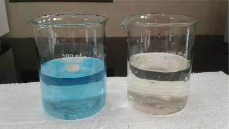
Glycerin. Addition of methylene blue gave a light blue color to the fixative.
This helped to scrutinize the color of fixatives and to shun the inclination
to smell the components.
Multiple human tissue materials from varying sites (skin, viscera,
lymph nodes, bones etc.,) were utilized. Fixation was done in formalin and
new compound fixative. Unfixed raw tissue slices were immediately fixed
in the prepared compound fixative after collecting from MOT and GOT.
Fixation time was titrated for both formalin & the new compound fixative.
NEW COMPOUND FIXATIVE: COMPOSITION9
Formalin–6%
Ethanol - 30%
Glycerin - 5%
Methylene Blue- 0.05%
Buffer:
Sodium dihydrogen phosphate monohydrate - 4g
Anhydrous disodium hydrogen phosphate - 6g
0.7% hypotonic saline was used to make up the volume to 100 ml.
The ph was maintained between 7. 2 - 7.4.
Tissue samples were taken from various specimens and they were
categorized into
Small biopsies
Soft
Intermediate
Hard
Decalcified tissues.
Two representative bits from each specimen was taken. The unfixed
tissues were placed in plastic cassettes. These cassettes were then
immersed in coplin jars, one containing formalin and the other with new
compound fixative.
Fixation of tissues in Formalin & new compound fixative solutions
PROTOCOL FOR TISSUE PROCESSING:30
The tissues were washed thoroughly in water.
Methanol - 7 Minutes at 40˚c
Methanol - 7 Minutes at 40˚c
Isopropyl Alcohol - 8 Minutes at 40˚c
Isopropyl Alcohol - 8 Minutes at 40˚c
Xylene - 10 Minutes
Wax I - 30 Minutes at 75˚c
Wax II -30 Minutes at 75˚c.
In our study we used xylene for better clearing which is not routinely
used in microwave processing. Embedding of processed tissues were done
using paraffin wax and 4 micron thickness sections were taken by means of
a microtome. Staining was done by routine hematoxylin and eosin staining.
STAINING PROCEDURE:
HEMATOXYLIN SOLUTION PREPARATION:5
Hematoxylin - 2.5 g
Potassium alum - 50 g
Sodium iodate - 0.5 g
Absolute ethanol - 25 cc
Glacial acetic acid - 20 cc
Hematoxylin is initially dissolved in absolute alcohol, and then alum
is added to it. The alum ought to be primarily dissolved in warm distilled
water. The mixture is allowed to boil followed by careful addition of
sodium iodate. The stain is quickly cooled with subsequent addition of
acetic acid and the resultant stain can be used immediately.
EOSIN PREPARATION5
Eosin Y - 1 g
95% ethanol - 80 cc
Glacial acetic acid - 0.2 cc
Distilled water - 20 cc
Eosin Y is first dissolved in distilled water followed by subsequent
addition of 95% ethanol and glacial acetic acid.
STAINING:
1. Xylene 3 changes–2 minutes each.
2. Graded alcohols (90%, 80%, 70%)–10 dips each.
3. Expose sections to water.
4. Harris hematoxylin–15 minutes.
5. Wash in clean water.
6. Differentiate by 1% acid alcohol.
7. Wash in clean water.
8. Bluing using 0.5% lithium carbonate.
10.Eosin–15 seconds to 2 minutes based on the age of eosin.
11.Wash in clean water.
12.Dehydration in absolute alcohol - two changes,10 dips each.
13.Xylene–two changes, 10 dips each.
14.Mount the slides using DPX mountant.
Stained slides were observed using light microscope and
analysed by two independent pathologists. Cellular Architecture,
Cytoplasmic, Nuclear details Staining characteristics and Fixation artifacts
were studied.
A scoring system based on the Nuclear, cytoplasmic and
architectural features were followed. Score 3 was given to nuclear,
cytoplasmic and architectural features of all tissues fixed in new
compound fixative that are comparable to those fixed in 10% NBF.
NUCLEAR FEATURES:
The following nuclear parameters were assessed:
Preservation of nucleus and nucleolus
Nuclear size
Regularity of nuclear membrane
Score 3 was assigned to sections fixed in new compound fixative
with comparable nuclear parameters to sections that were fixed in 10%
details. Score 1 was assigned to tissues having more than 2 less defined
nuclear features. Score 0 was assigned to sections with poor conservation
of details, not suitable for diagnosis.
CYTOPLASMIC FEATURES:
Assessment was done by considering the following cytoplasmic
features.
Color of cytoplasm
Volume
Nuclear cytoplasmic contrast
Erythrocyte integrity.
Score 3 was assigned to tissues fixed in new compound fixative with
comparable cytoplasmic details to tissues fixed in 10% NBF. Score 2 was
assigned to sections exhibiting cytoplasmic shrinkage. Score 1 was
assigned to sections having greater than two less specific cytoplasmic
details. Score 0 was assigned to sections with poor conservation of features
not suitable for diagnosis.
ARCHITECTURAL FEATURES:
Assessment was based on the following architectural features:
Shrinkage artifacts
Formalin pigments.
Score 3 was assigned to tissues fixed in compound fixatives with
comparable architectural features to tissues that were fixed in conventional
10% NBF. Score 2 was assigned to sections having one to two ill defined
architectural features. Score 1 was assigned to sections having more than
two ill defined architectural details. Score 0 was assigned to sections with
poor conservation of features, not suitable for diagnosis.
The microwave processed tissues fixed in new compound fixative
was evaluated and compared with formalin fixation. Tabulation of results
were done. Chisquare test, Z score test and Fisher exact test were used for
statistical analysis of differences between formalin fixed tissues and
specimens fixed in new compound fixative. p -values less than 0.05 were
OBSERVATION AND RESULTS
A total of 100 specimens were included in this study. Tissues from
various systems were taken into consideration in this study. They were
categorized into Soft, Intermediate, Hard, Small biopsy and Decalcified
tissue. One bit was placed in formalin and another in new compound
fixative.
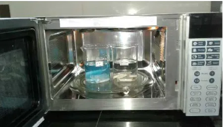
A domestic microwave was used. Initially, the tissues were fixed at
60˚c for 90 seconds in 2 pulses. Mild boiling of tissues was observed.
Alcoholic fumes were present. Hence fixation temperature was titrated to
[image:67.595.97.526.491.695.2]50˚c for 90 seconds in 2 pulses.
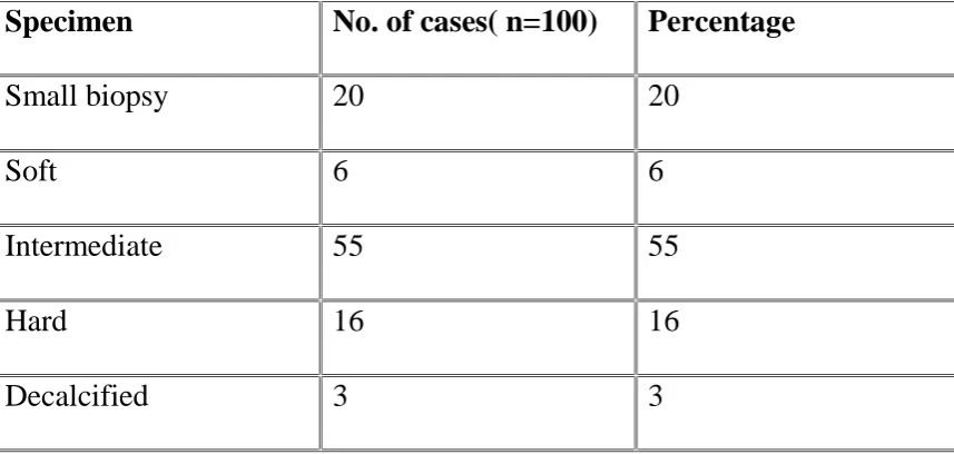
TABLE 1: CATEGORY OF SPECIMENS
Specimen No. of cases( n=100) Percentage
Small biopsy 20 20
Soft 6 6
Intermediate 55 55
Hard 16 16
CHART 1: CATEGORY OF SPECIMENS
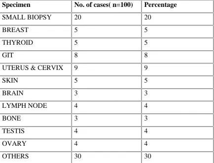
TABLE 2 : DISTRIBUTION OF SPECIMENS
Specimen No. of cases( n=100) Percentage
SMALL BIOPSY 20 20
BREAST 5 5
THYROID 5 5
GIT 8 8
UTERUS & CERVIX 9 9
SKIN 5 5
BRAIN 3 3
LYMPH NODE 4 4
BONE 3 3
TESTIS 4 4
OVARY 4 4
OTHERS 30 30
20
6
55 16 3
CATEGORY OF SPECIMENS
small biopsy soft
intermediate hard
CHART 2: DISTRIBUTION OF SPECIMENS
GROSS APPEARANCE:
Consistency of tissues after fixation in new compound fixative was same as
tissues fixed in 10% NBF.
No cutting and sectioning difficulties were encountered.
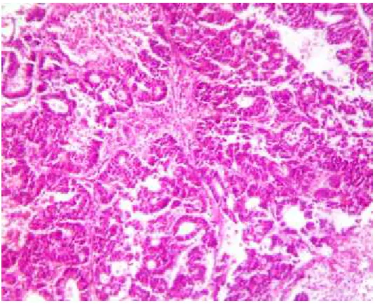
ANALYSIS OF HISTOMORPHOLOGICAL CHARACTERISTICS: Stained slides were analysed by two independent pathologists
using light microscope. Cellular Architecture, Cytoplasmic, Nuclear
details, Staining characteristics and Fixation artifacts were studied. 0
5 10 15 20 25
COMPARISON OF NUCLEAR CHARACTERISTICS: The following nuclear parameters were assessed:
Nuclear size
Preservation of nucleus and nucleolus
Regularity of nuclear membrane
Score 3 was assigned to sections fixed in new compound fixative
with comparable nuclear parameters to sections that were fixed in 10%
NBF. Score 2 was assigned to tissues having 1 to 2 less defined nuclear
details. Score 1 was assigned to tissues having more than 2 less defined
nuclear features. Score 0 was assigned to sections with poor conservation
of details, not suitable for diagnosis.
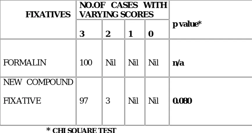
COMPARING NUCLEAR FEATURES OF NEW COMPOUND FIXATIVE AND 10% NBF:
Nuclear features of tissues fixed in new compound fixative were compared
with formalin fixed tissues, both being subjected to microwave fixation and
processing. Out of the 100 specimens, 97 specimens presented with nuclear
features comparable to formalin and scored 3. Three of the specimens
especially the decalcified tissues showed nuclear shrinkage compared to
CHART 3: NUCLEAR FEATURES OF NEW COMPOUND FIXATIVE VS 10% NBF
0 20 40 60 80 100 120
FORMALIN NEW COMPOUND FIXATIVE
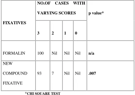
TABLE 3: COMPARING NUCLEAR FEATURES OF NEW COMPOUND FIXATIVE AND 10% NBF:
FIXATIVES
NO.OF CASES WITH VARYING SCORES
p value*
3 2 1 0
FORMALIN 100 Nil Nil Nil n/a
NEW COMPOUND
FIXATIVE 97 3 Nil Nil 0.080
*CHI SQUARE TEST
The above table shows that the p value is 0.080, which is not
significant, thereby showing that there is no significant difference between
10% NBF and New compound fixative. Hence the new compound fixative
is equivalent to 10% NBF in preserving nuclear features.
COMPARISON OF CYTOPLASMIC FEATURES:
Cytoplasmic features that were assessed:
Color
Volume
Nuclear cytoplasmic contrast
[image:72.595.94.528.147.377.2]Score 3 was assigned to cytoplasmic features of sections fixed in new
compound fixative that were comparable to those fixed in 10% NBF.
Score 2 was assigned to sections with cytoplasmic shrinkage. Score 1 was
assigned to tissues having more than 2 less defined characteristics. Score 0
was assigned to sections with poor conservation of details, not suitable for
diagnosis.
COMPARISON OF CYTOPLASMIC CHARACTERISTICS OF NEW COMPOUND FIXATIVE AND 10% NBF:
In this chart and table, cytoplasmic features were compared between
new compound fixative and formalin. Of the total specimens fixed in new
compound fixative, 97 have been assigned score 3 as they exhibited
features akin to sections fixed in 10% NBF. Remaining three cases were
assigned score 2 as they exhibited cytoplasmic shrinkage and poor nuclear
CHART 4: CYTOPLASMIC FEATURES OF NEW COMPOUND FIXATIVE VS 10% NBF
0 20 40 60 80 100 120
FORMALIN NEW COMPOUND FIXATIVE