A Dissertation on
ANTI- HELICOBACTER PYLORI THERAPY RESPONSE IN
EROSIVE AND NON EROSIVE GASTRITIS
Dissertation Submitted to
THE TAMILNADU DR. M.G.R. MEDICAL UNIVERSITY
CHENNAI – 600 032
With partial fulfillment of the regulations for the award of the degree of
M.D. GENERAL MEDICINE
BRANCH – I
COIMBATORE MEDICAL COLLEGE COIMBATORE
DECLARATION
I solemnly declare that this dissertation entitled “ANTI- HELICOBACTER PYLORI THERAPY RESPONSE IN EROSIVE AND
NON EROSIVE GASTRITIS” was done by me at Coimbatore Medical College and Hospital during the academic year 2013-2016 under the guidance and supervision of Prof. Dr. M. RAVEENDRAN M. D. This dissertation is submitted to The Tamil Nadu Dr. M. G. R. Medical University, towards the fulfillment of requirement for the award of M.D. Degree in General Medicine (Branch -I)
Place: Coimbatore
Date:
ACKNOWLEDGEMENT
At the outset, I would like to express my sincere gratitude to our Dean,
Prof. Dr. EDWIN JOE, M.D., for his permission to conduct this study in Coimbatore Medical College.
I’m immensely grateful and indebted to my Prof. Dr. KUMAR NATARAJAN M.D., Professor and Head, Department of Medicine, Coimbatore Medical College.
It gives me immense pleasure to express my sincere and deep gratitude to my guide, Prof. Dr. M. RAVEENDRAN M.D., Professor, Department of General Medicine, Coimbatore Medical College, for his guidance and constant encouragement throughout the study.
It is with deepest sense of gratitude and respect that I would like to thank my co- guide, Prof. Dr. S. RAJA M.D., D.M., Assistant Professor, Department of Gastro enterology, Coimbatore Medical College, for his constant support, guidance, invaluable suggestions and help that he has rendered through out the study.
I would like to acknowledge with gratitude, the help provided by our professors Dr Usha M.D. and Dr. Sivakumar M.D., our unit Assistant Professors - Dr. Geetha M.D, Dr. Sivakumar M.D. and Dr Akila M.D. I would like to thank them for their valuable suggestions throughout the study. I’m indebted to them for being a constant source of inspiration.
study and helping me with the study results, without which I would not have completed my study.
I would also like to thank Mr. Jayakumar, Lab assistant cum statistician , Department of Biochemistry,Trivandrum Medical College, Trivandrum ,Kerala in helping me with the statistical analysis for the study. I would always like to remember with extreme sense of thankfulness for the cooperation and criticism from my fellow post- graduates, my dear seniors and juniors .
I would like to thank my wife, Dr. Rashma Mohammed P for her constant support. I would like to take this opportunity to show my gratitude to my father,
Dr Humayoon Kabir S, my mother Dr. M. Saboora Beegum, my father in law
Dr. P. A Mohammed Kunju, my mother in law Mrs Shameema and my brother
Mohammed Samir H and daughter Imana Ahamed for their never ending support and prayers.
I’m ever grateful to the ALMIGHTY GOD for showering his blessings on me and my family and I pray Almighty God to give me the strength to achieve all my endeavors.
ABBREVIATIONS
GIT - Gastro intestinal Tract
OGD - Oesophago gastro duodenoscopy APCGH – Asia Pacific Consensus Guidelines APD – Acid Peptic Disease
RUT – Rapid Urease Test UBT - Urease Breath test
OLGA - Operative Link for Gastritis assessment PPI - Proton Pump Inhibitors
H2 blocker - Type 2 Histamine receptor blockers ITP - Idiopathic thrombocytopenic pupura GERD – Gastro Esophageal Reflux disease RF – Rheumatoid factor
ESR – Erthrocyte sedimentation rate CRP – C reactive protein
HBV – Hepatitis B virus HCV – Hepatitis C virus
WBC – White blood cell CBC – Complete blood count LFT – Liver function tests RFT – Renal function tests DNA – Deoxy ribonucleic acid OPD – Out patient department
CLO - Campylobacter – like organism MALT - Mucosa Associated lymphoid Tissue Hcp - Helicobacter cysteine rich proteins Vac A - Vacoulating cytotoxin A
Cag A - Cytotoxin associated gene A SES - Socioeconomic Status
LIST OF TABLES
S. No. Details of the tables Page No.
1 Socioeconomic status distribution 41 2 Table showing relationship between RUT &
gastritis
46
3 Follow up patients & the gastritis type they belonged
49
4 Endoscopic Findings after treatment 51 5 Clinical symptoms post treatment 54 6 Comparison of endoscopic finding pre and post
treatment
55
[image:9.612.139.534.121.460.2]LIST OF FIGURES
S. No. Details of the figures Page No.
1 Helicobacter pylori with its lophotrichous flagellae 6 2 Helicobacter pylori as seen in scanning electron microscope 7 3 Mechanism of H.pylori infection 9 4 Types and progression of H.pylori infection 10 5 Flowchart of sequence of infection 11 6 Immunohistochemical staining of H.pylori gastritis biopsy 15
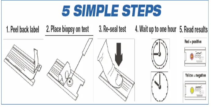
7 Rapid Urease Test –mechanism 20
8 Steps of doing Rapid Urease Test 21
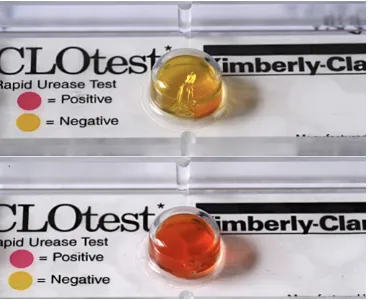
9 Rapid Urease test results 22
10 Endoscopic findings 31
CONTENTS
S. No. Content Page No.
1. INTRODUCTION 1
2. AIMS AND OBJECTIVES 4
3. REVIEW OF LITERATURE 5
4. MATERIALS AND METHODS 34
5. RESULTS 38
6. DISCUSSION 58
7. SUMMARY 71
8. CONCLUSION 74
9. REFERENCES 10. ANNEXURES
INTRODUCTION
Helicobacter pylori is a gram negative micro-aerophilic bacteria found
in the stomach which is one of the human infections with a global coverage.
It has been reported that about 50% of the total human population harbors
this organism.[1] First discovered in 1982 by Australian scientists Barry
Marshall and Robin Warren, after which the organism has been postulated to
be a cause of very many diseases related to the gastrointestinal tract and has
revolutionized the field of gastroenterology.[2] Even though the prevalence is
worldwide the infections due to Helicobacter pylori vary in different
countries and even in different regions within a country. But a definite
increased prevalence in the developing countries is proven [1].
Helicobacter pylori infection is a proven etiology for peptic ulcer
disease and gastritis[3].Infection if persisting was found to be a factor of
definite risk for adenocarcinoma of stomach and MALToma (mucosa
associated lymphoma). Studies have shown that chronic gastritis and
duodenal ulcer has an association of 100% compared to 50% in the controls
who did not have ulcer [4]. Increased association of Helicobacter pylori was
dependent on the type of gastritis and also depended on site of infection as
shown by a prevalence of 57% in erosive gastritis, 28.7% in superficial
and 80% in case of gastric ulcer [5]. Antral infection was associated with
increased severity in gastritis [6,7].
For research purpose modified Sydney system of classification is used
wherein features as seen in endoscopy of gastric mucosa like erythema or
exudation, erosion, mosaic pattern or cobble stone appearance, hypertrophic
rugae, nodular and atrophic appearance are considered as abnormal. Erosive
gastritis has been defined by white base lesions, either raised or flat,
surrounded by a margin of intense erythema. Similarly unequivocal erythema
or exudation, mosaic pattern, hypertrophic rugae , nodular and atrophic
appearance in endoscopy are features suggestive of non erosive gastritis [8].
Non invasive methods of detection of Helicobacter pylori is with
blood antibody test, carbon urea breath test, stool antigen test, urine ELISA
test etc. Invasive methods include endoscopic biopsy and histological
examination .When this is combined with either microbial culture or rapid
urease test it is considered as one of the most reliable detection method [9].
The Rapid urease test detects Helicobacter pylori infection within an hour
with an accuracy of 90% and is widely accepted to initiate the eradication
The determinants of infection particularly the socioeconomic standard
of living and heterogeneity of infection even within a country and with
different risk groups affects the success of a given therapeutic regimen.
Increased rate of resistance to organisms and difference or even failure to
respond pointed out the need to have a specific therapy regimen tailored for a
particular region within a state or a country[2].
There is a requirement for prospective epidemiological data of high
quality as emphasized in second Asia Pacific consensus guidelines for
Helicobacter pylori infection(APCGH) especially from India[2]. This study
aims at finding the relation of Helicobacter pylori infection with erosive and
non erosive gastritis, also to evaluate the effects of the treatment with a
fourteen day regimen of Amoxicillin, Metronidazole and Proton pump
AIMS & OBJECTIVES
1. To find out the relationship of Helicobacter pyloriinfection with erosive and
non-erosive gastritis.
2. Effect of Anti Helicobacter pylori therapy on both types of gastritis.
3. To compare the effects of Anti Helicobacter pylori therapy on erosive and
REVIEW OF LITERATURE
Helicobacter pylori was linked to chronic gastritis and gastric ulcer
following its discovery 1982.Previously they were not believed to be from a
microbial cause. But since then many of the gastroenterological disorders
were postulated to be caused by this organism. Even extra-gastrointestinal
diseases have been researched for Helicobacter pylori as their cause. The
organism is thought to be an essential component of the natural ecology of
stomach. Even when more than half of the population harbours this organism
only 20% of the individuals are causing disease or infection.
Helicobacter pylori though global, its more in the developing than in
the developed countries. The sole source of the organism is the human gastric
mucosa. The exact mechanism of transmission is not clear but it has been
postulated to be or -oral or feco-oral .Poverty, over crowding and poor
hygiene favours transmission and thus explains its increased prevalence in
the developing countries.
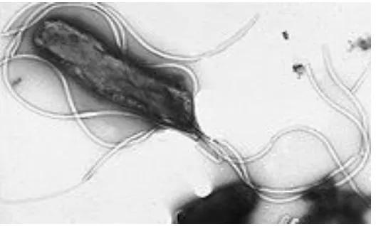
Helicobacter pylori is a helical gram negative microaerophilic
bacteria about 3 to 4 micrometres long and 0.5 micrometer in diameter .It
hydrogen with the help of hydrgenases. It has ability to from biofilms which
help to avoid the acidic environment and various adaptations which adds to
[image:17.612.171.435.193.352.2]its survival, epidemiology and pathogenicity.
Fig 1. Helicobacter pylori with its lophotrichous flagellae
The bacterial structure consists of proteins like adhesions, porins, iron
transporters, flagellum associated proteins. Being a gram negative organism
they are composed of phospholipids and lipopolysaccharides. They have
unipolar tuft of about four to six lophotrichous flagella and thus makes these
organisms highly motile. Helicobacter pylori shows considerable genetic
diversity as evident in its molecular typing. The complete genome of the
bacterium has been mapped. The scientists have identified a 40 kilo base pair
long Cag (cytotoxicity associated gene) pathogenicity island which has about
40 genes responsible for the pathogenicity of Helicobacter pylori. These
asymptomatic individuals. Virulence of Helicobacter pylori has been
associated with certain alleles in genes like vac( vacoulating cytotoxin gene)
in addition to the cag gene .
The genome study of Helicobacter pylori is under progress with
increasing emphasis attempts to understand its pathogenicity and the various
diseases for which it has been postulated as the cause.
PATHOPHYSIOLOGY
This bacteria belonging to phylum -Proteeobacteria, order-
Campylobacterales and family Helicobacteraceae has a large diversity of
strains. They have to tide over the extremely acidic environment of the
gastrointestinal tract. It has various adaptations to avoid the acidic
environment and burrows beneath the gastric mucosal lining where there is a
neutral pH with the help of its lophotrichous flagella. They adhere to the cells
with adhesins. By means of chemotaxis they avoid areas of acidic pH and
they produce increased amounts of enzyme urease, which will break down
urea to ammonia and carbon dioxide ,they neutralize the acidic environment.
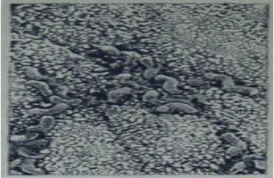
The ammonia being basic neutralizes the acid in the gastric mucosal lining of
Fig 2. Helicobacter pylori as seen in scanning electron microscope
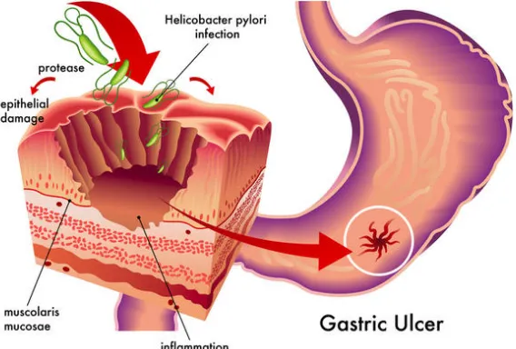
Certain subtle changes occur in the gastric mucosal lining due to
several mechanisms during an infection. Firstly ammonia which is produced
to neutralize the pH is toxic to the mucosal layer. Secondly proteases,
vacoulating cytotoxin A (VacA) and phospholipases. Thirdly cytotoxin
associated gene (CagA) causes inflammation and is potentially a carcinogen.
These changes in the mucosa are used to predict the presence of the
organism. The strains of Helicobacter pylori that produces high levels of
VacA and CagA causes greater tissue damage and is evidenced in the
Fig 3 : Mechanism of H.pylori infection
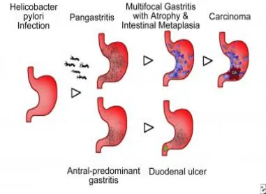
Colonization of the gastric mucosa results in chronic gastritis. The
Helicobacter cysteine rich (Hcp) proteins are a trigger to an immune response
leading to inflammation. Thus ulcers of stomach and duodenum develops and
the breach in gastric mucosal barrier will result in worsening of the
symptoms .Further the G cells in the antrum produce gastrin in response to
the inflammatory response which results in stimulation of the parietal cells
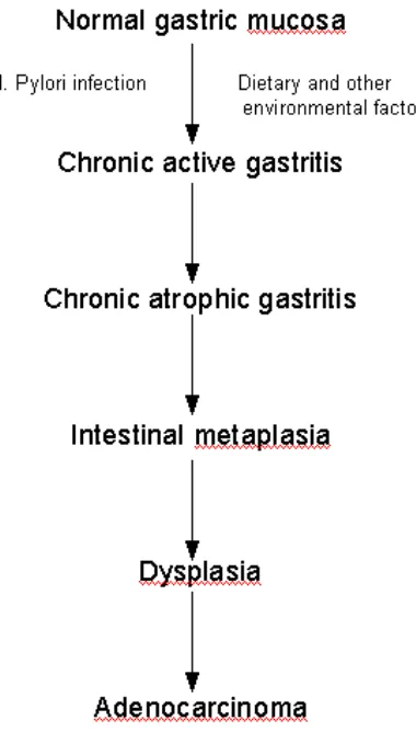
Fig 5: Flowchart showing sequence of development of cancer
Persistent inflammatory response induced by the bacteria will result in
the atrophy of gastric mucosal lining and will result in gastric
adenocarcinoma. The Mucosa associated lymphoid tissue(MALT)
lymphomas are antigen driven and regress with elimination of the
organism.Two mechanisms have been proposed by which the organism could
cause cancer .It may involve the increased production of free radicals which
results in increased rate of mutations to the host cells in the vicinity of
adhesion proteins resulting in the enhancement of the transformed cell
phenotype and this has been termed as “perigenetic pathway”.
SIGNS AND SYMPTOMATOLOGY
About 80% of the population infected with Helicobacter pylori is
asymptomatic. Acute gastritis with abdominal pain or nausea may be the
presenting feature[11]. When the disease progresses and develops chronic
gastritis it causes severe abdominal pain, bloating, belching and vomiting.
Dyspepsia which is defined as an abdominal or retrosternal chest pain is a
major health problem. Gastroesophageal reflux disease(GERD) ,peptic ulcer
disease and gastric carcinoma are usually associated with dyspepsia .Most
common type of dyspepsia is functional dyspepsia and is defined as
persistent abdominal and retrosternal discomfort in whom reasonable clinical
evaluation has failed to reveal a definite cause for the symptoms [12,13].
Helicobacter pylori is proven to play a significant role in causing symptoms
Association of the organism in gastropathy associated with analgesics
like NSAIDS( Non steroidal anti inflammatory drugs), gastroesophageal
reflux disease and functional dypepsia are under research with the completed
studies have proven the association [2] . Significant association with Portal
hypertensive gastropathy in cirrhotic patients and its severity has been
established7. Even association with extra-gastrointestinal disease has been
established as in case of skin diseases like chronic urticaria, rosacea,
psoriasis, Sjogrens syndrome lichen planus , behcets disease , systemic
sclerosis etc [2,3] . Rarely, symptoms of all these diseases can occur in case of
DIAGNOSIS
Testing for infection with Helicobacter pylori is recommended in case
of peptic ulcer disease, a suspicion of low grade MALT lymphoma, after
endoscopic resection of gastric carcinoma, in 1st degree relatives with gastric
cancer patients. There are numerous tests available to detection of the
organism. All the diagnostic tests available can be classified to two types
either, invasive or non invasive [14].
Invasive tests : They involve the endoscopic biopsy of the gastric
mucosa and examination of the sample with culture ,microscopy and urease
tests. Microscopy of biopsy sections by silver staining or gram staining is
useful. Other staining methods used are with Giemsa ,Haematoxylin –eosin
,acridine orange and even the use of a phase contrast microscopy. Culture is
more sensitive but requires expertise and takes nearly three to seven days.
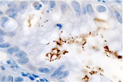
FIG 6 . IMMUNOHISTOCHEMICAL STAINING OF H.PYLORI
GASTRITIS BIOPSY SPECIMEN
Non invasive tests include serology with ELISA and urease breath
test. The urease breath test is sensitive and reliable but needs better isotope
The Second Asia- Pacific Consensus Guidelines for Helicobacter
pylori infection (APCGH) has stated that when there is no need for an
endoscopy ,C14 urease breath test is approved as an accurate non invasive
test for the assessment of results and initial diagnosis of the anti Helicobacter
pylori eradication treatment. But the test shows greater variability because of
the non uniformity of the test parameters used in the various labs [2].C13 urea
breath test is superior in this aspect. Breath tests had shown consistent high
diagnostic accuracy among the non invasive tests and results were almost
comparable with invasive tests like the biopsy based tests.
APCGH had recommended for outcome assessment after eradication
treatment as there was high failure rate among significant proportion of
population in terms of achieving eradication. Persistent undetected infection
with Helicobacter pylori makes the patient prone to complications of
infection .The importance of determining outcome will depend on the
indication of treatment. Discordant opinion regarding need for assessment
was there in the consensus but since the practice varies between countries it
was agreed to customize according to that particular region. Re assessment of
the infected patients should be done in 4 weeks following the completion of
eradication treatment .It was suggested to withhold the proton pump
Least accurate diagnostic tests are the serological tests for
Helicobacter pylori infection and are not that useful in determining the
outcome of therapy as stated in Statement 14 of APCGH15. In case of
bleeding patients the biopsy based tests may give a false negative value and
in that case serological tests plays an important role. Also with the use of
proton pump inhibitors and antibiotic use there is an increased false negative
rate in biopsy, stool antigen and breath tests and in this scenario serological
tests were proven to be useful.
Statement 3 of APCGH had emphasized that in Helicobacter pylori
infective patients who have uninvestigated dyspepsia without alarm features
the appropriate strategy would be to “Test and Treat”. The statement 4
recommend that infection in case of Gastroesophageal reflux disease it is not
recommended to test and treat the patient. But in case of an erosive
esophagitis which was endoscopically proven it is advisable give the therapy
because in Asia, peptic ulcer disease and gastric carcinoma are more
common and there will be commensurately greater benefit. Section 6 of
APCGH states that to screen and treat the Helicobacter pylori infection in
populations with higher incidence of gastric carcinoma. This would
Prospective evidence of decrease in cancer was available in an eight year
prospective study from China. The study had revealed that eradication in
those who are in early stages and had not developed intestinal metaplasia
and also patients with gastric atrophy had lower rate of gastric cancer when
compared to those who were not on eradication therapy.
In the consensus there was agreement over the value of screen and treat
strategy even in populations with low prevelance based on the new data
which was available from various studies done in the last ten years. Also
recent data from intervention studies showed a lesser risk for metachronous
gastric cancer post eradication treatment and have thus emphasized the
value of therapy in secondary prevention.
Thus even when the non invasive tests are considered to be better than
the invasive tests according to the consensus guidelines, various factors taken
into consideration proves the superiority of the invasive tests. Firstly the cost
of non invasive tests like urea breath test limits its use in the developing
countries .Secondly ,the use of invasive methods gives the added benefit of
This is useful as more than giving the idea of the site of ulceration and
classification of the gastritis as erosive and non erosive ,it helps in
identifying any gastric atrophy or features suggestive of early gastric cancer .
Thirdly the time required for diagnosis is less as rapid urease test just takes
minutes to prove the presence of an infection. In case of severe infection the
result is conclusive with in seconds but the non invasive tests takes days for
RAPID UREASE TEST
Rapid urease test is synonymous with CLO test ( Campylobacter –
like organism test) and has a high sensitivity and specificity and accurately
(>90%) diagnose the infection .But active intestinal bleeding reduces the
accuracy.
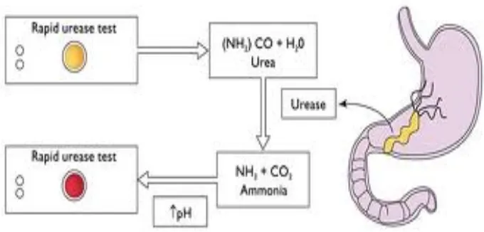
[image:31.612.166.506.303.466.2]Principle of Rapid urease test
Fig 7 : Rapid Urease Test -mechanism
The diagram given below illustrates the steps to be taken for doinga
Rapid Urease test(RUT).After endoscopically obtaining the sample from
Fig 8: Steps of doing Rapid Urease Test
The interpretation is based on the colour which turns pink in the
presence of the urease enzyme and in turn detects the Helicobacter pylori in
TREATMENT
Various permutations and combinations of antibiotics, proton pump
inhibitors along with other drugs like bismuth sulphate are used as therapy.
Success of therapy depends primarily on the antibiotic sensitivity.
Controversies over the use of various regimens have been addressed in
various guidelines and consensus statements world wide like North America,
Europe and the two editions of Asia –pacific consensus10.
Before going into the treatment we will look upon some details which
came in the 2nd Asia pacific consensus guidelines regarding the indications
for treatment.
Indications for treatment
Gastric ulcer disease (A)
MALT Lymphoma (A)
Atrophic gastritis (B)
gastric cancer-post resection (B)
Patients’ choice (after consultation with their personal physician) (A)
Non-ulcer dyspepsia (A)
To decrease the risk of peptic ulcer and upper gastrointestinal bleeding in
non-steroidal anti-inflammatory drug(NSAID) users (A)
Before starting long-term aspirin therapy for patients with higher risk for
ulceration and ulcer related complications (B)
Patients on long-term low-dose aspirin therapy and who have a past history
of upper gastrointestinal bleeding and perforation (B)
GERD patients requiring long-term proton pump inhibitor (B)
As a strategy for prevention of gastric cancer in communities with higher
incidence of gastric carcinoma (A)
Unexplained iron-deficiency anemia, or ITP(idiopathic thrombocytopenic
purpura) (C)
Statement 15 of APCGH states that in asia the currently recommended
first line therapy for Helicobacter pylori is the use of a protonpump inhibitor
with Amoxicillin and clarithromycin for a duration of seven days.
Metronidazole has been considered as an acceptable alternative to
amoxicillin or clarithromycin in the triple therapy regimens. In Asia where
Amoxicillin is preferred metronidazole is less used.
In case of penicillin allergy metronidazole is considered the first line
substitute .Another option is to start on bismuth based quadruple therapy as
first line. As mentioned earlier various options can be worked out and
selected as regimen to that particular area based on certain characteristics
pertaining to the population in that area.
The various treatment regimens in treatment are discussed further
Treatment regimens for Helicobacter pylori as in 2nd APCGH
Triple therapy (Standard proton pump inhibitor therapy- (PPI)based):for 7– 14 days
2. Metronidazole 400 mg, clarithromycin 500 mg with PPI twice daily
3. Amoxicillin 1 g, metronidazole 400 mg with PPI twice daily
Quadruple therapy for 7–14 days
Bismuth 240 mg twice daily, metronidazole 400 mg twice daily or three
times daily, tetracycline 500 mg four times daily and PPI twice daily
Levofloxacin-based triple therapy for 10 days
levofloxacin 250 mg (or 500 mg), amoxicillin 1 g with PPI twice daily
Rifabutin-based triple therapy for 7–10 days
PPI, rifabutin 150 mg, amoxicillin 1 g twice daily
The second Asia Pacific consensus in its statement 15 had recommended first
line therapy with Proton Pump Inhibitor(PPI) ,Amoxycillin and
clarithromycin for 7 days10. Metronidazole was mentioned as an acceptable
alternative to amoxicillin or clarithromycin.Further statements 16 and 17 in
the consensus had highlighted about clarithromycin resistance and the
fourteen day therapy10. Although the statement 17 states that the fourteen day
therapy gives only limited advantage over seven day triple therapy regimen
Asian studies and none were Indian. A 5% increase in eradication rate with
fourteen day regimen was addressed10. Salvage therapies in case of resistance
were included in the consensus.
Section 20 of the 2nd APCGH has recommended various choices for
salvage therapy:
1. Triple therapy which has not been earlier used in the patient
2. Bismuth based quadruple therapy
3. Levofloxacin based triple therapy
4. Rifabutin based triple therapy
Quadruple therapy as a salvage therapy is time tested and experience
with it is more than for other salvage therapies which has been used
worldwide . The determination of use of salvage therapy depends on
various factors like antibiotic resistance in the given area,, previous
treatment, drug availability and suggested to depend on the local prevalence
of tuberculosis in view of the rifabutin use. For example ,levofloxacin-based
triple therapy may be an excellent second line treatment in areas with low
levofloxacin resistance. Rifabutin is considered only in tuberculosis low
GASTRITIS
Gastritis is defined as the inflammation of the mucosal lining of the
gastric region. There are very many causes leading to gastritis ,one of the
most frequent causes is Helicobacter pylori infection and use of analgesics. Other causes include alcohol, smoking, drug abuse, critical illness,
autoimmune conditions, radiation etc.
Majority of the population affected by gastritis are asymptomatic. But if
symptomatic, abdominal pain is the most common presentation. It is
described as a dull, vague, burning, aching, sore, or sharp type of pain which
is localized to the upper central portion ,[11]but can be diffuse affecting any
part of the abdomen or even around to the back.
Signs and symptoms associated with gastritis are listed below:
• Nausea
• Vomiting
• Bloating sensation
• Early satiety
• Unexplained weight loss
Classification of gastritis was first put forward by Whitehead in 1972
based of the mucosa type and severity of disease activity .This was followed
by Strickland –Mackay classification and later the Glass classification. In
1990 Sydney classification of gastritis was approved .This classification was
based on endoscopic findings, etiology ,site of ulceration etc
Based on the endoscopic findings it was classified into:
1. erythematous or exudative type
2. superficially erosive
3. polypoid gastritis with erosions
4. atrophic
5. hemorrhagic gastritis
6. bile gastritis
Classification according to etiology
1. Autoimmune gastritis (type A)
2. Bacteria related gastritis (type B)
3. induced by Chemotoxic agents (type C)
4. distinct forms of gastritis
Other histological classifications are the Houston variation of Sydney system
and the Operative Link for Gastritis assessment (OLGA) system which came
in 2005.
Modified Sydney system of classification is where in endoscopic
features of gastric mucosa like erythema or exudation, mosaic pattern or
cobble stoning, erosion ,hypertrophic rugae, nodular and atrophic
appearance are considered as abnormal and are used to classify gastritis It
came into existence in 1996 and classifies gastritis into erosive and non
Erosive gastritis has been defined by raised or flat ,white base lesions, with
intense erythema surrounding the margin. Similarly unequivocal erythema or
exudation, hypertrophic rugae , mosaic pattern, atrophic or nodular appearance in
[image:43.612.129.514.257.552.2]endoscopy are suggestive features of non erosive gastritis.
MATERIALS AND METHODOLOGY
PLACE OF THE STUDY
This study undertaken on the outpatients in the Gastroenterology
department of Coimbatore Medical College Hospital ,Coimbatore.
PERIOD OF STUDY
July 2014 to July 2015.
DESIGN OF STUDY
METHODOLOGY
Patients aged between fifteen to sixty years having dyspeptic
symptoms and willing to undergo for upper gastrointestinal endoscopy and
anti Helicobacter pylori treatment were enrolled in this study. Every ethical
issues which could arise were discussed with and informed written consent
was taken from all patients.
After taking a detailed history and physical examination, patients were
submitted to upper Gastrointestinal endoscopy and Rapid Urease Test(RUT)
was done with one of the specimens taken from the predominant site of
gastritis. Patients who were on Proton pump inhibitor or H2 blocker therapy
were taken for endoscopy only after stopping these drugs for atleast 2 weeks.
Repeat testing for checking the eradication was done four weeks after the
completion of anti-H. pylori therapy.
Anti-H. Pylori therapy consisting of Metronidazole (500 mg bd) and
Amoxycillin (1 gm) bd with omeprazole (20 mg bd)) for 14 days was used .
Follow-up visits were made for assessment of compliance and side effects.
After completion of therapy, clinical history was again taken and compared
against the pretreatment symptoms. Frequency of infection to be calculated
among endoscopically proven gastritis patients.
Follow up visits were arranged for increasing compliance and for
identifying any side effects. Follow up endoscopy was performed 4 weeks
after completion of the therapy. Biopsy specimens were collected from
antrum of stomach for the rapid urease test.
SELECTION CRITERIA
(a) INCLUSION CRITERIA
• Adults aged between 15 to 60 years having symptoms of dyspepsia.
• Those willing to undergo upper gastrointestinal endoscopy and anti-
Helicobacter pylori therapy.
(b) EXCLUSION CRITERIA
Patients who were regular users of NSAID and steroids, had peptic
ulcer and its complications.
Patients with history of previous Helicobacter pylori eradication
therapy.
Patients with coexisting gastric cancer, pregnancy or lactation and
concomitant other severe diseases.
Any acute bleeding episodes
Data consists of primary data collected by the principal investigator
directly from the patients who had approached Government Medical College
Hospital,Coimbatore. The subjects consists of outpatients attending the
Gastroenterology Outpatient Department.
STATISTICAL ANALYSIS
Clinical features, symptoms, endoscopic findings, pre- and post treatment
disease status were compared with the help of. SPSS (Statistical Package for
Social Services) 16.0 for statistical calculations.
• Total of 92 patients were enrolled in the study, who presented with
dyspeptic symptoms and were willing for the study between the period of
July 2004 to July 2015.
• 81 patients out of the 92 enrolled had endoscopically proven gastritis.
• Out of the 81 endoscopically proven gastritis patients, 72 patients were
proven to be infected with helicobacter pylori based on the Rapid Urease
test.
• 72 patients who were H.pylori infected were treated and were followed up
and planned for repeat testing after 4 weeks of therapy.
• Out of the 72 only 48 patients could be successfully called back for a repeat
testing following the therapy .Dropout percentage was 33%.(24 out of 72).
AGE DISTRIBUTION
FIG 12. AGE WISE DISTRIBUTION OF GASTRITIS PATIENTS
o Endoscopically proven gastritis changes in the mucosa were more
in the age middle age group with about 64.2% (52) of the study
population.
o About 1/3rd were of age group above 50 years.
SEX DISTRIBUTION
Male : Female ratio was nearly 3:1
[image:51.612.152.479.216.427.2]58 males(71.6%) : 23 females (28.4%)
FIG 13: PIE CHART SHOWING THE GENDER DISTRIBUTION
SOCIO ECONOMIC STATUS (SES)
Being a tertiary care centre in the government setup we had more than half of
TABLE 1. SOCIOECONOMIC STATUS DISTRIBUTION
SES Frequency Percent
Lower 44 54.3
Lower middle 29 35.8
Upper middle 7 8.6
Upper 1 1.2
Total 81 100.0
FIG14. PIE CHART SHOWING SOCIOECONOMIC STATUS
DISTRIBUTION
Thus lower middle and lower socio economic class had higher prevalence of
gastritis among the study group (90.1%).
This histogram shows the various symptoms and their frequency in gastritis
[image:53.612.145.487.201.405.2]patients.
FIG 15. HISTOGRAM SHOWING FREQUENCY OF THE
PRESENTING SYMPTOMS
• Abdominal pain is the commonest symptom (69%) followed by bloating
sensation(51%).
• As in literature abdominal pain which is due to gastric mucosal damage in
ENDOSCOPIC FINDINGS
FIG 16: PIE CHART SHOWING ENDOSCOPIC FINDING
DISTRIBUTION
About 3/4th of the gastritis patients presented with erosive type of
gastritis which causes severe symptoms and also persistent infection ,if left
untreated leads to the complications.
Out of the 81 endoscopically proven gastritis patient 59 (72.8%)had
SITE OF LESION
FIG 16 . CONE HISTOGRAM SHOWING DISTRIBUTION OF THE
SITE OF GASTRITIS
• Major site of infection leading to gastiritis was at pylorus (77.8%) and least
involved region was the lesser curvature (7.4%).
• About 10 times more prediliction for the pyloric region to be affected than
RAPID UREASE TEST
FIG 17 : PIE CHART SHOWS THE DISTRIBUTION OF THE RAPID
UREASE TEST
• Out of the 81 endoscopically proven gastritis patients 72 (88.9%) had Rapid
urease test positivity .
• Prevalence of Helicobacter pylori infection in endoscopically proven gastritis
patients were as 88.9%.
RUT & TYPE OF GASTRITIS
TABLE 2: TABLE SHOWING RELATIONSHIP BETWEEN RUT &
GASTRITIS
RUT Endoscopic Findings
Erosive Non erosive
N % N %
Positive 56 94.9 16 72.7
Negative 3 5.1 6 27.3
FIG 18: HISTOGRAM SHOWING RELATIONSHIP BETWEEN RUT
& GASTRITIS
df=1 p=0.015
Among the 59 erosive gastritis patients 56 (95%) had H.pylori infection
and in case of the 22 non erosive gastritis patients 16(73%) had H.pylori
infection
Among the 72 H.pylori positive patients 56 (77.7%) had erosive gastritis
and 16 (22.3%) had non erosive gastritis.
erosive
[image:59.612.190.352.167.332.2]non erosive FIG 20: SHOWS RELATIONSHIP BETWEEN H.PYLORI INFECTION
AND TYPE OF GASTRITIS
22.3%
77.7%
FOLLOW UP PATIENTS
• Out of the 72 H.pylori infected patients who were instituted therapy 48 had
came for post therapy testing to assess eradication.
• 37 of the 48 were from the erosive group and 11 were from the non erosive
TABLE 3: FOLLOW UP PATIENTS & THE GASTRITIS TYPE
THEY BELONGED
Follow up patients Frequency Percent
Erosive 37 77.1
Non erosive 11 22.9
FOLLOW UP RAPID UREASE TEST
FIG 21: PIE CHART SHOWING FOLLOW UP RAPID
UREASE TEST
Out of the 48 patients who were followed up in the study 41 became
POST TREATMENT ENDOSCOPIC FINDINGS
TABLE 4: ENDOSCOPIC FINDINGS AFTER TREATMENT
Post treatment findings Frequency Percent
Erosive 5 10.4
Non Erosive 2 4.2
Cured 41 85.4
Total 48 100.0
• Post treatment endoscopic findings revealed a cure rate of 85.4%
(41/48) with about 15% of patients having persistent gastritis.
CLINICAL SYMPTOMS POST TREATMENT
TABLE 5: TABLE SHOWING CLINICAL SYMPTOMS POST
TREATMENT Clinical features Before treatment After treatment
N % Resolved Not resolved
N % N %
Abdomina
l pain
3
6
75.0 2
5 52. 1 1 1 22. 9
Bloating 2
6
54.2 1
8
37.
5
8 16.
7
Early
satiety
1
2
25.0 8 16.
7
4 8.3
Nausea 1
0
20.8 8 16.
7
2 4.2
Anorexia 5 10.4 5 10.
4
0 .0
Vomiting 7 14.6 7 14.
6
0 .0
• There was drastic reduction in the clinical symptoms with abdominal
• Also other symptoms of bloating ,early satiety and nausea showed more than
30% resolution.
• There was total absence of symptoms of anorexia and vomiting in patients
post therapy.
The effective resolution of symptoms in case of Helicobacter pylori infection
in gastritis patients can be better understood with help of this histogram
ENDOSCOPIC RESOLUTION POST THERAPY
TABLE 6. COMPARISON OF ENDOSCOPIC FINDING PRE AND
POST TREATMENT
Post treatment Pre treatment
Endoscopic
findings
Erosive Non Erosive Cured
N % N % N % N %
Erosive 5 100 0 0 32 78 37 77.1
Non
erosive
0 0 2 100 9 22 11 22.9
Total 5 100 2 100 41 100 48 100
• Out of the 37 erosive gastritis patients who had came for followup 32
patients had endoscopic clearance of the lesions following therapy and
only 5 had persistent lesions.Of the 22 non erosive patients only 2 had
persistent non erosive gastritis.
• Thus endoscopic cure rate was 86.4% in case of erosive gastritis and
HELICOBACTER PYLORI ERADICATION
TABLE 7. SHOWING H.PYLORI ERADICATION RATE
& GASTRITIS TYPES
Endoscopic
findings
Follow-up RUT Total
Positive Negative
N % N % N %
Erosive 5 13.5 32 86.5 37 100.0
Non erosive 2 18.2 9 81.8 11 100.0
Total 7 14.6 41 85.4 48 100.0
χ2 = 0.000 df=1 p=1.000
• Eradication rate for both erosive and non erosive gastritis patients
infected with Helicobacter pylori were comparable and p value was not
significant.
• Similar to the endoscopic cure rate ,the eradication rate was also 86.4%
FIG 23: COMPARISON OF FOLLOW UP RUT WITH TYPE OF
GASTRITIS
• This Histogram shows that the eradication rate was equally good in both
DISCUSSION
In our study, a total of 92 patients had fulfilled the inclusion criteria
and were enrolled in the study. These were mainly the dyspeptic patients
who had attended our gastroenterology out patient department and who were
willing for the study which was done between the time period of July 2004
to July 2015.
92 patients
81
81 patients out of the 92 enrolled had endoscopically proven gastritis.
Out of the 81 endoscopically proven gastritis patients, 72 patients were
proven to be infected with helicobacter pylori based on the Rapid Urease test.
Seventy two patients who were Helicobacter pylori infected were
treated and were followed up and planned for repeat testing after 4 weeks of
therapy.
Out of the 72 only 48 patients could be successfully called back for a
repeat testing following the therapy .
Further study and comparison were done with these 48 patients to
come to a conclusion.
Helicobacter pylori eradication in people infected results in
reduction of gastric atrophy and gastritis. Subsequently the malignancy risk
is drastically reduced with even near total cure of the low grade MALT
lymphomas [9] .
AGE DISTRIBUTION
Our study population were mainly from the middle age group who were
about 64.2% (52) of the total study population and one third were of age
group above 50 years. This finding was similar to the world wide studies and
WHO had stated that infection rate was more in middle age groups of 25
to50 years age when compared to other age groups.
SEX
There was a male preponderance in our study group owing to
socio-economic status, literacy, awareness among females and decreased
accessibility to health care as they are decreased to household activities.In
our study there were 58 males(71.6%) and 23 females (28.4%) with a male
SOCIOECONOMIC STATUS
More than half of the study population were from the lower socio
economic strata(54.3%).Socio economic status is significant because there is
increased colonization in the poor socioeconomic status group and those
with lesser education apart from considering the genetic factors in case of
developing and underdeveloped countries.
The results from our study are in agreeing with the proven results of
other studies done around the world which were determined by the place of
the study,socio-economic status as well as the mode of transmission which
causes the spread infection from person to person or by oro-oral or the
SITE OF INFECTION
In most persons, H. pylori infection is gastric antrum is the most affected site. Studies have shown that H. pylori infection occurs in the antrum in about 85% of patients with the disease, and in about 15% of
patients orpus is the most affected site.11,12 Our study found out that nearly
78% of RUT positive gastritis were seen in the pylorus and 11.8% in the
antrum of gastric mucosa.
ENDOSCOPIC FINDINGS BEFORE TREATMENT
Erythema as an endoscopic finding are frequently labeled as gastritis
despite a lack of evidence supporting a correlation between endoscopic
features and histologic gastritis.14
In our study, we found out that 77.7% had erosive gastritis and 22.3%
non-erosive gastirits among the Rapid urease test positive Helicobacter
pylori gastritis. Erosive gastritis has been defined by raised or flat ,white base
lesions, with intense erythema surrounding the margin. Similarly unequivocal
erythema or exudation, hypertrophic rugae , mosaic pattern, atrophic or
gastritis.About 40% or more of patients have a false negative endoscopic
reporting with endoscopically normal mucosa having histological gastritis
visible in a biopsy specimen [18] . Almost always corresponding histologic
inflammatory changes are present when the endoscopic changes are more
pronounced and in case of erosions or frank atrophic gastritis . So
generalizing the concept a more severe the endoscopic gastric defect, the
better the correlation with the histology report from the biopsy[15,16] .
Our study had incorporated the Sydney system- endoscopic
appearance of gastritis for diagnosis of gastritis without taking into
consideration the histology. Khan et al observed that the erythematous
gastritis was the single most common endoscopic finding in gastritis and
Helicobacter pylori was detected in 74% of the patients. Stolte and Edit stated that antral erosion was a sequelae of Helicobacter pylori infection and
that these chronic erosions caused by the organism in future would be
INFECTION RATE
H. pylori had a strong association with gastritis2 which has been
proven in various studies done worldwide. In this study, H. pylori infection rate in gastritis was 88.8%.
SYMPTOMATOLOGY PRE &POST THERAPY
Regarding symptomatology , abdominal pain is the commonest
symptom (69%) followed by bloating sensation(51%) in our study
population.Pre-treatment symptoms of abdominal pain, bloating sensation
,early satiety, nausea ,anorexia and vomiting had decreased from
75%,54.2%,25%,20.8%,10.4%, 14.6% respectively in the study population to
a frequency of 22.9%,16.7%,8.3%,4.2% ,0%,0% respectively in the follow
up patients post eradication therapy.Post therapy patients were completely
In eradication studies18 there is an ongoing debate on whether
dyspeptic symptoms decrease with anti-H. pylori treatment. This is explained with possibility of the high placebo-response rate and also that
many therapy regimens have failed to cure the infection. Slowing of
bacterial growth cannot affect symptoms of gastritis significantly, if these are
due to mucosal inflammation, and symptom resolution may take many weeks
or months following the eradication of Helicobacter pylori and the associated gastritis.
But our study proves the other studies wrong and shows that in areas
of high prevalence eradication will result in more symptom relief as
evidenced by resolution of abdominal pain, bloating sensation, early satiety,
nausea, anorexia and vomiting in 52.1%, 37.5%, 16.7%, 16.7%, 10.4% and
14.6% patients respectively after treatment. Hence out treatment of the triple
drug regimen of antibiotics and proton pump inhibitors were effective in
DETECTION WITH RUT
According to the Maastricht III consensus conference held in 2005,
there was a recommendation that diagnosis can be confirmed and treatment
can be started if Rapid urease test is positive [29] . There are a numerous RUT
kits which are commercially available with an overall pretreatment
sensitivities of >90% and specificities of >95%19 and these are sufficient
The Rapid urease test being simple, cost effective, and quick in
providing results makes it a practical and economic means of testing for H.
pylori infections in patients not taking antibiotics or proton pump inhibitors
who need an upper Gastrointestinal endoscopy. Hence RUT was used as
single best test for diagnosis of H. pylori gastritis in our study. As there is resolution of infection and as the distribution of H. pylori infection becomes patchy after antibiotics or proton pump inhibitors, biopsy for the Rapid
urease test should be taken from two sites, the body and the antrum at area of
greater curvature [30] . Due to limitation in facilities, we took biopsy from
only predominant site of gastritis before treatment and only from one site
after treatment for diagnosis and assessment of eradication .
In this study, Helicobacter pylori infection status was considered to
be positive by a positive RUT test result. Based on this criterion, out of 81
endoscopically proven gastritis patients, 88.8% had H. pylori gastritis. The remaining 11.2% patients were negative for Rapid urease test.
Non-invasive tests can be employed for confirming the eradication of
Helicobacter pylori like urea breath test or stool antigen test except in
patients where repeat endoscopy is indicated, as in case of patients with
gastric ulcer.19 As post therapy endoscopy was performed to identify the
changes of gastric mucosa after triple therapy, Rapid urease tests were done
for confirmation of the eradication of bacteria.
The treatment of Helicobacter pylori infection is a challenging
clinical problem due to the increase in antimicrobial resistance and
decreasing eradication rates. The third Maastricht Consensus Report agreed
that effective treatment for H. pylori should achieve an intention-to-treat (ITT) eradication rate of over 80%. [6]
In clinical practice eradication rates are lesser than 80% for many of
the standard therapy regimes. Lots of factors such as duration of therapy,
type and combinations of the antibiotics and other supportive drugs used,
,more patient compliance and awareness may help to improve the rate of
CURE RATE and TYPE OF GASTRITIS
Our treatment regimen with PPI-amoxycillin-metronidazole for 14
days was used in this study. This regimen was chosen as bulk of our patients
came from lower or lower middle class of the society. Out of the 48 infected
patients 41 were Rapid urease test negative which confirmed the eradication.
So the eradication rate from the study was 85.4%. In clinical trials using
anti-H. pylori treatment, the global eradication rate was only 64%.21 .
Out of the 37 erosive gastritis patients who had came for follow up 32
patients had endoscopic clearance of the lesions following therapy and only 5
had persistent lesions. Of the 11 non erosive patients only 2 had persistent
non erosive gastritis.
Thus endoscopic cure rate was 86.4% in case of erosive gastritis and
81.8% in case of non erosive gastritis. In our study the eradication rate of
In the present study, strong relationship between H. pylori infection and gastritis was found. Majority of cases had pyloric erosive gastritis. After
treatment with H. pylori eradication therapy, significant improvement of endoscopic feature of gastritis occurred and the erosive group responded only
slightly better than non-erosive group. But what was significant in this study
was that both groups showed a very high response rate in terms of
endoscopic clearance of lesion and in terms of eradication of Helicobacter
pylori.
Prevalence of Helicobacter pylori infection among endoscopically
proven gastritis patients in our study was nearly 89%. Hence proves that
our part of country is a high prevalence zone after taking into
consideration the effect of hospital bias.
High prevalence could be also due to the socioeconomic status.
Our study had 54.3% belonging to lower class and 35.8% from the lower
middle class. Thus about 90% of the study population were from low
income group and proves the higher rate of infection among the lower
socioeconomic groups.
Endoscopically proven gastritis changes in the mucosa were more
in the age middle age group with about 64.2% (52) of the study
population.
Abdominal pain is the commonest symptom (69%) followed by
bloating sensation(51%) in case of an infection which was proven in
other studies.
Among the 72 H.pylori positive patients 56 (77.7%) had erosive
gastritis and 16 (22.3%) had non erosive gastritis. Shows the increased
Dropout rate in our study was just 33% .About 67% of study
population had returned for post therapy evaluation
Out of the 48 patients who were followed up in the study 41 became
RUT negative(85.4%).Thus the eradication rate in our study was about
86%.This denotes the effectiveness of the Standard triple therapy
instituted in our tertiary care centre. This was high when compared to
H.pylori eradication studies done world wide.
Thus endoscopic cure rate was 86.4% in case of erosive gastritis
and 81.8% in case of non erosive gastritis. In our study the eradication
rate of Helicobacter pylori and the endoscopic cure rate were going hand
in hand.
Even though the cure rate in both types of gastritis were high there
was no statistically significant difference in eradication rate or
So effective treatment and follow up of patients with gastritis will
CONCLUSION
Our part of country is a high prevalence zone of Helicobacter
pylori infection as proven with 89% positive cases among gastritis
patients in our study.Also this increased prevalence could be attributed
to the low socioeconomic status as majority of our study population were
lower socioeconomic group. Since this infection affects the middle age
population the most, who are the bread winners and this in turn affects
the quality of life and economic status forming a vicious cycle.
Early detection and institution of eradication therapy is the need
of the hour. Our study concludes that gastritis has strong association with
Helicobacter pylori infection and with triple therapy we follow we have
the highest eradication rate of about 86% both in case of erosive(87%) as
well as non erosive gastritis(82%).There was no significant difference in
Along with eradication of Helicobacter pylori there is drastic
improvement of the symptoms as well as increased endoscopic clearance
of the lesion. The increased symptom relief in these patients also
improves the patient compliance and helps in attaining such
CONSENT FORM
Yourself Mr/Mrs/Ms__________________________________________
are being asked to be a participant in the research study titled “ Anti
Helicobacter Pylori therapy response in Erosive and Non erosive Gastritis –
A Prospective Study in CMC Hospital, Coimbatore Medical College. You
satisfy eligibility as per the inclusion criteria. You can ask any questions you
may have before agreeing to participate.
Research being done
Anti Helicobacter Pylori therapy response in Erosive and Non erosive
Gastritis – A Prospective Study
Purpose of Research
To identify the relationship of Helicobacter pylori with erosive and non
erosive gastritis ,to study the effects of Anti H. pylori therapy and their effects
on both types of gastritis.
Procedures involved
Adult patients attending the outpatient department of Coimbatore
Medical College hospital with complaints of dyspepsia for more than 3 months
are selected. Detailed history including personal history will be taken. Thorough
clinical examination is done.
After getting informed consent ,they are subjected to upper GI endoscopy
After making Endoscopic diagnosis,3 biopsy samples are taken .2 samples from
gastric antrum & the 3rd sample from lesion if any.
First sample is subjected to rapid urease test Second sample is stained
using Giemsa special stain for Helicobacter pylori demonstration
The third sample is graded pathologically & the patient is treated with
Amoxycillin ,Clarithromycin and PPI for 2 weeks followed by PPI for 4 weeks.
Patient is regularly followed up in each visit and 4 weeks after completion of
treatment patient’s history regarding dyspeptic symptoms noted and also
endoscopic biopsy done and Rapid urease test repeated to detect H.pylori ,also
the nature of gastritis reassessed whether resolved or not.
Decline from Participation
You have the option to decline from participation in the study
Privacy and Confidentiality
Privacy of individuals will be respected and any information about you
or provided by you during the study will be kept strictly confidential.
Authorization to publish Results
Results of the study may be published for scientific purposes and/or
presented to scientific groups, however you will not be identified.
I volunteer and consent to participate in this study.Ihave read the consent
or it has been read to me.The study has been fully explained to me,and I may
ask questions at any time.
---
Signature/left thumb impression
Date
(volunteer)
---
Signature of witness
xg;g[jy; gotk;
xg;g[jy; gotk;
xg;g[jy; gotk;
xg;g[jy; gotk;
bgah; :
ghypdk; :
Kfthp : taJ :
muR nfhit kUj;Jtf; fy;Y}hpapy; bghJ kUj]Jtk] kUj;Jt Jiwapy; gl;l nkw;gog;g[ gapYk; khzth; _____________________________________________
mth;fs;______________________________________________________________________
Ma;tpy; nkw;nfhs;Sk; bra;Kiw kw;Wk; midj;J tptu';fisa[k; nfl;Lf; bfhz;L vdJ re;njf';fis bjspt[gLj;jpf; bfhz;nld; vd;gij bjhptpj;Jf; bfhs;fpnwd;.
ehd; ,e;j Ma;tpy; KG rk;kjj;Jld;/ Ra rpe;jida[lDk; fye;J bfhs;s rk;kjpf;fpnwd;.
,e;j Ma;tpy; vd;Dila midj;J tpgu';fs; ghJfhf;fg;gLtJld; ,jd; Kot[fs; Ma;tpjHpy; btspaplg;gLtjpy; Ml;nrgid ,y;iy vd;gij bjhptpj;Jf;bfhs;fpnwd;. ve;j neuj;jpy; me;j Ma;tpypUe;J ehd; tpyfpf; bfhs;s vdf;F chpik cz;L vd;gija[k; mwpntd;.
,lk; : ifbahg;gk; / nuif
ABBREVIATIONS FOR MASTER CHART
SES - Socioeconomic status
CF – Clinical features
EF- Endoscopic findings
RUT- Rapid urease test
Sex : M- Male F- Female
Socioeconomic status
L- Lower class
LM – Lower Middle class
UM- Upper Middle class
U- Upper class
Clinical features
A- Abdominal pain
B- Bloating
E- Early satiety
N – Nausea
R – Anorexia
V – Vomiting
AB – Abdominal pain & Bloating
AE – Abdominal pain & early satiety
AN – Abdominal pain & nausea
ALL – All features
Site of lesion
1-Pylorus
2-Antrum’
3-Lesser curvature
4-Diffuse
RUT & follow up RUT
82
MASTER CHART
sl no
age Sex ses CF EF site RUT Follow up
2nd RUT Abd pain Bloating Early satiety
Nausea Anorexia Vomiting Erosive
1 37 M L AB E 1 1 1 2 1 1 3
2 44 M LM N N 1 1
3 42 M L AE E 2 1 1 2 1 1 3
4 38 M LM B E 1 1 1 2 1 3
5 39 F L V N 2 2 1
6 37 M UM AB E 1 1 1 2 2 1 3
7 43 F LM B E 1 1
8 42 M L ALL E 2 1 1 2 1 1 1 1 1 1 3
9 40 M LM AB E 1 1
10 41 M L N N 1 1 1 2 2 3
11 36 F LM AB E 1 1 1 2 1 1 3
12 38 F L ALL E 1 1 1 1 1 2 2 1 1 1 1
13 40 M LM B E 1 2
14 44 M L ALL E 1 1 1
15 41 M LM AB E 3 1
16 37 F L AE E 1 1 1 2 2 1 3
17 39 M LM AE N 1 1 1 2 1 2 3
18 40 M L V E 3 1 1 2 1 3
19 36 M L N E 1 1
20 42 M L A N 1 2