Copyright © 2005, American Society for Microbiology. All Rights Reserved.
Penetration of Enveloped Double-Stranded RNA Bacteriophages
13 and
6 into
Pseudomonas syringae
Cells
Rimantas Daugelavicˇius,
1,2Virginija Cvirkaite
˙,
1,2Ausˇra Gaidelyte
˙,
1,2Elena Bakiene
˙,
2Rasa Gabre
˙naite
˙-Verkhovskaya,
1† and Dennis H. Bamford
1*
Department of Biological and Environmental Sciences and Institute of Biotechnology, University of Helsinki, Helsinki,
Finland,1and Department of Biochemistry and Biophysics, Vilnius University, Vilnius, Lithuania2
Received 20 September 2004/Accepted 22 November 2004
Bacteriophages6 and13 are related enveloped double-stranded RNA viruses that infect gram-negative
Pseudomonas syringaecells.6 uses a pilus as a receptor, and13 attaches to the host lipopolysaccharide. We compared the entry-related events of these two viruses, including receptor binding, envelope fusion, pepti-doglycan penetration, and passage through the plasma membrane. The infection-related events are dependent on the multiplicity of infection in the case of13 but not with6. A temporal increase of host outer membrane permeability to lipophilic ions was observed from 1.5 to 4 min postinfection in both virus infections. This enhanced permeability period coincided with the fast dilution of octadecyl rhodamine B-labeled virus-associ-ated lipid molecules. This result is in agreement with membrane fusion, and the presence of temporal virus-derived membrane patches on the outer membrane. Similar to6,13 contains a thermosensitive lytic enzyme involved in peptidoglycan penetration. The phage entry also caused a limited depolarization of the plasma membrane. Inhibition of host respiration considerably decreased the efficiency of irreversible virus binding and membrane fusion. An active role of cell energy metabolism in restoring the infection-induced defects in the cell envelope was also observed.
During infection many bacteriophages deliver only their nu-cleic acid, leaving the virus capsid outside (for a review, see reference 42). Double-stranded RNA (dsRNA) viruses must also bring virion-associated RNA-dependent RNA polymer-ases into the cell, since cells do not possess enzymes that are capable of transcribing and replicating viral dsRNA templates. Accordingly, the infection mechanisms of dsRNA bacterio-phages differ considerably from bacteriobacterio-phages that bring only their genome into the host cytosol.
The RNA-dependent RNA polymerase of 6, a dsRNA bacteriophage belonging to theCystoviridaefamily, is a com-ponent of the viral polyhedral inner capsid (core or polymerase complex), which is surrounded by a shell of protein P8 making the nucleocapsid (NC) (10).6 has a lipid-protein envelope that surrounds the NC (28, 50). Bacteriophage6 was initially isolated by usingPseudomonas syringaepathovar phaseolicola (50), but it is able to infect several otherP. syringaepathovars (14). The primary receptor of6 is a chromosomally encoded type IV pilus ofP. syringae(2, 50). The host bacteria use these pili to adsorb to the leaf surface of the target plant (44, 45). In contrast to filamentous single-stranded DNA phages that at-tach to the pilus tip, many RNA phages, such as bacteriophage 6, attach to the sides of the pilus.6 attaches to the pilus with its spike protein P3 (34, 44, 45).
To carry the viral polymerase complex into the host cyto-plasm,6 must traverse the gram-negative cell’s envelope, a multilayer barrier composed of the outer membrane (OM), the
peptidoglycan layer in the periplasm, and the plasma mem-brane (PM). The integral phage envelope protein P6 mediates the fusion between the viral membrane and the OM. The fusion leads to the release of the phage NC into the periplasm without leakage of periplasmic markers (4). The NC-associ-ated peptidoglycan-digesting endopeptidase P5 is required to deliver the viral core across the peptidoglycan layer (12, 35). During the final stage of entry, NC penetrates the PM to release the viral core into the host cytosol. This event is as-sisted by the NC surface protein P8 (2, 25, 41, 46). Based on electron microscopic data, penetration of the NC particle into the cytosol occurs via a membrane invagination and an intra-cellular vesicle, a process similar to endocytic entry of animal viruses (40, 48). The actual mechanisms of how the NC-con-taining PM vesicle is pinched off into the cytosol and how the NC is uncoated are currently not known. However, the mem-brane voltage (⌬) plays a key role in this process (41). The6 entry mechanism is unique among prokaryotes and in many aspects resembles the penetration mechanisms used by both enveloped and nonenveloped animal viruses (20, 40, 48), such as6 membrane fusion with the host OM (4), and endocytosis, as the pathway for NC penetration (41, 46).
Bacteriophage13 was isolated from the leaves of the radish plant (Raphanus sativum) (36). Based on cryo-electron micros-copy, the 13 virion is very similar to that of 6 (51). The diameter of both virions is⬃86 nm, and the NCs appear as ⬃58 nm-diameter particles. Spikes protrude from the envel-oped surface of both virions. The genome organization of13 is also similar to bacteriophage6, and there are similarities in the amino acid sequences of some proteins. However, the products of genes 3, 5, 9, and 10 have no detectable similarity to the corresponding proteins in6 (43).
The preliminary morphological analysis of cells infected with
* Corresponding author. Mailing address: Viikki Biocenter, P.O. Box 56 (Viikinkaari 5) FI-00014, University of Helsinki, Helsinki, Finland. Phone: 358-9-191-59100. Fax: 358-9-191-59098. E-mail: dennis [email protected].
† Present address: Department of Applied Biology, University of Helsinki, Helsinki, Finland.
5017
on November 8, 2019 by guest
http://jvi.asm.org/
13 also indicates that it uses an entry mechanism similar to that of6. However, the Mindich laboratory (36) showed that these viruses use an lipopolysaccharide (LPS) receptor. Phage 13 does not infect the normal6 host but is able to infect a strain that is resistant to6 with no type IV pilus but with a truncated O chain of LPS (36).
In addition to 6-mediated envelope fusion, many gram-negative bacteria release OM vesicles packed with periplasmic components (6, 23). These vesicles are able to fuse with the OM of the target cell and introduce their content into the periplasm. In this case two LPS-covered membrane surfaces come into contact and fuse. Small unilamellar phospholipid vesicles are also able to fuse with the surface of gram-negative bacteria but only after the cells have been treated with EDTA (33). Lipid bilayer patches in the OM and bacterial OM pro-teins seem to be involved in this type of fusion. The enveloped dsRNA phage fusion is unique since the viral phospholipid membrane fuses with the LPS-containing bacterial OM.
A number of fluorescent probes have been used to monitor membrane fusion events in animal virus infections (7, 49). Currently, the lipophilic fluorescent dye octadecyl rhodamine B (R18), developed by Keller et al. (27), is the most popular fluorophore used. It has been shown that R18 can be incor-porated into intact virions to concentrations inducing self-quenching without redistribution of the probe into the target membrane under conditions where no fusion occurs. After fu-sion, the virion-bound fluorophore diffuses into the cell mem-brane, resulting in relief of self-quenching (24, 30, 37), and an increase in fluorescence signal is subsequently observed.
We developed an R18-based fusion assay to monitor the entry of phages6 and13. In addition, membrane-associated events during the initial stages of infection were monitored by potentiometric studies of lipophilic ion distribution to gain information on the changes inP. syringaeenvelope character-istics during6 and13 entry.
MATERIALS AND METHODS
Phage, bacterial strains, and cultivation.P. syringaepv. phaseolicola HB10Y and phage6 were obtained from Anne K. Vidaver in 1974 (50) and propagated in the D. H. Bamford laboratory since then.P. syringaestrains LM2489 and LM2509 and phage13 were obtained from L. Mindich (Public Health Research Institute, Newark, N.J.). Strain LM2489 is a derivative of LM2333 which, in turn, is a mutant of HB10Y but resistant to several DNA phages and sensitive to13 (36). LM2509 is a derivative of LM2489 that is resistant to6, due to loss of the receptor (type IV pilus), but sensitive to13.
All strains were grown in Luria-Bertani (LB) medium (47) with decreased NaCl content (5 g/liter) at 28°C. Soft agar contained 7 g of agar per liter of LB medium, whereas nutrient agar contained 15 g of agar.
To obtain phage agar stocks, soft agar from semiconfluent plates was removed to a flask, and 3 ml of LB medium was added per plate. The culture was incubated for 4 h at 28°C with aeration. The debris was removed by low-speed centrifugation (Sorvall GSA rotor, 20 min at 4°C, 8,000 rpm). The titers of the obtained phage stocks were ca. 2⫻1011
and 5⫻1011
PFU/ml for13 and6, respectively.
For phage purification, host cells (P. syringaeHB10Y for6 orP. syringae
LM2509 for13) were grown in LB medium at 28°C with aeration to a density of⬃2⫻108
CFU/ml. Phage was added to obtain a multiplicity of infection (MOI) of 10. After lysis, phage particles were collected, concentrated, and purified to get a 1⫻purified virus preparation as previously described (5, 51). Phage titers and specific infectivities were determined after every step during virus purification. Virus suspensions were used fresh or stored at⫺80°C.
Phage labeling with R18.For the phage labeling with R18, the host cells were grown and infected as described above. After lysis, cell debris was removed by centrifugation (Sorvall GSA rotor, 20 min, 4°C, 8,000 rpm), and the fluorescent
probe R18, dissolved in ethanol, was added to the supernatant to obtain the final probe and ethanol concentrations of 5⌴and 0.5% (vol/vol), respectively. The mixture was stirred at 4°C overnight. The phage suspension was further clarified (Sorvall GSA rotor, 15 min, 4°C, 6,000 rpm), and the purification was performed as indicated for unlabeled virus (see above). R18-labeled phage was used fresh or stored at⫺80°C. R18 chloride was obtained from Molecular Probes.
One-step growth experiment.The bacteria were grown in LB broth at 28°C with aeration to⬃1.4⫻108CFU/ml, transferred to 24°C, grown to a density of
⬃2 ⫻108
CFU/ml, and infected by using different MOIs. The turbidity of infected cultures was monitored (with or without aeration) at 24°C until lysis occurred. The cell debris was removed by low-speed centrifugation (Sorvall GSA rotor, 20 min, 5°C, 8,000 rpm), and the titer of the supernatant was determined.
13 adsorption kinetics assay.LM2489 host cells were grown to a density of
⬃1.5⫻108
CFU/ml in LB broth at 28°C, transferred to 24°C, and grown to a density of⬃2⫻108CFU/ml. Cells were then infected by adding the purified virus suspension to obtain MOIs of⬃10⫺5
. An assay of unadsorbed phage particles over time was performed as described by Adams (1).
Receptor saturation assay.For the receptor saturation assay, the cells were grown to a density of⬃2⫻108CFU/ml as described above and infected with MOI values between 0.1 and 1,000 in equal volumes by using a fresh purified virus preparation. After 10 min of incubation at 24°C (with aeration), the cells were collected by centrifugation (Biofuge, 13,000 rpm, 2 min) and washed once with LB medium. The number of PFU in the supernatant fractions was deter-mined.
Assay of lytic activity.The virion-associated lytic activity was assayed on agar plates as previously described (12) with the following modifications.P. syringae
lawns were grown overnight in 0.6% agar overlay on LB plates at 28°C. After exposure of the lawn to chloroform vapor for 30 min, several dilutions of 1⫻
purified Triton X-100-treated phages were spotted on the lawn. After incubation of the plates at 28°C for 3 h, the lytic activity was evaluated on the basis of the formation of clear zones caused by bacterial lysis. Triton X-100-treated phages that were incubated at 43°C for 20 min to inactivate the lytic enzymes were used as negative controls.
Transmission electron microscopy.LM2509 cells were infected with13 using MOIs of 6, 15, and 30 with samples taken 7.5, 15, 30, 60, and 90 min postinfection (p.i.). The infection mixtures (3 ml) were fixed with 3% glutaraldehyde (20 min, 24°C), collected by centrifugation (Sorvall SS34 rotor, 6,000 rpm, 15 min, 4°C), washed with 1 ml of 20 mM potassium phosphate buffer (pH 7.0), and pelleted in a microcentrifuge (13,000 rpm, 1 min at 24°C). The samples for transmission electron microscopy were prepared as described by Bamford and Mindich (3). The micrographs were taken with a JEOL 1200EX II electron microscope op-erating at 60 kV.
Fluorescence measurements.For fluorescence measurements, bacteria were grown in LB medium at 28°C with aeration to a density of⬃2⫻108
CFU/ml, collected by low-speed centrifugation (Sorvall GSA rotor, 6,500 rpm 12 min at 4°C), and resuspended in 100 mM sodium phosphate buffer (pH 7.0) to obtain 2
⫻1011cells/ml. The concentrated cells were kept on ice until used (maximally 4 h). The fluorescence (theemissionis 580 nm when theexcitationis 550 nm) was measured by a MOS-5 spectrofluorimeter connected to a computer. Cells were added to the medium, preincubated for 5 min, before the addition of R18-labeled phage and/or other supplements. A stable fluorescence level was ob-tained 10 to 15 s after the addition of the R18-labeled phage. The absence of considerable amounts of soluble R18 in the medium was confirmed in experi-ments with the resistant cells (HB10Y for13 and LM2509 for6) in the presence of 0.5 mM EDTA.
Electrochemical measurements.The concentrations of tetraphenylphospho-nium (TPP⫹), phenyldicarbaundecaborane (PCB⫺), and K⫹ions in the medium were monitored by selective electrodes as described previously (15–18). The measurements of ion fluxes were performed simultaneously in four reaction vessels. A typical registration course of ion movements is presented in the figures. The internal TPP⫹and K⫹concentrations were calculated from the external ones, assuming that the dimensions ofP. syringaecells are equal to those ofEscherichia colicells, 120 Klett units (A540) correspond to 2⫻108cells/ml, 1.25⫻109
cells correspond to 1 mg of dry mass, and the intracellular water volume ofP. syringaeis 1.1l/mg of dry mass (8). The membrane voltage (⌬) values were calculated by using a modified Nernst equation as described previ-ously (16, 26). Tetraphenylphosphonium chloride, polymyxin B sulfate (PMB), 7730 U of PMB base/mg), and gramicidin D (GD) were purchased from Sigma, EDTA disodium salt from Serva, potassium salt of phenyldicarbaundecaborane was synthesized and purified by A. Beganskiene˙ (Vilnius University).
Determination of ATP content.The ATP content of the cells was determined by the luciferin-luciferase method as described previously (16). The cell suspen-sion (50l) was withdrawn and mixed with 750l of 100 mM Tris-acetate, 2 mM
on November 8, 2019 by guest
http://jvi.asm.org/
EDTA buffer (pH 7.75), and 200l of ATP-monitoring reagent (BioOrbit, Turku, Finland). The total ATP content of the cells was measured by adding 200
l of ATP-releasing reagent to the bacterial suspension. Intracellular ATP con-centration of the cells was calculated from the external one as described in the discussion of electrochemical measurements above.
Analytical methods.Protein concentrations were measured by using the Coo-massie blue method with bovine serum albumin as the standard (9). The proteins in the phage preparations were analyzed by sodium dodecyl sulfate-polyacryl-amide electrophoresis as described by Olkkonen and Bamford (39).
RESULTS
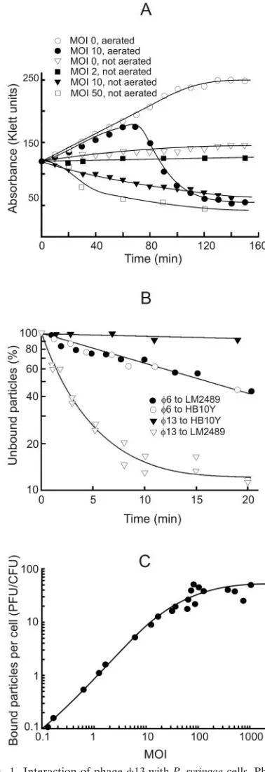
Characteristics of the 13 infection cycle. The highest phage titer in a one-step growth experiment was obtained by using a cell density of 2⫻108CFU/ml (120 Klett units) and an
MOI of 10. Cell lysis started 70 min p.i. and continued for 40 to 50 min (Fig. 1A). The obtained titer of the lysate was ca. 3 to 4⫻1010PFU/ml. On average 150 to 200 infective progeny
phage particles were released per cell. Starting 35 to 40 min p.i., it was possible to induce a premature lysis of infected cells by adding EDTA (0.5 mM final concentration) and diluting the infection mixture 1:10 with distilled water. However, the first PFU in the lysate appeared almost at the same time as with normal cell lysis, as also observed with6 (50). If the infected cultures were not aerated, the cell turbidity decreased in an MOI-dependent manner (Fig. 1A). In the case of uninfected cells, lack of aeration did not induce lysis but there was only a slight increase in the turbidity.
Kinetics of the phage adsorption was determined by using very low MOIs (⬃10⫺5). We studied HB10Y cells, which are
sensitive to only6 (pili and long LPS O chains); LM2489, which are sensitive to6 and13 (pili and truncated LPS); and LM2509 cells, which are sensitive to only 13 (no pili and truncated LPS). LM2489 and LM2509 cells adsorbed13 par-ticles at a similar rate (shown for LM2489 in Fig. 1B). During the first 10 min of the infection, LM2489 cells adsorbed ca. 85% of the added13 but only 40% of the6 particles (Fig. 1B). The rate of6 adsorption was the same for HB10Y and LM2489 cells. About 45% of adsorbed phage6 particles but only 3% of infective13 particles were released when infected LM2489 cells were pelleted, resuspended, and pelleted again at 10 min p.i. The adsorption rate constants (1) for6 and13 at 3 min p.i. were ca. 5⫻10⫺9and 1⫻10⫺8, respectively (Fig.
1B).
The maximal number of infective13 particles irreversibly bound per one LM2489 cell was around 60 and was achieved at MOI ⬃100 (Fig. 1C). Taking into account that the specific infectivity of 1⫻purified6 is as close to maximal as possible (⬃9 ⫻ 1010PFU/g of protein; calculations were based on
data from Day and Mindich [19]) and that the protein content of13 virions is the same as in6 but the specific infectivity is ⬃10 times lower (results not shown), we found that only about 1 of 10 of the13 particles are infectious. Noninfectious par-ticles or some fraction of them might irreversibly bind to the cell surface and, consequently, there may be more than 60 particles bound to each cell.
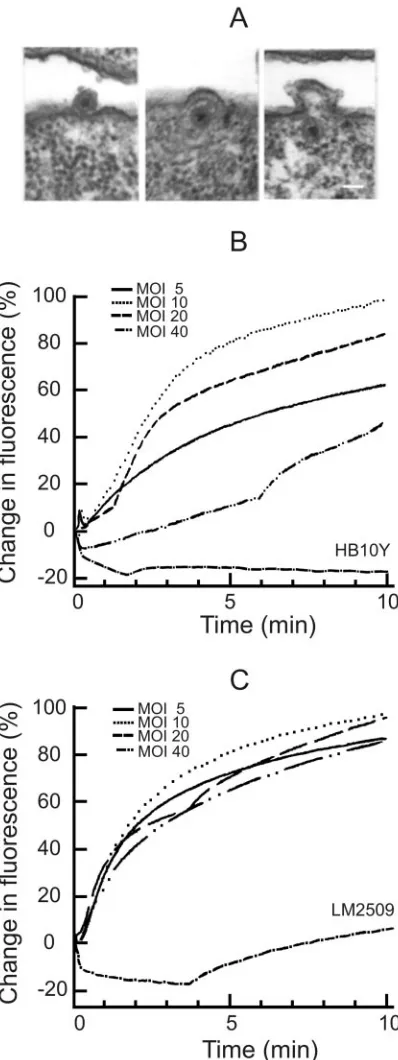
13 envelope fusion and labeling of the virus.Thin sections of infected LM2489 cells showed virus particles associated with the cell surface at 7.5 min p.i. (Fig. 2A). The phage envelope fusion with the host OM can be clearly seen, and intracellular enveloped NC particles can be readily distinguished, as in the
case of phage 6 infection (2, 4, 41, 46). The first progeny enveloped virus particles appeared⬃45 min p.i., similarly to 6 (not shown).
[image:3.585.323.512.69.619.2]Fluorescent lipid probe R18 was incorporated into phage envelopes to a high enough concentration to obtain significant
FIG. 1. Interaction of phage13 withP. syringaecells. Phage13 one-step growth experiments (A), phage6 and13 adsorption kinet-ics measurements (B), and LM2489 cell surface receptor saturation with phage13 (C). The experiments were performed as described in Materials and Methods.
on November 8, 2019 by guest
http://jvi.asm.org/
self-quenching. The infectivity of R18-labeled phage particles was almost equal to unlabeled ones before the purification. Although the specific infectivity of 1⫻purified R18-labeled6 particles was about half of the unlabeled ones, there was prac-tically no effect of labeling in the case of13. The addition of detergent (Triton X-100) to the labeled particles caused an immediate increase in fluorescence, and approximately one-tenth of6 particles gave the same amplitude of Triton X-100-induced increase of fluorescence compared to13 (results not shown). The high accumulation of R18 into 6 envelopes might explain a higher sensitivity of labeled 6 particles to purification.
When labeled 13 or6 particles were added to the sus-pension of sensitive cells, the fluorescence increase started at 10 to 15 s and leveled at 3.5 to 6 min p.i. Resistant HB10Y cells did not induce any fluorescence increase in the case of phage 13. However, there was a weak late (starting⬃3.5 min p.i.) increase in fluorescence after phage 6 addition to LM2509 cells (Fig. 2C), although6 did not form any plaques when titers were determined on the lawn of these cells (results not shown).
Phage 13 infection-induced increase of fluorescence was MOI dependent (Fig. 2B), whereas that induced by phage 6 was MOI independent (Fig. 2C). The infection-induced increase of fluorescence was biphasic. An initial fast increase (⬃3.5 min) was followed by a slow increase (Fig. 2B and C) with similar fluorescence kinetics for both viruses.
Energy-depleting agents block6 and13 adsorption.We observed that the metabolic state of the cells affected 13 adsorption. The phage bound better to fresh cells versus cells kept on ice for several hours, and the final level of fluorescence was higher when labeled phage was added to the suspension of preincubated cells compared to the situation when cells were added to the phage-containing medium. Therefore, effects of energy-depleting agents on viral adsorption and phage enve-lope fusion were studied. The relief of self-quenching was considerably lower when NaN3(blocking respiration and ATP
hydrolysis by membrane H⫹-ATPase) treated cells were in-fected with6 (Fig. 3B). However, the 6 infection-induced increase in fluorescence was only slightly lower when arsenate (reducing intracellular ATP concentration by arsenolyzation of acetyl phosphates) was present. The control experiments showed that an increase in the ionic strength of the medium (addition of NaCl to 30 mM) had no considerable effect on the infection-induced increase of the fluorescence (not shown).
NaN3 had less of an effect on the 13 infection-induced
increase in the fluorescence compared to 6 (Fig. 3A), and arsenate had no considerable effect on membrane fusion (not shown). However, an inhibitor of cytochrome oxidase, KCN, blocked the fluorescence increase very effectively. Adsorption studies with13 confirmed the role of energy metabolism at the initial stages of infection. KCN and NaN3 inhibited the
irreversible adsorption in a concentration-dependent manner (Fig. 3C). The effects of KCN and NaN3on phage13
infec-tion were stronger when the infecinfec-tion was performed in LB medium compared to 50 mM sodium phosphate buffer (not shown).
OM permeability and membrane voltage changes during
[image:4.585.64.263.83.613.2]13 and 6 infections. P. syringae cells accumulate low amounts of TPP⫹due to the low permeability of the OM (Fig.
FIG. 2. Phage envelope fusion assays performed by using electron microscopy and R18 fluorescence measurements. Thin-section micro-graphs (A) of LM2509 cells infected with13 (MOI 15) and collected 7.5 or 15 min p.i. The scale bar represents 50 nm. (B and C) Decreased self-quenching of R18-labeled phage13 (B) and6 (C) at different MOIs. The fluorescence experiments were performed in 50 mM so-dium phosphate buffer (pH 7.0), at room temperature. The MOI was changed by adding different amounts of cells (LM2509 [B]; HB10Y [C]) while using the same amount of phage particles. 100% corre-sponds to the fluorescence level at MOI 10 at 12 min p.i.
on November 8, 2019 by guest
http://jvi.asm.org/
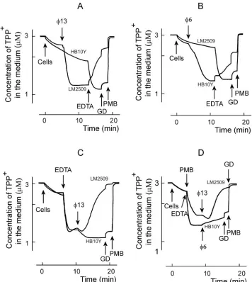
4). EDTA removes divalent cations from the LPS layer, in-creasing the cell envelope permeability to lipophilic com-pounds. The treatment ofP. syringaecells with EDTA at con-centrations higher than 0.04 mM induces the uptake and distribution of TPP⫹between the cytosol and external medium according to membrane voltage (⌬, Fig. 4C). The maximal accumulation of TPP⫹was observed when the concentration of EDTA was⬎0.2 mM.P. syringaecells were, however, rather sensitive to this chelator. At concentrations of ⱖ0.7 mM, EDTA induced only a transient accumulation of TPP⫹due to a slow depolarization of the PM (not shown).
Polycationic antibiotic PMB displaces inorganic divalent cat-ions from the outer surface of the OM. Being bulkier than cations it replaces, PMB changes the packing order of LPS and increases the permeability of the OM to a variety of molecules, including GD (17). At concentrations of⬎4g/ml PMB in-duced an accumulation of TPP⫹(Fig. 4D), but at 40g/ml and higher concentrations PMB depolarized the PM and induced a leakage of accumulated TPP⫹(results not shown).
Changing the cell/permeabilizer concentration ratio, it was possible to choose proper concentrations of EDTA or PMB to permeabilize the cells without depolarization. In the following series of experiments, we studied the fluxes of TPP⫹across the
P. syringaeenvelope upon interaction with bacteriophages6
and13 (Fig. 4). The addition of phage induced a very active accumulation of TPP⫹to the phage-sensitive cells but had no effect on the resistant cells (Fig. 4A and B). This indicates that upon adsorption bacteriophages6 and13 effectively perme-abilized the OM to TPP⫹. The TPP⫹accumulation continued for⬃3 and⬃6 min with13 and6, respectively (Fig. 4A and B). If EDTA was added after the phage, it induced a leakage of accumulated TPP⫹from infected cells but caused accumu-lation of TPP⫹in the case of phage-resistant cells (Fig. 4A and B). The subsequent addition of the channel-forming antibiotic GD had a rather weak effect on accumulation of TPP⫹in both uninfected and infected cells. PMB addition depolarized the cells very effectively and induced a fast efflux of accumulated TPP⫹. The phage-induced depolarization of the cells in the presence of EDTA was considerably stronger in the case of 13 compared to6 (compare Fig. 4C and D).
Phage 6 or 13 and EDTA, but neither of them alone, caused leakage of TPP⫹from infected cells (Fig. 4). There are two explanations for these results. (i) Phage-induced perme-abilization of the OM to TPP⫹is temporal, and TPP⫹stays locked in the cytosol of the infected cell in the absence of EDTA in spite of PM depolarization. (ii) EDTA depolarizes the PM (see above), and phages only increase the sensitivity of the cells to this compound. Experiments with low concentra-tions of PMB showed (Fig. 4D) that EDTA is not obligatory for the depolarizing action of phage13 but that the phage-induced increase of OM permeability to lipophilic compounds is temporal.
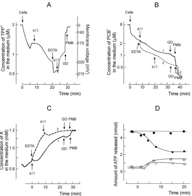
Physiological changes during early stages of infection.More detailed analysis of phage entry-induced changes in bacterial energetics was performed at pH 8 (Fig. 5). In spite of higher membrane voltage, 13-induced accumulation of TPP⫹ was less effective under these conditions (Fig. 5A). The addition of EDTA induced an additional accumulation of TPP⫹into in-fected cells, but leakage of accumulated TPP⫹started imme-diately after the second13 addition. These results indicate that a superinfection-exclusion type phenomenon is expressed only weakly during phage13 infection.
Intact P. syringae cells bound high amounts of PCB⫺, a lipophilic anion accumulating in biological and artificial mem-branes (Fig. 5B). The infection-induced PCB⫺ binding was rather weak and proceeded simultaneously with the phage-induced permeabilization of the OM to TPP⫹(compare Fig. 5A and B). The addition of EDTA to the cell suspension caused considerably stronger binding of PCB⫺than the infec-tion (Fig. 5B, curve 2).
P. syringaecells were not able to maintain a stable
concen-tration of intracellular K⫹ in sodium phosphate buffer (Fig. 5C), and at pH 8.0 the spontaneous leakage of K⫹was stronger than at pH 7.0 (not shown). The maximal concentration of intracellular K⫹was ca. 230 mM.13 infection stimulated the K⫹leakage starting 1.5 to 2 min p.i., and the phage-stimulated leakage was observed at almost the same time period as accu-mulation of TPP⫹ and PCB⫺. The addition of EDTA very effectively depleted the intracellular K⫹, and the cell
depolar-FIG. 3. Effects of energy-depleting agents on the infection-induced increase in fluorescence (A and B) and13 adsorption (C). Experi-ments were performed at room temperature in LB medium. LM2509 (A and C) or HB10Y (B) cells were incubated with drugs for 5 min and then infected with R18-labeled13 (A) and6 (B) or unlabeled13 (C) particles at an MOI of 10 (A and B) or 0.1 (C). (A and B) 100% indicates the level of fluorescence 12 min p.i. in the absence of drugs. The final drug concentrations were 20 mM (A and B) or as indicated (C).
on November 8, 2019 by guest
http://jvi.asm.org/
ization by GD and PMB did not have any considerable effect on K⫹equilibration between the cytosol and the medium (Fig. 5C). Phage6-induced effects on accumulation of PCB⫺and K⫹were kinetically similar to those induced by13, but the amplitudes of6-induced ion fluxes were considerably weaker (results not shown).
The intracellularP. syringaeATP concentration was 5 to 6 mM before infection (Fig. 5D). Bacteriophages 6 and13 (MOI 10) induced a small but reproducible leakage of intra-cellular ATP starting 1 to 1.5 min p.i. In spite of the leakage, 6 infection did not considerably affect the level of ATP in the infected cells, but a decrease⬎50% was observed in the case of 13 during the first 10 to 12 min of infection.
Prevention of virus entry by treating the virus particles at elevated temperature. In the case of6, the NC-associated lytic enzyme, digesting of a local opening in the peptidoglycan layer, is temperature sensitive (4, 12). The protein composition of13 indicates the presence of an equivalent protein (43),
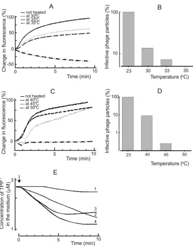
and we detected lytic activity associated with13 particles (not shown, but see Materials and Methods). Although only⬃10% of the heat-inactivated (10 min, 30°C) phage13 particles were able to form infective centers (Fig. 6B), the fusion of viral envelope with the OM ofP. syringaecells was not considerably affected (Fig. 6A). However, after more extensive inactivation (10 min, 35°C),13 particles were no more able to induce fluorescence increase and did not form plaques. Phage6 was less sensitive to heating, and therefore a control study was performed at temperature intervals of 23 to 50°C (Fig. 6C).
[image:6.585.114.470.67.465.2]To get insights to the mechanism of heat inactivation of13 particles, the accumulation of TPP⫹byP. syringaeduring the initial stages of infection was studied (Fig. 6E). Phage 13 heated 10 min at 30 or 33°C was able to induce an accumula-tion of TPP⫹into the host cells, indicating a normal fusion event. The final level of cell-accumulated TPP⫹ in the pres-ence of EDTA indicated that the heated phage particles did not cause a considerable PM depolarization (not shown). This
FIG. 4. Effects of phages13 (A, C, and D) or6 (B and D) on TPP⫹uptake byP. syringaecells. The experiments were performed at 24°C in 50 mM sodium phosphate buffer (pH 7.0) with 3⫻108cells/ml. R18-labeled phage particles were added to obtain an MOI of 10. EDTA was
used at the final concentration of 0.2 mM, and GD was used at 4g/ml. PMB was added to obtain the final concentration of 100g/ml, except for panel D, where the final PMB concentration was 5g/ml.
on November 8, 2019 by guest
http://jvi.asm.org/
shows that the entry prevention took place after the fusion but before the particle reached the PM, being in line that the penetration of the peptidoglycan layer is inactivated.
DISCUSSION
Phage6 binds to the sides of the pilus and is brought to the cell surface by pilus retraction. Consequently, since there are only one to two pili per cell, only a few particles eventually enter the cell regardless of the MOI used (4, 44). The retrac-tion process is cell energy dependent (32), thus complicating the study of6 entry-associated energetic events in detail. In contrast, the LPS receptor of13 (36) allows the virus to bind randomly on the cell surface, leading to MOI-dependent entry events. The measured maximal number of irreversibly bound active13 particles was about 60 per cell (Fig. 1C) but, due to low specific infectivity, the actual number of particles may be
higher.13 can enter to several unrelated bacteria (36), sug-gesting that host envelope proteins may not play a significant role during the entry. However, we assume that the LPS re-ceptor used by13 is a vital component of the host cell and that its alteration may lead to cell death. Screening of 150 putative phage-resistant cell lines did not lead to any true 13-resistant cells, whereas phage6 resistance was commonly obtained (results not shown).
[image:7.585.106.480.66.448.2]Interestingly, when MOIs higher than 10 were used, reduc-tion of the amplitude of infecreduc-tion-induced increase in fluores-cence was detected for13 (Fig. 2B). In the case of6 (4), the irreversible step in the entry is considered to be the fusion of the viral membrane with the OM, linking the irreversible bind-ing to the fusion event. However, there may be yet another step after the receptor binding and before the fusion that irrevers-ibly binds the virus to the cell. The difference between the maximum number of particles bound (⬃60, Fig. 1C) and the
FIG. 5. Phage and EDTA effects on TPP⫹(A), PCB⫺(B), K⫹(C), and ATP (D) content ofP. syringaecells. The experiments were performed at 28°C in 100 mM sodium phosphate buffer (pH 8.0). The cells were added to the final concentration of 109cells/ml (A to C). Unlabeled phage 13 particles were added to obtain an MOI of 10 after each addition (A to C). EDTA was added to the final concentration of 0.2 mM, and GD and PMB were added to the final concentrations of 4 and 100g/ml, respectively. The ATP measurements in panel D were performed as described in Materials and Methods. The cells were infected at time point “0.” Open circles (6) and squares (13) indicate the amount of free ATP in the cell incubation medium, and the filled circles (6) and squares (13) indicate the total amount of ATP in the cell suspension. The amount of ATP shown corresponds to 109of the cells.
on November 8, 2019 by guest
http://jvi.asm.org/
number that cause a maximal increase in fluorescence (⬃10, Fig. 2B) suggests the presence of an irreversible binding step different from fusion. However, these measurements were per-formed under different conditions (no aeration during fluores-cence measurements), possibly explaining some of the differ-ence.
The13 adsorption/fusion is dependent on the physiological state of the cell. Inhibition of respiration rapidly renders the cells resistant to infection (Fig. 3A and C). The usage of high
MOI also leads to the inhibition of fusion. The consequences of both of these events—blocked respiration and the increase of ion permeability of the PM after infection—are the depo-larization of the PM and the decrease of H⫹concentration in the cell envelope (11, 13, 29). Depolarization of the PM by using incubation buffers with pH 6.0 prevented neither the irreversible binding of13 nor the infection-induced increase of the fluorescence (not shown). As in many animal viruses (21, 37), an acidic environment probably promotes the binding/
FIG. 6. Infection by heat-treated phage particles. Fusion kinetics were studied in 50 mM sodium phosphate buffer (pH 7.0) at room temperature. In the case of unheated phage13 (A) and6 (C) particles, the infection MOI was 10, and the same amounts of viruses were used after heating. The capability of the phage13 (B) and6 (D) particles to form infective centers was assayed after virus incubation at different temperatures for 10 and 15 min, respectively. (E) The TPP⫹uptake experiments were performed at 28°C in 50 mM sodium phosphate buffer (pH 7.0). The cells were added to obtain the final concentration of 2⫻108CFU/ml. The MOI was 10 in the case of unheated phage infection, and
the same amounts of particles were used after the heating. Curves: 1, HB10Y cells infected with unheated13; 2 and 3, LM2509 cells infected with 13 heated for 10 min at 33 and 30°C, respectively; 4, LM2509 cells infected with unheated13. The arrow indicates the addition of phage.
on November 8, 2019 by guest
http://jvi.asm.org/
[image:8.585.105.481.63.541.2]fusion of bacteriophages belonging to theCystoviridaefamily. Since EDTA does not prevent infection-induced permeabili-zation of the membranes (Fig. 4 and 5) or increased fluores-cence (not shown), divalent cations seem to be an unimportant factor for phage to traverse the OM.
It appears that in the13 system the irreversible binding is autoregulated by the number of infecting particles. When an increasing amount of phage particles interact with the host envelope, the PM permeability to H⫹increases above some threshold value and prevents the membrane fusion events. This is a superinfection immunity-related event (31) by using a unique mechanism.
A temporal increase of the OM permeability to lipophilic ions (TPP⫹ and PCB⫺) between ⬃1.5 and 4 min p.i. was observed (Fig. 4 and 5). This permeability coincided with the fast dilution of fluorophore-labeled virus-associated lipid mol-ecules after infection (Fig. 2). These observations are in agree-ment with membrane fusion and the presence of temporal virus-derived membrane patches on the OM. The closure of the lipophilic ion-permeable window apparently occurs due to the lateral movement of LPS in the outer layer of the OM, closing the passage of lipophilic molecules (for a review, see reference 38).
Bacteriophage6 has a lytic enzyme, protein P5, associated with the NC surface (22). This enzyme is active after mem-brane fusion and digests an opening to the peptidoglycan layer (12, 35). Phage 13 has a related protein, and we demon-strated here the presence of lytic activity associated with13 virions. It has been shown that P5 of6 is temperature sensi-tive (12), and the lytic protein of 13 is also temperature sensitive, only more so (Fig. 6). This suggests that both6 and 13 use a similar mechanism to penetrate the peptidoglycan layer.
Penetration of the6 NC through the PM is an endocytosis-like event that depends on membrane voltage (41). The NC penetration of6 was studied by using host cell spheroplasts (cells devoid of OM and peptidoglycan). This system was not optimal for studying the electrophysiology of the NC penetra-tion due to the spontaneous leakage of intracellular indicator ions into the medium. Here we used intact cells and an MOI of 10 to assess the PM-associated events during 6 and 13 penetration (Fig. 4 and 5).
At 3 min p.i., K⫹ions were detected leaking from the cells in both the6 and the13 infections. However, this leakage was weak and gradually ceased at ca. 10 min p.i. Also, similar amounts of ATP leaked in both infections, but intracellular ATP concentrations continued to decrease in 13-infected cells, whereas the ATP levels remained stable in the case of6 infection (Fig. 5D). We attribute these observations to the lower membrane voltage of 13-infected cells (Fig. 4) that prevented ATP synthesis. Using an MOI of 10, the amplitudes of the virus-induced effects of 13 are several times higher than those for6-infected cells. This is in line with different numbers of virus particles entering the cell.
In6 infection when only one to two particles enter the cell, the energy state of the host is not considerably altered. The PM is only slightly depolarized, and the intracellular ATP concen-tration is not considerably changed. In the case of13, MOIs higher than 10 had an adverse effect on virus production (not shown) and exhibited decreased entry-associated events (Fig.
2B). In addition, if the infected cells (MOI 10) were not aer-ated, lysis occurred (Fig. 1A). This suggests an active role of cell energy metabolism in repairing the infection-caused cell envelope defects.
Enveloped dsRNA bacterial viruses share entry principles (membrane fusion and PM penetration with an endocytosis-type mechanism) thought to occur only in animal viruses. This suggests that such mechanisms evolved early in the bacterial domain and were spread with the diversification of life.
ACKNOWLEDGMENTS
This study was supported by research grants 1202108 and 1202855 from the Academy of Finland (to D.H.B.; Finnish Center of Excel-lence Program [2000-2005]) and by Lithuanian State Science and Stud-ies Foundation research grant 575 (to R.D.). R.D. is a Lithuanian State Fellowship holder. V.C. and E.B. were supported by EC project “Ce-biola” (ICAI-CT-2000-70027).
REFERENCES
1.Adams, M. H.1959. Bacteriophages. Interscience Publisher, Inc., New York, N.Y.
2.Bamford, D. H., E. T. Palva, and K. Lounatmaa.1976. Ultrastructure and life cycle of the lipid-containing bacteriophage6. J. Gen. Virol.32:249–259. 3.Bamford, D. H., and L. Mindich.1980. Electron microscopy of cells infected
with nonsense mutants of bacteriophage6. Virology107:222–228. 4.Bamford, D. H., M. Romantschuk, and P. J. Somerharju.1987. Membrane
fusion in prokaryotes: bacteriophage6 membrane fuses with the Pseudo-monas syringaeouter membrane. EMBO J.6:1467–1473.
5.Bamford, D. H., P. M. Ojala, M. Frilander, L. Wallin, and J. K. H. Bamford. 1995. Isolation, purification, and function of assembly intermediates and subviral particles of bacteriophage PRD1 and6. Methods Mol. Genet.6: 455–474.
6.Beveridge, T. J.1999. Structures of gram-negative cell walls and their derived membrane vesicles. J. Bacteriol.181:4725–4733.
7.Blumenthal, R., S. A. Gallo, M. Viard, Y. Raviv, and A. Puri.2002. Fluores-cent lipid probes in the study of viral membrane fusion. Chem. Phys. Lipids 116:39–55.
8.Boulanger, P., and L. Letellier.1988. Characterization of ion channels in-volved in the penetration of phage T4 DNA intoEscherichia colicells. J. Biol. Chem.263:9767–9775.
9.Bradford, M. M.1976. A rapid and sensitive method for the quantification of microgram quantities of protein utilizing the principle of protein-dye bind-ing. Anal. Biochem.72:248–254.
10.Butcher, S. J., T. Dokland, P. M. Ojala, D. H. Bamford, and S. D. Fuller. 1997. Intermediates in the assembly pathway of the double-stranded RNA virus6. EMBO J.16:4477–4487.
11.Calamita, H. G., W. D. Ehringer, A. L. Koch, and R. J. Doyle.2001. Evidence that the cell wall ofBacillus subtilisis protonated during respiration. Proc. Natl. Acad. Sci. USA98:15260–15263.
12.Caldentey, J., and D. H. Bamford.1992. The lytic enzyme of the Pseudomo-nasphage6: purification and biochemical characterization. Biochim. Bio-phys. Acta1159:44–50.
13.Cherepanov, D. A., B. A. Feniouk, W. Junge, and A. Y. Mulkidjanian.2003. Low dielectric permittivity of water at the membrane interface: effect on the energy coupling mechanism in biological membranes. Biophys. J.85:1307– 1316.
14.Cuppels, D. A., A. K. Vidaver, and J. L. van Etten.1979. Resistance to bacteriophage6 byPseudomonas phaseolicola. J. Gen. Virol.44:493–504. 15.Daugelavicˇius, R., E. Bakiene, J. Berzinskiene, and D. H. Bamford.1997.
Binding of lipophilic anions to microbial cells. Bioelectrochem. Bioenerget. 42:263–274.
16.Daugelavicˇius, R., J. K. H. Bamford, and D. H. Bamford.1997. Changes in host cell energetics in response to bacteriophage PRD1 DNA entry. J. Bac-teriol.179:5203–5210.
17.Daugelavicˇius, R., E. Bakiene, and D. H. Bamford.2000. Stages of polymyxin B interaction withEscherichia colicell envelope. Antimicrob. Agents Che-mother.44:2969–2978.
18.Daugelavicˇius, R., E. Bakiene, J. Berzinskiene, and D. H. Bamford.2001. Use of lipophilic anions for estimation of biomass and cell viability. Biotech-nol. Bioeng.71:208–216.
19.Day, L. A., and L. Mindich.1980. The molecular weight of bacteriophage6 and its nucleocapsid. Virology103:376–385.
20.Dimitrov, D. S.2004. Virus entry: molecular mechanisms and biomedical applications. Nat. Rev. Microbiol.2:109–122.
21.Gaudin, Y., R. W. Ruigrok, and J. Brunner.1995. Low-pH induced confor-mational changes in viral fusion proteins: implications for the fusion mech-anism. J. Gen. Virol.76:1541–1556.
on November 8, 2019 by guest
http://jvi.asm.org/
22.Hantula, J., and D. H. Bamford.1988. Chemical crosslinking of bacterio-phage6 nucleocapsid proteins. Virology165:482–488.
23.Henry, T., S. Pommier, L. Journet, A. Bernadac, J.-P. Gorvel, and R. Lloubes.2004. Improved methods for producing outer membrane vesicles in gram-negative bacteria. Res. Microbiol.155:437–446.
24.Hoekstra, D., T. de Boer, K. Klappe, and J. Wilschut.1984. Fluorescence method for measuring the kinetics of fusion between biological membranes. Biochemistry23:5675–5681.
25.Kakitani, H., H. Iba, and Y. Okada.1980. Penetration and partial uncoating of bacteriophage6 particle. Virology101:475–483.
26.Kalasauskaite, E. V., D. L. Kadisaite, R. J. Daugelavicˇius, L. L. Grinius, and A. A. Jasaitis.1983. Studies on energy supply for genetic process: require-ment for membrane potential inEscherichia coliinfection by phage T4. Eur. J. Biochem.130:123–130.
27.Keller, P. M., S. Person, and W. Snipes.1977. A fluorescence enhancement assay of cell fusion. J. Cell Sci.28:167–177.
28.Kenney, J. M., J. Hantula, S. D. Fuller, L. Mindich, P. M. Ojala, and D. H. Bamford.1992. Bacteriophage6 envelope elucidated by chemical cross-linking, immunodetection, and cryoelectron microscopy. Virology190:635– 644.
29.Koch, A. L.1986. The pH in the neighborhood of membranes generating a protonmotive force. J. Theor. Biol.120:73–84.
30.Loyter, A., V. Cytovsky, and R. Blumenthal.1988. The use of fluorescence dequenching measurements to follow viral membrane fusion events. Meth-ods Biochem. Anal.33:129–164.
31.Lu, M. J., and U. Henning.1994. Superinfection exclusion by T-even-type coliphages. Trends Microbiol.2:137–139.
32.Maier, B., L. Poter, M. So, H. S. Seifert, and M. P. Sheetz.2002. Single pilus motor forces exceed 100 pN. Proc. Natl. Acad. Sci. USA99:16012–16017. 33.Marvin, H. J. P., M. B. A. ter Beest, D. Hoekstra, and B. Witholt.1989.
Fusion of small unilamellar vesicles with viable EDTA-treatedEscherichia colicells. J. Bacteriol.171:5268–5275.
34.Mindich, L., J. F. Sinclair, and J. Cohen. 1976. The morphogenesis of bacteriophage6: particles formed by nonsense mutants. Virology75:224– 231.
35.Mindich, L., and J. Lehman.1979. Cell wall lysin as a component of the bacteriophage6 virion. J. Virol.30:489–496.
36.Mindich, L., X. Qiao, J. Qiao, S. Onodera, M. Romantschuk, and D. Hoog-straten.1999. Isolation of additional bacteriophages with genomes of seg-mented double-stranded RNA. J. Bacteriol.181:4505–4508.
37.Mittal, A., E. Leikina, J. Bentz, and L. V. Chernomordik.2002. Kinetics of influenza hemagglutinin-mediated membrane fusion as a function of tech-nique. Anal. Biochem.303:145–152.
38.Nikaido, H.2003. Molecular basis of bacterial outer membrane permeability revisited. Microbiol. Mol. Biol. Rev.67:593–656.
39.Olkkonen, V. M., and D. H. Bamford.1989. Quantitation of the adsorption and penetration stages of bacteriophage6 infection. Virology171:229–238. 40.Peisajovich, S. G., and Y. Shai.2002. New insights into the mechanism of
virus-induced membrane fusion. Trends Biochem. Sci.27:183–190. 41.Poranen, M., R. Daugelavicˇius, P. M. Ojala, M. W. Hess, and D. H. Bamford.
1999. A novel virus-host cell membrane interaction: membrane voltage-dependent endocytic-like entry of bacteriophage6 nucleocapsid. J. Cell Biol.147:671–681.
42.Poranen, M. M., R. Daugelavicˇius, and D. H. Bamford.2002. Common principles in viral entry. Annu. Rev. Microbiol.56:521–538.
43.Qiao, X., J. Qiao, S. Onodera, and L. Mindich.2000. Characterization of
13, a bacteriophage related to6 and containing three dsRNA genomic segments. Virology275:218–224.
44.Romantschuk, M., and D. H. Bamford.1985. Function of pili in bacterio-phage6 penetration. J. Gen. Virol.66:2461–2469.
45.Romantschuk, M., and D. H. Bamford.1986. The causal agent of halo blight in bean,Pseudomonas syringaepv. phaseolicola, attaches to stomata via its pili. Microb. Pathog.1:139–148.
46.Romantschuk, M., V. M. Olkkonen, and D. H. Bamford.1988. The nucleo-capsid of bacteriophage 6 penetrates the host cytoplasmic membrane. EMBO J.7:1821–1829.
47.Sambrook, J., and D. W. Russell.2001. Molecular cloning: a laboratory manual, 3rd ed. Cold Spring Harbor Laboratory Press, Cold Spring Harbor, N.Y.
48.Smith, A. E., and A. Helenius.2004. How viruses enter animal cells. Science 304:237–242.
49.Stegmann, T., P. Schoen, R. Bron, J. Wey, I. Bartoldus, A. Ortiz, J. L. Nieva, and J. Wilschut.1993. Evaluation of viral membrane fusion assays. Com-parison of the octadecylrhodamine dequenching assay with the pyrene exci-mer assay. Biochemistry32:11330–11337.
50.Vidaver, A. K., R. K. Koski, and J. L. van Etten.1973. Bacteriophage6: lipid-containing virus ofPseudomonas phaseolicola. J. Virol.11:799–805. 51.Yang, H., E. V. Makeyev, S. J. Butcher, A. Gaidelyte, and D. H. Bamford.
2003. Two distinct mechanisms ensure transcriptional polarity in double-stranded RNA bacteriophages. J. Virol.77:1195–1203.