Dissertationsubmitted to
THETAMILNADUDr. M.G.R.MEDICAL UNIVERSITY
Inpartialfulfillment fortheDegree of
M
AS
T
ER OF
DENT
A
L
S
U
R
G
ERY
BRANCH I
PROSTHODONTICSAND CROWN AND
BRIDGE APRIL2018
BRANCH I
PROSTHODONTICS AND CROWN AND BRIDGE
2018
COMPARISON OF FRACTURE RESISTANCE OF SEVERELY ATTRITED
ENDODONTICALLY TREATED TEETH WITH DIFFERENT
CERTIFICATE
This is to certify that this dissertation titled “COMPARISON OF FRACTURE
RESISTANCE OF SEVERELY ATTRITED ENDODONTICALLY TREATED TEETH
WITH DIFFERENT CORE BUILD-UP MATERIALS – AN IN VITRO STUDY’’ is a bona fide record of work done by Dr. SRUTHI PRIYA M. under my guidance and to my satisfaction during her postgraduate study period of 2015-2018.
This dissertation is submitted to THE TAMILNADU Dr. M.G.R MEDICAL UNIVERSITY, in partial fulfilment for the degree of MASTER OF DENTAL SURGERY
in Prosthodontics including Crown and Bridge and Implantology. It has not been submitted (partially or fully) for the award of any other degree or diploma.
Dr. V. PRABHAKAR M.D.S.,
PRINCIPAL
Sri Ramakrishna dental college and hospital,
Coimbatore
Date: Place:
Dr. V. R. THIRUMURTHY M.D.S.,
Vice Principal, Guide,
Professor and Head of Department
Department of Prosthodontics Including
Crown and Bridge and Implantology
Sri Ramakrishna dental college and
DECLARATION
TITLE OF DISSERTATION
COMPARISON OF FRACTURE
RESISTANCE OF SEVERELY
ATTRITED ENDODONTICALLY
TREATED TEETH WITH DIFFERENT
CORE BUILD-UP MATERIALS – AN IN
VITRO STUDY
PLACE OF STUDY
SRI RAMAKRISHNA DENTAL
COLLEGE AND HOSPITAL,
COIMBATORE – 641006
DURATION OF THE COURSE 3 YEARS
HEAD OF THE DEPARTMENT DR. V.R.THIRUMURTHY
NAME OF THE GUIDE DR. V.R.THIRUMURTHY
I hereby declare that no part of the dissertation will be utilized for gaining financial assistance/ any promotion without obtaining prior permission of the Principal, Sri Ramakrishna Dental College and Hospital, Coimbatore. In addition, I declare that no part of this work will be published either in print or in electronic media without the permission of the guide who has been actively involved to dissertation. The author has the right to reserve for publish of work solely with the prior permission of the Principal, Sri Ramakrishna Dental College and Hospital, Coimbatore.
CERTIFICATE II
This is to certify that this dissertation work titled COMPARISON OF FRACTURE RESISTANCE OF SEVERELY ATTRITED ENDODONTICALLY TREATED
TEETH WITH DIFFERENT CORE BUILD-UP MATERIALS – AN IN VITRO
STUDY of the candidate Dr. Sruthi Priya M. with registration Number 241511353 for the award of Masters of Dental Surgery in the branch of Prosthodontics including Crown and Bridge and Implantology. I personally verified the urkund.com website for the purpose of plagiarism Check. I found that the uploaded thesis file contains from introduction to
conclusion pages and result shows 9 percentage of plagiarism in the dissertation.
ACKNOWLEDGEMENT
“It is part of my thesis that all our knowledge grows
only through the correcting of our mistakes”
Karl Popper
This thesis becomes a reality with the kind support and help of many individuals. Thank you all for your part in my journey.
Foremost, I want to offer this endeavor to God Almighty for the wisdom he bestowed upon me, the strength to keep going, peace of my mind and good health in order to finish this dissertation.
The dream begins with a teacher who believes in you.
I owe a ton of thanks to my respected Guide, Prof. Dr. V. R. Thirumurthy, Head of the Department, who is my mentor and a dedicated scholar. Without his continued
motivation, enthusiasm for teaching and constructive criticism, none of this would have been possible. I am privileged to be his student and grateful to have been associated with him. As a teacher, he affects eternity and it cannot be told where his influence stops. His valuable guidance, suggestions for improvement and constant motivation contributed to the completion of this task.
been etched in my heart and mind forever. My heartfelt thanks for her invaluable advice, lots of good ideas and ever approachable nature.
With immense gratitude I acknowledge Dr. Y. A. Bindhoo for her unsurpassed skill, devotion, scientific insight and proficient guidance which have helped me complete my study.
I extend my thanks to Dr. Arun, Dr. Sriram Balaji, Dr. Vandana N, Dr. Ashwin Devanarayanan, for their timely guidance and words of encouragement.
I convey my special thanks to Professor and Director, Lt. Gen. Dr. S. Murali Mohan and also to the Principal and Prof. Dr. Prabhakar for giving me this opportunity and their experienced guidance.
I would like to thank my colleagues, Dr. Parvathy and Dr. Monicasri for their whole hearted co-operation, valuable feedback and perfect understanding.
I express my heartfelt thanks to my seniors, Dr. Vishnu Manohar, Dr. Kumaran, Dr. Megasham, Dr. Geetha kumari, Dr. Priyankaa, Dr. Vijayapriya and also my juniors Dr. Lawrence, Dr. Deepak, Dr. Muthukeerthana, Dr. Cathryn, Dr. Sridevi and Dr. Prabha for their support and active synergy.
I thank Mr. Muthukumar, PSG Industrial Institute for helping me with the testing of my study sample. I’m thankful Dr. Deepta Kumaran and Dr. Sadhana Kandavel for
generously sharing their knowledge with the statistical analysis.
encouragement which helped me in completion of this disseratation.
I thank all the non-teaching members for their unconditional service.
CONTENTS
TITLE
PAGE No.
1. INTRODUCTION
01
2. AIM AND OBJECTIVES
05
3. REVIEW OF LITERATURE
06
4. MATERIALS AND METHODS
17
5. RESULTS
41
6. DISCUSSION
47
7. CONCLUSION
51
LIST OF TABLES
S.No.
TABLES
PAGE No.
1. Materials 17
2. Equipments 19
3. Failure loads of 30 specimens 42
4. Distribution of the mean compressive load among the groups A, B and C
43
5. Tests of normality 44
6. Mean (±S.D.) among the three groups 44
7. Mean difference among groups A, B and C 45
8. Mean value comparison of groups A and B 46
9. Mean value comparison of groups B and C 46
LIST OF FIGURES
S.No.
FIGURES
PAGE No.
1. Metal ring 26
2. Mounting device 27
3. Materials 28
4. Equipment 28
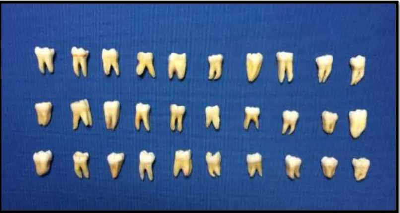
5. Sample of 30 molar teeth 29
6. Biomechanical preparation 30 7. Obturation of specimen with gutta percha
cone
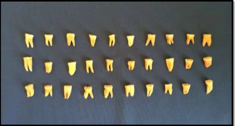
30 8. Crown height reduced upto 2mm from CEJ 31 9. Crown height measured 2mm 31 10. Specimens after crown height reduction 32
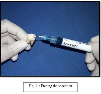
11. Etching the specimen 33
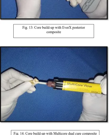
12. Applying of bonding agent 33 13. Core build-up with EverX posterior
composite
34 14. Core build-up with Multicore dual cure
composite
16. Light polymerization of the resin 35
17. Group A 36
18. Group B 36
19. Group C 36
20. Metal ring 37
21. Specimens mounted in metal ring with auto-polymerizing resin
37
22. Mounting device 38
23. Specimen positioned at 450 angle in the mounting device
38
24. Metal jig 39
25. Universal testing machine 39
26. Specimen under load 40
LIST OF BAR DIAGRAMS
S.No.
BAR DIAGRAM
PAGE No.
1. Distribution of the mean compressive load among the groups A, B and C
Fractures occurring in endodontically treated teeth pose a persistent problem in clinical dentistry. This is complicated because of the substantial loss of coronal tooth structure and the ability to predict restorative success. For the success of the restoration of endodontically treated teeth, numerous techniques have been suggested with criteria based on the variations in length, diameter, shape and surface configuration, quantity of dentinal structure and materials used in the reconstruction.
Fractures are more common in endodontically treated teeth than in the vital teeth. Endodontically treated teeth are considered to be more brittle due to structural changes in the dentin, with loss of water and collagen cross-linking. On the contrary, Huang et al compared the physical and mechanical properties of dentin specimens from teeth with and without endodontic treatment at different levels of hydration and concluded that neither dehydration nor endodontic treatment caused degradation of the physical or mechanical properties of dentin. In most endodontically treated teeth there are missing tooth structure caused by caries, fracture or existing restorations, associated to endodontic access preparation, hence it is difficult to establish if higher occurrence of fractures is depending on the structural change in the dentin, missing of tooth structure or both. It is suggested to use bonded restorations to avoid coronal microleakage and bacterial contamination that can also cause endodontic failure.
Remaining dentin thickness (RDT) is a major factor to be considered for the fracture resistance of endodontically treated teeth. Greater remaining tooth structure means greater longevity for the teeth. A loss of tooth structure greater than 50% would necessitate the use of posts to retain a core and to distribute stress. The post in an endodontically treated tooth contributes significantly towards the fracture resistance, where the RDT is less than 1.5mm.
treated teeth need post restoration. The fracture resistance mainly depends on the preservation of residual dentin. Post space preparation remove some root dentin which could increase the risk of root fracture. A clear guideline has not been suggested so far for whether a post should be applied to teeth with different coronal wall defects. Studies show that fracture resistance is not affected by the presence or absence of posts. Also, the over-preparation of post space and larger posts produce no greater reinforcement but actually decrease the resistance to fracture. The failure of post system is a risk of irreversible damage to the endodontically treated teeth.
The position of the tooth in the arch is an aspect to be considered when selecting materials and techniques to restore endodontically treated teeth because the masticatory force is different in anterior and posterior regions. The posterior teeth are subject to vertical forces while the anterior have lateral and shearing types of forces. Studies show that the incidence of fractures is two times higher in the mandibular first molars than in maxillary first molars.
The techniques for reinforcement of endodontically treated, or multi-rooted posterior teeth are from dowels and cores fabricated with direct wax or pattern resin to amalgam cores.
if necessary, with an additional post. But core resins have to be used in combination with dentin and post conditioning. However, clinical research on pin placement shows that the technique has a few complications. The most frequently faced complications are loose pins or inadequate post space length in the canal. The mercury content of amalgam is a detriment to the patients.
Some dentists favour glass ionomer cements for core build up, in view of the apparent ease of placement, adhesion, fluoride release, and nearly matched coefficient of thermal expansion. The ‘miracle mix’ of GIC and unreacted amalgam alloy have been especially popular. Some believe that the silver within the material enhances its physical and mechanical properties. A study where cermet cement was used to fill deciduous teeth showed that it performed less efficiently in fracture toughness than a conventional GIC. Nevertheless, many workers regard GICs as inadequately strong to support major core build-ups. Hence it is recommended that a tooth should have at least two structurally intact walls if a GIC core is to be considered. GIC can be an excellent filler but a relatively weak build up material. To overcome this, GIC has been modified with metal by the addition of filler particles to improve strength, fracture toughness and resistance to wear.
composite is chosen. To overcome this difficulty composites have been introduced with titanium filler particles. These are chemically cured materials, which despite their impressively strong appearance have lower values of diametric strength, compressive strength and fracture toughness than regular light cured composite. They are however stronger than GIC.
The amount of tooth structure that remains after endodontic treatment and the properties of the core build up material, especially its efficiency in resistance to fracture are important considerations in the success of restoration of endodontically treated teeth.
AIM OF THE STUDY
To evaluate and compare the fracture resistance of severely attrited endodontically treated molar teeth restored with three different core build-up materials.
OBJECTIVES
The present study is designed with the following objectives:
1. To evaluate the fracture resistance of severely attrited endodontically treated molar teeth withGC EverX posterior fibre reinforced composite.
2. To evaluate the fracture resistance of severely attrited endodontically treated molar teeth with Ivoclar Multicore dual cure composite.
3. To evaluate the fracture resistance of severely attrited endodontically treated molar teeth withVOCO Rebilda DC dual cure composite.
Sorensen JA et al45 (1984) studied the effect of intracoronal reinforcement and coronal
coverage with amalgam in endodontically treated teeth and reported that there was no significant increase in resistance to fracture or dislodgment nor an improvement in the rate of clinical success of the teeth.
McLean31 (1985) reported that Cermet cement has superior mechanical properties to
traditional glass-ionomer cements, especially in terms of compressive strength. Accordingly it has been advocated for core build-up. However, its low compressive strength is of concern and it is therefore suggested that the use of a cermet as core build-up material, based on strength, remains suspect compared to other restorative filling materials.
Pilliar RM et al40 (1986) carried out a variety of experimental specimen configurations for
Goldman, Davis and Waters11,19 (1987) used the double torsion test instead of the single-edge
specimen test and concluded that the critical stress intensity values of glass ionomer materials were lower than the composite resins.
Hunter et al25 (1989) stated that preparation of a post space adds a certain degree of risk to a
restorative procedure. Procedural accidents may occur during post-space preparation. Though very rarely, these accidents include perforation in the apical portion of the root or into the lateral fluted areas of the midroot, a so-called “strip perforation.” Placing a post may increase the chances of root fracture and treatment failure, especially if an oversized post channel is prepared. So, posts should only be used when there are no other options available to retain a core.
Sorensen and Engleman44 (1990) found that 1mm of parallel dentin above the shoulder
preparation increased the fractured resistance of endodontically treated teeth, whereas a contrabevel design did not improve the fracture resistance.
Lloyd and Butchart28 (1990) investigated the fracture toughness of 6 composite resins, 2 glass
Millstein PI33 (1991) in his in vitro study compared resin-based composite, amalgam and cast
gold as core material under a crown in endodontically treated teeth and found no significant difference in the fracture and failure characteristics among these materials, provided there existed a 2-mm ferrule on the margin of healthy tooth substance. Glass ionomer cement, on the other hand, was shown to be weak in tensile and compressive strengths, and it had low fracture resistance as a core material in another study.
Kane JJ et al27 (1991) suggested that endodontically treated, molar teeth should receive cuspal
coverage, but in most cases, do not require a post. Unless there is an extensive destruction of coronal tooth structure, the pulp chamber and canals provide adequate retention for a core build-up.
Miyawaki et al34 (1993) determined the critical stress intensity values for a variety of
composite resin and glass ionomer core materials, using a single-edge notched-beam test specimen configuration. He used five core build-up materials (1) glass ionomer (2) resin modified glass ionomer (3) titanium-reinforced composite (4) composite resin with fluoride and (5) amalgam. The two glass ionomer core materials had much smaller critical stress intensity values at around 0.6 MN · m23/2, whereas the composite resin core materials showed values 2 to 3 times higher.
Donald et al12 (1997) tested the fracture strength of endodontically treated mandibular molar
Mendoza DB et al32 (1997) presented evidence that resin cements give additional resistance
to fracture compared to brittle and nonbonding zinc phosphate cement. With composite resin cements there is a lower risk of fracture and loss of retention. Resin bonding of posts seems to lower microleakage significantly as compared to posts cemented with zinc phosphate cement.
Alves J et al1 (1998) reported that the oral environment is rich in microorganisms, and dental
restorations must withstand repeated exposure to physical, chemical, and thermal stressors. It is a difficult environment to maintain a hermetically sealed system. In vitro studies have shown that exposure of coronal gutta-percha to bacterial contamination can cause migration of bacteria to the apex in a few days. Bacterial by-products and endotoxins can penetrate to the apex in an even shorter time than bacteria and cause endodontic failure. In addition, recurrent caries or restorations that are fractured may lead to recontamination of the root canal system.
Steele and Johnson46 (1999) evaluated the fracture resistance of endodontically treated
maxillary premolars presenting different design preparations and restorative materials in a laboratorial study, and noted that teeth with endo access only were more resistant to fracture. Also, the fact of restoring the teeth, with amalgam or composite resin improved the fracture resistance, independent of using bonding agents or not.
Chan CP et al8(1999) stated that the tooth placement in the arch is an aspect to be considered
more than 2 times higher in mandibular first molars than in maxillary first molars, maxillary first premolars, maxillary second premolars and mandibular second molars and attributed this fact to the heavier masticatory force and thin or flat roots in this region. They concluded that canines were the teeth least susceptible to fracture and incisors were susceptible after subjected to endodontic treatment. The force incidence in anterior and posterior teeth is different because posterior teeth are subject to vertical forces while the anterior must resist to lateral and shearing types of forces, increasing the post requirement to provide force distribution in the coronal and root parts of the teeth, avoiding fractures.
Glazer18 (2000) from his 5-year period study, reported that patients were submitted to
endodontic treatment whose remaining coronal structure was inferior to 50%. Carbon fiber posts and metal ceramic crowns were used to restore their teeth. The overall failure rate was 7.7% and the cumulative survival rate was 89.6%, demonstrated at the end of the follow-up period. The results demonstrated that these posts were used in the upper anterior teeth with a higher success rate and longer life than in premolars, especially lower premolars, because of the narrow mesiodistal dimension of these roots. Based on this information, occlusion should be considered in a choice of a post because canines and upper incisors, responsible for guide disocclusion and cut food, are subjected to oblique forces, indicating a root post use.
Ferrari M16 (2001) reported that the fourth-generation adhesive systems (3-step systems)
Bader JD5 (2001) in a 15-week study involving 11 dentists reported 543 tooth fractures, of
which 85.6% were complete cusp fractures, 13.4% incomplete or suspected cusp fractures, and 0.9% root fractures. Some 3% of the fractured teeth had not been restored previously, and most of the 377 cusp fractures that were not associated with caries occurred in teeth with vital pulps. This study also reported that during any given year some 6% of adult patients with at least 1 posterior tooth at risk would experience a complete posterior cusp fracture in the absence of caries, with an incidence rate of 72.7 percent at risk.
Wolanek GA et al51 (2001) suggested that the root canal system should be protected by sealing
the canals and floor of the pulp chamber with intracoronal barriers. Bonded materials such as glass-ionomer cement or composite resin are preferred. The canal orifices are countersunk with a round bur, and the floor of the chamber is cleaned of excess gutta-percha and sealer. The chamber floor is etched and primed if a resin material is used or “conditioned” if using glass-ionomer cement or resin-modified glass glass-ionomer. The barrier material is then placed over the floor of the chamber and light cured, and a temporary restoration is placed with or without a cotton pellet in the chamber. The intracoronal barrier protects the root-canal system from contamination during the period of temporization and while the restorative dentistry is performed.
Mollersten et al35 (2002) concluded from their study on the comparison of core and post-core
Howdle MD et al23 (2002) reported that amalgam has been used as a build-up material, with
well recognized strengths and limitations. It has good physical and mechanical properties and works well in high-stress areas. In many cases, it requires the addition of pins or other methods to provide retention and resistance to rotation. Placement can be clumsy when there is minimal coronal tooth structure, and the crown preparation must be delayed to permit the material time to set. Amalgam can cause aesthetic problems with ceramic crowns and sometimes makes the gingiva look dark. There also is a risk of tattooing the cervical gingiva with amalgam particles during the crown preparation. For these reasons, and potential concern about mercury, it is no longer widely used as a build-up material. Amalgam has no natural adhesive properties and should be used with an adhesive system for build-up.
Pilo et al41 (2002) showed that composite cores have fracture resistance comparable to
Varela et al49 (2003) reported that eugenol-containing root-canal sealers inhibit the
polymerization of resin cements and this problem can be avoided by thorough cleaning and etching of the canal walls. In their study, the concerns about negative effects of sodium hypochlorite irrigants on resin adhesion to dentin also are not reported.
Malfarrari S et al29 (2003) discussed about fiber post failures that are associated to
displacement or detachment of the post and crown or prosthesis decementation than root fractures, which is a common failure related to conventional metal cast posts. Because metal cast posts present high rigidity, they appear to vibrate at high frequencies when loaded with lateral forces, which achieving critical points, may determine longitudinal fractures of the root.
Nagasiri and Chitmongkolsuk36 (2005) study proves that greater remaining tooth structure
means greater longevity for the teeth. One example is that molars with maximum tooth structure remaining after endodontic treatment had a survival rate of 78% at 5-year evaluation.
Tan et al48 (2005) demonstrated that teeth restored with post and core using 2mm uniform
ferrule presented fracture resistance similar to endodontically treated tooth restored without posts. In addition, this study related that fracture resistance increases proportionally to quantity of remaining coronal tooth structure once 2mm ferrule group and non-uniform ferrule groups were more fracture resistant than the group that lacked a ferrule.
Grandini et al20 (2005) stated that fiber posts associated to direct resin restorations is a faster
and direct resin restoration longevity by 6, 12, 24 and 30-month recall and satisfactory results were found although any comparison with teeth without posts had been made.
Mannocci F et al30 (2005) compared the longevity of endodontically treated teeth restored
with amalgam or fiber posts and composite resin and concluded that restorations with fiber posts were more effective than amalgam in preventing tooth fractures but less effective in preventing secondary caries.
Dumbrigue et al39 (2006) evaluated the effect of remaining coronal tooth structure location on
the fracture resistance of endodontically treated tooth and demonstrated that palatal walls were more resistant to fractures than labial walls. The authors concluded it based on the fact that the median load necessary to cause failures were 607N, 782N, 358N, 375N and 172N for the complete palatal, labial, proximal and no retained coronal tooth structure incisal to the finish line, respectively.
Hayashi et al21 (2006) studied the mode of fractures when tooth restored with fiber post,
when fractures happen, tooth are lost because of the position of the fracture while cervical root fractures of teeth restored with fiber posts did not necessarily represent loss of the teeth.
Stuart CH et al47 (2006) studied 60 endodontically treated immature maxillary teeth reported
that no significant differences were found among any of the treatment groups when comparing the reinforcement and strengthening ability of gutta-percha, Resilion and a self-cure flowable resin.
Wilkinson KI et al50 (2007) studied 72 mandibular sheep incisors and evaluated the fracture
resistance achieved by obturating the root canals of simulated immature teeth with MTA and then gutta-percha, Resilion, or self-cure flowable and hybrid resin composites. The hybrid resin composite was the only material that showed significantly more fracture resistance than when the canals contained only MTA plugs.
Andreasen JO, Andreasen FM2 (2007) reported that normal functional stresses might result
in coronal and/or radicular tooth fractures in instances of reduced mechanical properties, from reduced tooth structure caused by incomplete root formation, caries, tooth wear, and operative dentistry procedures, and from changes in tooth structure caused by aging, vital pulp tissue loss, and endodontic therapy.
Schmage et al42 (2009) reported that core composites should not be used as permanent fillings,
lower than those of restorative composites. Therefore, an alternative may be to build up the coronal defect with a restorative composite. It should be taken into account that—apart from wear—the clinical success of a composite filling depends on an effective dentin bonding, a high fracture strength of the material, and the extension of the filling. The wear of Rebilda DC was improved compared to Rebilda D, probably by changing filler sizes and through the use of smaller fillers. Decreased hardness for Rebilda DC may be due to a higher amount of matrix to keep the flowability. The same observation was made for Multicore Flow.
El-Deeb HA et al14 (2016) reported that the dual-cured resin composite core build-up materials
The study compares the fracture resistance of endodontically treated molar teeth with severe attrition that has been restored with three different core build up materials. The materials and methods used in this study are sequentially described in this section.
TABLE 1: MATERIALS
PROCEDURE S.No. MATERIALS BRAND,
MANUFACTURER
STORAGE OF SPECIMEN
1 0.9% Normal Saline Claris Ostuka Pvt. Ltd., Ahmeddabad,
India
ACCESS OPENING 2 Straight fissure bur Mani Inc., Japan
BIOMECHANICAL PREPARATION
3 Sodium hypochlorite Dentsply-Maillefer, Ballaigues, Switzerland
OBTURATION
4 Gutta-percha points Dentsply India Pvt. Ltd., Haryana, India
OBTURATION 6 Eugenol
Prime Dental Products Pvt. Ltd.,
Thane, India
CROWN HEIGHT REDUCTION
7 Carbide burs Edenta, Switzerland
ETCHING AND BONDING
8 Etching gel Ivoclar Vivadent, Liechtenstein,
9 Bonding agent Ivoclar Vivadent,
CORE BUILD UP
10 EverX posterior GC Dental Products Corp., Japan
11 Multicore DC Ivoclar Vivadent, India
12 Rebilda DC VOCO, Germany MOUNTING OF
SPECIMEN 13 Auto-polymerizing resin DPI, Dental Products of India, Mumbai,
TABLE 2: EQUIPMENTS
PROCEDURE S.No. INSTRUMENT BRAND,
MANUFACTURER
ACCESS OPENING OF MOLARS
1 Airotor NSK, Japan
BIOMECHANICAL PREPARATION
2 K files Prime Dental Products Pvt. Ltd., Thane, India
OBTURATION
3 Spreaders Prime Dental Products Pvt. Ltd., Thane, India
4 Glass slab Samit products, India
5 Cement mixing spatula GDC, India
6 Tweezer GDC, India
CROWN HEIGHT MEASUREMENT
7 Divider
Universal Trading Company, Delhi,
India
8 Metal scale
ETCHING AND BONDING
9 Applicator sticks Ivoclar Vivadent, India
10 Dappen dish Samit products, India
11 Light-cure unit Prime Dental Products Pvt. Ltd., Thane, India
CORE BUILD UP
CORE BUILD UP
12 Plastic instrument GDC, India
13 Catridge Ivoclar Vivadent, India
14 Dental gun Ultradent products Inc., South Jordan
15 Mixing pad GC Dental Products Corp., Japan
MOUNTING OF SPECIMEN
MOUNTING OF SPECIMEN
17 Wax knife
Prime Dental Products Pvt. Ltd., Thane, India
18 Wax carver Prime Dental Products Pvt. Ltd., Thane, India
COMPRESSIVE LOAD MEASUREMENT
19 Universal testing machine WANCE, Germany
METHODOLOGY
STUDY DESIGN
SELECTION OF 30 EXTRACTED HUMAN MAXILLARY AND MANDIBULAR MOLARS
ROOT CANAL TREATMENT FOR 30 SAMPLES
SPECIMEN PREPARATION BY REDUCING CROWN HEIGHT UPTO 2MM FROM CEJ
SPECIMEN GROUPING
GROUP A 10 SAMPLES WITH CORE BUILD UP
USING EVERX POSTERIOR COMPOSITE
GROUP B 10 SAMPLES WITH CORE BUILD UP
USING MULTICORE DUAL CURE COMPOSITE
GROUP C 10 SAMPLES WITH CORE BUILD UP
USING REBILDA DC DUAL CURE COMPOSITE
SPECIMEN EMBEDDING IN CUSTOM-MADE METAL MOULDS
FRACTURE RESISTANCE TESTING UNDER UNIVERSAL TESTING MACHINE LOADING
RESULTS
SAMPLE SIZE
STEPS FOLLOWED
1. Teeth selection 2. Root canal treatment
3. Specimen grouping and preparation
a) Group A – core build up with EverX posterior composite b) Group B – core build up with Multicore dual cure composite c) Group C – core build up with Rebilda DC composite
4. Specimen embedding 5. Specimen testing
30 ROOT CANAL TREATED
MOLARS WITH REDUCED
CROWN HEIGHT
GROUP A
(10 SAMPLES)
GROUP B
(10 SAMPLES)
1. TEETH SELECTION
Thirty extracted human molars were obtained and each tooth was examined thoroughly to ensure that there were no carious lesions, cracks and previous restorations. Dental plaque, calculus and periodontal tissues were removed. Teeth were stored in 0.9% saline solution during all subsequent procedures.
2. ROOT CANAL TREATMENT
Access opening was done for all the specimens. The pulp chamber of each tooth was opened using a straight fissure bur. The root canals were instrumented using 0.02 tapered hand files to International Standardization Organization (ISO) size 35 at 1mm above the apex. Conventional step-back technique was used. After intermitted rinsing with Sodium hypochlorite, the canals were dried with paper points. The roots were obturated with gutta-percha points and endodontic sealer (Zinc oxide eugenol; Dentsply-Maillefer, Ballaigues, Switzerland). An ISO 35 primary gutta-percha master cone was coated with sealer and seated in the canal to full working length. Lateral condensation with a finger spreader (Dentsply Maillefer) and fine gutta-percha points was performed until the entire canal was obturated. Teeth were inspected for cracks after the endodontic treatment using a magnifying glass.
3. SPECIMEN GROUPING AND PREPARATION
surfaces. The crown height was reduced upto this marking by using a contra-angled hand-piece with diamond bur (TF 12, ISO 179/014; MANI, Tochigi, Japan). Now the specimens resemble severely attrited molars. The CEJ on the buccal or lingual sides were not preferred to make the marking in order to avoid too much loss of crown height. After the procedure, the specimens were stored in 0.9% saline solution.
The gutta-percha points 2mm below the CEJ were removed from all the teeth. The prepared surfaces of the teeth were etched for 20 seconds and rinsed with water. After drying the etched surfaces by blow drying, the bonding agent was applied to the exposed coronal structure with an applicator stick. It was then light polymerized for 40 seconds.
All the teeth were randomly divided into three groups (A-C) of 10 teeth each. The specimens were prepared as follows:
Group A: endodontically treated teeth with core build up using EverX posterior
Group B: endodontically treated teeth with core build up using Multicore dual cure composite Group C: endodontically treated teeth with core build up using Rebilda DC
To the specimens of group A, EverX posterior was added into the pulp chamber first and then it was used to shape the core layer by layer. The resin was light polymerized for 40 seconds with 2mm resin increment each.
To the specimens of group B, Multicore dual cure composite was used to shape the core, which was then light-cured upto 40 seconds.
4. SPECIMEN EMBEDDING
All specimens were embedded 1mm below the CEJ in auto-polymerizing acrylic resin contained in custom-made metal rings, orienting the long axes of all teeth perpendicular to the horizontal plane.
5. SPECIMEN TESTING
After embedding in auto-polymerizing resin, each intact specimen was positioned in the mounting device and aligned at a 45- degree angle with respect to the long axis of the tooth. A universal testing machine was used to apply a constant load at a crosshead speed of 0.5mm/minute until fracture occurred. The load was measured in Newton. Failure was defined as the point at which the loading force reached a maximum value by either fracturing the root or core, or debonding the cement. The loading site was at the central fissure of the occlusal surface. Force data applied over time were recorded. The fracture of specimens under compressive loading was determined when the force-versus-time graph showed an evident load drop. Both the failure load and failure mode were recorded.
[image:44.595.135.460.80.762.2]
Fig. 6: Biomechanical Preparation
[image:45.595.131.462.426.754.2]
Fig. 8: Crown height reduced upto 2mm from CEJ
Fig. 11: Etching the specimen
Fig. 3: Materials
[image:50.595.111.477.73.396.2]
[image:50.595.110.482.263.719.2]
Fig. 13: Core build-up with EverX posterior composite
[image:51.595.112.481.71.401.2]
[image:51.595.110.485.387.741.2]
Fig. 15: Core build-up with Rebilda DC composite
Fig. 17: Group A
Fig. 18: Group B
[image:53.595.174.428.106.367.2]
[image:53.595.79.503.367.680.2]
Fig. 20: Metal ring
[image:54.595.169.453.74.345.2]
Fig. 22: Mounting device
Fig. 23: Specimen positioned at 450 angle in the
[image:54.595.172.456.357.730.2][image:55.595.131.457.103.750.2]
[image:55.595.185.410.396.756.2]
Fig. 24: Metal jig
This study was designed to evaluate the fracture resistance of endodontically treated molars with severe attrition restored with three different core build-up materials.
Null hypothesis
Null hypothesis H0: There is no significant difference in the efficiency of three different
core build-up materials on the fracture resistance of endodontically treated molars with reduced crown heights.
A sample size of 30 extracted molars that were endodontically treated and had reduced crown heights were used in the study. 10 specimens out of the 30 were restored with EverX posterior composite (Group A), 10 specimens with Multicore dual cure composite (Group B) and 10 specimens with Rebilda DC composite (Group C). All the specimens were mounted in a metal rings and were subjected to loading in a universal testing machine at a 450 angle to the longitudinal axis of the tooth with a crosshead speed of 0.5mm mm-1 until fracture. The load
was measured in Newtons. The loading site was at the centre of the occlusal surface.
TABLE 3: FAILURE LOADS OF 30 SPECIMENS
(GROUPS A, B AND C)
SPECIMEN No. GROUP A GROUP B GROUP C
1 224N 612N 387N
2 257N 652N 399N
3 212N 649N 462N
4 232N 609N 434N
5 212N 662N 411N
6 182N 649N 421N
7 185N 622N 456N
8 251N 631N 412N
9 172N 645N 431N
STATISTICAL ANALYSIS
[image:60.595.73.537.387.683.2]Data were statistically analysed with SPSS 17.0 (SPSS Inc., Chicago, IL, USA). One-way analysis of variance was used to reveal difference in failure load among the three groups to detect significant differences in the efficiency of three core build-up materials on resistance to fracture. Failure mode was analysed by Kruskal Wallis and Mann-Whitney tests.
TABLE 4: DISTRIBUTION OF THE MEAN COMPRESSIVE LOAD AMONG THE
GROUPS A, B AND C
S.No. Groups Compressive Load
Mean
( ± S.D.)
Level of significance
p value
1 1A 212.80 (±28.739)
O.OO1
2 2B 635.20 (±18.546)
TABLE 5: TESTS OF NORMALITY
Kolmogorov-Smirnov Shapiro-Wilk
COMPRESSIVE
LOAD
Statistic Df Sig. Statistic df Sig.
0.186 30 0.010 0.876 30 0.001
TABLE 6: MEAN (± S.D.) AMONG THE THREE GROUPS
Group A Group B Group C
Mean 212.80 635.20 423.40
[image:61.595.73.537.422.600.2]KRUSKAL WALLIS TEST
TABLE 7: MEAN DIFFERENCE AMONG GROUPS A, B AND C
GROUP N MEAN RANK
COMPRESSIVE
LOAD
A 10 5.50
B 10 25.50
C 10 15.50
TOTAL 30
BAR DIAGRAM 1: DISTRIBUTION OF THE MEAN COMPRESSIVE LOAD
AMONG THE GROUPS A, B AND C
0 100 200 300 400 500 600 700
TABLE 8 : MEAN VALUE COMPARISON OF GROUPS A AND B
GROUP N MEAN RANK SUM OF
RANKS
COMPRESSIVE
LOAD
A 10 5.50 55.00
B 10 15.50 155.00
TOTAL 20
TABLE 9: MEAN VALUE COMPARISON OF GROUPS B AND C
GROUP N MEAN RANK SUM OF
RANKS
COMPRESSIVE LOAD
B 10 15.50 155.00
C 10 5.50 55.00
TOTAL 20
TABLE 14: MEAN VALUE COMPARISON OF GROUPS A AND C
GROUP N MEAN RANK SUM OF
RANKS
COMPRESSIVE LOAD
A 10 5.50 55.00
C 10 15.50 155.00
This study was designed to evaluate the fracture resistance of molar teeth with different core build-up materials. The null hypothesis was rejected since the fracture resistance showed significant differences in terms of the restorative materials used for core build-up.
When an endodontically treated tooth was restored with a post, the post could redistribute the stress to increase the fracture resistance. Nam37 et al’s study demonstrated that in the zero-wall group without posts, stress was concentrated at the coronal and lingual area of CEJ, but no obvious stress concentration was found in that with posts. To the contrary, the post effect was not significant in groups with two, three or four walls, on condition that the thickness of dentin walls after tooth preparation was 1mm, and there were facial and lingual walls in two-wall group.
In this study, gutta percha point 2mm below CEJ was removed. It seemed like composite resin core fabrication with a 2mm well in the root canal could not only enhance retention but also protect adequately against root fracture.
A study on 6-year survival rate of premolars15 revealed that teeth with one, two and three coronal walls had significantly lower failure risks than those without coronal walls. These may explain why the post effect was not significant in groups with substantial coronal structure. So it can be concluded that not all endodontically treated teeth need post restoration and the number of residual dentin walls need to be considered.
Increasing the ferrule length does not significantly increase the fracture resistance of endodontically treated teeth. This could be explained because of the minimal differences in the elastic moduli of carbon fiber post and dentin, such that the forces could be distributed along the length of the post. Duret et al13 stated that the presence of, at least 1mm of coronal structure
Zhi-yue and Yu-Xing52 supports the idea that the loss of structural integrity associated with access preparation may lead to a higher occurrence of fractures in endodontically treated teeth. The findings of the present study are in agreement with Sorensen and Engleman44, who found that 1 mm of remaining coronal tooth structure was able to resist compressive load. This may suggest that the strength of the tooth was directly related to the remaining bulk of dentin and is more important than the type of material that the core and post are made from.
However, it is important to note that the forces responsible for failure in the present study were considerably higher than the maximal physiologic forces acting on the teeth intraorally. A careful review of literature points to one obvious fact that the most common cause of failure is the fracture of the restorative material or the coronal structure, as observed in the present study.
In this study three different core build-up materials were used to restore the molar teeth resembling severe attrition. A total of thirty specimens were used which were divided into three groups of ten specimens each.
Each specimen was mounted in a metal ring using auto-polymerizing resin. Each intact specimen was positioned in the mounting device and aligned at a 450 angle with respect to the long axis of the tooth. The specimens were subjected to a constant load applied at a crosshead speed of 0.5 mm/min in a universal testing machine. The load was applied until fractured occurred. After loading, the mode of failure (composite resin core, root, and/or coronal structure and composite resin core) was observed.
More of root fracture was observed in the specimens of group B. Debonding as well as coronal structure fracture was observed in the specimens of group C.
Kruskal Wallis test demonstrated that there was a significant difference in between the three groups load level stress (p value = 0.001). Hence the Null hypothesis H0 was rejected.
From post-hoc analysis, (Mann Whitney U Test) it was clear that there was statistically significant difference in the fracture resistance between the groups, i.e. A and B; A and C and between B and C. Descriptive analysis showed that the highest mean value was seen in group B and the least value was in group A.
The results showed that,
1. On comparing groups A and B, the maximum fracture resistance was observed in group B. GROUP B > GROUP A
2. On comparing groups B and C, the maximum fracture resistance was observed in group B. GROUP B > GROUP C
3. On comparing groups A and C, the maximum fracture resistance was observed in group C. GROUP C > GROUP A
4. Among the three groups, maximum fracture resistance was observed in group B specimens and fracture resistance was minimum in group A specimens.
GROUP B > GROUP C > GROUP A
It was observed that under incremental loading, though stresses increased in all three groups, and were susceptible to fracture, the amount of remaining coronal tooth structure and the bonding between the tooth and the restorative material plays an important role in the failure of an endodontically treated tooth.
Precautions can be taken to reduce the incidence of the failure of endodontically treated teeth with maximum bonding of the composite resin to the tooth structure, enhancing retention for the resin by removing 2mm of gutta-percha and thus creating a well for the core build-up material, preserving at least one-wall of the remaining coronal tooth structure.
Limitations:
1. It was an in vitro study, which could not fully replicate oral conditions. For clinically relevant results, in vivo studies should be performed.
The failure of endodontically treated teeth is a common problem when the teeth are not restored with crowns. Giving a crown would be not possible when the remaining coronal tooth structure is minimal. In such cases, a core build-up with better fracture resistance is a good option.
In this study, the fracture resistance of three different core build-up materials (EverX posterior, Multicore DC and Rebilda DC) on severely attrited teeth were studied. Load was applied to 30 specimens of endodontically treated molar teeth, 10 in each group. Load testing wasUniversal testing machine was used for load testing. Statistical analysis of the obtained results showed that there was significant variation in the fracture resistances of the three core build-up materials used and the assumed null hypothesis was hence rejected.
Within the limitations of the present study it can be concluded that
1. There was a significant variation in the fracture resistances of the three different core build-up materials that were used in the study.
1. Alves J, Walton R, Drake D. Coronal leakage: endotoxin penetration from mixed bacterial communities through obturated, post-prepared root canals. J Endodon 1998;24:587–91. 2. Andreasen JO, Andreasen FM. Textbook and color atlas of traumatic injuries to the teeth. Copenhagen: Munksgaard; 2007.
3. Aquilino SA, Caplan DJ. Relationship between crwon placement and the survival of
endodontically treated teeth. J Prosthet Dent 2002;87(3):256-63.
4. Assif D, Gorfil C. Biomechanical considerations in restoring endodontically treated teeth. J Prosthet Dent 1994;71(6):565-7.
5. Bader JD, Martin JA, Shugars DA. Incidence rates for complete cusp fracture, Community Dent Oral Epidemiol 2001;29:346-53.
6. Bonilla ED, Mardirossian G, Caputo AA: Fracture toughness of various core build-up materials. J Prosthod 2000;9:14-18.
7. Caplan DJ, Weintraub JA. Factors related to loss of root canal filled teeth. J Public
Health Dent 1997;57:31-39.
8. Chan CP, Lin CI, Tseng SC, Jeng JH. Vertical root fracture in endodontically versus nonendodontically treated teeth: a survey of 315 cases in Chinese patients. Oral Surg Oral Med Oral Pathol Oral Radiol Endod 1999;87:504-7.
9. Combe EC, Shaglouf AMS, Watts DC, Wilson NHF: Mechanical properties of direct core build-up materials. Dental Materials 1999;15:158-165.
10. Costa LCS, Pegoraro LF, Bonfante G. Influence of different metal restorations bonded with resin on fracture resistance of endodontically treated maxillary premolars. J
11. Davis DM, Waters NE: An investigation into the fracture behavior of a particulate-filled Bis-GMA resin. J Dent Res 1987;66:1128-1133.
12. Donald HL, Jeansonne BG, Gardiner DM, Srkar NK. Influence of dentinal adhesives
and a prefabricated post on fracture resistance of silver amalgam cores. J Prosthet Dent
1997;77:17-22.
13. Duret B, Duret F, Reynaud M. Long-life physical property preservation and postendodontic rehabilitation with the composipost. Compend Contin Educ Suppl 1996;20:50-6.
14. El-Deeb HA et al. Repair bond strength of dual-cured resin composite core buildup materials. J Adv Res 2015;6(3):1-7.
15. Ferrari M, Vichi A, Fadda GM et al. A randomized controlled trial of endodontically treated and restored premolars. J Dent Res 2012; 91(7): 72–78.
16. Ferrari M, Vichi A, Mannocci F, Mason PN. Retrospective study of the clinical performance of fiber posts. Am J Dent 2000;13(Spec No):9B-13B.
17. Fusayama T, Maeda T. Effect of pulpectomy on dentin hardness. J Dent Res 1969;48(3):452-60.
18. Glazer B. Restoration of endodontically treated teeth with carbon fibre posts – a prospective study. J Can Dent Assoc 2000;66(11):613-8.
19. Goldman M: Fracture properties of composite and glass ionomer dental restorative materials. J Biomed Mater Res 1985;19:771-783.
21. Hayashi M, Takahashi Y, Imazato S, Ebisu S. Fracture resistance of pulpless teeth restored with post-cores and crowns. Dent Mater 2006;22(5):477-85.
22. Heydecke G. Fracture strength after dynamic loading of endodontically treated teeth
restored with different post-and-core systems. J Prosthet Dent 2002;87:438-45.
23. Howdle MD, Fox K, Youngson CC. An in vitro study of coronal microleakage around bonded amalgam coronal-radicular cores in endodontically treated molar teeth. Quintessence Int 2002;33:22–9.
24. Hu YH. Fracture resistance of endodontically treated anterior teeth restored with
four post-and-core systems. Quint Int 2003;34:349-53.
25. Hunter AJ, Feiglin B, Williams JF. Effects of post placement on endodontically treated teeth. J Prosthet Dent 1989;62:166–72.
26. J.W. McLean, O. Gasser, Glass-cermet cements, Quint. Int. 5 (1985) 333.
27. Kane JJ, Burgess JO. Modification of the resistance form of amalgam coronal-radicular restorations. J Prosthet Dent 1991;65:470–4.
28. Lloyd CH, Butchart DGM: The retention of core composites, glass ionomers, and cermets by a self-threading dentin pin: The influence of fracture toughness upon failure. Dent Mater 1990;6:185-188.
29. Malferrari S, Monaco C, Scotti R. Clinical evaluation of teeth restored with quartz fiber-reinforced epoxy resin posts. Int J Prosthodont 2003;16(1):39-44.
31. McLean JW, Gasser O. Glass-cermet cements. Quintessence hit 1985;16:333-43.
32. Mendoza DB, Eakle WS, Kahl EA, Ho R. Root reinforcement with a resin-bonded preformed post. J Prosthet Dent 1997;78:10-4.
33. Millstein PL, Ho J, Nathanson D. Retention between a serrated steel dowel and
different core materials. J Prosthet Dent 1991;65:480-2.
34. Miyawaki H, Taira M, Toyooka H, Wakasa K, Yamaki M. Hardness and fracture
toughness of commercial core composite resins. Dent Mater J 1993;12(1):62-8.
35. Mollersten L, Lockowandt P, Linden LA. A comparison of strengths of five core and post-and-core systems. Quintessence Int 2002;33:140–9.
36. Nagasiri R, Chitmongkolsuk S. Long-term survival of endodontically treated molars without crown coverage: a retrospective cohort study. J Prosthet Dent 2005;93(2):164-70. 37. Nam SH, Chang HS, Min KS et al. Effect of the number of residual walls on fracture resistances, failure patterns, and photoelasticity of simulated premolars restored with or without fiber-reinforced composite posts. J Endod 2010; 36(2): 297–301.
38. Nayyar A. An amalgam coronal-radicular dowel and core technique for
endodontically treated posterior teeth. J Prosthet Dent 1980;43:511.
39. Ng CCH, Dumbrigue HB, Al-Bayat MI, Griggs JA, Wakefield CW. Influence of remaining coronaal tooth structure location on the fracture resistance of restored endodontically treated anterior teeth. J Prosthet Dent 2006;95(4):290-6.
41. Pilo R, Cardash HS, Levin E, Assif D. Effect of core stiffness on the in vitro fracture of crowned, endodontically treated teeth. J Prosthet Dent 2002;88: 302–6.
42. Schmage P et al. Wear and hardness of different core build-up materials. Journal of Biomedical Research Part B: Applied Biomaterials 2009;April:71-9.
43. Schwartz RS, Robbins JW. Post placement and restoration of endodontically treated
teeth: a literature review. J Endod 2004;30(5):289-301.
44. Sorensen JA, Engleman MJ. Ferrule design and fracture resistance of endodontically-treate dteeth. J Prosthet Dent 1990;63:529-36.
45. Sorensen JA, Martinoff JT. Intraccoronal reinforcement and coronal coverage: a study of endodontically-treated teeth. J Prosthet Dent 1984;51:780-4.
46. Steele A, Johnson BR. In vitro fracture strength of endodontically treated premolars. J Endod 1999;25(1):6-8.
47. Stuart CH, Schwartz SA, Beeson TJ. Reinforcement of immature roots with a new resin filling material. J Endod 2006;32:350-3.
48. Tan PLB, Aquilino SA, Gratton DG, Stanford CM, Tan SC, Johnson WT, et al. In vitro fracture resistance of endodontically treated central incisors with varying ferrule heights and configurations. J Prosthet Dent 2005;93(4):331-6.
49. Varela SG, Rabade LB, Lombardero PR, Sixto JM, Bahillo JD, Park SA. In vitro study of endodontic post cementation protocols that use resin cements. J Prosthet Dent 2003;89:146– 53.
51. Wolanek GA, Loushine RJ, Weller RN, Kimbrough WF, Volkmann KR. In vitro bacterial penetration of endodontically treated teeth coronally sealed with a dentin bonding agent. J Endodon 2001;27:354–7.