P16INK4a/KI-67 IMMUNOCYTOCHEMISTRY IN IMPROVING THE
PREDICTIVE VALUE FOR HIGH GRADE CERVICAL INTRAEPITHELIAL (≥
CIN2) LESIONS IN PAP SMEAR.
A DISSERTATION SUBMITTED IN PART FULFILLMENT OF THE
REGULATION FOR THE AWARD OF THE DEGREE OF M.D. PATHOLOGY
BRANCH III.
THE TAMIL NADU DR. M.G.R. UNIVERSITY
CHENNAI, TAMIL NADU
P16
INK4a
/ki67 immunocytochemistry in
improving the predictive value for high
grade cervical intraepithelial (
≥
CIN2)
lesions in pap smear.
A dissertation submitted in part fulfillment of
the regulation for the award of the degree of
CERTIFICATE
This is to certify that this dissertation “P16INK4a/ki67 immunocytochemistry in improving the predictive value for high grade cervical intraepithelial (≥ CIN2) lesions in pap smear’’ is the bonafide work done by Dr. Vinoth Kumar.G, in part fulfillment of the rules and regulations for the M.D. Branch III (Pathology) Degree Examination of Tamil Nadu Dr.M.G.R. Medical university, to be held in May 2018.
Dr. Vivi M. Srivastava, MD,
Professor and Head, Department of Pathology, Christian Medical College,
Vellore.
Dr. Anna B. Pulimood, MD, PhD, Principal,
CERTIFICATE
This is to certify that this dissertation “P16INK4a/ki67 immunocytochemistry in improving the predictive value for high grade cervical intraepithelial (≥ CIN2) lesions in pap smear” is the bonafide work done by Dr. Vinoth Kumar.G, in part fulfillment of the rules and regulations for the M.D. Branch III (Pathology) Degree Examination of Tamil Nadu Dr.M.G.R. Medical university, to be held in May 2018. The candidate has independently reviewed the literature, standardized the data collection methodology and carried out the evaluation towards completion of the thesis.
Dr. Anne Jennifer Prabhu, MBBS, MD,
Professor of Pathology,
Department of General Pathology,
Christian Medical College,
CERTIFICATE
This is to certify that this dissertation “P16INK4a /ki67 immunocytochemistry in improving the predictive value for high grade cervical intraepithelial (≥ CIN2) lesions’’ is the bonafide work done by Dr. Vinoth Kumar.G, in part fulfillment of the rules and regulations for the M.D. Branch III (Pathology) Degree Examination of Tamil Nadu Dr.M.G.R. Medical university, to be held in May 2018. I have independently reviewed the literature, standardized the data collection methodology and carried out the evaluation towards completion of the thesis.
Dr. Vinoth Kumar.G,
PG Registrar,
Department of General Pathology,
Christian Medical College,
CERTIFICATE
This is to certify that this dissertation work titled “P16INK4a /ki67 immunocytochemistry in improving the predictive value for high grade cervical intraepithelial (≥CIN2) lesions’’ of the candidate with registration Number 201513357 for the award of the M.D in the Branch III (Pathology). I personally verified the urkund.com website for the purpose of plagiarism Check. I found that the uploaded thesis file contains from introduction to conclusion pages and result shows two percent of plagiarism in the dissertation.
ACKNOWLEDGEMENT
I would like to thank the Almighty, my family and friends for their help in pursuing my thesis.
I express my warm thanks to Dr. Anne Jennifer Prabhu (General Pathology), Dr. Abraham Peedicayil (Department of Gynaecologic Oncology) and Dr. Priya Abraham (Department of Virology) for their guidance and persistent help for the completion of this dissertation.
I would also like extend my thanks to Dr. Ajith Sebastian, Dr. Priya, Dr. Sandeepan from department of Gynaecologic Oncology for helping me in collecting study samples and all the staff nurses from gynaecology OPD for helping me to collect consent forms.
ABBREVIATIONS
ASC-US - Atypical squamous cells - of undetermined significance
ASC-H - Atypical squamous cells - cannot exclude HSIL
LSIL - Low-grade squamous intraepithelial lesion
HSIL - High-grade squamous intraepithelial lesion
SCC - Squamous cell carcinoma
CIN -Cervical intraepithelial neoplasia
hr-HPV - high risk - Human papilloma virus
ICC - Immunocytochemistry
HC-2 - Hybrid capture-2
RLU - Relative lights unit
PPV - Positive predictive value
NPV - Negative predictive value
Contents
Introduction ... 1
Aims and Objectives ... 4
Review of literature ... 6
Materials and Methodology ... 40
Results ... 48
Discussion ... 84
Conclusions ... 97
Limitations ... 99
Bibliography ... 101
Appendix ... 108
Appendix:01 ... 109
Appendix:02 ... 109
1
2 Cervical cancer remains an important problem, most importantly in developing countries where it is associated with high incidence and increased mortality. It has a potential for prevention with effective screening program. Cervical cancer is the fourth most common cancer in women, and the seventh overall, with an estimated 528,000 new cases in 2012(1).Based on report by Indian council of Medical Research, 20-35 per 1,00,000 women were affected by cervical cancer in India between age group of 33 to 65 years, whereas in developed countries it varies from 1 to 8 cases per 1,00,000 women.
Pap smear is considered as one of the best screening tests in medicine and it has reduced the incidence of cervical cancer significantly to more than 50% (2). However, the limitation of Pap test is its wide range of sensitivity to detect cervical precancerous lesions. Infection with high-risk human papillomaviruses (hr-HPV) for longer period of time has been identified as the important causal factor for developing high-grade cervical intraepithelial neoplasia (CIN) and increase the chance for progression to cervical carcinoma.
3 current standard protocol being followed in the Gynecology outpatient department of CMC is to test for HPV DNA if cervical cytology is suggestive of ASC-US or LSIL. This further directs the clinician to recommend colposcopy.
hr-HPV DNA tests increase the identification of number of women with high grade lesions. This testing has improved sensitivity compare to cervical cytology but has lower specificity. A Few host cell biomarkers were assessed to improve the specificity of cervical screening(3). P16INK4A is one such host cell biomarker which has been recently identified. This is also called as P16 which is a cyclin dependent kinase inhibitor. This is used as a cellular protein marker(4).
4
5
AIM:
• To assess the significance of P16INK4a/ki67 immunocytochemistry in improving the predictive value for high grade cervical intraepithelial (≥ CIN2) lesions on Pap smears.
Objectives:
• To identify 100 consecutive cases of ASC-US/LSIL/ASC-H/HSIL on thin prep pap smears in women aged more than 25 years.
6
7 The earliest description regarding cervical intraepithelial pre-cancerous lesion was given by Sir John Williams in 1888(6). Originally the term carcinoma in situ was coined by Broders to describe lesions on cervical biopsies that was composed of cells that morphologically similar to carcinoma, but without invasion of the basement membrane(7).
However, by the early 1950s, it became evident that there were surface lesions that had abnormal histopathological features which did not fulfil the carcinoma in situ criteria. These lesions had less risk for subsequent development for cancer. In 1952, Reagen and Hicks coined the term 'atypical hyperplasia' to describe these histopathological lesions. However in the subsequent year, they replaced this term with dysplasia, which was further graded as mild, moderate and severe(8). Albeit, the most profound change in cervical histopathological terminology came about in 1969, when Richart proposed that carcinogenesis in the cervix was a continuum of disease that begins as mild dysplasia and progresses through mild, moderate, and severe dysplasia to carcinoma in situ. He therefore proposed that the artificial terminological distinction between dysplasia and carcinoma in situ be abandoned and coined the term 'cervical intraepithelial neoplasia' to emphasize its association with cancer(9).
8 precancerous lesions, which shared common etiology and histological changes. Infection with high-risk types of HPV plays an important etiology for the development of cervical cancer. Thus, a new model of cervical carcinogenesis has been developed. This model has three discrete steps: Initial infection with a high-risk type of HPV, progression to a histologically defined precursor lesion that requires persistence of the HPV infection, and invasion(10,11).
Based on above model, the spectrum of histological changes that are referred to as CIN do not represent a single disease process at different stages in its development but instead two distinct biological entities, one a productive viral infection and the other a true neoplastic process confined to the epithelium(12). The screening test for cervical cancer in the last 70 odd years has been the Papanicolaou test which is also known as Pap smear or Pap test. It was developed by Georgios Papanicolaou in 1940. The cytopathology also had a similar terminology problem before the Bethesda conferences. The previous terminology in cytology was mild, moderate and severe dysplasia. These conferences proposed a new terminology for reporting cervical cytology. Now this is commonly known as The Bethesda System (TBS) which created standard reporting terms and criteria for each interpretive category(13).
9 Squamous intraepithelial lesion (SIL) with only two gradations (low and high grade) reflects the different biologic states of productive HPV infections versus lesions with a higher risk of transitioning to precancer and ultimately cancer. The Bethesda System also enabled the development of clinical management guidelines linked to these standardized terminologies. The Bethesda System(TBS) for cervical cytology supported a two-tiered classification.
Promotion of a 2-tiered terminology for histology in the 1990s was not supported by professional organizations, hence this terminology for histopathological report was never widely adopted. The 2001 and 2006 American society of colposcopy and cervical pathology (ASCCP) Consensus Guidelines and World Health Organization (WHO) used a 2-tiered terminology for cervical precancerous lesion, except in adolescents and young women with CIN 2 and CIN 3 (16,17). At present, WHO and 2012 ASCCP Consensus Guidelines for the clinical management of cervical histological abnormalities, uses a 2-tiered terminology for precancerous cervical lesions(18) i.e. LSIL and HSIL.
10
Discordance between cytology and histopathology:
In USA, the median reporting rate of ASCUS is 4.7% according to a 2003 survey by College of American Pathology and median reporting rate of ASC-H in 2003 was 0.4%(20). Data obtained from the College of American Pathologists (CAP) show that, in 2006 the median rate for LSIL was 2.5 % and HSIL was 0.5%. They also found that, there is an overall 10–15 % inter- pathologist discordance rate between the diagnosis of LSIL and HSIL interpretations in cervical cytology (21). It was found that, 15–25 % of women with LSIL in cytology are found to have CIN 2+ lesions in histopathology(22).
Etiology:
Major risk factors for cervical cancer are presence of high-risk types of HPV and lack of cervical screening. Factors such as smoking, use of oral contraceptive and immunosuppression may result in increase in the risk for cervical carcinoma in women infected with high-risk types of HPV(23).
Pathogenesis:
11 The human papilloma viruses belong to the genus alpha-papillomavirus. The most important of these are HPV 16, HPV 18 and 45. Papilloma viruses are epitheliotropic and predominantly infect skin and mucous membranes. These produce abnormal epithelial proliferations at the site of HPV infection. These benign epithelial proliferations or papillomas can undergo malignant transformation in certain circumstances. HPV infections occur on the skin, mucous membranes, oral cavity, conjunctiva, larynx, tracheobronchial tree, urinary bladder, esophagus, anus and genital tract. HPVs replicate only in the nucleus of infected cells(25).
More than 40 types of HPV can infect the anogenital tract and based on their associations with anogenital and cervical cancers, 13 different anogenital HPVs have been identified and classified by the International Agency for Research on Cancer(IARC) as oncogenic. These include HPV 16, 18, 31, 33, 35, 39,45, 51, 52, 56, 58, 59 and 66 (25). HPV types 68 and 82 are also considered oncogenic by few others(26). HPV 6 and 11, which are the two types that are most commonly associated with condyloma acuminata, are not implicated in the development of cervical cancer. But they are associated with carcinomas of the larynx, vulva, penis and anus (25).
13
Genomic Organization of HPV:
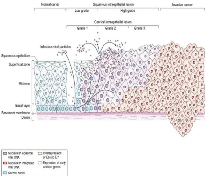
Fig 1: Pathogenesis of HPV in precancerous and cancerous cervical lesion
Adapted from Woodman et al, Nat Rev Cancer 2007;7:11-22
14 region is transcribed early in the viral life cycle and encodes proteins that are needed for viral replication. The late region encodes for viral structural proteins that are produced late in the life cycle. Open reading frames (ORFs) of early region encode proteins needed for viral replication and maintenance in infected cells. The early region of HPV genome contains E5, E6 and E7 transforming regions. The E6 and E7 ORFs encode the major transforming genes of HPV (28). E6 can bind to p53 and result in rapid proteolytic degradation of p53, through ubiquitin-dependent mechanism, thus preventing apoptosis. E7 binds to the retinoblastoma(Rb) gene product and also to ‘‘Rb-like proteins’’. Binding of E7 to Rb blocks the cell proliferation by inhibiting the function of these endogenous tumor suppressors. The final result of over expression of E6 and E7 within infected cells is unrestricted cell proliferation.
15 infection. This latent infection does not produce characteristic morphological changes in the epithelial cell. They can only be detected by molecular testing. Productive viral infection occurs independent of host chromosomal DNA synthesis and results in replication of viral DNA in the cervical epithelium. The infected cervical epithelial cells move toward epithelial surface as they mature. Transcriptional factors produced by the epithelium stimulate the production of viral capsid protein. Viral associated effects of HPV produce characteristic cytological and histological changes seen in the cervical epithelial cells due to large amounts of virions. These viral-associated effects include nuclear atypia, multinucleation, acanthosis, cytoplasmic vacuolization and koilocytosis.
16 Approximately, 10 % of high risk HPV infections will stay for more than two years. In a few women, recurrent HPV infections can occur after the clearance of the previous infection. There is strong association between immunodeficiency and detection of HPV in patients with HIV infection detected in one study(32). The above facts suggest that women who appear to have cleared the HPV infection continue to harbor low copy number of latent HPV in the epithelium. HPV has high prevalence in sexually active population. Prevalence of high risk HPV infection is high in late teens and drops with increasing age, in women with normal cervical cytology(33). High HPV prevalence is seen in less developed countries due to unknown reasons, but this may be related to poor personal hygiene, sexual practices and increase in burden of comorbid conditions in the general population.
17 Due to understanding of the pathogenesis in squamous intraepithelial lesion, researchers have proposed that LSIL is multicellular in origin whereas HSIL is unicellular in origin. LSIL develops within latently infected cervical epithelial cells and is linked with multiple types of HPV infection. HSIL is frequently aneuploid associated with a single type of HPV and may have integrated HPV DNA(36). Squamous intraepithelial lesions can be monoclonal or polyclonal. Low risk HPV types in LSIL are polyclonal whereas high risk HPV types in LSIL are monoclonal(37). This shows that LSIL associated with low risk HPV types are biologically different from lesions identified as low grade by histology, but harbor high risk HPV types. Studies on CIN1 and CIN2 have shown that 83 % of monoclonal lesions progress and 64% polyclonal lesions regress(38).
Cellular origin of squamous intraepithelial lesions:
18 high grade precursors have the capacity to progress to invasive cervical cancers(41). Studies have shown that on long term follow up, partially treated or untreated HSIL had 30-50 % chance for progression to invasive cervical cancer over 30 years(42).
Clinical features:
SIL is seen more commonly on the posterior lip of cervix and it involves rarely the lateral cervical regions(43). It may spread horizontally and involve the entire transformation zone. Rarely endocervical extension beyond the endocervical canal and then into the uterus can be seen. Based on the severity of the lesion, the size and endocervical distribution tend to vary.
Screening tests:
Pap smear:
19
Criteria for precancerous cervical lesion:
According to the Bethesda 2014, squamous cell abnormalities are classified as following:(45)
1) Atypical squamous cells
o of undetermined significance (ASC-US) o cannot exclude HSIL (ASC-H)
2) Low-grade squamous intraepithelial lesion (LSIL) 3) High-grade squamous intraepithelial lesion (HSIL) 4) Squamous cell carcinoma.
Table 1: Criteria used to diagnose squamous cell abnormalities
20 Bethesda System subdivides the ASC category into two subdivisions.
o Atypical squamous cells of undetermined significance (ASCUS) refers to samples in which the cytological changes are suggestive of LSIL, but lack sufficient cytological abnormalities to allow a definitive diagnosis.
21
1.
Atypical squamous cells of undetermined significance(ASC-US)
Criteria:
• In ASC-US cells nuclei are enlarged from 2.5to 3 times of the normal intermediate cell nucleus or more than 2 times the nucleus size of a squamous metaplastic cell.
• Mildly increased N/C ratio with minimal nuclear hyperchromasia and irregularly distributed chromatin.
• Atypical parakeratosis and incomplete koilocytosis.
2.
Low grade squamous Intraepithelial lesion (LSIL)
Criteria:
• Nuclear enlargement > 3times of normal intermediate nuclei with mildly increased N/C ratio.
• Cytoplasmic and nuclear changes usually seen in intermediate or superficial squamous cells.
• Overall cell size is enlarged with abundant cytoplasm.
• Nuclei are usually hyperchromatic with coarsely granular to densely opaque chromatin, mild anisonucleosis and absent nucleoli.
• Smooth to irregular nuclear membranes.
22
Figure 2: Atypical squamous cell of undetermined significance at 200x
23
Fig 3: Low grade Squamous Intraepithelial Lesion(LSIL)at 400X
24
Atypical squamous cells – cannot exclude HSIL (ASC-H):
Criteria:
• Atypical squamous cells – cannot exclude HSIL (ASC-H) - cytological changes are suggestive of HSIL but not insufficient to allow a definitive interpretation.
• Cells are size of metaplastic cells usually seen singly or arranged in small groups.
•
Nuclei about 1.5–2.5 times more than normal metaplastic squamous cells with highN/C ratio.
High-Grade Squamous Intraepithelial Lesion (HSIL):
Criteria:
• HSIL cells are smaller and show less cytoplasmic maturity as compared to cells of LSIL but higher N/C ratio.
• Cells occur singly, sheets, or in syncytial-like aggregates may result in hyperchromatic crowded groups.
• Nuclei are generally hyperchromatic with irregular nuclear grooves and frequent indentation.
•
Nucleoli are usually not seen, but may be present when high grade lesion extends intoendocervical gland.
•
Cytoplasm is usually immature but occasional “mature” and densely keratinized25
Figure 4: Atypical Squamous cell – Can not exclude HSIL (ASC-H) at 400x
26
Figure 5: High grade squamous intraepithelial lesion (HSIL) at 400x
27
Management of precancerous cervical lesions:
The American Society for Colposcopy and Cervical Pathology (ASCCP)guidelines(18):
Atypical squamous Cells of Undetermined Significance: (ASC-US)
• Women with ASC-US who are HPV-negative should come for repeat HPV testing or co-testing at 3 years.
• Women with ASC-US who are HPV-positive should be referred for colposcopy examination to look for precancerous lesion.
• If hr-HPV test is not available, suggested follow-up at 12 months.
• If colposcopy is negative, co-testing is recommended at 12 months. If both tests are negative (Cytology as well as HPV), suggested follow up testing in 3 years.
• Diagnostic excisional tests like loop electrosurgical excision is not acceptable in women with ASC-US cytology with absence of high grade (CIN2+) cervical intraepithelial lesion.
28
Low-Grade Squamous Intraepithelial Lesion (LSIL)
• ASCUS-LSIL Triage Study (ALTS) suggested that high-risk -HPV testing is not recommended particularly in women with less than thirty years due to the high prevalence of HPV infection(46).
• HPV testing is advisable for LSIL in postmenopausal women because of more specificity in this group of women.
• HPV co-testing in women with LSIL cytology is recommended above 30 years of age.
• Follow-up is recommended in women with LSIL cytology less than 25 years of age at the interval period of 12 months
29
Atypical Squamous cells – Cannot exclude HSIL(ASC-H)
• Colposcopy guided cervical examination is recommended for women with ASC-H cytology irrespective of HPV result. Reflex HPV testing is not advisable in
ASC-H cytology.
High-Grade Squamous Intraepithelial Lesion (HSIL)
• Colposcopy guided cervical examination is recommended for women with HSIL cytology. HPV testing is not recommended.
• Most of the women with a cytologic result of HSIL will have identifiable lesion in colposcopy and biopsy of lesion should be taken to confirm high grade (CIN 2+) cervical intraepithelial lesion(47).
• Women aged 25 years and older with cytologic HSIL, immediate excisional procedure may be performed at the time of colposcopy if a lesion is identified.
30
Treatment:
Colposcopy with biopsy helps in planning the management of patients with precursor lesions of cervix. It allows the gynaecologist to rule out invasive cancer. These precancerous cervical lesions will be treated by conservative methods like laser ablation, cryosurgery and loop electrical excision procedure (LEEP) or cone biopsy.
Prognosis:
High grade precursor lesions are more likely to persist. Approximately 57% of CIN 1 lesions regress spontaneously without any intervention and 11% progress to carcinoma in situ. About 43% of CIN 2 lesions regress while 22% progress to carcinoma in situ. 32% of CIN III lesions regress and 12 % progress to carcinoma in situ(49). Recent studies by Castle et al. shows that rates of spontaneous regression of biopsy confirmed CIN II after 24 months of follow-up was 43% which is identical to older studies(50).
HPV DNA testing.
31 Since the knowledge of HPV pathophysiology has been established, the identification of new biomarkers with ability to distinguish those at risk of disease progression becomes necessary. Therefore several host cell biomarkers were evaluated to improve the specificity for the screening of cervical intraepithelial lesions(3). One such bio marker which has been recently identified is dual P16INK4A/ki67.
p16 INK4A
:
P16 INK4A (also referred to as p16) is a tumor suppressor protein that helps in cell cycle regulation. It acts by inhibiting cyclin D – cyclin dependent kinase 4 complex formation, thereby preventing cell cycle progression. Numerous studies have demonstrated p16 to be down regulated in many tumors. However, interestingly, increased expression of p16 has also been described in few tumors (51).
Physiological role of p16Ink4a
:
32
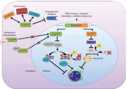
Fig 6: Molecular mechanism of cervical cancer by HPV
Adapted from Romagosa et al, Oncogene (2011)
33 cyclin D – cyclin dependent kinase 4/6 complex, it releases the E2F transcription factors, and thereby facilitates cell cycle progression.(53). P16Ink4a acts as a CDK inhibitor, blocks the cyclin D – CDK4/6 mediated phosphorylation of Rb and induces cell cycle arrest. In HPV-related neoplasms, the molecular mechanism if p16 INK4a can be explained by the presence of HPV oncoproteins E6 and E7. The integration of the viral DNA into host DNA causes overexpression of these viral oncoproteins. E6 protein binds and causes degradation of p53. E7 binds to RB protein and displaces the E2F transcription factors promoting cell cycle progression(54,55). This inactivation of the RB protein releases p16 INK4a from its negative feedback control, which in turn causes a paradoxical increase in the levels of p16. Therefore, the overexpression of p16 INK4a in HPV related tumors is considered to be due to an unsuccessful attempt to inhibit cell replication(56). In addition to its role in cell cycle regulation, p16 INK4a has also been implicated in few other cellular processes including apoptosis, cell invasion and angiogenesis and it has been hypothesized that this role may contribute to its overexpression in some neoplasms(57).
Subcellular location of p16Ink4a overexpression:
34 hypothesized that p16 INK4a has varied roles in varied subcellular locations, and that the nuclear p16 INK4a mainly regulates cell cycle. This has been supported by studies that show downregulation of nuclear p16 INK4a was associated with E2F overexpression. However, overexpression of cytoplasmic p16 INK4a exhibited no relation with E2F expression(59). Many theories have been postulated to explain the presence of cytoplasmic p16 INK4a, and it seems this is unrelated to p16 INK4a gene alteration. Proteomic and post translational studies have shown that p16 INK4a is expressed in different subcellular location, depending on post-translational modification or its ability to form complexes with other proteins(60). In quiescent cells, p16 INK4a forms a complex with CDK4 and prevents its subsequent kinase function.
35 Adapted from Romagosa et al, Oncogene (2011)
Figure 7: Molecular difference between expression of p16 and Ki-67 among
different tumors.
36
Ki-67:
Cell proliferation is strictly associated with high expression of ki-67 protein in human beings. This ki-67 antigen is exclusively identified within the nucleus during interphase but in mitosis majority of this antigen is seen over the surface of chromosomes. Hence in all active phases of cell cycle ki-67 protein is expressed (G1, S, G2 & mitosis). Ki67 protein is absent during resting phase(G0). So, it is used to calculate the growth fraction of cells. Even though this ki-67 protein is well identified in molecular levels as good indicator of proliferation, functional characteristics still remains unclear. Ki-67 protein expression is a must for progression through the cell-division cycle(62).
P16/ki-67 dual immunocytochemistry:
37 There are several studies done on the performance of dual immunocytochemistry for p16 and Ki-67 for the identification of CIN2+ lesion(63–67). Bergeron et al assessed performance of P16/Ki-67 dual immunocytochemistry in a group of 28,000 women, from 5 European countries. In that study, performance of hr-HPV test and p16/Ki-67 dual immunocytochemistry were compared among women with the diagnosis of ASCUS and LSIL. Sensitivity of p16/Ki-67 in ASCUS for the detection of CIN II+ was 92.2% (90.9% for HR HPV test), specificity was 80.6% (36.3% for HPV test). In a group of LSIL, sensitivity was 94.2% (96.4% for hr-HPV test), specificity was 68.0% (19.1% for hr- HPV).
38 In all the above studies co-expression of cytoplasmic p16 and nuclear Ki-67 in at least one cell was considered as positive ICC test which indicates transformed infection. The concept of single positive cell as a cutoff has been used due to the monoclonal nature of any malignant neoplasm. But these studies also showed that false positivity for p16/Ki-67 resulted in decrease in its positive predictive value to identify underlying high-grade lesion.
39 Clinical utility of p16/ki-67 in urothelial malignancies has also been assessed in a few studies. These studies propose that dual immunocytochemistry is used in evaluation of high grade urothelial cancer and for the follow-up management after conservative treatment for non- invasive urothelial carcinoma (69,70).
40
41
After approval from the Institutional Review Board (IRB) min no: 10175, this prospective diagnostic study was conducted in the Department of Pathology, in conjunction with Department of Virology and Department of Gynaecologic oncology.
Study population:
A total of 175 patients from gynecology/ gynecologic oncology, whose cervical smears were reported by the Department of general pathology as ASC-US/LSIL/ASC-H and HSIL, were identified for the study, over a period of 8 months from August 2016 to March 2017. Information sheet and consent form were given to all patients willing to participate in this study.
Exclusion criteria:
All patients, who were
• Less than 25 years,
• Pregnant women,
• Patients unwilling for colposcopy/ biopsy,
• Seropositive patients and
42
Study design
Figure 08: Detailed diagrammatic Algorithm of the study.
All sexually active women >25 years who come to gynecology OPD
Cervical cytology screening by thin prep Negative
positive (ASC-US/LSIL/ASC-H/HSIL) Excluded from study
the study
Hr-HPV DNA test Colposcopy p16/Ki67 on pap smears
43
Sample collection and processing:
Thin prep cervical smear:
The residual Thin prep sample material of all study patients (Hologic vials) were stored at 2-4 degree Celsius for up to 6 weeks. If the samples exceeded 6 weeks of storage period, they were processed in the Thin Prep® 2000 Processor (Hologic™ Inc.) and the unstained smears were stored in ethanol fixative at 2-4degree Celsius.
The dual immunocytochemistry was performed in batches. In view of inadequacy and poor cellularity of some smears that were stored, the third batch of smears were prepared from the refrigerated samples, on the day of the ICC procedure.
Technical procedure as described in Appendix-1.
P16/ki67 dual immunocytochemistry
:
Immunocytochemistry analysis using the CINtec PLUS Cytology kit (Roche MTM Laboratories, Heidelberg, Germany) were done on these smears according to the manufacturer's instructions. All slides were examined by the principal investigator and the guide.
44
Hr-HPV DNA test
:
HPV DNA tests for hr-HPV were also done on all study patients as per the standard management protocol followed in the Gynecology outpatient department. The material for this test was collected at the time of colposcopy in using the digene HC2 DNA collection device which consisted of a cervical brush and digene Specimen Transport Medium(STM). In vitro nucleic acid assay with signal amplification by Hybrid Capture 2 assay (HC2; Digene Corp., Gaithersburg, MD) was used.
Technical procedure as described in Appendix-3.
Interpretation of HPV DNA
:
The hr - HPV DNA data was interpreted based on the results (positive/ negative) obtained from Hybrid Capture (HC2) in the department of virology. Serial amplification assay for hr- HPV DNA in cervical specimen can detect upto13 different hr-HPV DNA types [16,18,31,33,35,39, 45,51,52,56,58,59 and 68] in the cervical cells. Detection of hr-HPV DNA in Hybrid Capture (HC2) technique is done by using microplate chemiluminescence. The result is obtained as relative light units (RLU). The digene
45
Colposcopy and biopsy:
Colposcopy and biopsy were also done on all the study patients as part of a routine protocol, in the Gynecology outpatient department, according to accepted diagnostic standards.
Histopathology:
Fixed tissue samples were processed to paraffin blocks in the histopathological laboratory. Slides(4μm) were cut on the microtome, stained with hematoxylin and eosin (H+E) and were assessed by histopathologists who were blinded to the ICC and HPV results of the study patients. Biopsy was considered as gold standard, against which the new dual immunocytochemistry and hr-HPV DNA results were correlated.
Interpretation of P16/ki-67 dual immunocytochemistry:
The dual immunocytochemistry was interpreted by the primary investigator and guide. The presence of one or more cervical epithelial cells with co-localization of brown cytoplasmic immunostaining and red nuclear immuno-staining within the same cell was regarded as a positive CINtec PLUS test result. We also assessed the performance of p16/Ki-67 dual immunocytochemistry, with a cut off for positivity as more than ten cells.
46
The presence of cervical epithelial cells that do show a single immuno-reactivity only for one of the two markers (e.g. – Brown staining for p16 only, or red staining for Ki-67 only)
is not considered as a positive test result for the CINtec PLUS kit. Strict criteria for positive and negative tests were followed to avoid discrepancy.
Smears from a known case of squamous cell carcinoma were used as a positive control, with colocalization of both brown cytoplasmic immunostaining (p16) and red nuclear immunostaining (ki67). Different cell types present in representative cervical cytology specimens, that are known to be negative for the expression of the p16 and ki-67 antigens (such as superficial cells) may serve as an additional internal negative control to assess any background staining.
Clinical details of the cases
:
47
Statistical method used
Sample size calculation:
The required samples size to show sensitivity of about 90% with 10% precision and 95% confidence interval was found to be 35 patients.
Clinical Research form:
The data obtained from each test and the necessary clinical information obtained from the clinical workstation were entered into the clinical research form for each case. (Form attached in annexure).
Data entry and analysis:
48
49
Study flow chart
Total number of study samples: 175
Lost to follow up =32
Biopsy not done = 11
HPV test not done = 08
Figure 9: Selection of cases included under study Total samples available for testing: 124
Adequate Scanty Inadequate(excluded)
76 18 30
50 A total of 175 cases were reported as ASCUS, LSIL, ASC-H and HSIL between August 2016 to March 2017. Thirty-two patients did not come for follow -up, eleven cases did not have biopsy and eight cases did not have HPV. Fifty-six cases were excluded from the study, leaving a total of 124 samples for further testing.
51
ABNORMAL PAP SMEAR GROUPS:
Figure 10: Number of cases in each abnormal pap smear groups
The median age was 42.73 years (standard deviation of 11.4 years) for overall abnormal pap smears with ASC-US, LSIL, ASC-H and HSIL. The youngest woman was 24 and the oldest patient was 71.
• ASC-US: 43 (45%)
• LSIL: 16 (17%)
• ASC-H: 14 (15%)
• HSIL: 22 (23%)
0 5 10 15 20 25 30 35 40 45 50 ASCUS
LSIL ASC-H HSIL
NUMBER
GR
52
AGE:
Figure 11: Different groups in accordance with age
ASC-US was the most common intra epithelial lesion identified in all age group of women.
There were 12cases (13%) less than 30 years of age and 82 cases (87%) above 30 years of age. 5 10 17 4 6 3 10 2 1 0 3 4 4 3 0 1 7 8 5 2 0 2 4 6 8 10 12 14 16 18
<30 31-40 41-50 51-60 >60
NUM
B
E
R
GROUP
53
SCREENING:
Figure 12: Indication for screening
Among 94 women, 75 were symptomatic (80%) whereas 19 were asymptomatic (20%) who were referred for routine screening.
80%
20%
54
SYMPTOMS:
Figure 13: Different clinical presentation of abnormal pap smears
Symptomatic women presented with following complaints:
Bleeding per vagina (59%), lower abdominal pain (16%), white discharge per vagina (11%), itching (7%), burning micturition (4%), post coital bleeding (3%) and cervical growth (1%). Bleeding per vagina was the most common clinical presentation of women presented to gynecology/ gynecologic oncology.
55
MENSTRUAL STATUS:
Fig 14: Number of reproductive and postmenopausal women
Among 94 women, 25 were in postmenopausal age group (27%) and 69 were reproductive age group (73%).
27%
73%
56
Hr-HPV TEST vs BIOPSY IN
≥CIN2 LESIONS:
Table 02: hr– HPV test vs biopsy in ≥ CIN 2 lesions cross tabulation:
The above table shows performance of hr-HPV test among abnormal Pap smears as compared with the gold standard of ≥ CIN2 lesions i.e. biopsy. There were 94 study cases. Hr-HPV test was positive in 58 cases and negative in 36 cases. Among positive cases 30 cases (32%) had ≥ CIN2 lesions (True positive) and 28cases (30%) biopsies were negative (False positive). Among hr-HPV test negative cases, 1 case (1%) had ≥ CIN2 lesion (False negative) and 35 cases (37%) were negative (True negative) on biopsy.
• Total number of cases: 94
• Number of true positive: 30 (32%)
• Number of false positive:28 (30%)
• Number of false negative: 01 (1%)
• Number of true negative: 35 (37%)
≥ CIN2 LESIONS
TOTAL
Positive
Negative
hr-HPV test
Positive
30 28 58Negative
1 35 3657
P16/Ki-67 IMMUNOCYTOCHEMISTRY vs BIOPSY IN
≥CIN2 LESIONS:
Table 03: p16/Ki-67 vs biopsy in ≥ CIN2 lesions cross tabulation
The above table shows the performance of dual immunocytochemistry test among abnormal Pap smears as compared with gold standard of ≥ CIN2 lesions i.e. biopsy. There were 94 study cases. Dual immunocytochemistry was positive in 49 cases and negative in 45 cases. Among positive cases 30 cases (32%) had ≥ CIN2 lesion (True positive) and 19 cases (20%) biopsies were negative (False positive). Among negative cases 1case (1%) had ≥ CIN 2 lesions (False negative) and 44 cases (47%) biopsies were negative (True negative).
▪ Total number of cases: 94
▪ Number of true positive: 30 (32%)
▪ Number of false positive: 19 (20%)
▪ Number of false negative: 1(1%)
▪ Number of true negative:44 (47%)
≥
CIN2 LESIONS
TOTAL
Positive
Negative
p16/Ki-67
Positive
30 1949
Negative
1 4445
58
PERFORMANCE OF P16/KI-67 AND HR-HPV TEST IN DETECTION OF
≥ CIN2 LESIONS:Table 04
:
p16/Ki-67 and Hr-HPV test in ≥ CIN 2 lesionsThis table summarizes the analysis for sensitivity, specificity, PPV and NPV with gold standard ≥ CIN2 lesions for p16/Ki-67 and hr-HPV tests for abnormal Pap cases. The sensitivity of dual-stained cytology and hr-HPV DNA testing was 96.8% for ≥ CIN2 in all abnormal groups. The specificity of dual-stained cytology was 70.2% for whereas hr-HPV test had specificity of 55.8% (table 04).
Sensitivity
%(95%CI)
Specificity
%(95%CI)
PPV
%(95%CI)
NPV
%(95%CI)
Hr-HPV test 96.8 55.8 51.7 97.6
P16/Ki-67≥ 1
positive cell
96.8 70.2 61. 97.6
P value 1.00 0.095 0.324 1.00
95% CI
Difference
59 Specificity for dual stained cytology was higher than hr-HPV test. The positive predictive value for dual stained cytology was 61.2% whereas hr-HPV test had 51.7%. The positive predictive value for dual stained cytology was higher than hr-HPV test. The negative predictive value for dual stained cytology and hr-HPV was similar 97.6%. P value and 95% CI difference for both tests were not significantly significant (table 04).
PERFORMANCE OF P16/KI-67 AND HR-HPV IN DETECTION OF ≥ CIN3
LESIONS:
Table 05
:
p16/Ki-67 and Hr-HPV test in≥ CIN 3 lesions Sensitivity%(95%CI)
Specificity %(95%CI)
PPV
%(95%CI)
NPV
%(95%CI)
Hr-HPV test 100 47.4 31 100
P16/Ki-67 ≥ 1 positive cell
100
59.2 36.7 100
P value 1.000 0.186 0.534 1.000
95% CI
60 This table summarizes the analysis for sensitivity, specificity, PPV and NPV with gold standard ≥ CIN3 lesions for p16/Ki-67 and hr-HPV tests for abnormal Pap cases. The sensitivity and negative predictive value for p16/Ki-67 dual immunocytochemistry and high-risk HPV test was 100% for ≥ CIN3 lesions. Specificity of p16/Ki-67 was 59.2 % and specificity of high risk HPV test was 47.4 %.
61
THRESHOLD FOR POSITIVE IMMUNOCYTOCEMISTRY IN≥ CIN2
LESIONS:
The presence of one or more cervical epithelial cells with co expression of brown cytoplasmic P16 immunostaining and red nuclear Ki-67 immunostaining within the same cell is regarded as a positive CINtec PLUS test result. However, we also looked at performance of dual immunocytochemistry with a threshold of positivity for more than 10 cells(68).
Figure 15: Performance of p16/Ki-67 immunocytochemistry with different
thresholds in detection of ≥CIN2 lesions
Increase in threshold showed higher specificity (95.1 vs 70.2) and positive predictive value (89.3 vs 61.2) but there was decrease in sensitivity (78.1 vs 96.8) and negative predictive value (89.2 vs97.6).
96.8
70.2
61.2
97.6
78.1
95.1
89.3 89.2
SENSITIVITY SPECIFICITY PPV NPV
62
Table 06
:
p16/Ki-67(≥10 cells) and Hr-HPV test in≥ CIN 2 lesionsThis table summarizes the statistical difference for sensitivity, specificity, PPV and NPV.
P value and 95% CI for sensitivity, specificity and positive predictive values were statistically significant. Negative predictive value was not statistically significant (table 06).
Sensitivity
%(95%CI)
Specificity
%(95%CI)
PPV
%(95%CI)
NPV
%(95%CI)
Hr-HPV test 96.8 55.8 51.7 97.6
P16/Ki-67≥ 10
positive cells
78.1 95.1 89.3 89.2
P value 0.026 <0.001 0.001 0.132
95% CI
difference
63
THRESHOLD FOR POSITIVE IMMUNOCYTOCEMISTRY IN
≥CIN3+
LESION:
The diagnostic capability of dual stained cytology for ≥ CIN 3 lesions with positivity for more than 10 cells showed higher specificity (82.7 vs 59.2) and higher positive predictive value (53.6 vs 36.7) but there was decrease in sensitivity (83.8 vs 100) and negative predictive value (100 vs 95.4).
Figure 16: Performance of p16/Ki-67 immunocytochemistry with different
thresholds in detection of ≥ CIN3 lesions
100
59.2
36.7
100
83.8 82.7
53.6
95.4
SENSITIVITY SPECIFICITY PPV NPV
64
Table 07: p16/Ki-67 (≥10 cells) and Hr-HPV test in ≥ CIN 3 lesions
This table summarizes the analysis for sensitivity, specificity, PPV and NPV. P value and 95% CI for sensitivity, specificity and negative predictive values were statistically significant. Positive predictive value was not statistically significant (table 07).
Sensitivity
%(95%CI)
Specificity
%(95%CI)
PPV
%(95%CI)
NPV
%(95%CI)
Hr-HPV 100 47.3 31 100
P16/Ki-67≥ 10
positive cells
83.8 82.7 53.6 95.4
P value <0.001 <0.001 0.051 <0.001
95% CI
difference
65
PERFORMANCE OF P16/KI-67 AND HR-HPV TEST IN DETECTION OF
≥ CIN2 LESIONS IN WOMEN <30 YEARS:
Table 08: p16/Ki-67 and Hr-HPV test in ≥CIN 2 lesions in women <30 years
This table summarizes the analysis for sensitivity, specificity, PPV and NPV with gold standard ≥ CIN2 lesions for p16/Ki-67 and hr-HPV tests for abnormal Pap cases in women < 30 years. There were 12 women less than 30 years of age and 82 women more than 30 years of age. The sensitivity and negative predictive values of hr-HPV and dual stained cytology were similar 100%. The specificity of hr-HPV was 45.5% and positive predictive value 12.1%. Sensitivity, specificity and negative predictive value and positive predictive value 95% CI difference and P value were not statistically significant (table 08). Sensitivity %(95%CI) Specificity %(95%CI) PPV %(95%CI) NPV %(95%CI)
Hr-HPV test 100 45.5 12.1 100
P16/Ki-67 ≥1
positive cell
100 36.4 14.3 100
P value 1.000 0.301 0.737 1.000
95% CI
difference
66
Figure 17: Performance of p16/Ki-67 immunocytochemistry with different
thresholds to detect ≥ CIN2 lesions in women <30 years
The dual immunocytochemistry showed, specificity of 36.4% and positive predictive value of 14.3% when a score of one or more cells was the positive threshold. Both tests had low specificity and positive predictive value. When the threshold was increased to more than ten cells there was significant increase in specificity (90 vs 30.6) and positive predictive value (50 vs 14.3). P value and 95% CI for specificity and positive predictive value were statistically significant. Sensitivity and negative predictive values were not statistically significant (table 09).
100
36.4
14.3
100 100
90
50
100
SENSITIVITY SPECIFICITY PPV NPV
67
PERFORMANCE OF P16/KI-67 AND HR-HPV TEST IN DETECTION OF ≥ CIN2 LESIONS IN WOMEN LESS THAN 30 YEARS.
Table: 09: p16/Ki-67(≥10 cells) and Hr-HPV test in ≥ CIN 2 lesions in women <30 years
This table summarizes the analysis for sensitivity, specificity, PPV and NPV. P value and 95% CI for specificity and positive predictive values were statistically significant. sensitivity and negative predictive value was not statistically significant.
Sensitivity
%(95%CI)
Specificity
%(95%CI)
PPV
%(95%CI)
NPV
%(95%CI)
Hr-HPV test 100 45.5 12.1 100
P16/Ki-67 >10
positive cells
100 90 50 100
P value 1.000 <0.001 <0.001 1.000
95% CI
Difference
68
PERFORMANCE OF P16/KI-67 AND HR-HPV TEST IN DETECTION OF ≥ CIN2 LESIONS IN WOMEN MORE THAN 30 YEARS.
Table10: P16/KI-67 and hr-HPV test in ≥ CIN2 lesions in women >30 years
This table summarizes the analysis for sensitivity, specificity, PPV and NPV for ≥ CIN2 lesions p16/Ki-67 immunocytochemistry and hr-HPV tests for women >30 years. The sensitivity of hr-HPV test and dual immunocytochemistry cytology were similar 100% in all 82 women >30 years of age. The negative predictive values of both hr-HPV test and dual stained cytology were relatively high (93.5 and 95.1). The specificity and positive predictive value for hr-HPV test were 56.9%. P value and 95% CI difference for specificity was statistically significant. Sensitivity, positive and negative predictive values were not statistically significant (table 10).
Sensitivity %(95%CI)
Specificity %(95%CI)
PPV
%(95%CI)
NPV
%(95%CI)
Hr-HPV test 93.5 56.9 56.9 93.5
P16/Ki-67≥ 1 positive
cell 93.5 76.5 70.7 95.1
P value 1.000 0.020 0.140 0.756
69
Figure 17: Performance of p16/Ki-67 immunocytochemistry with different
thresholds to detect ≥ CIN2 lesions in women >30 years
The dual stained cytology showed, specificity of 76.5% and positive predictive value of 70.7% when the threshold was taken as ≥ 1 cell. When the threshold was more than ten cells, there was increase in specificity (96.1 vs 76.5) and positive predictive value (92.3vs 70.7).
93.5
76.5
70.7
95.1
77.4
96.1
92.3
87.5
SENSITIVITY SPECIFICITY PPV NPV
70
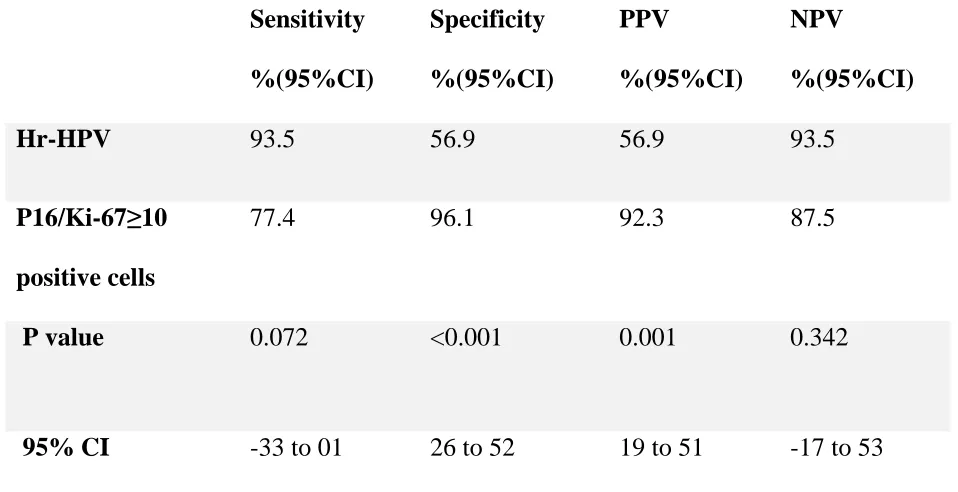
Table 11: P16/KI-67 (≥ 10 cells) and hr-HPV test in ≥ CIN2 lesions in women >30
years
This table summarizes the analysis for sensitivity, specificity, PPV and NPV. P value and 95% CI difference for specificity and positive predictive value were statistically significant. Sensitivity and negative predictive value was not statistically significant (table:11).
Sensitivity
%(95%CI)
Specificity
%(95%CI)
PPV
%(95%CI)
NPV
%(95%CI)
Hr-HPV 93.5 56.9 56.9 93.5
P16/Ki-67≥10
positive cells
77.4 96.1 92.3 87.5
P value 0.072 <0.001 0.001 0.342
95% CI
difference
71
AGREEMENT BETWEEN HR-HPV TEST AND P16/KI-67:
The kappa for hr-HPV test to detect ≥ CIN2 lesions was 0.406. When > 1 cell was used
as the threshold, the kappa was 0.559 but the agreement was increased to 0.754, when more than 10 cells were used as the threshold. Thus, the kappa agreement for dual
[image:81.612.71.525.247.596.2]immunocytochemistry for ≥ CIN2 lesions was higher when compared to hr-HPV test.
Figure 18: Agreement between p16/Ki-67 and hr-HPV test ≥ CIN2 lesions with different thresholds.
0.406
0.559
0.754
0 0.1 0.2 0.3 0.4 0.5 0.6 0.7 0.8
CIN2+
72
PERFORMANCE OF P16/KI-67 AND HR-HPV TEST IN DETECTION OF
≥ CIN2 LESIONS IN ASC-US.
Table 12: p16/Ki-67 and hr-HPV in CIN ≥ 2 lesions for ASC-US
This table summarizes the analysis for sensitivity, specificity, PPV and NPV with gold standard ≥ CIN2 lesions for ASC-US group. Sensitivity and negative predictive value for p16/Ki-67 dual immunocytochemistry and hr-HPV test were similar (100%) to detect high grade lesions among ASCUS group. P16/Ki-67 specificity was 82 % and positive predictive value was 30%. Hr-HPV test specificity was 71.8% and positive predictive value was 21.4%. P value and 95% CI difference for both tests were not significantly significant (table 12).
Sensitivity %(95%CI) Specificity %(95%CI) PPV %95%CI) NPV %(95%CI)
Hr-HPV test 100 71.8 21.4 100
P16/Ki-67≥ 1 Positive cell
100 82.1 30 100
P value 1.000 0.280 0.631 1.000
95% CI difference
73
Figure 19: Performance of p16/Ki-67 immunocytochemistry with different
thresholds to detect ≥ CIN2 lesions in ASC-US.
When the threshold was more than ten cells, there was increase in specificity (82.1 vs 94.9) and positive predictive value (30 vs 50) and decrease in sensitivity (100 vs 66.7) and negative predictive value (100 vs 97.4). Sensitivity, specificity and positive predictive value P value and 95% CI were not statistically significant. Negative predictive value was not statistically significant (table:13)
100
82.1
30
100
66.7
94.9
50
97.4
SENSITIVITY SPECIFICITY PPV NPV
74
Table 13: p16/Ki-67(≥ 10 cells) and hr-HPV in CIN ≥ 2 lesions for ASC-US
This table summarizes the analysis for sensitivity, specificity, PPV and NPV. P value and 95% CI difference for specificity was statistically significant. Sensitivity positive predictive value and negative predictive value were not statistically significant (table:13)
Sensitivity %(95%CI)
Specificity %(95%CI)
PPV
%(95%CI)
NPV
%(95%CI)
Hr-HPV test 100 71.8 21.4 100
P16/Ki-67 ≥ 10 positive cells
66.7 94.9 50 97.4
P value 0.185 0.006 0.260 <0.001
95% CI difference
75
PERFORMANCE OF P16/KI-67 AND HR-HPV IN DETECTION OF ≥ CIN2
LESIONS IN LSIL:
Table 14: p16/Ki-67 and hr-HPV test in ≥ CIN2 lesions for LSIL
This table summarizes the analysis for sensitivity, specificity, PPV and NPV with gold standard ≥ CIN2 lesions for LSIL group. Sensitivity and Negative Predictive Value for p16/Ki-67 dual immunocytochemistry and hr-HPV test were similar (100%) to detect high grade lesions among LSIL group. P16/Ki-67 specificity was 64.3 % and positive predictive value was 28.6%. Hr-HPV test specificity was 21.4% and positive predictive value was 8.3%. P value and 95% CI difference for sensitivity, specificity, PPV and NPV values were not statistically significant. (table:14)
Sensitivity %(95%CI)
Specificity %(95%CI)
PPV
%(95%CI)
NPV
%(95%CI)
Hr-HPV test 100 21.4 8.3 100
P16/Ki-67≥ 1 positive cell
100 64.3 28.6 100
P value 1.000 0.184 0.242 1.000
76
Figure 20: Performance of p16/Ki-67 immunocytochemistry with different
thresholds to detect ≥ CIN2 lesions in LSIL.
When the threshold was more than ten cells, there was increase in specificity (64.3 vs 100) and positive predictive value (28.6 vs 100) and similar in sensitivity (100) and negative predictive value (100).
100
64.3
28.6
100
100 100 100 100
SENSITIVITY SPECIFICITY PPV NPV
77
Table 15: p16/Ki-67(≥ 10 cells) and hr-HPV in CIN ≥ 2 lesions for in LSIL.
This table summarizes the analysis for sensitivity, specificity, PPV and NPV. P value and 95% CI difference for sensitivity, specificity, PPV and NPV values were not statistically significant.
Sensitivity %(95%CI)
Specificity %(95%CI)
PPV
%(95%CI)
NPV
%(95%CI)
Hr-HPV test 100 21.4 8.3 100
P16/Ki-67 ≥ 10 positive cells
100 100 100 100
P value 1.000 0.407 0.937 1.000
95% CI difference
78
MENOPAUSE STATUS:
[image:88.612.72.509.308.602.2]There were 25 women in postmenopausal age group. The sensitivity and negative predictive value for hr-HPV test and dual stained cytology were similar 100%. The specificity for hr-HPV test was 58.3 and PPV was 72.5. Dual stained cytology had specificity of 66.7% and PPV 76.5% when cutoff was one or more cells but with cutoff of more than 10 cells showed 100 % specificity and PPV for ≥ CIN2 lesions.
Figure 18: Performance of p16/Ki-67 immunocytochemistry and hr-HPV test to detect ≥ CIN2 lesions in postmenopausal women.
100 58.3 72.2 100 100 66.7 76.5 100 84.6 100 100 85.7 0 20 40 60 80 100 120
Sensitivity Specificity PPV NPV
79 There were 69 women in reproductive age group. The sensitivity for both tests were similar (89%). The negative predictive value for hr-HPV test was 93.1 and p16/Ki-67 was 94.6%. The specificity for hr-HPV test was 48.3 and PPV was 22.5. p16/Ki-67 Dual immunocytochemistry with more than ten cells cutoff showed significantly improved specificity (93.9 vs 70), PPV (82.4 vs 53.1) and decrease in sensitivity (73.7 vs 89.5) and NPV (90.2 vs 94.6%) as compare to a threshold of one or more cells.
Figure 19: Performance of P16/Ki-67 immunocytochemistry and hr-HPV test in detection of ≥ CIN2 lesions in reproductive age group women
89.5 48.3 22.5 93.1 89.5 70 53.1 94.6 73.7 93.9 82.4 90.2 0 10 20 30 40 50 60 70 80 90 100
sensitivity specificity PPV NPV
80
[image:90.612.73.463.97.386.2]Images:
[image:90.612.72.466.406.683.2]Figure 20: Positive control of p16/Ki-67 dual ICC at 40x
81
Figure 22: Negative control of p16/Ki-67 dual ICC at 200x
[image:91.612.75.480.404.671.2]82
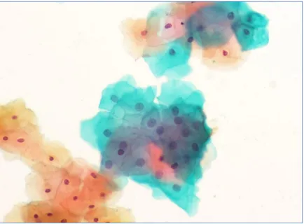
Figure 24: P16/ki67 dual ICC- ASCUS at 200x.
[image:92.612.73.481.401.671.2]83
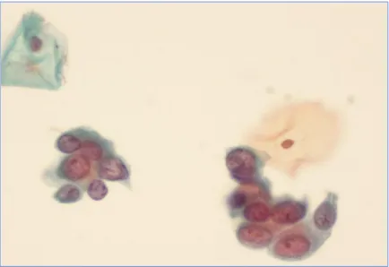
Figure 27: P16/Ki-67 dual ICC- ASC-H at 200x
84
85 Cervical cancer is still one of the major causes of death worldwide, particularly more common in developing countries. Pap smear is still one the most efficient tests used for screening of cervical carcinoma in many countries. The disadvantage of using Pap smear is that it has low sensitivity and relatively high specificity. Since the awareness about association between cervical carcinoma and human papilloma virus, high risk HPV DNA test was implemented as a potential screening test to improve the sensitivity and to identify 13 high risk types of HPV.
This test had shown good sensitivity when compared to Pap smear screening but it has low specificity. Presence of persistent hr- HPV was associated with more risk of developing precancerous cervical lesion followed by invasive malignancy due to dysregulation of cell cycle by incorporation oncogenic viral proteins into the host genome.
86 Thus, it was important to find out a better triaging test for hr-HPV positive women or women with minor cytological abnormalities to reduce the number of false positive cases and thereby to reduce the number of unwanted colposcopies and biopsies. One of the relatively recently introduced triage system for these cases is immunocytochemistry p16/Ki-67. p16 is a protein which triggers the cell cycle arrest under normal physiological state and Ki-67 indicates proliferating index of a cell. Co-expression of both p16 and Ki-67 in a same cell indicates dysregulated cell cycle due to incorporation viral oncoprotein into the host genome suggestive of an underlying high-grade lesion.
Our study is a prospective diagnostic study which evaluated performance of dual stained immunomarker p16/ki-67 with HC2 HPV assay in cervical screening for detection of high grade cervical intraepithelial lesion in abnormal Pap smears. This study included 42 ASC-US, 16 LSIL, 14 ASC-H and 22 HSIL cases of abnormal Pap smears from August 2016 to March 2017 for analysis.
87
Performance of p16/Ki-67 and hr-HPV test:
In our study the performance of dual immunocytochemistry for high grade (≥ CIN2) lesion was as follows: sensitivity 96.8%, negative predictive value 97.6%, specificity 70.2% and positive predictive value of 61.2%. Thus, for detecting high grade lesions (≥ CIN2) among all abnormal Pap smears, sensitivity and negative predictive value of dual immunocytochemistry was similar to HC2 HPV DNA testing, but with improved specificity and positive predictive values.
These results were comparable to You-Lin Qiao et al study (71). You-Lin Qiao et al in 2015performed cytology-based screening in total population of1290 women including pre -cancerous and cancerous lesions in china. You-Lin Qiao et al highlighted performance of p16/Ki-67 dual immunocytochemistry as following; sensitivity was 90.2% (94.4% hr- HPV test), specificity was 79.5% (76.9% for hr-HPV test), positive predictive value was 49.2% (47.1 for hr- HPV test), and negative predictive value was 97.6% (98.4% for hr- HPV test). You-Lin Qiao et al study highlighted high sensitivity and negative predictive value for hr-HPV test as compare to dual immunocytochemistry whereas our study showed similar results for sensitivity and negative predictive value for both tests (Table:10) The present study showed better specificity and positive predictive value for dual immunocytochemistry as compared to hr-HPV test which is similar to
88 Our study:
N=94 P16/Ki-67
You-Lin Qiao
et al, N=1079
p16/Ki-67
Our study:
N=94
Hr-HPV
You-Lin Qiao
et al, N=1079
Hr-HPV
Sensitivity 96.8% 90.9% 96.8% 94.4%
Specificity 70.2% 79.5% 55.8% 76.9%
PPV 61.2% 49.2% 51.7% 47.1%
[image:98.612.67.560.68.515.2]NPV 97.6% 97.6% 97.6% 98.4%
Table 16: Comparison of performance of p16/Ki-67 vs hr-HPV test in our study
89 A few studies have highlighted the difference between the performance of dual immunocytochemistry for less than 30 and more than 30 years of age (Table 11).Bergeron et al showed performance of p16/Ki-67 in LSIL for women < 30 years and >30 years as follows: sensitivity(84.6% vs 86.5%), specificity (50%vs56%), PPV (23.2vs29.4%) and NPV (94.8 vs 95.1) whereas Peter Ziemke 2017 study results were following, sensitivity(81.6 vs 84.6), specificity(61.1 vs 75.7), PPV(42.5 vs 52.8) and NPV (90.4 vs 93.8%). Above studies show that performance of p16/Ki-67 for women more 30 years was better as compared to women less than 30 years.
Table:17: p16/Ki-67 immunocytochemistry and age dependency for LSIL cytology
in different studies
Author Age number Sensitivity Specificity PPV NPV
90