T E C H N I C A L N O T E
Open Access
Establishment of a PCR analysis method for
canine BRCA2
Yasunaga Yoshikawa
1*, Masami Morimatsu
2, Kazuhiko Ochiai
3, Kento Okuda
1, Takahiro Taoda
4, Seishiro Chikazawa
5,
Asako Shimamura
6, Toshinori Omi
3, Makoto Bonkobara
6, Koichi Orino
1and Kiyotaka Watanabe
1Abstract
Background:Mammary tumors are the most common tumor type in both human and canine females. In women, carriers of mutations in BRCA2, a tumor suppressor gene product, have a higher risk of breast cancer. Canine BRCA2has also been suggested to have a relationship with mammary tumors. However, clearly deleterious BRCA2 mutations have not been identified in any canine mammary tumors, as appropriate methods to detect mutations or a consensus BRCA2 sequence have not been reported.
Findings:For amplification and sequencing of BRCA2, we designed 14 and 20 PCR primer sets corresponding to the BRCA2 open reading frame (ORF) and all 27 exons, respectively, including exon-intron boundaries of the canine BRCA2 regions, respectively. To define the consensus canine BRCA2 ORF sequence, we used established methods to sequence the full-length canine BRCA2 ORF sequence from two ovaries and a testis obtained from individual healthy mongrel dogs and partially sequence BRCA2 genomic sequences in 20-56 tumor-free dogs, each aged over 6 years. Subsequently, we compared these sequences and seven previously reported sequences, and defined the most common base sequences as the consensus canine BRCA2 ORF sequence. Moreover, we established a detection method for identifying splicing variants. Unexpectedly, we also identified novel splicing variants in normal testes during establishment of these methods.
Conclusions:The present analysis methods for determining the BRCA2 base sequence and for detecting BRCA2 splicing variants and the BRCA2 ORF consensus sequence are useful for better understanding the relationship between canine BRCA2 mutation status and cancer risk.
Findings
Mammary tumors are the most common tumor type in both human and canine females, constituting about half of all tumors in female dogs [1-4]. Furthermore, approxi-mately half of canine mammary tumors are malignant [5,6]. In humans, heritable breast cancers have been linked with mutations in the breast cancer susceptibility geneBRCA2. Genetic analysis, including detection of deleterious mutations and splicing variants, to identify BRCA2 mutation carriers is strongly advocated, as the lifetime risk of breast cancer is high (81-88%) for females carrying a BRCA2 mutation [7,8].
In a recent study, it was suggested that the canine
BRCA2gene locus is associated with mammary tumors
based on single nucleotide polymorphism analysis of an intronic marker [9,10]. Consistent with this notion, we previously showed that loss of heterozygosity, which is
one of the mechanisms ofBRCA2inactivation, was
pre-sent in a mammary tumor [11]. Canine BRCA2 missense mutations have also been reported in mammary tumors [11-13]. However, clearly deleterious mutations in the canine BRCA2 sequence have not been identified in mammary tumors due to the lack of appropriate methods to detect such mutations. Furthermore, a full-length con-sensus canine BRCA2 open reading frame (ORF) sequence has not been defined, as full-length canine BRCA2 has only been identified in a single sample [14].
Determination of the base sequence ofBRCA2in a
tumor sample and of this sequence comparison with the BRCA2consensus sequence is the most standard method for detecting mutations in tumor samples in humans. During the course of our present study, one study * Correspondence: [email protected]
1
Laboratory of Veterinary Biochemistry, School of Veterinary Medicine, Kitasato University, Aomori 034-8628, Japan
Full list of author information is available at the end of the article
Yoshikawaet al.BMC Research Notes2012,5:173 http://www.biomedcentral.com/1756-0500/5/173
reported the mutation analysis of full-length of canine BRCA2, but they used many primer sets (about 50 sets) and amplified sequence only from genomic DNA [15]. To establish a more efficient mutation analysis method for cDNA and genomic DNA that requires fewer primer sets, we designed 14 and 20 primer sets in order to sequence the BRCA2 ORF and all 27 exons, respectively, including the exon-intron boundaries of the canine BRCA2 regions. All PCR targets were successfully ampli-fied, and were sufficient to determine DNA base sequences (Figure 1A and 1B).
Some splicing variants of tumor suppressor genes (e.g., BRCA2) in tumor tissue have been associated with tumori-genesis because these splicing variants often lead to frame-shift mutations [16,17]. Thus, we next designed five primer sets for detecting splicing variants from cDNA
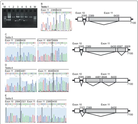
(Figure 1C). All PCR targets were successfully amplified, and the predicted sizes of PCR products were confirmed. During the establishment of this method, we unexpectedly identified splicing variants between exon 10 and exon 14 in normal testes (Figure 1C and 2). These transcripts skipped most of exon 11, leading to frameshift mutations (Figure 2).
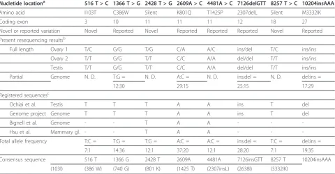
To define the consensus canine BRCA2 ORF, we sequenced the full-length canine BRCA2 ORF in two ovar-ies and a testis obtained from individual healthy mongrel dogs using the method described here. We identified six single nucleotide variations (516 T > C, 1366 T > G, 2428 T > G, 2609A > C, 4481A > C and 8257 T > C) and two insertion/deletion polymorphisms (7126ins/delGTT and 10204ins/delAAA) (Accession numbers: AB622997, AB622998 and AB622999). None of these variations
M 1 2 3 4 5 6 7 8 9 10 11 12 13 14 15 16 17 18 19 20 M
B
*
*
*
*
*
*
A
1 2 3 4 5 6 7 8 9 10 11 12 13 14
M
M
*
*
C
[image:2.595.60.542.295.680.2]M
1 2 3 4 5
Figure 1PCR products amplified by each primer set. (A) cDNA samples prepared from total RNA of each mammary gland were amplified. (B) Genomic DNA from each mammary gland was amplified. (C) Splicing variants of the cDNA from total RNA of the mammary gland and testis were amplified. Primer sets for each lane are shown in Table 1. The“M”indicates the molecular size marker (1-kbp DNA ladder; New England Biolabs). Arrowhead and“*”indicate novel BRCA2 transcript and non-specific PCR products, respectively.
Yoshikawaet al.BMC Research Notes2012,5:173 http://www.biomedcentral.com/1756-0500/5/173
resulted in nonsense or frameshift mutations. To deter-mine the most common base sequences and generate a consensus canine BRCA2 ORF sequence, we compared these three new sequences (six alleles) and the seven pre-viously reported sequences (Accession numbers: AB043895.5, NC_006607.2, Z75664 and FJ464397-FJ464400) (Table 2). The four variations (516 T (103I), 2428 T (740 G), 4481A (1425 T), and 8257 T (2683I)) could be defined as consensus base sequences, but the other four variations (1366 T > G, 2609A > C (K801Q), 7126ins/delGTT, and 10204ins/delAAA) could not be
defined as such because the frequencies between the major and minor alleles in each variation were nearly iden-tical. We therefore sequenced these four variations in genomic DNA from 20-23 normal blood samples from tumor-free dogs aged over 6 years; the methods described here were used (Tables 3 and 4). We finally defined the most common base sequences as the consensus canine BRCA2 ORF sequence (Table 2). The 10204insAAA varia-tion was consensus sequence in dogs, but in four minia-ture Dachshunds this variation was determined to be a minor variation (allele ratio; del:ins = 6:2, Table 4).
1 2 3 4 5 6
M M
A B
2389 6430 Exon 11
Testis-1 Testis-1
Exon 11 Exon 10
2085 2389
7100 6430
C
Exon 11 Exon 10
2085 2389
7100 6430 6587 6909
2389 6430
Exon 11 Exon 11 6587 6909 Testis-3
2389 4481
Exon 11 Exon 11 4648 6430 Testis-4
Exon 11 Exon 10
2085 2389
7100 4481 4648 6430
D
2084 2321
Exon 10 Exon 11 Exon 112389 6430 Testis-5
E
Exon 11 Exon 10
2389
7100 6430
[image:3.595.56.540.87.519.2]2321
Figure 2Identification of splicing variants within exon 11 in normal testes. The splicing variants identified in normal testes lacked a large portion of exon 11. (A) To confirm the presence of a novel BRCA2 transcript, splicing variants of the cDNA from total RNA of the testes were amplified using nested PCR. (B-E) Electropherogram showing the direct sequencing data and overview of the novel BRCA2 transcript that lacked nucleotides 2390 to 6429 (B), 2390 to 6429 and 6588 to 6908 (C), 2390 to 3380, and 4649 to 6429 (D) and 2085 to 2321 and 2390 to 6429 (E) from Testis-1, -3, -4, and -5, respectively. The Testis-1 was the same sample used to generate the data in Figure 1C. Primer sets for each lane are shown in Table 1. The“M”indicates the molecular size marker (1-kbp DNA ladder). Arrowheads indicate novel BRCA2 transcripts.
Yoshikawaet al.BMC Research Notes2012,5:173 http://www.biomedcentral.com/1756-0500/5/173
Table 1 Nucleotide base sequences of primers
Primer sets Forward Reverse Annealing
temperature
Elongation time
Lane Number
Expected sizes
For amplification of cDNA 1 5’-GCGGCACCTCGGAAGGC-3’ 5’-CCCCAAACTTTGACTTTTAGC-3’ 60°C 1 min Figure 1 A 1
834 bp
2 5’-GATCGGTTTATCCCTTGTGGTC-3’ 5’-CTTCAGGTTCTTTAAAGTTTGG-3’ 60°C 1 min Figure 1
A 2
865 bp
3 5’-CTGAAGGGATGTCCAATGC-3’ 5’-ATATTTTATATGATTCTTTTCCTC-3’ 56.1°C 1 min Figure 1 A 3
850 bp
4 5’-CCAGTCTGTTAACTCCTAGC-3’ 5’-GGATAATGTTCCTCAATATCTTTG-3’ 60°C 1 min Figure 1
A 4
826 bp
5 5’-ACAGCTTCTAATAAAGAGATAAA AC-3’ 5’-GCCGGCATTTATTATTTTTC-3’ 56.1°C 1 min Fig. 1 A 5
850 bp
6 5’-GTTTCTCCTCAAGCAAATACAA-3’ 5’-ATTTTTTACTTTGTCCAAAGATTCC-3’ 60°C 1 min Figure 1 A 6
873 bp
7 5’-CTGATCCTGCAGCAAAGACC-3’ 5’-GAAAAACCAATGTTTTTTCTCTCTC-3’ 59.2°C 1 min Figure 1A 7
908 bp
8 5’-CATTCTAGTGAAGTGTATAATAA ATCAG-3’
5’-CTGTCCTAAATCCAGAGAAAGC-3’ 50.8°C 1 min Figure 1 A 8
919 bp
9 5’-AGTATCACTTAAAGATAATGAAG AAC-3’
5’-CTTTTAGGATGCCGTCTGG-3’ 50°C 1 min Figure 1
A 9
887 bp
10 5’-CCCCCAATTAAAAGAAACTTG-3’ 5’-GCCAATTGTATTCCTTCTCC-3’ 53.7°C 1 min Figure 1
A 10
905 bp
11 5’-CCTCTGCATGTTCTCATAAAC-3’ 5’-GGGTATGCTCTTTGAACAACTAC-3’ 60°C 1 min Figure 1 A 11
886 bp
12 5’-CATGGAGCAGAACTGGTAGG-3’ 5’-GTGTAAGGTTTAATAATGTCTTCA-3’ 50°C 1 min Figure 1 A 12
1094 bp
13 5’-CCTATCCCAAGTTTATCAGCC-3’ 5’-CAGACACAAGTTGATGTTCTCC-3’ 60°C 1 min Figure 1
A 13
959 bp
14 5’-GAAGGCATTTCAGCCACCACG-3’ 5’-CAATCACACTAGAATCATAAAAAGG-3’ 60°C 1 min Figure 1 A 14
978 bp
For amplification of genomic DNA
exon 1-2 5’-GCCCCCTGCCCAGAACCC-3’ 5’-CTTTTCAGCAAGCATGCACAGTTACG-3’ 60°C 2 min Figure 1 B 1
1193 bp
exon 3 5’-CTACAGTCAAAATGTCAAGCG-3’ 5’-CACAATTAACAATAGATCTGGGAG-3’ 60°C 1 min Figure 1 B 2
430 bp
exon 4-7 5’-ATAAGAATAAAAACTTCCAGAGAATG-3’ 5’-ATTATCTTTTCATATATTCTTTTTGTC-3’ 60°C 2 min Figure 1 B 3
1384 bp
exon 8-9 5’-GTAGTATATGTGACTTTTGATGTCTG-3’ 5’-GGAAAAGCAATGTATTTTCTCTTT-3’ 60°C 2 min Figure 1 B 4
615 bp
exon 10 5’-CTTTAAATACTGCCTTATGGGCTA-3’ 5’-GTCACCATCCCTAAAACTATATGC-3’ 60°C 2 min Figure 1 B 5
1311 bp
exon 11-a 5’-GTCACTTTGTGTCTTCATGC-3’ 5’-GGATAATGTTCCTCAATATCTTTG-3’ 56.4°C 2 min Figure 1 B 6
1246 bp
Yoshikawa
et
al
.
BMC
Research
Notes
2012,
5
:173
http://ww
w.biomedcen
tral.com/1756
-0500/5/173
Page
4
of
Table 1 Nucleotide base sequences of primers(Continued)
exon 11-b (same as primer set 5)
5’-ACAGCTTCTAATAAAGAGATAAAAC-3’ 5’-GCCGGCATTTATTATTTTTC-3’ 56.4°C 1 min Figure 1 B 7
850 bp
exon 11-c(same as primer set 6)
5’-GTTTCTCCTCAAGCAAATACAA-3’ 5’-GATTTTTTACTTTGTCCAAAGATTCC-3’ 60°C 1 min Figure 1 B 8
873 bp
exon 11-d (same as primer set 7)
5’-CTGATCCTGCAGCAAAGACC-3’ 5’-GAAAAACCAATGTTTTTTCTCTCTC-3’ 59.2°C 1 min Figure 1 B 9
908 bp
exon 11-e 5’ -CATTCTAGTGAAGTGTATAATAAATCAG-3’
5’-ATTCCCCTAAACTATACATAAAAG-3’ 56.4°C 2 min Figure 1 B 10
1720 bp
exon 12 5’-CAATTCTTTAGTTTTAAAAAATGG GC-3 5’
- GAAAAAGCTTAGAAAAAGAACTTAAAAAATAC-3’
59.2°C 1 min Figure 1
B 11
275 bp
exon 13 5’
- GTAAATGTTTATAATGTGTAATATACAGGC-3’
5’-CTGTACCTTCCCTACACTATATTAGTAG-3’ 60°C 1 min Figure 1 B 12
230 bp
exon 14-15 5’-CCAAACTTAAATATTTTCTCCTC-3’ 5’-CAGGGATCCCAGTCTATTC-3’ 60°C 2 min Figure 1 B 13
1213 bp
exon 16 5’-GCAGCAAACCCTTGAATGTAG-3’ 5’-GTCAGGTGAACCGTAATGTG-3’ 60°C 1 min Figure 1 B 14
552 bp
exon 17-8 5’- GGTCTTGTACAGTGTAGTGTTTG-3’ 5’-GTTTTTAAGCAATGGAGCATC-3’ 59.2°C 2 min Figure 1 B 15
1258 bp
exon 19-20 5’- CCATCATGTTTAAATTGAAGTCTC-3’ 5’-CAATTACAGAGGTTAAATCAGAAGCC-3’ 59.2°C 2 min Figure 1 B 16
739 bp
exon 21-24 5’-CTCGATATTTGATTCACCAGC-3’ 5’-CAACAGTCCCTTCCTAACCC-3’ 60°C 2 min Figure 1 B 17
1739 bp
exon 25 5’- CAGTATCACTTTTTCTACATTTTG GTC-3’
5’-CCCAATTTTCACAGAAGCCAC-3’ 59.2°C 1 min Figure 1 B 18
471 bp
exon 26 5’-GGCTTCCATAGATGTTAGATG-3’ 5’-GGACAACTTGGGATCATTTGC-3’ 50.8°C 1 min Figure 1 B 19
337 bp
exon 27 5’- GCTAAATTGCTGATGTTTTCTAC-3’ 5’-CTGCTGAGTCCTCTAATAAGGC-3’ 60°C 2 min Figure 1 B 20
1437 bp
exon 25 5’- CAGTATCACTTTTTCTACATTTTGGTC-3’ 5’-CCCAATTTTCACAGAAGCCAC-3’ 59.2°C 1 min Figure 1 B 18
471 bp
exon 26 5’-GGCTTCCATAGATGTTAGATG-3’ 5’-GGACAACTTGGGATCATTTGC-3’ 50.8°C 1 min Figure 1 B 19
337 bp
exon 27 5’- GCTAAATTGCTGATGTTTTCTAC-3’ 5’-CTGCTGAGTCCTCTAATAAGGC-3’ 60°C 2 min Figure 1 B 20
1437 bp
For detection of splicing variants
exon 1-11 5’-CGAATTTGTTAGCCGTCTCC-3’ 5’-GGATCCTGAGATATTATTTTATTATTAG-3’ 60°C 2.5 min Figure 1 C 1
2118 bp
exon 10-14 5’-CTGAAGGGATGTCCAATGC-3’ 5’-GAAATTTGGATTCTGTATTTCTTG-3’ 58°C 6 min Figure 1 C 2
5594 bp and 1554 bp
Yoshikawa
et
al
.
BMC
Research
Notes
2012,
5
:173
http://ww
w.biomedcen
tral.com/1756
-0500/5/173
Page
5
of
Table 1 Nucleotide base sequences of primers(Continued)
exon 11-18 5’-CTTCCTGTGAAAACAAATATAG-3’ 5’-GCTGATCTTCTGCTTTTATC-3’ 50.8°C 2 min Figure 1 C 3
1417 bp
exon 15-25 5’-CCTCTGCATGTTCTCATAAAC-3’ 5’-GTGTAAGGTTTAATAATGTCTTCA-3’ 60°C 2 min Figure 1 C 4
1759 bp
exon 24-27 (same as primer set 13)
5’-CCTATCCCAAGTTTATCAGCC-3’ 5’-CAGACACAAGTTGATGTTCTCC-3’ 60°C 2 min Figure 1 C 5
959 bp
For nested PCR of the transcripts lacking most of exon 11
exon 10-13 (1735-7280)
5’ -GTTCTCAAATAATATGACTAATCCAAAC-3’
5’-GTTCCTCAGTTGTGCGAAAG-3’ 58°C 6 min Figure 2
A
5546 bp and 1506 bp, 1185 bp, 1674 bp or 1270 bp
For DNA sequence cB2 seq1 5’-CAATAGAGGTGTTTTCTCCATC-3’
cB2 seq2 5’ -GGATCCTGAGATATTATTTTATTATTAG-3’
5546 bp and 1506 bp, 1185 bp, 1674 bp or 1270 bp
cB2 seq3 5’-CCAGCTTTGTCTTTAACCAG-3’
cB2 seq4 5’-CTGTGTGACCACTTTCACTATC-3’
cB2 seq5 5’-CCCTCCTTCATAAACTGGC-3’
cB2 seq6 5’-CTTTCTGAGAGGCATGATCTG-3’
cB2 seq7 5’-GCATGGCAAGTGTCTGATTTAC-3’
cB2 seq8 5’-GTGAACAAACTTCACAACTTAACC-3’
cB2 seq9 5’-GCTGATCTTCTGCTTTTATC-3’
cB2 seq10 5’-GGTATGTTTTACAATGATGC-3’
cB2 ex14 R (exon)
5’-CTAAAGGTTCTTTTTCATTCTTTG-3’
cB2 ex15 F 5’- GCTTTTTAAATGTTACATGGAGG-3’
cB2 ex17 R 5’-GTACCAGTCAGGGATGTGAG-3’
cB2 ex18 F (exon)
5’-ATATGATGTGGAAATTGATAAAA G-3’
cB2 ex22F 5’-CTTTTTAAAGGGATTCATTTACAG TGG-3’
cB2 ex23 F (exon)
5’-CCATCACCAGATTTATATTCCC-3’
cB2 ex26 R (exon)
5’-CAGAAATTTATTTCCTATGCC-3’
cB2 ex23 F (exon)
5’-CCATCACCAGATTTATATTCCC-3’
cB2 ex26 R (exon)
5’-CAGAAATTTATTTCCTATGCC-3’
Yoshikawa
et
al
.
BMC
Research
Notes
2012,
5
:173
http://ww
w.biomedcen
tral.com/1756
-0500/5/173
Page
6
of
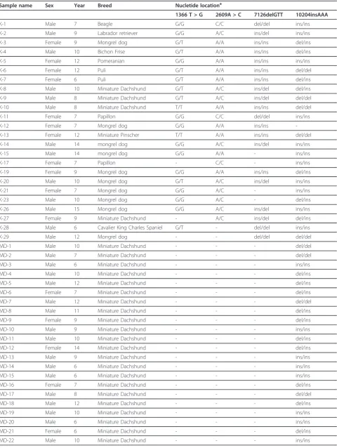
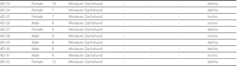
[image:6.794.63.712.68.513.2]To confirm the consensus sequence in miniature Dachs-hunds, we sequenced BRCA2 DNA from an additional 32 blood samples, and the assembled allele ratio was del:ins = 30:42 (Table 4).
We established a PCR analysis method for canine BRCA2 in order to determine the base sequence from cDNA and genomic DNA, and to detect splicing variants. We identified novel splicing variants in normal canine testes. The functions of these splicing variants were not assessed in this study; nevertheless, these results indi-cated that the established method was a useful tool for detecting splicing variants.
We also defined the consensus sequence using meth-ods established and described here. During the
[image:7.595.58.539.102.352.2]definition of the consensus BRCA2 ORF, we identified three novel (516 T > C, 2428 T > G, and 8257 T > C) and three reported (1366 T > G, 2609A > C and 4481A > C) single nucleotide variations and two reported inser-tion/deletion polymorphisms (7126ins/delGTT and 10204ins/delAAA) (Accession numbers: AB622997, AB622998 and AB622999) [11,12,15,18]. The variations 1366 T > G (C386W), 2609A > C (K801Q), 4481A > C (T1425P), and 10204ins/delAAA (M3332IV) are located in the histone acetyltransferase domain, the FANCG binding domain, BRC repeat 3, and nuclear localization signal 2, respectively [13,19-21]. The effects of these var-iations on BRCA2 function were not understood, with the exception of 10204insAAA; nuclear localization Table 2 Comparison between our sequences from the canine BRCA2 open reading frame with registered sequences
Nucletide locationa 516 T > C 1366 T > G 2428 T > G 2609A > C 4481A > C 7126delGTT 8257 T > C 10204insAAA
Amino acid I103T C386W Silent K801Q T1425P 2307delL Silent M3332IK
Coding exon 3 10 11 11 11 12 18 27
Novel or reported variation Novel Reported Novel Reported Reported Reported Novel Reported
Present resequencing resultsb
Full length Ovary 1 T/C G/G T/G C/A A/C ins/del T/C ins/ins
Ovary 2 T/T G/G T/T C/C A/A del/del T/T ins/ins
Testis T/T G/G T/T C/C A/A del/del T/T ins/ins
Partial Genome N. D. T:G = N. D. A:C = N. D. ins:del = N. D. del:ins =
12:30 29:15 25:15 17:29
Registered sequencesc
Ochiai et al. Testis T T T A A ins T del
Genome project Genome T T T A A ins T del
Bignell et al. Genome - - T A A - -
-Hsu et al. Mammary gl. - - T A A - -
-Total allele frequency T:C = T:G = T:G = A:C = A:C = ins:del = T:C = del:ins =
7:1 14:36 12:1 37:20 12:1 28:20 7:1 19:35
Consensus sequence 516 T 1366 G 2428 T 2609A 4481A 7126insGTT 8257 T 10204insAAA
(103I) (386 W) (740 G) (801 K) (1425 T) (2307insL) (2638I) (3332IK)
N. D., not determined.
a
Nucleotide and amino acid location is based on AB043895.5
b
Full-length sequence was determined by cDNA sequencing (Accession number; AB622997, AB622998 and AB622999). When frequencies of major and minor alleles were nearly equal or were inconsistent with reported sequences, alleles were further analyzed by partial sequencing of blood genome DNA from 20-23 dogs (Table 3 and 4).
c
Sequence from one dog was regarded as one allele because allele type analyses have not been described in these reports. The study by Hsu et al. examined three dogs, while others studied only one dog. Accession numbers for sequences reported by Ochiai et al., the Genome Project, Bignell et al. and Hsu et al. are AB043895.5, NC_006607.2, Z75664 and FJ464397-FJ464400, respectively.
Table 3 Genotype frequency of four variations in normal blood samples
Nucletide locationa 1366 T > G 2609A > C 7126delGTT 10204insAAA
Amino acida C386W K801Q 2307delL M3332IK
Coding exon 10 11 12 27
Genotype frequency 1366 T homozygosity 2/21 2609A homozygosity 10/22 insGTT homozygosity 9/20 delAAA homozygosity 5/23
1366 G homozygosity 11/21 2609 C homozygosity 3/22 delGTT homozygosity 4/20 insAAA homozygosity 11/23
Heterozygosity 8/21 Heterozygosity 9/22 Heterozygosity 7/20 Heterozygosity 7/23
a
Nucleotide and amino acid locations are based on AB043895.5
Yoshikawaet al.BMC Research Notes2012,5:173 http://www.biomedcentral.com/1756-0500/5/173
[image:7.595.57.538.641.723.2]Table 4 Information of blood samples and allele type of the four frequently found variations
Sample name Sex Year Breed Nucletide locationa
1366 T > G 2609A > C 7126delGTT 10204insAAA
K-1 Male 7 Beagle G/G C/C del/del ins/ins
K-2 Male 9 Labrador retriever G/G A/C ins/del ins/ins
K-3 Female 9 Mongrel dog G/T A/A ins/ins del/ins
K-4 Male 10 Bichon Frise G/T A/A ins/ins del/ins
K-5 Female 12 Pomeranian G/G A/A ins/ins ins/ins
K-6 Female 12 Puli G/T A/A ins/ins del/del
K-7 Female 6 Puli G/T A/A ins/ins del/ins
K-8 Male 10 Miniature Dachshund G/T A/C ins/del del/ins
K-9 Male 8 Miniature Dachshund G/T A/C ins/del del/del
K-10 Male 8 Miniature Dachshund T/T A/A ins/ins del/del
K-11 Female 7 Papillon G/G C/C del/del ins/ins
K-12 Female 7 Mongrel dog G/G A/A ins/ins
-K-13 Female 12 Miniature Pinscher T/T A/A ins/ins del/del
K-14 Male 14 mongrel dog G/G A/C ins/del ins/ins
K-15 Male 14 mongrel dog G/G A/A - ins/ins
K-17 Female 7 Papillon - C/C - ins/ins
K-19 Female 9 Mongrel dog G/G A/A ins/ins del/ins
K-20 Male 10 Mongrel dog G/T A/C ins/del ins/ins
K-21 Female 7 Mongrel dog G/G A/C - ins/ins
K-23 Male 10 Mongrel dog G/G A/C - del/ins
K-26 Male 15 Mongrel dog G/G A/C ins/del ins/ins
K-27 Female 9 Miniature Dachshund - A/C ins/del del/ins
K-28 Male 6 Cavalier King Charles Spaniel G/T - del/del ins/ins
K-29 Male 12 Mongrel dog - - del/del del/del
MD-1 Male 10 Miniature Dachshund - - - del/del
MD-2 Male 7 Miniature Dachshund - - - del/del
MD-3 Male 6 Miniature Dachshund - - - ins/ins
MD-4 Male 10 Miniature Dachshund - - - del/ins
MD-5 Male 12 Miniature Dachshund - - - del/ins
MD-6 Female 7 Miniature Dachshund - - - del/ins
MD-7 Male 12 Miniature Dachshund - - - del/del
MD-8 Male 11 Miniature Dachshund - - - del/ins
MD-9 Female 9 Miniature Dachshund - - - del/ins
MD-10 Male 9 Miniature Dachshund - - - ins/ins
MD-11 Male 10 Miniature Dachshund - - - del/ins
MD-12 Female 14 Miniature Dachshund - - - del/ins
MD-13 Male 9 Miniature Dachshund - - - ins/ins
MD-14 Male 6 Miniature Dachshund - - - ins/ins
MD-15 Male 6 Miniature Dachshund - - - ins/ins
MD-16 Female 7 Miniature Dachshund - - - del/ins
MD-17 Male 8 Miniature Dachshund - - - del/del
MD-18 Male 12 Miniature Dachshund - - - del/ins
MD-19 Male 10 Miniature Dachshund - - - ins/ins
MD-20 Male 6 Miniature Dachshund - - - ins/ins
MD-21 Female 6 Miniature Dachshund - - - del/ins
MD-22 Male 10 Miniature Dachshund - - - ins/ins
Yoshikawaet al.BMC Research Notes2012,5:173 http://www.biomedcentral.com/1756-0500/5/173
signal 2 harboring the 10204insAAA variation showed enhanced nuclear localization [13]. The other nonsynon-ymous variations were not located in previously known functional domains.
We identified four variations (1366 T > G, 2609A > C, 7126ins/delGTT, and 10204ins/delAAA), in which the allele frequency of minor variations in genomic DNA from normal blood samples was very high (28.5-37.5%).
Such frequent variations in theBRCA2 gene have not
been reported in other species. These highly frequent variations thus appear to be a canine BRCA2-specific feature, and should be considered when studying canine BRCA2. These four variations were found in the homo-zygous state in some blood samples from elderly tumor-free dogs. Homozygous mutations in BRCA2 are assumed to be embryonic-lethal mutations or responsi-ble for Fanconi anemia, which is characterized by bone marrow failure, developmental abnormalities, and pre-disposition to cancer [22,23]. Thus, these four variations were probably neutral variations, although the 10204insAAA variation is reportedly a candidate malig-nant mutation in dogs [11].
In this study, we established a PCR analysis method and defined the consensus sequence of BRCA2 ORF to identify canine BRCA2 mutations. Using these methods, we are now able to perform BRCA2 mutation analysis and search for abnormal BRCA2 splicing variants from mammary tumors in dogs, as is done in human cases.
Methods
Specimens
Two ovaries (from two mongrel dogs), six testes (from a mongrel dog and five Beagles), a mammary gland (from a female Beagle) and 56 blood samples (Table 4) from tumor-free dogs were kindly provided by Dr. Takashi Kubo and Dr. Go Honda. All experimental procedures were approved by and conducted in accordance with the Guidelines for Institutional Laboratory Animal Care and
Use of the School of Veterinary Medicine at Kitasato University, Japan (Approval Number: 11-065).
Total RNA and genomic DNA extraction, and preparation of cDNA
Total RNA was isolated from ovaries, six testes, and one mammary gland, which each were stored in RNAlater solution (Life Technologies, Grand Island, NY), using a TRIzol and PureLink RNA micro kit (Life Technolo-gies). First-strand cDNA was synthesized from 1-5μg of total RNA using SuperScript III (Life Technologies). Genomic DNA samples were extracted using a Gentra Puregene tissue kit (Qiagen, Hilden, Germany).
PCR and sequencing
For PCR amplification of the full-length canine BRCA2 ORF from cDNA and all 27 exons from genomic DNA, we designed 14 and 20 primer sets, respectively (Table 1). We also designed five primer sets to detect splicing var-iants and a primer sets to confirm a novel BRCA2 tran-script that lacked most of exon 11 using nested PCR (Table 1). Each reaction mixture contained 0.1μL of first-strand cDNA reaction products or 10-50 ng of genomic DNA as a template, each forward and reverse primer at
300 nM, 200μM dNTPs, 0.02 U of KOD FX DNA
[image:9.595.58.540.102.237.2]poly-merase (Toyobo, Japan), and 1× PCR buffer, which was supplied with the enzyme, in a total volume of 10μL. PCR included one cycle of 2 min at 94°C, followed by 35 cycles of 10 s at 98°C, 30 s at the optimal temperature shown in Table 1 the optimal time shown in Table 1 at 68°C, and a final extension step of 7 min at 68°C. PCR products were treated with shrimp alkaline phosphatase (Affymetrix, Santa Clara, CA) and Exonuclease I (New England Bio-Labs, Beverley, MA) before sequencing, which was performed with the BigDye Terminator Cycle Sequencing kit Version 3.1 and a ABI PRISM 3100-Avant DNA sequencer (Life Technologies). Direct DNA sequencing was performed at least twice for each amplicon. When we Table 4 Information of blood samples and allele type of the four frequently found variations(Continued)
MD-23 Female 10 Miniature Dachshund - - - del/ins
MD-24 Female 7 Miniature Dachshund - - - del/ins
MD-25 Female 7 Miniature Dachshund - - - ins/ins
MD-26 Male 8 Miniature Dachshund - - - ins/ins
MD-27 Female 9 Miniature Dachshund - - - del/ins
MD-28 Male 9 Miniature Dachshund - - - ins/ins
MD-29 Male 8 Miniature Dachshund - - - del/ins
MD-30 Male 8 Miniature Dachshund - - - del/ins
MD-31 Male 9 Miniature Dachshund - - - ins/ins
MD-32 Female 12 Miniature Dachshund - - - del/ins
a
Nucleotide and amino acid locations are based on AB043895.5
“-”indicates not determined.
Yoshikawaet al.BMC Research Notes2012,5:173 http://www.biomedcentral.com/1756-0500/5/173
attempted to define the consensus canine BRCA2 ORF sequence, two or three amplicons from each sample were sequenced. Because we detected only three electrophero-gram patterns among the PCR products with the inser-tion/deletion mutation sites, we were able to determine the heterozygous insertion/deletion mutations by direct sequencing (Additional file 1: Figure S1).
Additional material
Additional file 1: Figure S1. Example of an electropherogram by direct sequencing from PCR products having the insertion/deletion mutation (7126ins/delGTT).
Acknowledgements
This work was supported in part by a Kitasato University Research Grant for Young Researchers and a Grant for the Encouragement of Young Scientists from the School of Veterinary Medicine, Kitasato University, Grant-in-Aid for Young Scientists (B) (No. 23780326 and No. 22791476) and for Scientific Research (C) (No. 23580399) from Japan Society for the Promotion of Science.
Author details
1Laboratory of Veterinary Biochemistry, School of Veterinary Medicine, Kitasato University, Aomori 034-8628, Japan.2Division of Disease Model Innovation, Institute for Genetic Medicine, Hokkaido University, Sapporo 060-0815, Japan.3Department of Basic Science, School of Veterinary Nursing and Technology, Nippon Veterinary and Life Science University, Tokyo 180-8602, Japan.4Department of Small Animal Surgery 2, School of Veterinary Medicine, Kitasato University, Aomori 034-8628, Japan.5Department of Small Animal Internal Medicine 2, School of Veterinary Medicine, Kitasato University, Aomori 034-8628, Japan.6Department of Veterinary Science, School of Veterinary Medicine, Nippon Veterinary and Life Science University, Tokyo 180-8602, Japan.
Authors’contributions
YY outlined the design of and coordinated the study, performed the experiments, and drafted the manuscript. MM, K. Ochiai, K. Orino, and WK participated in the design of the study and interpretation of the data and helped to draft the manuscript. K. Okuda, TT, SC, SA, TO, and MB performed several experiments.
Competing interests
The authors declare that they have no competing interests.
Received: 2 February 2012 Accepted: 3 April 2012 Published: 3 April 2012
References
1. DeSantis C, Siegel R, Bandi P, Jemal A:Breast cancer statistics, 2011.CA Cancer J Clin2011,61(6):409-418.
2. Egenvall A, Bonnett BN, Ohagen P, Olson P, Hedhammar A, von Euler H:
Incidence of and survival after mammary tumors in a population of over 80,000 insured female dogs in Sweden from 1995 to 2002.Prev Vet Med
2005,69(1-2):109-127.
3. Moe L:Population-based incidence of mammary tumours in some dog breeds.J Reprod Fertil Suppl2001,57:439-443.
4. Moulton JE, Rosenblatt LS, Goldman M:Mammary tumors in a colony of beagle dogs.Vet Pathol1986,23(6):741-749.
5. Gilbertson SR, Kurzman ID, Zachrau RE, Hurvitz AI, Black MM:Canine mammary epithelial neoplasms: biologic implications of morphologic characteristics assessed in 232 dogs.Vet Pathol1983,20(2):127-142. 6. Moulton JE, Taylor DO, Dorn CR, Andersen AC:Canine mammary tumors.
Pathol Vet1970,7(4):289-320.
7. Evans DG, Shenton A, Woodward E, Lalloo F, Howell A, Maher ER:
Penetrance estimates for BRCA1 and BRCA2 based on genetic testing in a Clinical Cancer Genetics service setting: risks of breast/ovarian cancer quoted should reflect the cancer burden in the family.BMC Cancer2008,
8:155.
8. King M-C, Marks JH, Mandell JB, Group NYBCS:Breast and ovarian cancer risks due to inherited mutations in BRCA1 and BRCA2.Science2003,
302(5645):643-646.
9. Rivera P, Melin M, Biagi T, Fall T, Haggstrom J, Lindblad-Toh K, von Euler H:
Mammary tumor development in dogs is associated with BRCA1 and BRCA2.Cancer Res2009,69(22):8770-8774.
10. Rivera P, von Euler H:Molecular biological aspects on canine and human mammary tumors.Vet Pathol2011,48(1):132-146.
11. Yoshikawa Y, Morimatsu M, Ochiai K, Nagano M, Tomioka Y, Sasaki N, Hashizume K, Iwanaga T:Novel variations and loss of heterozygosity of BRCA2 identified in a dog with mammary tumors.Am J Vet Res2008,
69(10):1323-1328.
12. Hsu W-L, Huang Y-H, Chang T-J, Wong M-L, Chang S-C:Single nucleotide variation in exon 11 of canine BRCA2 in healthy and cancerous mammary tissue.Vet J2010,184(3):351-356.
13. Yoshikawa Y, Morimatsu M, Ochiai K, Nagano M, Yamane Y, Tomizawa N, Sasaki N, Hashizume K:Insertion/deletion polymorphism in the BRCA2 nuclear localization signal.Biomed Res2005,26(3):109-116.
14. Ochiai K, Morimatsu M, Tomizawa N, Syuto B:Cloning and sequencing full length of canine Brca2 and Rad51 cDNA.J Vet Med Sci2001,
63(10):1103-1108.
15. Borge KS, Børresen-Dale AL, Lingaas F:Identification of genetic variation in 11 candidate genes of canine mammary tumour.Vet Comp Oncol
2011,9(4):241-250.
16. Sugiura T, Matsuyama S, Akiyosi H, Takenaka S, Yamate J, Kuwamura M, Aoki M, Shimada T, Ohashi F, Kubo K:Expression patterns of the BRCA1 splicing variants in canine normal tissues and mammary gland tumors.J Vet Med Sci2007,69(6):587-592.
17. Walker LC, Whiley PJ, Couch FJ, Farrugia DJ, Healey S, Eccles DM, Lin F, Butler SA, Goff SA, Thompson BA,et al:Detection of splicing aberrations caused by BRCA1 and BRCA2 sequence variants encoding missense substitutions: implications for prediction of pathogenicity.Hum Mutat
2010,31(6):E1484-1505.
18. Yoshikawa Y, Morimatsu M, Ochiai K, Nagano M, Yamane Y, Tomizawa N, Sasaki N, Hashizume K:Analysis of genetic variations in the exon 27 region of the canine BRCA2 locus.J Vet Med Sci2005,67(10):1013-1017. 19. Hussain S, Witt E, Huber PAJ, Medhurst AL, Ashworth A, Mathew CG:Direct
interaction of the Fanconi anaemia protein FANCG with BRCA2/FANCD1.
Hum Mol Genet2003,12(19):2503-2510.
20. Ochiai K, Yoshikawa Y, Oonuma T, Tomioka Y, Hashizume K, Morimatsu M:
Interactions between canine RAD51 and full length or truncated BRCA2 BRC repeats.Vet J2011,190(2):293-295.
21. Siddique H, Zou JP, Rao VN, Reddy ES:The BRCA2 is a histone acetyltransferase.Oncogene1998,16(17):2283-2285.
22. Howlett NG, Taniguchi T, Olson S, Cox B, Waisfisz Q, De Die-Smulders C, Persky N, Grompe M, Joenje H, Pals G,et al:Biallelic inactivation of BRCA2 in Fanconi anemia.Science2002,297(5581):606-609.
23. Sharan SK, Morimatsu M, Albrecht U, Lim DS, Regel E, Dinh C, Sands A, Eichele G, Hasty P, Bradley A:Embryonic lethality and radiation hypersensitivity mediated by Rad51 in mice lacking Brca2.Nature1997,
386(6627):804-810.
doi:10.1186/1756-0500-5-173
Cite this article as:Yoshikawaet al.:Establishment of a PCR analysis method for canine BRCA2.BMC Research Notes20125:173.
Yoshikawaet al.BMC Research Notes2012,5:173 http://www.biomedcentral.com/1756-0500/5/173