R E S E A R C H A R T I C L E
Open Access
Plasma cell-free DNA (cfDNA) as a
predictive and prognostic marker in
patients with metastatic breast cancer
Daniel Fernandez-Garcia
1, Allison Hills
2, Karen Page
1, Robert K. Hastings
1, Bradley Toghill
1, Kate S. Goddard
2,
Charlotte Ion
3, Olivia Ogle
4, Anna Rita Boydell
3, Kelly Gleason
3, Mark Rutherford
5, Adrian Lim
3, David S. Guttery
1,
R. Charles Coombes
2and Jacqueline A. Shaw
1*Abstract
Background:Breast cancer (BC) is the most common cancer in women, and despite the introduction of new screening programmes, therapies and monitoring technologies, there is still a need to develop more useful tests for monitoring treatment response and to inform clinical decision making.
The purpose of this study was to compare circulating cell-free DNA (cfDNA) and circulating tumour cells (CTCs) with conventional breast cancer blood biomarkers (CA15-3 and alkaline phosphatase (AP)) as predictors of response to treatment and prognosis in patients with metastatic breast cancer (MBC).
Methods:One hundred ninety-four female patients with radiologically confirmed MBC were recruited to the study. Total cfDNA levels were determined by qPCR and compared with CELLSEARCH® CTC counts and CA15-3 and alkaline phosphatase (AP) values. Blood biomarker data were compared with conventional tumour markers, treatment(s) and response as assessed by RECIST and survival.
Non-parametric statistical hypothesis tests were used to examine differences, correlation analysis and linear regression to determine correlation and to describe its effects, logistic regression and receiver operating characteristic curve (ROC curve) to estimate the strength of the relationship between biomarkers and clinical outcomes and value normalization against standard deviation to make biomarker values comparable. Kaplan–Meier estimator and Cox regression models were used to assess survival. Univariate and multivariate models were performed where appropriate.
Results:Multivariate analysis showed that both the amount of total cfDNA (pvalue = 0.024, HR = 1.199, CI = 1.024– 1.405) and the number of CTCs (pvalue = 0.001, HR = 1.243, CI = 1.088–1.421) are predictors of overall survival (OS), whereas total cfDNA levels is the sole predictor for progression-free survival (PFS) (pvalue = 0.042, HR = 1.193, CI = 1.007–1.415) and disease response when comparing response to non-response to treatment (HR = 15.917, HR = 12.481 for univariate and multivariate analysis, respectively). Lastly, combined analysis of CTCs and cfDNA is more informative than the combination of two conventional biomarkers (CA15-3 and AP) for prediction of OS.
Conclusion:Measurement of total cfDNA levels, which is a simpler and less expensive biomarker than CTC counts, is associated with PFS, OS and response in MBC, suggesting potential clinical application of a cheap and simple blood-based test.
Keywords:Breast cancer, Liquid biopsy, Biomarkers, cfDNA, CTCs
© The Author(s). 2019Open AccessThis article is distributed under the terms of the Creative Commons Attribution 4.0 International License (http://creativecommons.org/licenses/by/4.0/), which permits unrestricted use, distribution, and reproduction in any medium, provided you give appropriate credit to the original author(s) and the source, provide a link to the Creative Commons license, and indicate if changes were made. The Creative Commons Public Domain Dedication waiver (http://creativecommons.org/publicdomain/zero/1.0/) applies to the data made available in this article, unless otherwise stated. * Correspondence:js39@le.ac.uk
1Leicester Cancer Research Centre, Department of Genetics and Genome
Biology, University of Leicester, Robert Kilpatrick Clinical Sciences Building, Leicester Royal Infirmary, Leicester LE2 7LX, UK
Introduction
Breast cancer (BC) is the most common cancer in women [1]. The introduction of screening programmes and the development of targeted therapies has signifi-cantly improved BC survival rates in the last 40 years [2,
3]. However, although many patients are initially respon-sive to therapies, resistance can develop and lead to re-lapse and ultimately death from metastatic disease [4]. Patients with metastatic breast cancer (MBC) are moni-tored by radiological imaging; primarily by computed tomography (CT) on average every 3 months, and scans are assessed using Response Evaluation Criteria in Solid Tumour (RECIST) criteria to determine disease response to treatment. FLT-PET and magnetic resonance imaging (MRI) is also carried out in some centres, but these tests are costly, insensitive and less readily accessible. Along-side imaging, cancer antigen 15-3 (CA15-3) and alkaline phosphatase (AP) are often measured, although these lack in both sensitivity and specificity [5, 6]. Therefore, there is a need to develop more useful tests for monitor-ing treatment response and to inform clinical decision making.
Blood-based biomarkers, including circulating cell-free DNA (cfDNA) and circulating tumour cells (CTCs) have attracted considerable attention in recent years due to their potential as minimally invasive tools for cancer monitoring. A CTC count of ≥5 CTCs/7.5 ml blood, as determined by CellSearch®, is an independent predictor of poor prognosis in MBC, irrespective of other clinical parameters [7]. CTC counts can also be used to guide therapy selection in newly diagnosed patients receiving first-line systemic treatment [7,8]. Previous studies have proposed that CTC counts are superior to conventional radiological measures as predictor of prognosis and re-sponse in patients with MBC [8,9], but as yet, the appli-cation of total cfDNA levels has not been widely studied in MBC [10, 11]. Circulating cfDNA is derived from a combination of apoptosis, necrosis and active secretion from cancer cells and is found at higher levels in patients with advanced cancer than in either healthy individuals [12] or patients with early-stage disease [13]. The tumour-derived fraction of this total cfDNA, termed cir-culating tumour DNA (ctDNA), is under wide investiga-tion as a prognostic biomarker in several types of cancer, including breast, lung and colon cancers [14– 17]. Although much ongoing research is focussed on profiling of ctDNA, these analyses are currently expen-sive and not yet established in the clinic. We therefore compared conventional breast cancer blood biomarkers (CA15-3 and alkaline phosphatase (AP)) with CTC counts and simple measurement of total cfDNA levels, rather than ctDNA profiles to assess the best predictor of response to treatment and prognosis in 194 patients with metastatic breast cancer. The results indicate that
measurement of total cfDNA levels is a good predictor of response and survival in patients with MBC suggest-ing potential clinical application of a cheap and simple blood-based test.
Methods
Patients and demographics
Between February 2012 and July 2016, 194 female pa-tients with radiologically confirmed MBC, attending the breast oncology clinic at Charing Cross Hospital, London, were recruited to this study. One hundred ninety-three patients had proven metastatic disease, and one had unresectable locally recurrent disease.
Thirty-two patients of the 194 patients were off treat-ment at the time of blood sampling, and the remainder was receiving treatment for MBC. Details of treatment(s) undergone by each patient at the time of blood sample collection were obtained from the Imperial College NHS electronic prescription system and where necessary, tient records. CT and MRI data were obtained from pa-tient records and results confirmed by a consultant radiologist to determine patient disease status at time of blood collection. There were three orthogonal measure-ments of the primary tumour and volume estimated using a volume calculator, as described previously [18].
The maximal dimensions of the largest metastases were provided—two max per organ as per RECIST cri-teria. If there were multiple widespread lesions, then an estimate of how much of the whole organ is infiltrated with tumour was estimated visually. These were typically lymph nodes, lungs, liver and occasional brain and ad-renal metastasis.
Only lytic bony lesions were counted. Pleural and peri-toneal diffuse disease was documented but only mea-sured when there was a sizable mass. Irradiated bony or CNS sites were followed where hopefully there was a baseline but the disease may not be measurable.
Data from scans undergone at a time-point, generally within 2 weeks, closest to that of sample collection were used to evaluate disease response. Response to treatment was assessed using RECIST criteria [19]. A total of 30 patients were responding to their treatment, 73 had stable disease and 91 were progressing. The ER, PR and HER2 status of the primary tumour and metastatic bi-opsy where available were obtained from histology re-ports (Table1; Additional file1).
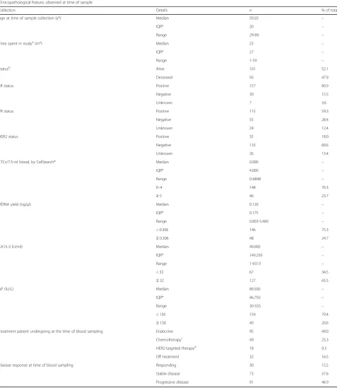
Table 1Descriptive statistics of variables and clinical characteristics of patients included in the study
Clinicopathological feature, observed at time of sample
Collection Details n % of total
Age at time of sample collection (y*) Median 59.50 –
IQR* 20 –
Range 29-89 –
Time spent in studya(m*) Median 23 –
IQR* 27 –
Range 1-59 –
Statusb Alive 101 52.1
Deceased 93 47.9
ER status Positive 157 80.9
Negative 30 15.5
Unknown 7 3.6
PR status Positive 115 59.3
Negative 55 28.4
Unknown 24 12.4
HER2 status Positive 35 18.0
Negative 133 68.6
Unknown 26 13.4
CTCs/7.5 ml blood, by CellSearch® Median 0.000 –
IQR* 4.000 –
Range 0-6848 –
0–4 148 76.3
≥5 46 23.7
cfDNA yield (ng/μl) Median 0.120 –
IQR* 0.175 –
Range 0.003-5.460 –
< 0.306 146 75.3
≥0.306 48 24.7
CA15-3 (U/ml) Median 49.000 –
IQR* 149.250 –
Range 1-6313 –
< 32 67 34.5
≥32 127 65.5
AP (IU/L) Median 89.500 –
IQR* 46.750 –
Range 30-555 –
< 130 154 79.4
≥130 40 20.6
Treatment patient undergoing at the time of blood sampling Endocrine 95 49.0
Chemotherapyc 49 25.3
HER2-targeted therapyd 18 9.3
Off treatment 32 16.5
Disease response at time of blood sampling Responding 30 15.5
Stable disease 73 37.6
Progressive disease 91 46.9
After screening and recruitment, patients were followed-up through the study. HER2 status was determined by immunohistochemical and fluorescence in situ hybridization assays. A patient was considered to have HER2-positive cancer if either assay was positive. CA15-3 and AP levels were determined form patient notes
IQRinterquartile range,yyears,mmonths aTime from collection to end of study or death
bStatus of patients at the end of the study (deceased or alive) cChemotherapy +/−endocrine therapy
response or survival. This small subset of the population was used as well to determine the necessary sample size to carry out the study (Additional file2).
Measurement of biomarkers
Twenty milligrams of venous blood samples were col-lected into K2EDTA tubes and processed by double
cen-trifugation to obtain plasma. Total cfDNA was extracted from 3 ml plasma using the Circulating Nucleic Acids kit (Qiagen) and quantified through a StepOnePlus Real-Time PCR System (Applied Biosystems) using a 96 bp single copy TaqMan assay as described previously [20]. 7.5 ml blood was separately collected into a CellSave Preservative tube and processed and counted within 96 h of collection using the CELLSEARCH® Circulating Tumor Cell Kit (Menarini Silicon Biosystems), as de-scribed previously [21, 22]. A threshold of 5 EpCAM+ CTC per 7.5 ml blood was selected to categorize the bio-marker to low count (< 5 CTCs) and high counts (≥5
CTCs), based on previous studies [7, 23]. The total cfDNA yield was categorized by optimizing the correl-ation with clinical outcome based on ROC curve analysis [10,24,25] and by analysing the significance of the cor-relation with survival [24]. Both methods showed a threshold of 0.306 ng/μl cfDNA above which levels were considered elevated (Fig.1).
CA15-3 and alkaline phosphatase (AP) values were ob-tained from patient records. The upper limits of normal values were 32 U/ml for CA15-3 and 130 IU/ml for AP, respectively, in accordance with the clinical reference ranges used routinely at Charing Cross Hospital.
Statistical analysis
Mann-Whitney-Wilcoxon and Kruskal-Wallis
[image:4.595.56.291.87.387.2]non-parametric tests were used, when appropriate, to exam-ine differences between baselexam-ine characteristics of the patients. Spearman’s rank correlation coefficient and lin-ear regression with Fishers testpvalue were used to de-termine whether the variables were correlated or not, and logistic regression with Wald statistic p value and receiver operating characteristic curve (ROC curve) with the area under the curve (AUC) were used to estimate the strength of the relationship between biomarkers and clinical outcomes. To make each of the 4 biomarkers comparable as a continuous variable, each biomarker was normalised against its own standard deviation. Uni-variate and multiUni-variate models were performed where appropriate. Kaplan–Meier estimator and Cox regression models were used to assess survival (overall survival (OS) and progression-free survival (PFS)). Each model was constructed using the counting process notation (start, end, event) [26], where the date of blood collec-tion was taken as the start and the date of last follow-up, Table 2Non-parametric correlation analysis of the four biomarkers included in the study
Non-parametric correlations (Spearman’s rho)
CTC count cfDNA yield CA15-3 ALP
CTC count
Correlation coefficient pvalue
cfDNA yield
Correlation coefficient 0.294
pvalue < 0.0001
CA15-3
Correlation coefficient 0.464 0.247
pvalue < 0.0001 0.001
AP
Correlation coefficient 0.392 0.193 0.370
pvalue < 0.0001 0.007 < 0.0001
[image:4.595.301.538.103.305.2]date of progression or date of death was considered the end, with an agreed administrative censoring date of 31 July 2017. Survival curves were compared using the log-rank test. Cox proportional-hazards regression analysis was used to estimate univariate and multivariate hazard ratios for progression-free survival and overall survival. All statistical analyses were performed using SPSS 25.0
software. Power and sample size analysis were performed using R package“pwr”version 1.2-2 (R version 3.5.1). Results
Patient characteristics, treatment and disease response
[image:5.595.59.539.109.303.2]An initial analysis was carried out using a subset of 36 patients to power the minimum number of patient
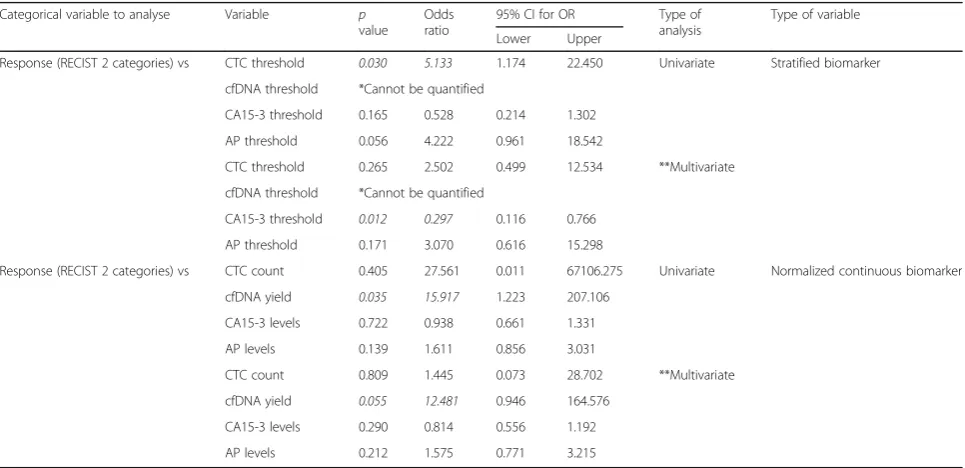
Table 4Logistic regression analysis between stratified clinical parameter (response to treatment) and stratified or normalized continuous biomarkers, showing thepvalue, odds ratio and the 95% confidence interval for this odds ratio. Univariate and multivariate analysis
Categorical variable to analyse Variable p value
Odds ratio
95% CI for OR Type of analysis
Type of variable Lower Upper
Response (RECIST 2 categories) vs CTC threshold 0.030 5.133 1.174 22.450 Univariate Stratified biomarker
cfDNA threshold *Cannot be quantified
CA15-3 threshold 0.165 0.528 0.214 1.302
AP threshold 0.056 4.222 0.961 18.542
CTC threshold 0.265 2.502 0.499 12.534 **Multivariate
cfDNA threshold *Cannot be quantified
CA15-3 threshold 0.012 0.297 0.116 0.766
AP threshold 0.171 3.070 0.616 15.298
Response (RECIST 2 categories) vs CTC count 0.405 27.561 0.011 67106.275 Univariate Normalized continuous biomarker
cfDNA yield 0.035 15.917 1.223 207.106
CA15-3 levels 0.722 0.938 0.661 1.331
AP levels 0.139 1.611 0.856 3.031
CTC count 0.809 1.445 0.073 28.702 **Multivariate
cfDNA yield 0.055 12.481 0.946 164.576
CA15-3 levels 0.290 0.814 0.556 1.192
AP levels 0.212 1.575 0.771 3.215
*Cannot be quantified as all patients in the group responding to treatment have low levels of cfDNA. No patients with high levels of cfDNA responding to treatment
[image:5.595.57.540.466.700.2]**Multivariate analysis represent the combination and analysis of all biomarkers (CTC count, cfDNA yield, CA15-3 levels and AP levels) together, as a joint analysis, to explore the possible interactions between them and how this interactions affects their effect on the clinical variables under study
Table 3Logistic regression analysis between stratified clinical parameter (response to treatment) and stratified biomarkers, showing thepvalue, odds ratio and the 95% confidence interval for this odds ratio
Categorical variable to analyse
Variable p value
Odds ratio
95% CI for OR Type of analysis
Type of variable Lower Upper
Response (RECIST 3 categories) vs
Responding CTC threshold Reference category Univariate Stratified biomarker
Stable disease 0.256 2.484 0.516 11.955
Progressive disease 0.007 7.966 1.783 35.587
Responding cfDNA threshold *Cannot be quantified Univariate Stratified biomarker
Stable disease
Progressive disease
Responding CA15-3 threshold Reference category Univariate Stratified biomarker
Stable disease 0.007 0.265 0.101 0.695
Progressive disease 0.977 1.014 0.382 2.694
Responding AP threshold Reference category Univariate Stratified biomarker
Stable disease 0.635 1.485 0.29 7.597
Progressive disease 0.01 7.233 1.616 32.373
samples needed for the study. To perform the analysis, we used the presence/absence of CTC counts against the sta-tus (alive/deceased) of 36 patients included in this prelim-inary study for whom tumour volume data was available. The effect size was calculated according to the means of the population and the pooled standard deviation of those means, which is the square root of the average of the two standard deviations. A total of 20 patients (10 patients in each group) are needed to achieve 80% power at two-sided 5% significance level; however, in order to maximize the effect and precision of the results of our study and in order to capture the full interaction between all the bio-markers and clinical parameters included, we opted to analyse all patients recruited over a fixed time period, be-tween February 2012 and July 2016, which increased the number of patients to 194.
In this pilot study over the subset of 36 patients, there was no association of the biomarkers with tumour bulk, either by the total tumour volume or the number of metastatic sites. Logistic regression analysis showed that none of the biomarkers, as categorical variables or as
continuous variables using a univariate or multivariate approach, nor response by RECIST criteria was associ-ated to tumour bulk (Additional file3).
Concerning the total cohort of 194 patients, the me-dian patient age was 59.5 years (ranges from 29 to 89; IQR = 20) and patients were from a range of breast can-cer sub-types as determined by ER, PR and HER2 status. Of the whole cohort, 95 (49.0%) patients were undergo-ing endocrine treatment, 49 (25.3%) were receivundergo-ing on chemotherapy, 18 (9.3%) were on HER2-targeted ther-apy, and 32 (16.5%) were off treatment at the time of blood sample (Table 1). Disease response data was ob-tained for all 194 patients using RECIST criteria [27] (Table 1; Additional file 4). There were 30 (15.5%) pa-tients showing a complete or partial response, 73 (37.6%) patients with stable disease and 91 patients (46.9%) with progressive disease.
Blood biomarkers
[image:6.595.60.538.88.420.2]a stable disease and 23 [18.1%] responding), AP was elevated (≥130 IU/L) in 40 patients (20.6%) (31 pro-gressing [77.5%], 7 [17.5%] with a stable disease and 2 [5.0%] responding) and both markers were elevated in 38 (19.6%) patients (29 progressing [76.3%], 7 [18.4%] with a stable disease and 2 [5.3%] responding) (Add-itional file 4) at the time of blood sampling. Levels of CA15-3 and AP were significantly correlated (p value < 0.0001; Table 2), confirming previous reports [6, 21,
28]. This correlation was confirmed by linear regres-sion analysis (p value < 0.0001; Additional file 5) and logistic regression analysis, where CA15-3 values were associated with AP values, and vice versa (p value < 0.0001, OR = 13.876, CI = 3.231–59.597; Additional files 6 and 7).
Eighty-nine of 194 patients (45.9%) had at least one
EpCAM+-positive CTC per 7.5 ml blood analysed
(range 1–6848) and 46 patients (23.7%) had ≥5 CTCs per 7.5 ml blood (defined as a high CTC count) [7] (33 progressing [71.7%], 11 [23.9%] with a stable dis-ease and 2 [4.4%] responding). The median plasma
cfDNA concentration was 0.112 ng/μl (IQR = 0.095; range 0.003 to 5.46 ng/μl), and 44 patients (22.7%) had high levels of cfDNA in plasma (≥0.306 ng/μ) (33 progressing [75.0%], 11 [25.0%] with stable disease). None of the patients with high cfDNA levels were responding to treatment at the time of blood sam-pling (Table 1; Additional file 4). The number of CTCs detected correlated positively with the total cfDNA level (p value < 0.0001), as reported previously [21] (Table 2). This correlation was confirmed by lo-gistic regression analysis, where high CTC counts were associated with cfDNA overall levels (p value = 0.003, OR = 2.140, CI = 1.305-3.510 and p value = 0.007, OR = 2.028, CI = 1.212–3.394; univariate and multivariate analysis; Additional file 6) and where high CTC counts were associated with high cfDNA levels, and vice versa (p value < 0.0001, OR = 8.083, CI = 3.803–17.180; Table 3). All 4 blood biomarkers included in the study were significantly correlated (Table 2), confirmed by regression analysis (Add-itional files 6 and 7).
[image:7.595.57.541.87.408.2]Comparison of biomarkers with molecular subtype and response to treatment
Based on data from the cohort, we also categorised each biomarker according to high and low cut-off points/ thresholds [7, 14, 23–25]. We then compared the four biomarkers against various clinical parameters using both the threshold values and using each biomarker as a continuous variable (Additional files8and9).
Regarding breast cancer molecular subtypes, analysis of each biomarker showed few significant results regard-ing hormone receptor (ER/PR) or HER2 status, or type of therapy administered. When using categorized vari-ables, CTCs and CA15-3 higher values were associated with HER2-negative status (p value = 0.048, OR = 0.284, CI = 0.082–0.989; p value = 0.003, OR = 0.311, CI = 0.145–0.670) and higher CA15-3 was associated with pa-tients receiving chemotherapy treatment. Results as con-tinuous variables showed CA15-3 was associated with ER and PR status (p value = 0.036, OR = 16.416, CI = 1.204–223.876; p value = 0.015, OR = 5.511, CI = 1.394– 21.792), as well as the use of chemotherapy treatment (p value = 0.027, OR = 1.495, CI = 1.046–2.136) by univari-ate and multivariunivari-ate analysis. Lastly, levels of cfDNA and AP were associated with patients receiving HER2 ther-apy (p value = 0.028, OR = 2.024, CI = 1.079–3.794; p value = 0.048, OR = 1.696, CI = 1.006–2.860) by multi-variate analysis (Additional files8and9).
According to RECIST criteria, patient response to treatment was classified as either complete or partial re-sponse, stable disease or progressive disease. To analyse the relationship with all four biomarkers, we compared the different categories against each biomarker as cat-egorical variables (Table 3). Results showed that high CTC counts and high AP levels are predictors of pro-gressive disease (pvalue = 0.007, OR = 7.996, CI = 1.783– 35.587 and p value = 0.01, OR = 7.233, CI = 1.616– 32.373, respectively), while CA15-3 is only associated with patients with a stable disease (pvalue = 0.007, OR = 0.265, CI = 0.101–0.695). It was not possible to quantify the effect of low/high levels of cfDNA because none of the 44 patients with higher levels of cfDNA were responding to treatment as all had either stable or pro-gressive disease. We therefore decided not to use this re-sponse stratification to draw any conclusions.
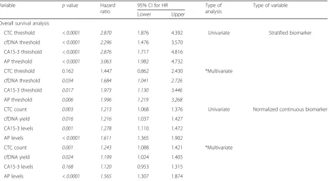
[image:8.595.59.541.109.374.2]To quantify accurately the effect of all biomarkers as a response predictor, we opted to group the response into two categories only. For that task, there were two differ-ent approaches: group categories by disease status (pro-gressing vs non-pro(pro-gressing disease) or focus the matter into the prediction of treatment response (responding vs non-responding to treatment). Based on clinical advice, we decided that assessing the efficiency of the treatment (responders vs non-responders) would be most useful in the follow-up of patients. Therefore, each biomarker was Table 5Overall survival analysis of metastatic breast cancer patients, showing thepvalue, hazard ratio and the 95% confidence interval for this hazard ratio. Stratified or normalized continuous biomarkers used as variables. Univariate and multivariate analysis
Variable pvalue Hazard
ratio
95% CI for HR Type of
analysis
Type of variable
Lower Upper
Overall survival analysis
CTC threshold < 0.0001 2.870 1.876 4.392 Univariate Stratified biomarker
cfDNA threshold < 0.0001 2.296 1.476 3.570
CA15-3 threshold < 0.0001 2.876 1.717 4.816
AP threshold < 0.0001 3.063 1.982 4.732
CTC threshold 0.162 1.447 0.862 2.430 *Multivariate
cfDNA threshold 0.034 1.684 1.041 2.726
CA15-3 threshold 0.017 1.973 1.130 3.446
AP threshold 0.006 1.996 1.219 3.268
CTC count 0.003 1.213 1.068 1.376 Univariate Normalized continuous biomarker
cfDNA yield 0.016 1.216 1.037 1.427
CA15-3 levels 0.001 1.278 1.110 1.472
AP levels < 0.0001 1.611 1.365 1.902
CTC count 0.001 1.243 1.088 1.421 *Multivariate
cfDNA yield 0.024 1.199 1.024 1.405
CA15-3 levels 0.168 1.120 0.953 1.315
AP levels < 0.0001 1.565 1.307 1.874
compared as a categorical and continuous variable against response to treatment (Table4).
The analysis of the 4 biomarkers as continuous vari-ables showed cfDNA levels as the sole predictor for treatment response, establishing a clear separation be-tween responding and non-responding patients, either by univariate (pvalue = 0.035, OR = 15.917) or multivari-ate analysis (p value = 0.055*, OR = 12.481; *borderline value) (Table4).
ROC curve analysis (Fig. 2) showed similar results for cfDNA yield, CTC counts and AP levels (AUC of 0.593, 0.585 and 0.573, respectively), whereas CA15-3 had a lower AUC (0.491). This suggests that CA15-3 was the poorest biomarker to discriminate patients according to their response to treatment. Although the AUC is mod-est, the cfDNA discrimination power is still 10% higher than CA15-3, which is one of the currently used markers in routine clinical practice. Concerning sensitivity and specificity analysis, AP levels had the highest sensitivity (68.3%) followed by cfDNA (59.8%), CA15-3 (54.9%) and CTC (47.6%), while CTC counts showed highest specifi-city (63.3%) followed by cfDNA, CA15-3 and AP, all with the same value (46.7%).
We interrogated the possible relationship between bio-markers and the presence/absence of any line of treat-ment. Results were not significant for any of the biomarkers (data not shown).
Comparison of biomarkers with patient survival
Stratification of patients according to the different thresh-old values for each biomarker demonstrated, as expected, that higher counts/values of all the biomarkers were sig-nificantly associated with poorer overall survival (OS) (Fig. 3; Table 5). A strong relationship was observed be-tween higher counts/levels of CTC (p value < 0.0001, HR = 2.870, CI = 1.876–4.392), cfDNA (p value < 0.0001, HR = 2.296, CI = 1.476–3.570), CA15-3 (pvalue < 0.0001, HR = 2.876, CI = 1.717–4.816) and AP (p value < 0.0001, HR = 3.063, CI = 1.982–4.732), with a poorer outcome of the metastatic breast cancer patients included in the study. However, when introducing the multivariate analysis ap-proach, CTC fails as prognostic factors.
We also compared the combined effect of cfDNA levels and CTC counts and CA15-3 and AP levels, with OS (Fig. 4; Additional file 10). Overall survival was im-proved (median survival > 59 months) when the CTC counts were low (< 5 CTCs/7.5 ml blood) regardless of cfDNA levels. However, when CTC counts were high the median survival was significantly affected by cfDNA levels, being 20 months when cfDNA levels were low (p value = 0.008, HR = 2.232, CI = 1.232–4.042) and just 6 months when cfDNA levels were high (pvalue < 0.0001, HR = 4.047, CI = 2.394–6.841). High levels of CA15-3 also significantly affected the median overall survival, re-ducing the median survival time from > 59 months to 31 months when AP levels were low or 12 months when AP levels were high. However, combined analysis of CTC counts and cfDNA levels showed the largest effect on the overall survival of patients.
Using a Cox regression model to analyse all bio-markers as continuous variables, we observe that all four biomarkers were significantly associated with patients
outcome by univariate analysis; however, when
Fig. 4Kaplan–Meier estimates of probability of overall survival in patients with metastatic breast cancer. Joint analysis ofAcirculating tumour cells and circulating free DNA (p< 0.0001, chi-square = 33.718; hazard ratio for patients with≥5 CTCs and < 0.306 ng/μl, 2.232 with 95% CI = 1.232–4.042; hazard ratio for patients with < 5 CTCs and≥0.306 ng/
[image:9.595.58.291.80.473.2]introducing multivariate analysis, only cfDNA levels (p value = 0.024, HR = 1.199, CI = 1.024–1.405), CTC counts (p value = 0.001, HR = 1.243, CI = 1.088–1.421) and AP levels (pvalue < 0.0001, HR = 1.565, CI = 1.307– 1.874), but not CA15-3, were able to predict OS (Table5).
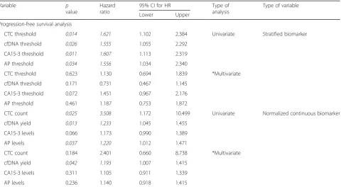
Stratification of patients according to the threshold values also showed as expected that higher counts/values of all the biomarkers were significantly associated with poorer progression-free survival (PFS) by univariate ana-lysis; however, none of them have significant results when using a multivariate approach (Fig. 5, Table 6). The analysis of all biomarkers as continuous variables showed that total cfDNA levels were the sole predictor of PFS by multivariate analysis (p value = 0.042, HR = 1.193, CI = 1.007–1.415) (Table 6). CTC counts and AP levels only had an effect as predictors in the univariate model, while CA15-3 levels did not have any effect.
Discussion
Many studies have shown that only approximately 50% of patients with MBC have high CTC counts or elevated
CA15-3 [5, 6, 29]. Hence, many MBC patients do not have an acceptable blood marker that allows the clin-ician to monitor the outcome of therapy without re-course to expensive imaging. Further, exome sequencing of the tumour, although offering a personalised approach to mutation profiling through ctDNA [30], has limita-tions since progression after therapy is often followed by the emergence of clones expressing other mutations.
CA15-3 and AP are the two routine biomarkers cur-rently used in the clinic. However, as neither appears to have a great ability to predict response to treatment, re-lapse or overall survival, in this study, we tried to com-pare these biomarkers with the newer biomarkers available (cfDNA and CTCs). Although the initial ana-lysis of all four biomarkers showed that their ability to discriminate response to treatment is not great, based on sensitivity and specificity of each biomarker, an over-all analysis of the area under the curve (AUC) by ROC curve analysis showed that cfDNA discriminatory power was 10% higher than CA15-3. This is, at least, a starting point to consider whether the markers we are using cur-rently in the clinic are the best available.
[image:10.595.63.539.88.393.2]The results of this study indicate that in patients with MBC, both the amount of total cfDNA and the number of EpCAM+CTCs in patient blood during the treatment period, are reflective of disease response and are indica-tors of overall survival, and importantly, cfDNA levels are the best predictor of disease response and PFS. Meanwhile, the conventional biomarker CA15-3 failed as a predictor of response and survival (OS and PFS), when analysed as a continuous variable. Overall, both CTC counts and cfDNA levels were associated with clin-ical outcomes in patients with MBC both individually and jointly, providing independent validation to a recent study [10].
Whilst we have shown that higher CTC counts and cfDNA levels are individually predictive and prognostic in patients with MBC, analysis of both, as a paired test, provides additional prognostic information. Overall sur-vival analysis in MBC patients based on CTC count alone showed a median survival > 59 months for patients with 0–4 CTCs/7.5 ml blood, compared with only 10 months for those with ≥5 CTCs/7.5 ml blood (Fig. 3). However, when patients have a CTC count ≥5 CTCs/ 7.5 ml blood, there was an increase in the median sur-vival time when the patient’s levels of cfDNA are low (< 0.306 ng/μl) and a decrease when the levels of cfDNA are high (≥306 ng/μl) (Fig.4). These results suggest that
[image:11.595.59.540.120.384.2]analysis of both CTC count and cfDNA level together may provide additional prognostic information and allow further stratification of patients with high CTC counts. One limitation of the current approach is the inability to detect CTCs in many patients even in the meta-static setting, and secondly that the majority of clin-ical service labs may not have access to a CTC platform, we therefore suggest that cfDNA measure-ment potentially represents an easier and less expen-sive biomarker than CTC counts in patients with MBC. Whilst our approach relies on detection of cfDNA levels rather than more specific mutation pro-filing through ctDNA, cfDNA measurement is a sim-ple and inexpensive test that could be set up as a routine test and done as an adjunct to radiological as-sessment. Importantly, the cost per test is in the tens of dollars rather than the hundreds of dollars re-quired currently for either CTC analysis or ctDNA profiling. Moreover, cfDNA measurement can be done more frequently than imaging through follow-up blood sampling to track disease response in real-time and alert clinicians as to when a change of treatment is needed. One other limitation to our study is that due to the high cost of CTC analysis, we analysed a single blood sample only from each patient; therefore, we cannot comment on dynamic responses over time. Table 6Progression-free survival analysis of metastatic breast cancer patients, showing thepvalue, hazard ratio and the 95% confidence interval for this hazard ratio. Stratified or normalized continuous biomarkers used as variables. Univariate and multivariate analysis
Variable p
value
Hazard ratio
95% CI for HR Type of
analysis
Type of variable
Lower Upper
Progression-free survival analysis
CTC threshold 0.014 1.621 1.102 2.384 Univariate Stratified biomarker
cfDNA threshold 0.026 1.555 1.055 2.292
CA15-3 threshold 0.011 1.607 1.113 2.319
AP threshold 0.034 1.556 1.034 2.340
CTC threshold 0.623 1.130 0.694 1.839 *Multivariate
cfDNA threshold 0.171 0.731 0.467 1.145
CA15-3 threshold 0.072 1.451 0.967 2.176
AP threshold 0.461 1.187 0.753 1.872
CTC count 0.025 3.508 1.172 10.499 Univariate Normalized continuous biomarker
cfDNA yield 0.013 1.233 1.045 1.455
CA15-3 levels 0.066 1.173 0.990 1.389
AP levels 0.037 1.220 1.012 1.471
CTC count 0.184 2.401 0.660 8.738 *Multivariate
cfDNA yield 0.042 1.193 1.007 1.415
CA15-3 levels 0.311 1.105 0.911 1.339
AP levels 0.236 1.140 0.918 1.415
Since we began this study in 2012, much research has focussed on characterising circulating tumour DNA (ctDNA), with the aim tracking somatic mutations with disease response. These studies have suggested that levels of ctDNA may be predictive of disease progres-sion, overall survival and progression-free survival in dif-ferent metastatic breast cancer patient populations, but these have not compared the results with other blood tests reflecting outcome. We explored the concordance between cfDNA and ctDNA levels in a previous study and showed that rising ctDNA is generally reflected by rising total cfDNA levels [19]. Some studies have re-ported that the dynamics of ctDNA show a better per-formance, when compared to the commonly assessed tumour protein biomarker CA15-3, in correlating with tumour burden, and provide a very early indication of treatment response [14, 31, 32]. In support of this, the results of this study indicate that simple measurement of total cfDNA levels is a good predictor of response, over-all survival and progression-free survival in patients with metastatic breast cancer suggesting potential clinical ap-plication of a cheap and simple blood-based monitoring test.
Conclusion
The results of this study indicate that in patients with MBC, both the amount of total cfDNA and the number of EpCAM+CTCs in patient blood during the treatment period, are reflective of disease response and are indica-tors of overall survival. Importantly, cfDNA levels are the best predictor of disease response and PFS; however, analysis of both cfDNA and CTC counts as a paired test, provides additional prognostic information and allows further stratification of patients. In conclusion, the re-sults of this study indicate that simple measurement of total cfDNA levels is a good predictor of response, over-all survival and progression-free survival in patients with metastatic breast cancer suggesting potential clinical ap-plication of a cheap and simple blood-based monitoring test.
Supplementary information
Supplementary informationaccompanies this paper athttps://doi.org/10. 1186/s13058-019-1235-8.
Additional file 1:Composite table with information of all the 194 samples included in the study. Information about the whole dataset included in the study.
Additional file 2:Composite table with information of all the 39 samples. Information about the small subset of the population used for preliminary analysis.
Additional file 3:Non-parametric correlation and linear regression ana-lysis of the four biomarkers included in the study, as continuous variables, against tumour volume and number of metastatic sites. Mann-Whitney-Wilcoxon non parametric test and logistic regression analysis between stratified biomarkers and response categories against tumour volume and
number of metastatics sites used as continuous variables, showing the p-value (multivariate analysis). Overall survival analysis of metastatic breast cancer patients, showing the p-value (univariate and multivariate analysis). Table including data regarding relationship between biomarkers, overall survival of patients and tumour bulk.
Additional file 4:Descriptive statistics of variables and clinical characteristics of patients included in the study, according to the type of disease response. Descriptive statistics of the whole population according to clinical parameters.
Additional file 5:Linear regression analysis between non stratified biomarkers, showing the p-value. Table containing information regarding the relationship between biomarkers as continuous variables.
Additional file 6:Logistic regression analysis between stratified biomarkers and normalized continuous biomarkers, showing the p-value, Odd ratio and the 95% confidence interval for this Odd ratio. Univariate and multivariate analysis. Table containing information regarding the rela-tionship between biomarkers as categorical and continuous variables.
Additional file 7:Logistic regression analysis between stratified biomarkers, showing the p-value, Odd ratio and the 95% confidence interval for this Odd ratio. Univariate and multivariate analysis. Table con-taining information regarding the relationship between biomarkers as categorical variables.
Additional file 8:Logistic regression analysis between stratified clinical, response and treatment parameters and stratified biomarkers, showing the p-value, Odd ratio and the 95% confidence interval for this Odd ratio. Univariate and multivariate analysis. Table containing information regard-ing the interactions between biomarkers, as categorical variables, and dif-ferent clinical parameters and treatment received by the patient.
Additional file 9:Logistic regression analysis between stratified clinical, response and treatment parameters and normalized continuous biomarkers, showing the p-value, Odd ratio and the 95% confidence interval for this Odd ratio. Univariate and multivariate analysis. Table con-taining information regarding the interactions between biomarkers, as continuous variables, and different clinical parameters and treatment re-ceived by the patient.
Additional file 10:Overall survival analysis of metastatic breast cancer patients, showing the p-value, Hazard ratio and the 95% confidence inter-val for this Hazard ratio. Comparison of joint analysis of new blood bio-markers (CTCs and cfDNA) against traditional biobio-markers (CA15-3 and AP). Stratified biomarkers as variables. Overall survival population results from joint analysis of biomarkers.
Acknowledgements
We thank the Imperial Cancer Research UK Centre and the Imperial and Leicester Experimental Cancer Medicine Centres (ECMC), the Imperial Biomedical Research Centre and the Imperial College Tissue Bank for supporting patient recruitment and sample collection. We thank David James Pinato for providing current CA15-3 and AP reference thresholds and Stephanie Marchese for support with CELLSEARCH.
Authors’contributions
RCC was the chief investigator on the study. RCC, DSG and JS conceived and supervised the study. KSG, CI, OO, ARB and KG recruited the patients, collected and processed the samples, and managed the clinical database supported by DFG. AH supported patient recruitment and carried out all CTC enumeration. DFG, KP, BT and RKH carried out all cfDNA analyses. DFG and RKH performed all statistical analyses, with advice from MR. AL provided response and tumour volume data. DFG and AH lead on drafting of the manuscript, and all authors read and approved the final manuscript.
Funding
Availability of data and materials
All data generated or analysed during this study are included in this published article [and its supplementary information files].
Ethics approval and consent to participate
The study protocol was approved by the Imperial College Healthcare Tissue Bank (ICHTB) Ethics Committee (project reference number: 12/WA/0196). Blood sample collection was conducted in accordance with the Declaration of Helsinki. All patients gave written informed consent prior to participation.
Consent for publication Not applicable
Competing interests
The authors declare that they have no competing interests.
Author details
1Leicester Cancer Research Centre, Department of Genetics and Genome
Biology, University of Leicester, Robert Kilpatrick Clinical Sciences Building, Leicester Royal Infirmary, Leicester LE2 7LX, UK.2Department of Surgery and Cancer, Imperial College London, Du Cane Road, Hammersmith, London W12 0NN, UK.3Imperial College London and Healthcare NHS Trust, Charing Cross Hospital, Fulham Palace Road, London W6 8RF, UK.4Welsh School of Pharmacy and Pharmaceutical Sciences, Redwood Building, King Edward VII Ave, Cardiff CF10 3NB, UK.5Department of Health Sciences, University of Leicester, George Davies Centre, Leicester LE1 7RH, UK.
Received: 16 July 2019 Accepted: 26 November 2019
References
1. Ferlay J, Soerjomataram I, Dikshit R, Eser S, Mathers C, Rebelo M, et al. Cancer incidence and mortality worldwide: sources, methods and major patterns in GLOBOCAN 2012. Int J Cancer. 2015;136(5):E359–86. 2. Cancer Research UK ¦ Breast cancer survival statistics. 2014. 3. UK CR. Breast cancer survival statistics; 2014.
4. Felipe Lima J, Nofech-Mozes S, Bayani J, Bartlett JM. EMT in Breast Carcinoma-A Review. J Clin Med. 2016;5(7):65.
5. Fu Y, Li H. Assessing clinical significance of serum CA15-3 and carcinoembryonic antigen (CEA) levels in breast cancer patients: a meta-analysis. Med Sci Monit. 2016;22:3154–62.
6. Keshaviah A, Dellapasqua S, Rotmensz N, Lindtner J, Crivellari D, Collins J, et al. CA15-3 and alkaline phosphatase as predictors for breast cancer recurrence: a combined analysis of seven International Breast Cancer Study Group trials. Ann Oncol. 2007;18(4):701–8.
7. Cristofanilli M, Budd GT, Ellis MJ, Stopeck A, Matera J, Miller MC, et al. Circulating tumor cells, disease progression, and survival in metastatic breast cancer. N Engl J Med. 2004;351(8):781–91.
8. Giuliano M, Giordano A, Jackson S, Hess KR, De Giorgi U, Mego M, et al. Circulating tumor cells as prognostic and predictive markers in metastatic breast cancer patients receiving first-line systemic treatment. Breast Cancer Res. 2011;13(3):R67.
9. Budd GT, Cristofanilli M, Ellis MJ, Stopeck A, Borden E, Miller MC, et al. Circulating tumor cells versus imaging--predicting overall survival in metastatic breast cancer. Clin Cancer Res. 2006;12(21):6403–9.
10. Ye Z, Wang C, Wan S, Mu Z, Zhang Z, Abu-Khalaf MM, et al. Association of clinical outcomes in metastatic breast cancer patients with circulating tumour cell and circulating cell-free DNA. Eur J Cancer. 2019;106:133–43. 11. Cristofanilli M, Pierga JY, Reuben J, Rademaker A, Davis AA, Peeters DJ, et al.
The clinical use of circulating tumor cells (CTCs) enumeration for staging of metastatic breast cancer (MBC): International expert consensus paper. Crit Rev Oncol Hematol. 2019;134:39–45.
12. Leon SA, Shapiro B, Sklaroff DM, Yaros MJ. Free DNA in the serum of cancer patients and the effect of therapy. Cancer Res. 1977;37(3):646–50. 13. Newman AM, Bratman SV, To J, Wynne JF, Eclov NC, Modlin LA, et al. An
ultrasensitive method for quantitating circulating tumor DNA with broad patient coverage. Nat Med. 2014;20(5):548–54.
14. Dawson SJ, Tsui DW, Murtaza M, Biggs H, Rueda OM, Chin SF, et al. Analysis of circulating tumor DNA to monitor metastatic breast cancer. N Engl J Med. 2013;368(13):1199–209.
15. Bettegowda C, Sausen M, Leary RJ, Kinde I, Wang Y, Agrawal N, et al. Detection of circulating tumor DNA in early- and late-stage human malignancies. Sci Transl Med. 2014;6(224):224ra24.
16. Spellman PT, Gray JW. Detecting cancer by monitoring circulating tumor DNA. Nat Med. 2014;20(5):474–5.
17. Ng SB, Chua C, Ng M, Gan A, Poon PS, Teo M, et al. Individualised multiplexed circulating tumour DNA assays for monitoring of tumour presence in patients after colorectal cancer surgery. Sci Rep. 2017;7:40737. 18. Gordon A, Palmieri C, Cleator SJ, Morden JP, Coombes RC. Tumour volume
analysis (TVA) as applied to a pilot randomized study of neoadjuvant endocrine versus neoadjuvant chemotherapy (NEOCENT). J Clin Oncol. 2015;33(15).https://doi.org/10.1200/jco.2015.33.15_suppl.e12003. 19. Page K, Guttery DS, Fernandez-Garcia D, Hills A, Hastings RK, Luo J, et al.
Next generation sequencing of circulating cell-free DNA for evaluating mutations and gene amplification in metastatic breast cancer. Clin Chem. 2017;63(2):532–41.
20. Page K, Guttery DS, Zahra N, Primrose L, Elshaw SR, Pringle JH, et al. Influence of plasma processing on recovery and analysis of circulating nucleic acids. PloS one. 2013;8(10):e77963.
21. Shaw JA, Guttery DS, Hills A, Fernandez-Garcia D, Page K, Rosales BM, et al. Mutation analysis of cell-free DNA and single circulating tumor cells in metastatic breast cancer patients with high circulating tumor cell counts. Clin Cancer Re. 2017;23(1):88–96.
22. Miller MC, Doyle GV, Terstappen LW. Significance of circulating tumor cells detected by the CellSearch System in patients with metastatic breast colorectal and prostate cancer. J Oncol. 2010;2010:617421.
23. Hayes DF, Cristofanilli M, Budd GT, Ellis MJ, Stopeck A, Miller MC, et al. Circulating tumor cells at each follow-up time point during therapy of metastatic breast cancer patients predict progression-free and overall survival. Clin Cancer Res. 2006;12(14 Pt 1):4218–24.
24. Budczies J, Klauschen F, Sinn BV, Gyorffy B, Schmitt WD, Darb-Esfahani S, et al. Cutoff Finder: a comprehensive and straightforward Web application enabling rapid biomarker cutoff optimization. PLoS One. 2012;7(12):e51862. 25. Valpione S, Gremel G, Mundra P, Middlehurst P, Galvani E, Girotti MR, et al.
Plasma total cell-free DNA (cfDNA) is a surrogate biomarker for tumour burden and a prognostic biomarker for survival in metastatic melanoma patients. Eur J Cancer. 2018;88:1–9.
26. Andersen PKG, R.D. Cox’s regression model for counting processes: a large sample study. Ann Stat. 1982;10(4):1100-20.
27. Eisenhauer EA, Therasse P, Bogaerts J, Schwartz LH, Sargent D, Ford R, et al. New response evaluation criteria in solid tumours: revised RECIST guideline (version 1.1). Eur J Cancer. 2009;45(2):228–47.
28. Briasoulis E, Andreopoulou E, Tolis CF, Bairaktari E, Katsaraki A, Dimopoulos MA, et al. G-CSF induces elevation of circulating CA 15-3 in breast carcinoma patients treated in an adjuvant setting. Cancer. 2001;91(5):909– 17.
29. Cheung KL, Graves CR, Robertson JF. Tumour marker measurements in the diagnosis and monitoring of breast cancer. Cancer Treat Rev. 2000;26(2):91– 102.
30. Abbosh C, Birkbak NJ, Wilson GA, Jamal-Hanjani M, Constantin T, Salari R, et al. Phylogenetic ctDNA analysis depicts early-stage lung cancer evolution. Nature. 2017;545(7655):446–51.
31. Diaz LA Jr, Williams RT, Wu J, Kinde I, Hecht JR, Berlin J, et al. The molecular evolution of acquired resistance to targeted EGFR blockade in colorectal cancers. Nature. 2012;486(7404):537–40.
32. Misale S, Yaeger R, Hobor S, Scala E, Janakiraman M, Liska D, et al. Emergence of KRAS mutations and acquired resistance to anti-EGFR therapy in colorectal cancer. Nature. 2012;486(7404):532–6.
Publisher’s Note