Erna Geessien Kroon,
aJônatas Santos Abrahão
aaDepartamento de Microbiologia, Instituto de Ciências Biológicas, Universidade Federal de Minas Gerais, Belo Horizonte, Minas Gerais, Brazil
bUniversidade Federal do dos Vales do Jequitinhonha e Mucuri, Diamantina, Brazil
ABSTRACT
Giant viruses are complex members of the virosphere, exhibiting
out-standing structural and genomic features. Among these viruses, the pandoraviruses
are some of the most intriguing members, exhibiting giant particles and genomes
presenting at up to 2.5 Mb, with many genes having no known function. In this
work, we analyzed, by virological and microscopic methods, the replication cycle
steps of three new pandoravirus isolates from samples collected in different regions
of Brazil. Our data indicate that all analyzed pandoravirus isolates can deeply modify
the
Acanthamoeba
cytoplasmic environment, recruiting mitochondria and
mem-branes into and around the electron-lucent viral factories. We also observed that the
viral factories start forming before the complete degradation of the cellular nucleus.
Various patterns of pandoravirus particle morphogenesis were observed, and the
as-sembly of the particles seemed to be started either by the apex or by the opposite
side. On the basis of the counting of viral particles during the infection time course,
we observed that pandoravirus particles could undergo exocytosis after their
mor-phogenesis in a process that involved intense recruitment of membranes that
wrapped the just-formed particles. The treatment of infected cells with brefeldin
af-fected particle exocytosis in two of the three analyzed strains, indicating biological
variability among isolates. Despite such particle exocytosis, the lysis of host cells also
contributed to viral release. This work reinforces knowledge of and reveals important
steps in the replication cycle of pandoraviruses.
IMPORTANCE
The emerging Pandoraviridae family is composed of some of the
most complex viruses known to date. Only a few pandoravirus isolates have been
described until now, and many aspects of their life cycle remain to be elucidated. A
comprehensive description of the replication cycle is pivotal to a better
understand-ing of the biology of the virus. For this report, we describe new pandoraviruses and
used different methods to better characterize the steps of the replication cycle of
this new group of viruses. Our results provide new information about the diversity
and biology of these giant viruses.
KEYWORDS
pandoravirus, giant virus, replication cycle, viral morphogenesis, viral
release, virus diversity
G
iant viruses are a group of complex viruses commonly referred to as
nucleocyto-plasmic large DNA viruses (NCLDV); the members of the group exhibit diverse
characteristics that have been astonishing the scientific community over the last few
years. Different groups of viruses described to date are able to replicate in amoeba cells,
expanding considerably our knowledge about their diversity, structure, genomics, and
evolution (1–5).
Five years ago, two complex giant viruses infecting
Acanthamoeba castellanii
cells
CitationPereira Andrade ACDS, Victor de Miranda Boratto P, Rodrigues RAL, Bastos TM, Azevedo BL, Dornas FP, Oliveira DB, Drumond BP, Kroon EG, Abrahão JS. 2019. New isolates of pandoraviruses: contribution to the study of replication cycle steps. J Virol 93:e01942-18.
https://doi.org/10.1128/JVI.01942-18.
EditorRozanne M. Sandri-Goldin, University of California, Irvine
Copyright© 2019 American Society for Microbiology.All Rights Reserved. Address correspondence to Jônatas Santos Abrahão, [email protected]. A.C.D.S.P.A. and P.V.D.M.B. contributed equally to this article.
Received1 November 2018 Accepted1 November 2018
Accepted manuscript posted online12 December 2018
Published19 February 2019
on November 6, 2019 by guest
http://jvi.asm.org/
Germany (7). Years after this discovery, analysis of this endosymbiont genome revealed
the viral nature of this organism, which was classified as a pandoravirus (8). This was the
third pandoravirus described, and it was named
P. inopinatum
(9, 10). In 2015 to 2016,
new pandoraviruses were described using a culture of
A. castellanii
cells belonging to
sewage and soda lake water samples. These viruses were named
P. massiliensis
,
P.
pampulha
, and
P. brasiliensis
(11–13). Another recent prospective study reported the
isolation of
Pandoravirus quercus
, isolated from samples of soil collected in Marseille
(France);
P. neocaledonia
, isolated from the brackish water around a mangrove near
Noumea Airport (New Caledonia), and
P. macleodensis
, isolated from a freshwater pond
near Melbourne (Australia) (14). Pandoraviruses represent a genome exceeding those of
some eukaryotic microorganisms, with a huge proportion of open reading frame (ORF)
genes without homologs (ORFans) in any database. The ORFans correspond to about
70% of the predicted genes of pandoraviruses (6).
Despite the plethora of novel characteristics revealed by analyses of the genomes
and evolution of the pandoraviruses, their replication cycle still needs further study to
be better understood. In the present report, we present an in-depth investigation of the
replication cycle steps of three new isolates of pandoraviruses. We observed that the
pandoraviruses are able to deeply modify the acanthamoeba cytoplasmic environment,
recruiting mitochondria and membranes into and around the electron-lucent viral
factories (VFs). The viral factory formation and viral particle morphogenesis were
analyzed in an in-depth manner by electron microscopy (EM), with results reinforcing
previously published data and revealing new features about pandoraviruses’ replication
cycles. We also demonstrated by microscopy and pharmacological inhibition of
mem-brane traffic that viral particles were released from infected cells both by exocytosis and
by cell lysis. This work contributes to the understanding of important steps in the
replication cycle of pandoraviruses.
RESULTS
New members of the emerging family Pandoraviridae.
Isolation of a new
pan-doravirus isolate, namely,
P. kadiweu
, was performed by culturing amoebas of the
A.
castellanii
species with water samples collected in the city of Bonito, Mato Grosso do
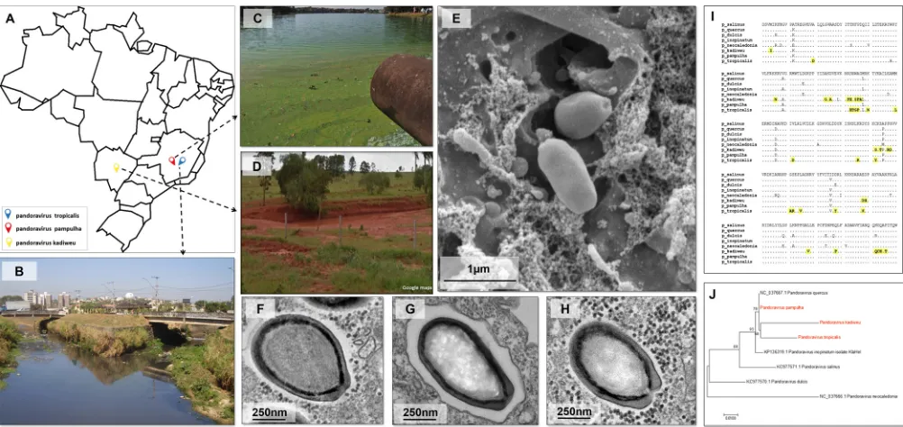
Sul, Brazil (Fig. 1A, D, and H). A prospective study conducted between 2015 and 2017
using culture of
A. castellanii
species with sewage samples from different environmental
and clinical samples reported the collection of two pandoravirus isolates that were
identified by real-time PCR and electron microscopy (12). The pandoravirus isolates
were obtained from samples of Mergulhão Creek and Bom Jesus Creek, in the region
of Pampulha Lake, Belo Horizonte, Brazil (Fig. 1A to C), and were named
Pandoravirus
pampulha
(12) (Fig. 1F) and
P. tropicalis
, respectively (Fig. 1E and G). The
P. kadiweu
isolate and the two isolates described by Andrade et al. in 2018 (12) are new members
of the emerging family Pandoraviridae.
The isolates were observed both by optical microscopy (data not shown) and by
electron microscopy, and the images indicated no evident morphological differences
among the three isolates (Fig. 1E to H). The isolates were
⬃
1.0
m in length and had
an ostiole-like apex at one end of the particle as previously described for other
pandoraviruses (6, 12–15). In order to evaluate whether our isolates were similar, we
on November 6, 2019 by guest
http://jvi.asm.org/
sequenced a fragment of the DNA polymerase subunit B gene. The analysis of predicted
amino acid sequences revealed that all of the isolates were different from each other.
In addition, we observed that
P. tropicalis
and
P. kadiweu
present unique amino acid
substitutions (Fig. 1I). The sequence of
P. pampulha
was more similar to that of
P.
quercus
(Fig. 1I). These results reveal the diversity among our isolates and other
pandoravirus isolates, and future genomic studies will determine whether
P. tropicalis
and
P. kadiweu
represent new clades among pandoraviruses (Fig. 1J). To date, there
have been no rules or parameters available to establish a new clade belonging to the
hypothetical family Pandoraviridae. The electron microscopy images obtained for these
isolates were used to assemble a collection of more than 200 images. This data set
allowed us to perform a comprehensive analysis of the replication cycle of these
viruses.
Pandoraviruses are phagocytosed and replicate in large and electro-lucent
viral factories.
As demonstrated by work published by Legendre et al. in 2018 (14), the
first steps involving the replication cycle of pandoraviruses seem to be similar for all
these viruses, independently of the virus isolate analyzed. We observed that the
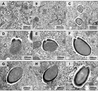
amphora-shaped viral particles enter into acanthamoeba cells, likely by phagocytosis,
which occurred within 30 min of infection (Fig. 2A and B). The particles were then
transported to the interior of the amoebal cytoplasm, being carried inside phagosomes
(Fig. 2C to E). This structure then seems to become fused with lysosome-like organelles,
which, upon releasing their content inside the phagosome, stimulate the uncoating of
the pandoravirus particles (Fig. 2C to F).
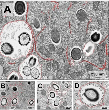
The viral factories (VFs) of the three analyzed isolates were wide, and electron-lucent
areas occupied approximately 1/3 of the amoeba cytoplasm, containing viral particles
in different stages of morphogenesis (Fig. 3). The VFs of the pandoraviruses seem to
have been homogeneous and were not clearly limited by any cell component.
Inter-estingly, we observed recruitment of mitochondria to regions inside and around the
VFs (Fig. 3) and membranes were also recruited to regions inside the VFs (Fig. 4). In
FIG 1Sites of collection and electron microscopy and phylogenetic analysis of the pandoravirus isolated in this work. (A) Map of Brazil showing where the samples were collected for the isolation of pandoraviruses. (B to D) Representative pictures from the areas of collection: Bom Jesus Creek (B), Mergulhão Creek (C), and the city of Bonito (D). (E)P. tropicalisparticles were analyzed using scanning electron microscopy at 24 h.p.i. and an MOI of 0.01. (F, G, and H) Transmission electron microscopy (24 h.p.i./MOI 0.01) for the viral particles corresponding to the isolates ofP. pampulha,P. tropicalis, andP. kadiweu, respectively. (I) Alignment of the sequences, showing thatP. kadiweuandP. tropicalisrepresent strains of pandoraviruses with many exclusive polymorphisms (highlighted in yellow), compared to the sequences of other isolated pandoraviruses. (J) Maximum likelihood tree constructed using predicted sequence of 251 amino acids of a DNA polymerase B subunit in different isolates of pandoraviruses. The giant viruses isolated in this work are highlighted in red.
on November 6, 2019 by guest
http://jvi.asm.org/
[image:3.585.42.543.70.307.2]addition, it is possible to observe an intense accumulation of structures that resemble
lysosomal vesicles near the VFs (Fig. 3, orange arrows).
We also analyzed the appearance of the nuclear and nucleolar structures during the
time course of infection of the three pandoravirus isolates (Fig. 5). The nuclear and
nucleolar structures, appearing in the typical manner, were promptly observed both by
transmission electron microscopy (TEM) and by Hemacolor staining in uninfected
FIG 2Initial steps of the pandoravirus replication cycle inside the amoebal host. (A and B) Scanning electron microscopy (A) and transmission electron microscopy (B) images show pandoravirus particles entering Acanthamoeba castellanii cells, likely as a consequence of phagocytosis. (C) The amoebas project pseudopods involving the viral particles and internalize them into vesicle-like structures known as phagosomes. (D and E) The phagosome then fuses with another component resembling a lysosome-like structure that, upon releasing their combined content, stimulates the uncoating of the pandoravirus particles (F). Although we used representative images in this figure, all the described steps were observed for all three isolated pandoraviruses. L, lysosome-like organelles; panels A and B,Pandoravirus tropicalis; panels C to F,Pandoravirus kadiweu.
FIG 3Characterization of pandoravirus viral factories. Viral factories of (A)Pandoravirus tropicalis, (B)P. pampulha, and (C)P. kadiweuwere observed by transmission electron microscopy. The region of the viral factories is highlighted in red, the mitochondria present in the interior of the viral factories are highlighted in green, and the lysosomes are pointed out by orange arrowheads.
on November 6, 2019 by guest
http://jvi.asm.org/
[image:4.585.45.368.71.369.2] [image:4.585.45.545.580.707.2]acanthamoeba cells (Fig. 5A). As expected, the same was observed during viral entry
(Fig. 5B). However, the nucleolar structure was no longer visible when the
pandoravi-ruses’ early VFs appeared, although we were still able to visualize the amoeba nucleus
with its membrane (Fig. 5C). At late infection, the nuclear structure was no longer
FIG 4Membranes recruited inside pandoravirus viral factories. (A) Transmission electron microscopy ofP. tropicalisviral factories. (B) Transmission electron microscopy ofP. pampulhaviral factories. (C) Transmission electron microscopy ofP. kadiweuviral factories. The membranes recruited inside the viral factories are highlighted in blue.
FIG 5The Acanthamoeba castellaniicell nucleus becomes disorganized and loses its natural shape during the course of pandoravirus infection. (A) Transmission electron microscopy image showing a noninfectedAcanthamoeba castellaniicell and how its nucleus is normally organized in this situation; it occupies about 2/3 of the cellular area, and it is delimited by a double-membrane layer known as the nuclear envelope (digitally highlighted in orange). The image at lower left represents the same conditions but visualized on a light microscope with Hemacolor staining. The nucleolus is observed as a dark spot surrounded by a bright area that represents the nucleus. (B) Image representing the amoeba observed just after the first steps of the pandoravirus replication cycle, as the virus (red arrow) is still harboring inside the amoebal phagosome. The nucleus does not yet seem to have suffered any modification at this stage. (C) At between h 3 and h 6 of infection, it seems that the nucleolus starts to be absent, as shown by one of the several images of transmission electron microscopy analyzed in this work. At lower left, the Hemacolor staining also shows the beginning of the appearance of the early viral factory. (D) The later steps of viral replication lead to the formation of the mature viral factory, marked by a bright area, easily recognizable in the images with Hemacolor staining. N, nucleus; Nc, nucleolus; eVF, early viral factory; mVF, mature viral factory.
on November 6, 2019 by guest
http://jvi.asm.org/
[image:5.585.45.545.71.210.2] [image:5.585.42.435.338.625.2]visible also, and the VFs occupied a substantial region in the cytoplasm (Fig. 5D). This
process was observed during the replication cycle of the three isolates.
Morphogenesis dynamics of pandoravirus particles.
After analysis of dozens of
TEM images of asynchronous cycles of the isolated pandoraviruses, we noticed that the
capsids of the pandoraviruses appeared to be formed from electron-dense semicircular
structures observed in the middle of the VF (Fig. 6A). These structures appeared to
become thicker and more electron dense as the cycle continued and to function as
crescent-shaped precursors (Fig. 6B and C). The crescent-shaped precursors underwent
a thickening of the apparent layer, followed by filling of the internal contents of the
particles. As the adjacent portions of the capsids formed, the internal content of the
particle continued to be filled simultaneously (Fig. 6D to I). As the particle enlarged,
the capsid became more electron dense until closure of the total capsid, which at that
stage was already filled with the particle’s internal contents (Fig. 6I). After careful
analysis of several images of our three isolates, we observed that particle
morphogen-esis/assembly could apparently start either at the ostiole-like apex or at the opposite
end (Fig. 6D to I).
Pandoravirus particles are released by exocytosis and cell lysis.
By studying the
infection cycles of the new pandoravirus isolates, we made a curious observation.
Analyses that have been done under a light microscope revealed that at early times of
infection (until 6 h postinfection [h.p.i.]), these viruses could already be detected in the
supernatant of infected cells, even at time points when the host cells had not yet
undergone lysis. We then hypothesized that the pandoravirus particles could have
started their release from the host by exocytosis, as suggested for some pandoravirus
isolates (14). The analyses of TEM images of the new isolates revealed intense
mem-FIG 6Morphogenesis of pandoravirus particles. Transmission electron microscopy images show stages of pandoravirus particle formation. (A to C) Crescent-like structures with different sizes, inside the viral factory, growing in thickness and complexity. (D to F) Particles being formed from the ostiolo-like apex. (G to I) Particles being formed from the end opposite the ostiolo-like apex. We used representative images ofP. tropicalis,P. pampulha, andP. kadiweuin this panel; all the described steps were observed for the three isolates.
on November 6, 2019 by guest
http://jvi.asm.org/
[image:6.585.44.368.70.369.2]brane traffic close to just-formed particles, in the periphery of the VF (Fig. 7A).
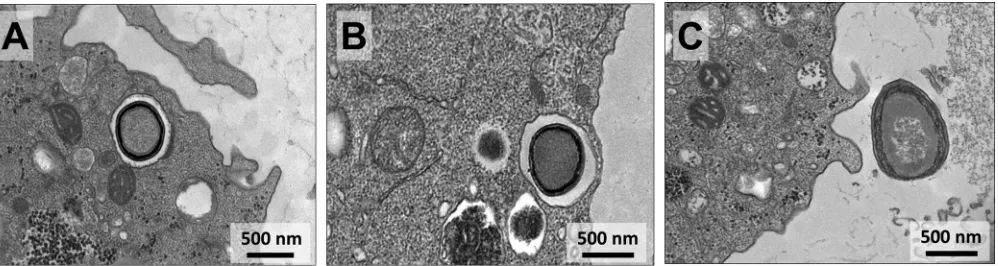
Interestingly, many particles were then wrapped inside such membranes, forming
exosomes containing various amounts of viral particles of different sizes (Fig. 7). These
exosome-containing particles then seemed to migrate to the periphery of the infected
cell, fusing with the host cell cytoplasmic membrane and releasing the particles to the
extracellular environment (Fig. 8).
To experimentally confirm that pandoraviruses can be released by exocytosis, we
counted the acanthamoeba cells and the number of pandoraviruses particles in the
supernatant through the viral cycle (multiplicity of infection [MOI] of 10). With this set
of data, we analyzed whether the increase in the number of viral particles in the
supernatant through the viral cycle could be observed before cell lysis was induced by
viral infection, which would indicate that these viruses were being released by
exocy-tosis at early times of infection. We observed that
P. tropicalis
caused the lysis of
infected amoebas at 12 h.p.i., while no significant decrease in cell numbers was
observed for cells infected with
P. pampulha
and
P. kadiweu
until 24 h.p.i. This indicates
differences in the time postinfection at which each pandoravirus can induce host lysis
(Fig. 9A to C). Cell lysis induced by
P. pampulha
and
P. kadiweu
was observed at 48 h.p.i.
FIG 7Transmission electron microscopy images showing pandoravirus particles being packaged into exosomes. (A) The late steps in pandoravirus replication are marked by intense membrane trafficking in the cytoplasm of the amoebal host (highlighted in red). This event is easily observed around the viral factory where the viral morphogenesis occurs. (B to D) Then, at around h 6 to h 9 postinfection, these double-membrane layers start to surround isolated or grouped viral particles, suggesting the beginning of exocytosis.
on November 6, 2019 by guest
http://jvi.asm.org/
[image:7.585.42.426.71.457.2](data not shown). Interestingly, we observed an increase in the level of viral particles
released in the supernatant from 6 h.p.i. for the three pandoravirus isolates, indicating
that exocytosis might indeed contribute to particle release (Fig. 9D to F).
Aiming to evaluate the impact of membrane traffic inhibition in pandoravirus
exocytosis, we pretreated infected amoebas with brefeldin A (BFA) (a
membrane-trafficking inhibitor). Viral particles were counted at 12 h.p.i. for
P. pampulha
and
P.
kadiweu
and at 6 h.p.i. for
P. tropicalis
. These time points were selected for each
pandoravirus isolate based on the experiments last described above (Fig. 9A to F),
whose results indicated the moment when the particles were undergoing exocytosis
and the cells were not undergoing lysis. It was observed that acanthamoeba cultures
treated with brefeldin A showed a reduction in the number of particles released for
P.
pampulha
and
P. kadiweu
viruses (Fig. 9G and H). Curiously, the same was not observed
for
P. tropicalis
(Fig. 9I). Future studies are needed to clarify why
P. tropicalis
can cause
lysis of cells earlier than
P. kadiweu
and
P. pampulha
and why its exocytosis does not
seem to be affected by brefeldin A treatment.
DISCUSSION
Giant virus prospective studies have revealed an outstanding universe of viral
diversity (3, 4, 6, 16–20). Metagenomic studies have indicated the presence of a giant
virus gene set in all continents (21–25). Some representatives, such as the mimivirus,
appear to be more abundant and ubiquitous, containing hundreds of isolates already
reported (21–26). Pandoravirus-like sequences were also detected in metagenomic data
from environmental samples (22, 27, 28) as well as from insects, simian bushmeat, and
human plasma (23–25, 27). Despite this, the amount of pandoravirus isolates is still
limited (4, 6, 8, 11–13). Therefore, the isolation of new pandoraviruses contributes to
the understanding of their biology, diversity, and distribution. The analyses of the
isolates obtained in this work add important information characterizing the steps in the
pandoravirus replication cycle.
It was hypothesized that pandoraviruses enter amoebas by phagocytosis (14). Our
data for
P. tropicalis
,
P. pampulha
, and
P. kadiweu
reinforce this previous observation, as
particles can be seen inside large vesicles in the amoebal cytoplasm within 30 min
postinfection (Fig. 2C to E). Korn and Weisman demonstrated in 1967 that only particles
larger than 500 nm can trigger phagocytosis in
Acanthamoeba
, a condition so far
fulfilled by pandoravirus particles (29). Our images clearly demonstrate the induction of
pseudopod formation when amoebas were kept in contact with pandoravirus particles
(Fig. 2A and B). Despite this evidence, the possibility of pandoravirus particles entering
amoebas by macropinocytosis could not be overruled, since this pathway also forms
endosomes larger than 1
m (30). However, the involvement of macropinocytosis in
virion entry is a rare phenomenon in the literature (30). After entry of amoebas and
release of the inner virion content (Fig. 2F), a short eclipse phase and an increase in
FIG 8Transmission electron microscopy images demonstrating sequential steps of pandoravirus particle exocytosis. The images demonstrate that in late stages of infection the particles of pandoravirus start being packaged inside vesicles (A and B), becoming closer to the cytoplasmic membrane of the host cell and being released to the external milieu (C).
on November 6, 2019 by guest
http://jvi.asm.org/
[image:8.585.46.545.72.205.2]growing pandoravirus VFs occur (Fig. 3 and 5). As for the distribution of cellular
organelles, the presence of many mitochondria inside and near the VF was seen (Fig.
3), along with the polarization of structures that resemble lysosomal vesicles in the
vicinity of the VF (Fig. 3), as previously reported for the cedratvirus (31). The presence
of mitochondria in this region could be related to the process of energy acquisition
during viral replication optimization (32). Lysosome polarization, corresponding to the
presence of vacuole-like structures occupying large portions of the host cell, might be
related to a cellular response to infection, such as autophagy, as suggested previously
in cedratvirus (31, 32). In addition, we observed gradual nucleolar and nuclear
degra-dation throughout the pandoravirus replication cycle (Fig. 4), as observed for other
pandoraviruses (6).
The onset of particle morphogenesis seems to occur from electron-dense
semicir-FIG 9Analysis of the time course of infection for the pandoravirus isolates inAcanthamoeba castellaniicultures. The course of infection ofP. tropicalis,P. pampulha, andP. kadiweuwas established. (A to C) First, we observed the kinetics for the diminishing of the number of amoebas during the replication cycles ofP. pampulha(A),P. kadiweu(B), andP. tropicalis(C) analyzed by cell counting. (D to F) Then, the number of viral particles present in the supernatant of these infected cells was also observed forP. pampulha(D),P. kadiweu(E), andP. tropicalis(F), at different time points. After setting a time point at which we observed an increase of more than 1 log of virus particles in the supernatant but without observing cellular lysis, the amoebal cultures were treated with an inhibitor of membrane trafficking (brefeldin A). (G to I) The cells were then infected withP. pampulha(G),P. kadiweu(H), andP. tropicalis(I) to check the influence of brefeldin A in the viral release. The number of exocyted viral particles in supernatant was counted. DMSO, dimethyl sulfoxide.
on November 6, 2019 by guest
http://jvi.asm.org/
[image:9.585.45.539.66.497.2]the NCLDV group reinforce the previously suggested idea that these viruses could share
an ancestor (38–41).
In a recent study, different strains of pandoravirus were seen to be initially released
during the viral replication cycle by exocytosis processes (14).
Pandoravirus quercus
,
P.
neocaledonia
, and
P. macleodensis
complete their entire replication cycle by between 8
to 12 h, starting the viral particles’ exocytosis in about 8 h.p.i. and finishing their
replicative cycle with lysis of the amoebal host cells, releasing hundreds of virions in the
supernatant. Although the particles of the pandoravirus isolates analyzed here were
seen to produce and were exocytosed from the cell as fast as those described by
Legendre et al. (14) (in about 6 to 12 h.p.i.), the lysis of cells was observed at the late
times of infection (12 to 48 h) (Fig. 9A to F). These results not only reinforce the
hypothesis that pandoraviruses can explore different pathways for the progeny release
but also demonstrate biological differences among viral isolates.
Many aspects of the replication cycle of these viruses still need to be clarified. This
work provides new data and reveals new questions that future studies, especially at the
molecular level, could answer. Prospective studies may also contribute in this regard by
revealing novel members within the NCLDV group and improving understanding of the
diversity, evolution, and biology of these complex viruses.
MATERIALS AND METHODS
Viral isolation, stock production, and titration.Three different pandoravirus isolates were used in this work. Two were coisolated with mimivirus from sewage samples collected in streams in the Pampulha region, Belo Horizonte, Brazil, in previous work and namedP. pampulhaandP. tropicalis(12). The other pandoravirus was isolated in the present work from water samples collected in Bonito, Mato Grosso do Sul, Brazil, and namedP. kadiweu. For viral isolation, we usedA. castellanii(ATCC 30234) as previously described (12). In order to produce the viral stocks,A. castellaniicells were grown in cell culture flasks and infected with 500l of isolates. After observation of typical cytopathic effect (cell rounding and lysis), the flask content was collected and the viruses were titrated. The titer was obtained by the endpoint method (42) and expressed as the number of 50% tissue culture infective doses (TCID50) per milliliter. Viral stocks were kept at⫺70°C until use in further experiments.
Sequencing, alignment, and phylogeny.A fragment of the DNA polymerase B subunit gene (from position 473404 to position 474507—referencePandoravirus quercus) was amplified (5=GCCCTCAAGCG GGGCCGCATG3=and 5=CATCCACTGGGTGATCGGCGCCT3=) and sequenced, in both orientations and in duplicate, using an automated capillary sequencer (MegaBACE sequencer; GE Healthcare, Buckingham-shire, United Kingdom). For the phylogenetic tree, the resulting predicted amino acid (aa) sequences (251 aa) were aligned with previously published sequences obtained from GenBank using ClustalW in MEGA 7.0 software. This gene is highly conserved among pandoravirus strains and has been used in other studies (6, 12). The tree was constructed using the maximum likelihood method and a bootstrap value of 1,000.
Amoebal and viral particle counting.Initial electron microscopy analyses raised the hypothesis that pandoravirus could be released by exocytosis. In that way, two experiments were coupled and performed in duplicate that involved (i) the counting of intact amoebas throughout the viral replication cycle and (ii) the counting of pandoravirus particles that were being released in the supernatant at the same time points of infection.A. castellaniicells were infected in 25-cm2culture flasks (Kasvi, Brazil) withP. tropicalis, P. pampulha, andP. kadiweuisolates using an MOI of 10, and analyses were carried out at the infection times of 0 h, 3 h, 6 h, 9 h, 12 h, and 24 h. The time point of 0 h corresponds to an adsorption step of 30 min after infection, when the monolayer of cells was washed with 1⫻phosphate-buffered saline (PBS) and the flasks were filled with 4 ml of peptone-yeast extract-glucose (PYG) medium. After each time point was reached, we separated 12l of the supernatant to count the number of pandoravirus particles released during infection. The particles were observed under light microscopy (OlympusBX41, Japan), at ⫻1,000 magnification, using a cell counting chamber (Kcell Olen Kasvi, Brazil). In parallel, 12l of whole content presented in the flasks (including cells) was used to count the number of intact amoeba cells
on November 6, 2019 by guest
http://jvi.asm.org/
in the previous section and fixed at various times postinfection with 2.5% glutaraldehyde in a 0.1 M sodium phosphate buffer for 1 h at room temperature. The amoebas were postfixed with 2% osmium tetroxide and embedded in Epon resin. Ultrathin sections were then analyzed using transmission electron microscopy (TEM; Spirit Biotwin FEI-120 kV) at the Center of Microscopy of Universidade Federal de Minas Gerais (UFMG).
Scanning electron microscopy.A. castellaniicells infected at an MOI of 0.01 were added to round glass coverslips covered with poly-L-lysine and fixed with 2.5% glutaraldehyde in 0.1 M cacodylate buffer for at least 1 h at room temperature. The samples were then washed three times with 0.1 M cacodylate buffer and postfixed with 1.0% osmium tetroxide for 1 h at room temperature. After the second fixation, the samples were washed three times with 0.1 M cacodylate buffer and immersed in 0.1% tannic acid for 20 min. The samples were then washed in cacodylate buffer and dehydrated by serial passages in ethanol solutions at concentrations ranging from 35% to 100%. Samples were then subjected to critical point drying using CO2, placed in stubs, and metalized with a 5-nm-particle-size gold layer. The analyses were completed using scanning electron microscopy (FEG Quanta 200 FEI) at the Center of Microscopy of UFMG.
Accession number(s).Sequences are available in GenBank under accession numbersMK131392(P. kadiweu),MK131393(P. pampulha), andMK131394(P. tropicalis).
ACKNOWLEDGMENTS
We thank our colleagues from Gepvig and the Laboratório de Vírus for their
excellent technical support. We also thank CNPq, Coordenação de Aperfeiçoamento de
Pessoal de Nível Superior (CAPES), FAPEMIG, and the Center of Microscopy of UFMG.
E.G.K. and J.S.A. are CNPq researchers. E.G.K. and J.S.A. are members of a
CAPES-COFECUB project.
We declare that we have no conflicts of interest.
REFERENCES
1. La Scola B, Audic S, Robert C, Jungang L, de Lamballerie X, Drancourt M, Birtles R, Claverie JM, Raoult D. 2003. A giant virus in amoebae. Science 299:2033.https://doi.org/10.1126/science.1081867.
2. Raoult D, Audic S, Robert C, Abergel C, Renesto P, Ogata H, La Scola B, Suzan M, Claverie JM. 2004. The 1.2-megabase genome sequence of Mimivirus. Science 306:1344 –1350.https://doi.org/10.1126/science.1101485. 3. Boyer M, Yutin N, Pagnier I, Barrassi L, Fournous G, Espinosa L, Robert C,
Azza S, Sun S, Rossmann MG, Suzan-Monti M, La Scola B, Koonin EV, Raoult D. 2009. Giant Marseillevirus highlights the role of amoebae as a melting pot in emergence of chimeric microorganisms. Proc Natl Acad Sci U S A 106:21848 –21853.https://doi.org/10.1073/pnas.0911354106. 4. Legendre M, Bartoli J, Shmakova L, Jeudy S, Labadie K, Adrait A, Lescot
M, Poirot O, Bertaux L, Bruley C, Coute Y, Rivkina E, Abergel C, Claverie JM. 2014. Thirty-thousand-year-old distant relative of giant icosahedral DNA viruses with a pandoravirus morphology. Proc Natl Acad Sci U S A 111:4274 – 4279.https://doi.org/10.1073/pnas.1320670111.
5. Rodrigues RAL, Andreani J, Andrade A, Machado TB, Abdi S, Levasseur A, Abrahao JS, La Scola B. 13 June 2018. Morphologic and genomic anal-yses of new isolates reveal a second lineage of cedratviruses. J Virol
https://doi.org/10.1128/JVI.00372-18.
6. Philippe N, Legendre M, Doutre G, Coute Y, Poirot O, Lescot M, Arslan D, Seltzer V, Bertaux L, Bruley C, Garin J, Claverie JM, Abergel C. 2013. Pandoraviruses: amoeba viruses with genomes up to 2.5 Mb reaching that of parasitic eukaryotes. Science 341:281–286. https://doi.org/10 .1126/science.1239181.
7. Scheid P, Zoller L, Pressmar S, Richard G, Michel R. 2008. An extraordi-nary endocytobiont in Acanthamoeba sp. isolated from a patient with
keratitis. Parasitol Res 102:945–950.https://doi.org/10.1007/s00436-007 -0858-3.
8. Scheid P, Balczun C, Schaub GA. 2014. Some secrets are revealed: parasitic keratitis amoebae as vectors of the scarcely described pandoraviruses to humans. Parasitol Res 113:3759 –3764.https://doi.org/10.1007/s00436-014 -4041-3.
9. Scheid P. 2016. A strange endocytobiont revealed as largest virus. Curr Opin Microbiol 31:58 – 62.https://doi.org/10.1016/j.mib.2016.02.005. 10. Antwerpen MH, Georgi E, Zoeller L, Woelfel R, Stoecker K, Scheid P. 2015.
Whole-genome sequencing of a pandoravirus isolated from keratitis-inducing acanthamoeba. Genome Announc 3:e00136-15. https://doi .org/10.1128/genomeA.00136-15.
11. Dornas FP, Khalil JY, Pagnier I, Raoult D, Abrahao J, La Scola B. 2015. Isolation of new Brazilian giant viruses from environmental samples using a panel of protozoa. Front Microbiol 6:1086.https://doi.org/10 .3389/fmicb.2015.01086.
12. Andrade ACDSP, Arantes TS, Rodrigues RAL, Machado TB, Dornas FP, Landell MF, Furst C, Borges LGA, Dutra LAL, Almeida G, Trindade G. d S, Bergier I, Abrahão W, Borges IA, Cortines JR, de Oliveira DB, Kroon EG, Abrahão JS. 2018. Ubiquitous giants: a plethora of giant viruses found in Brazil and Antarctica. Virol J 15:22.https://doi.org/10.1186/s12985-018 -0930-x.
13. Aherfi S, Andreani J, Baptiste E, Oumessoum A, Dornas FP, Andrade A, Chabriere E, Abrahao J, Levasseur A, Raoult D, La Scola B, Colson P. 2018. A large open pangenome and a small core genome for giant pandoraviruses. Front Microbiol 9:1486.https://doi.org/10.3389/fmicb.2018.01486. 14. Legendre M, Fabre E, Poirot O, Jeudy S, Lartigue A, Alempic JM, Beucher
on November 6, 2019 by guest
http://jvi.asm.org/
18. Bajrai LH, Benamar S, Azhar EI, Robert C, Levasseur A, Raoult D, La Scola B. 2016. Kaumoebavirus, a new virus that clusters with faustoviruses and Asfarviridae. Viruses 8:E278.https://doi.org/10.3390/v8110278. 19. Schulz F, Yutin N, Ivanova NN, Ortega DR, Lee TK, Vierheilig J, Daims H, Horn
M, Wagner M, Jensen GJ, Kyrpides NC, Koonin EV, Woyke T. 2017. Giant viruses with an expanded complement of translation system components. Science 356:82– 85.https://doi.org/10.1126/science.aal4657.
20. Abrahao J, Silva L, Silva LS, Khalil JYB, Rodrigues R, Arantes T, Assis F, Boratto P, Andrade M, Kroon EG, Ribeiro B, Bergier I, Seligmann H, Ghigo E, Colson P, Levasseur A, Kroemer G, Raoult D, La Scola B. 2018. Tailed giant Tupanvirus possesses the most complete translational apparatus of the known virosphere. Nat Commun 9:749.https://doi.org/10.1038/ s41467-018-03168-1.
21. Hingamp P, Grimsley N, Acinas SG, Clerissi C, Subirana L, Poulain J, Ferrera I, Sarmento H, Villar E, Lima-Mendez G, Faust K, Sunagawa S, Claverie JM, Moreau H, Desdevises Y, Bork P, Raes J, de Vargas C, Karsenti E, Kandels-Lewis S, Jaillon O, Not F, Pesant S, Wincker P, Ogata H. 2013. Exploring nucleo-cytoplasmic large DNA viruses in Tara Oceans micro-bial metagenomes. ISME J 7:1678 –1695.https://doi.org/10.1038/ismej .2013.59.
22. Kerepesi C, Grolmusz V. 2017. The “Giant Virus Finder” discovers an abundance of giant viruses in the Antarctic dry valleys. Arch Virol 162:1671–1676.https://doi.org/10.1007/s00705-017-3286-4.
23. Atoni E, Wang Y, Karungu S, Waruhiu C, Zohaib A, Obanda V, Agwanda B, Mutua M, Xia H, Yuan Z. 2018. Metagenomic virome analysis of Culex mosquitoes from Kenya and China. Viruses 10:E30.https://doi.org/10 .3390/v10010030.
24. Temmam S, Monteil-Bouchard S, Robert C, Pascalis H, Michelle C, Jardot P, Charrel R, Raoult D, Desnues C. 2015. Host-associated metagenomics: a guide to generating infectious RNA viromes. PLoS One 10:e0139810.
https://doi.org/10.1371/journal.pone.0139810.
25. Temmam S, Davoust B, Chaber AL, Lignereux Y, Michelle C, Monteil-Bouchard S, Raoult D, Desnues C. 2017. Screening for viral pathogens in African simian bushmeat seized at a French airport. Transbound Emerg Dis 64:1159 –1167.https://doi.org/10.1111/tbed.12481.
26. Aherfi S, Colson P, La Scola B, Raoult D. 2016. Giant viruses of amoebas: an update. Front Microbiol 7:349.https://doi.org/10.3389/fmicb.2016 .00349.
27. Verneau J, Levasseur A, Raoult D, La Scola B, Colson P. 2016. MG-Digger: an automated pipeline to search for giant virus-related
se-.org/10.1038/s41598-018-22398-3.
32. Novoa RR, Calderita G, Arranz R, Fontana J, Granzow H, Risco C. 2005. Virus factories: associations of cell organelles for viral replication and morpho-genesis. Biol Cell 97:147–172.https://doi.org/10.1042/BC20040058. 33. Suarez C, Andres G, Kolovou A, Hoppe S, Salas ML, Walther P, Krijnse
Locker J. 2015. African swine fever virus assembles a single membrane derived from rupture of the endoplasmic reticulum. Cell Microbiol 17: 1683–1698.https://doi.org/10.1111/cmi.12468.
34. Suarez C, Welsch S, Chlanda P, Hagen W, Hoppe S, Kolovou A, Pagnier I, Raoult D, Krijnse Locker J. 2013. Open membranes are the precursors for assembly of large DNA viruses. Cell Microbiol 15:1883–1895.https://doi .org/10.1111/cmi.12156.
35. Mutsafi Y, Zauberman N, Sabanay I, Minsky A. 2010. Vaccinia-like cyto-plasmic replication of the giant Mimivirus. Proc Natl Acad Sci U S A 107:5978 –5982.https://doi.org/10.1073/pnas.0912737107.
36. Andrade A, Rodrigues RAL, Oliveira GP, Andrade KR, Bonjardim CA, La Scola B, Kroon EG, Abrahao JS. 2017. Filling knowledge gaps for mimi-virus entry, uncoating, and morphogenesis. J Virol 91:e01335-17.https:// doi.org/10.1128/JVI.01335-17.
37. Legendre M, Lartigue A, Bertaux L, Jeudy S, Bartoli J, Lescot M, Alempic JM, Ramus C, Bruley C, Labadie K, Shmakova L, Rivkina E, Coute Y, Abergel C, Claverie JM. 2015. In-depth study of Mollivirus sibericum, a new 30,000-y-old giant virus infecting Acanthamoeba. Proc Natl Acad Sci U S A 112:E5327.https://doi.org/10.1073/pnas.1510795112. 38. Filee J, Chandler M. 2010. Gene exchange and the origin of giant viruses.
Intervirology 53:354 –361.https://doi.org/10.1159/000312920. 39. Iyer LM, Aravind L, Koonin EV. 2001. Common origin of four diverse
families of large eukaryotic DNA viruses. J Virol 75:11720 –11734.https:// doi.org/10.1128/JVI.75.23.11720-11734.2001.
40. Yutin N, Wolf YI, Raoult D, Koonin EV. 2009. Eukaryotic large nucleo-cytoplasmic DNA viruses: clusters of orthologous genes and reconstruc-tion of viral genome evolureconstruc-tion. Virol J 6:223.https://doi.org/10.1186/ 1743-422X-6-223.
41. Iyer LM, Balaji S, Koonin EV, Aravind L. 2006. Evolutionary genomics of nucleo-cytoplasmic large DNA viruses. Virus Res 117:156 –184.https:// doi.org/10.1016/j.virusres.2006.01.009.
42. Reed LJ, Muench H. 1938. A simple method of estimating fifty per cent endpoints. Am J Epidemiol 27:493–497.https://doi.org/10.1093/oxfordjournals .aje.a118408.
on November 6, 2019 by guest
http://jvi.asm.org/