Inhibition of Ongoing Influenza A Virus Replication Reveals
Different Mechanisms of RIG-I Activation
GuanQun Liu,a,bYao Lu,a,c Qiang Liu,a,b,c Yan Zhoua,b,c
aVaccine and Infectious Disease Organization-International Vaccine Centre (VIDO-InterVac), University of Saskatchewan, Saskatoon, Saskatchewan, Canada bVaccinology and Immunotherapeutics Program, School of Public Health, University of Saskatchewan, Saskatoon, Saskatchewan, Canada
cWestern College of Veterinary Medicine, University of Saskatchewan, Saskatoon, Saskatchewan, Canada
ABSTRACT Pattern recognition receptors provide essential nonself immune
surveil-lance within distinct cellular compartments. Retinoic acid-inducible gene I (RIG-I) is one of the primary cytosolic RNA sensors, with an emerging role in the nucleus. It is involved in the spatiotemporal sensing of influenza A virus (IAV) replication, leading to the induction of type I interferons (IFNs). Nonetheless, the physiological viral li-gands activating RIG-I during IAV infection remain underexplored. Other than full-length viral genomes, cellular constraints that impede ongoing viral replication likely potentiate an erroneous viral polymerase generating aberrant viral RNA species with RIG-I-activating potential. Here, we investigate the origins of RIG-I-activating viral RNA under two such constraints. Using chemical inhibitors that inhibit continuous viral protein synthesis, we identify the incoming, but not de novo-synthesized, viral defective interfering (DI) genomes contributing to RIG-I activation. In comparison, deprivation of viral nucleoprotein (NP), the key RNA chain elongation factor for the viral polymerase, leads to the production of aberrant viral RNA species activating RIG-I; however, their nature is likely to be distinct from that of DI RNA. Moreover, RIG-I activation in response to NP deprivation is not adversely affected by expression of the nuclear export protein (NEP), which diminishes the generation of a major sub-set of aberrant viral RNA but facilitates the accumulation of small viral RNA (svRNA). Overall, our results indicate the existence of fundamentally different mechanisms of RIG-I activation under cellular constraints that impede ongoing IAV replication.
IMPORTANCE The induction of an IFN response by IAV is mainly mediated by the
RNA sensor RIG-I. The physiological RIG-I ligands produced during IAV infection are not fully elucidated. Cellular constraints leading to the inhibition of ongoing viral replication likely potentiate an erroneous viral polymerase producing aberrant viral RNA species activating RIG-I. Here, we demonstrate that RIG-I activation during chemical inhibition of continuous viral protein synthesis is attributable to the incom-ing DI genomes. Erroneous viral replication driven by NP deprivation promotes the generation of RIG-I-activating aberrant viral RNA, but their nature is likely to be dis-tinct from that of DI RNA. Our results thus reveal disdis-tinct mechanisms of RIG-I acti-vation by IAV under cellular constraints impeding ongoing viral replication. A better understanding of RIG-I sensing of IAV infection provides insight into the develop-ment of novel interventions to combat influenza virus infection.
KEYWORDS Influenza A virus, RIG-I, aberrant RNA, defective interfering RNA, innate
immunity, interferon
N
onself discrimination of invading pathogens relies on a multitude of pattern recognition receptors (PRRs) that specifically recognize and respond to pathogen-associated molecular patterns (PAMPs) (1). The virus-derived RNA molecules are a class of PAMPs whose intracellular deposition in vertebrate cells is detected by RNA sensors,CitationLiu G, Lu Y, Liu Q, Zhou Y. 2019. Inhibition of ongoing influenza A virus replication reveals different mechanisms of
RIG-I activation. J Virol 93:e02066-18.https://
doi.org/10.1128/JVI.02066-18.
EditorRozanne M. Sandri-Goldin, University of California, Irvine
Copyright© 2019 American Society for
Microbiology.All Rights Reserved.
Address correspondence to Yan Zhou, yan.zhou@usask.ca.
This article is published with permission of the VIDO-InterVac Director as manuscript no. 853.
Received18 November 2018
Accepted18 December 2018
Accepted manuscript posted online2 January 2019
Published
crossm
5 March 2019
on November 6, 2019 by guest
http://jvi.asm.org/
such as the RIG-I-like receptors (RLRs). RLRs are the major cytoplasmic RNA sensors recognizing nonself RNA derived from diverse virus families (2, 3). Their activation leads to the phosphorylation of transcription factors, such as interferon regulatory factor 3 (IRF3), which drives the expression of type I and/or type III interferon (IFN) (4). Influenza A virus (IAV) bears a segmented, negative-strand RNA genome whose replication takes place exclusively in the cell nucleus (5). This unique site of replication differs from that of most RNA virus families but does not thereby endow IAV with RIG-I evasion. For over a decade, it has been clear that RIG-I plays an indispensable role in type I IFN induction by IAV (2, 3). Recent identification of RIG-I in the nuclear milieu sheds further light on its spatiotemporal detection of IAV replication (6). In contrast, the physiological RIG-I agonists produced from IAV remain underexplored. It is widely acknowledged that the panhandle promoter structure residing in the IAV genomic or subgenomic RNA is mainly responsible for RIG-I activation and IFN induction (7–11). However, controversies remain in regard to the relative contributions of the full-length genome, defective-interfering (DI) RNA, and other aberrant viral RNA species to RIG-I activation during IAV infection (7, 8, 12).
Ongoing IAV genome replication requires continuous viral protein synthesis to mediate the assembly of nascent viral ribonucleoprotein (vRNP) complexes (13). There-fore, ongoing IAV replication is sensitive to protein synthesis inhibitors, such as cycloheximide (CHX) (12, 14), and transcription inhibitors, such as actinomycin D (ActD), since IAV mRNA synthesis relies on the snatching of a 5=cap structure from the donor RNA molecules transcribed by host RNA polymerase (Pol) II (14–18). These inhibitors also abolish the synthesis of full-length viral genomes by suppressing the expression of viral nucleoprotein (NP), which serves as the key RNA chain elongation factor for the viral polymerase (19). Despite these similarities, the effects of these inhibitors on RIG-I activation by IAV remain controversial. CHX treatment during infection has been shown to either abrogate the generation of RIG-I-stimulatory viral RNA species (7) or promote RIG-I activation through unclear mechanisms (12, 20, 21). ActD treatment completely blocked IRF3 phosphorylation induced by IAV infection; the use of other transcription inhibitors, including alpha-amanitin and 5,6-dichloro-1--D
-ribofuranosyl-benzimida-zole (DRB), resulted in a similar effect (12). Nonetheless, the immunostimulatory viral RNA species generated in the presence of CHX, or inhibited for synthesis by ActD, were undetermined. It also remains obscure whether these RNAs represent known viral subgenomic RNA, such as the DI genomes, or a class of novel aberrant RNA with a unique genetic origin.
DI RNA is the major subset of IAV subgenomic RNA species produced during natural infection (22). It was originally discovered throughin vitropassages of IAV stocks at a high multiplicity of infection (MOI) but was also recently detected in clinical human nasopharyngeal specimens in vivo(23, 24). Having a heterogeneous nature, DI RNA species primarily interfere with IAV genome replication by competing in vRNP assembly and genome packaging (22, 23). Additionally, DI RNA contributes to the induction of innate immune responses (25); the prophylactic effects conferred by a cloned DI IAV on heterologous infections with non-IAV respiratory viruses, but not on homologous infections with IAV, are critically dependent on its stimulation of type I IFN responses (26, 27). Moreover, reduced DI RNA accumulation has been correlated with impaired antiviral response and fatal cases of IAV infection (28). Intriguingly, IAV DI-like RNA species associate with RIG-I during in vitro infections and are potential viral RNAs erroneously produced in the presence of CHX (8, 12).
In this study, we investigate the origins of immunostimulatory viral RNA under distinct cellular constraints that inhibit ongoing viral replication. Chemical inhibition of continuous viral protein synthesis reveals the incoming, but notde novo-synthesized, DI RNA species contributing to RIG-I activation. Erroneous viral polymerase activity trig-gered by NP deprivation promotesde novosynthesis of viral RNA species activating RIG-I, but their nature is likely to be distinct from that of DI RNA. Expression of nuclear export protein (NEP) diminishes the generation of a major subset of small aberrant viral RNAs under NP deprivation yet has minimum effect on RIG-I activation, likely owing to
on November 6, 2019 by guest
http://jvi.asm.org/
the emergence of small viral RNA (svRNA). Overall, our results indicate the existence of fundamentally different mechanisms of RIG-I activation under cellular constraints im-peding ongoing IAV replication.
RESULTS
IAV strains propagated in embryonated chicken eggs, but not in tissue cul-tures, activate RIG-I in the presence of CHX.Inhibition of viral protein synthesis by CHX revealed the contributions of unknown RNA species other than progeny IAV genomic RNA to IFN induction (12, 20). In an attempt to identify the nature of such immunostimulatory RNA, we examined the CHX effect on IRF3 phosphorylation in-duced by multiple IAV strains of different viral subtypes. They included human seasonal A/Victoria/3/75 (H3N2) (Vic75-H3N2) and pandemic A/Halifax/210/2009 (H1N1) (Hfx09-H1N1pdm) strains, as well as strains isolated from swine A/Swine/Texas/4199-2/98 (H3N2) (Tx98-H3N2) and chicken A/Chicken/British Columbia/CN-6/2004 (H7N3) (BC04-H7N3). Infection of A549 cells with any of the strains showed efficient accumulation of viral proteins at 8 h postinfection (p.i.), whereas CHX treatment blocked viral protein synthesis (Fig. 1A). In the absence of CHX, none of the strains tested stimulated IRF3
WT KO WT KO WT KO
PB2 PB1 PA NA PB2 PB1 PA NA
PR8(af) PR8(tcs)
FL
DI 1000
500 3000 Segments:
(bp) 1 2 3 4 5 6 7 8
5 10 50 50
Mock PR8(af) Vic75(tcs)
p-IRF3 (S396)
IRF3
PB1
NP
NS1 CHX:
1 55
55
86
56
26
-(kDa) 2 3 4 5 6 7 8 9 10
m.o.i: Infection:
45 - -actin
Mock PR8 Tx98
CHX:
Infection: Hfx09 Vic75 BC04
p-IRF3 (S396)
IRF3
PB1
NP
NS1
-actin 55
55
86
56
26
-(kDa) 45
-1 2 3 4 5 6 7 8 9 10 11 12
m.o.i = 10
p-IRF3 (S396)
IRF3
PB1
NP
NS1
-actin Mock
CHX: Infection:
m.o.i = 10
PR8(af) PR8(tcs)
A549:
RIG-I 102
55
55
88
56
26
45
-(kDa) 1 2 3 4 5 6 7 8 9 10 11 12
A
B
C
D
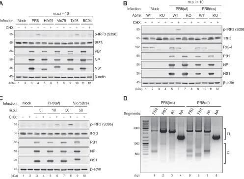
FIG 1CHX unmasking of RIG-I activation by IAV strains propagated in embryonated chicken eggs, but not in tissue culture. (A) A549 cells were infected with
a panel of IAV strains (PR8-H1N1, Hfx09-H1N1pdm, Vic75-H3N2, Tx98-swH3N2, and BC04-chH7N3) at an MOI of 10 in the absence or presence of CHX (50g/ml)
for 8 h. (B) WT orRIG-IKO A549 cells were infected with PR8 stocks propagated in embryonated chicken eggs (af) or MDCK cells (tcs) (MOI⫽10) in the absence
or presence of CHX for 8 h. (C) A549 cells were infected with increasing doses of allantoic fluid stock of PR8 (MOI⫽5, 10, and 50) or tissue culture stock of Vic75
(MOI⫽50) in the absence or presence of CHX for 8 h. The cell lysates were subjected to immunoblotting for phosphorylated (p) IRF3 (S396), total IRF3, RIG-I,
PB1, NP, NS1, and-actin. (D) Total RNA extracted from the tissue culture (tcs) or allantoic fluid (af) stock of PR8 was subjected to S-RT-PCR to amplify the DI
RNA species derived from the three viral polymerase genes. Amplification of the NA segment served as a negative control for DI presence, since it is not a major source of DI RNA generation. FL, full length.
on November 6, 2019 by guest
http://jvi.asm.org/
[image:3.585.44.541.73.437.2]phosphorylation (Fig. 1A, lanes 3, 5, 7, 9, and 11), indicating efficient suppression by the respective NS1 protein. In contrast, CHX treatment revealed IRF3 activation by the A/Puerto Rico/8/34 (H1N1) (PR8), Tx98, and BC04 strains (Fig. 1A, lanes 4, 10, and 12). Surprisingly, no IRF3 phosphorylation was detected for infection with the Hfx09 and Vic75 strains even in the presence of CHX (Fig. 1A, lanes 6 and 8), suggesting that CHX unmasking of IRF3 activation is not a universal effect.
In a search for the common feature shared by the three IRF3-activating strains (PR8, Tx98, and BC04), we found that they were phylogenetically unrelated but were all propagated in embryonated chicken eggs (allantoic fluid stocks). In comparison, the other two strains (Hfx09 and Vic75) were propagated in Madin-Darby canine kidney (MDCK) cells and had no passage history in eggs. To confirm this effect, we directly compared PR8 stocks derived from chicken eggs or MDCK cells for IRF3 activation in the presence of CHX. IRF3 phosphorylation was readily detectable in CHX-treated A549 cells infected with the allantoic fluid stock (designated “af” here), but not the MDCK tissue culture supernatant (designated “tcs” here) (Fig. 1B, lanes 6 and 10). Meanwhile, we determined the RIG-I dependency of PR8(af)-induced IRF3 phosphorylation. PR8(af) infection of A549RIG-Iknockout (KO) cells in the presence of CHX failed to stimulate IRF3 phosphorylation (Fig. 1B, lanes 6 and 8), demonstrating strict RIG-I-mediated IRF3 activation by the allantoic fluid virus stock. It has been acknowledged that virus stocks propagated in chicken eggs or tissue cultures contain various levels of DI particles (29, 30). We rationalized that the incoming DI genomes from the allantoic fluid virus stock might contribute to RIG-I activation. Indeed, in the presence of CHX, infection of A549 cells with increasing amounts of input PR8(af) led to dose-dependent IRF3 phosphor-ylation, whereas Vic75(tcs) infection at the highest tested MOI did not activate RIG-I (Fig. 1C). Moreover, direct amplification of DI contents within the allantoic versus tissue culture stocks of PR8 showed that PR8(af) contained more DI genomes derived from the three polymerase (PB2, PB1, and PA) genes than PR8(tcs). As a control, no DI genome was detected for the NA gene from either source (Fig. 1D). Taken together, these results indicated that the incoming DI genomes derived from chicken allantoic fluid virus stocks contributed to RIG-I activation in the presence of CHX.
Allantoic-fluid-derived DI genomes activate RIG-I in the presence of CHX or ActD.Another possibility for RIG-I activation by the allantoic fluid virus stocks is that some chicken RNA species contained in the allantoic fluid, rather than viral RNAs, were sensed by RIG-I in the presence of CHX. To disprove this, the PR8(af) stock was consecutively passaged in MDCK cells at a low MOI (0.001) in an attempt to dilute the presence of chicken RNAs. During the three passages (PL1to PL3), each virus stock was still able to induce similar levels of IRF3 phosphorylation in CHX-treated A549 cells (Fig. 2A, lanes 5, 8, and 11), suggesting the carryover of immunostimulatory RNA species. This effect conformed to the fact that viral DI genomes are capable of propagating along with the full-length genomes, whereas chicken RNAs should not. As such, the DI genomes contained in each passage served as incoming viral agonists activating RIG-I in the presence of CHX. In comparison, the PR8(af) stock was also passaged in MDCK cells at a high MOI to facilitate DI genome generation (Fig. 2B). The stock of the second passage (PH2) induced detectable IRF3 phosphorylation in A549 cells in the absence of CHX, concomitant with reduced viral protein accumulation (Fig. 2A, lane 19) and loss of most genome segments in the virus stock, as determined by multisegment reverse transcription (M-RT)-PCR (Fig. 2B, lane 3). CHX treatment further enhanced IRF3 acti-vation by PH2(Fig. 2A, lane 20). The PH3stock (from the third high-MOI passage) had a dramatic reduction in hemagglutinin (HA) titers due to interference by DI genomes (G. Liu and Y. Zhou, unpublished data), leading to low rates of virus infection and IRF3 activation (Fig. 2A, lanes 22 and 23). To provide direct evidence for DI-mediated RIG-I activation, the allantoic fluid virus stock was purified through a sucrose gradient, and the virus band corresponding to defective or complete viral particles was tested for immunostimulation in A549 cells. In the presence of CHX, infection with the defective, but not the complete, viral particles led to strong IRF3 phosphorylation, like that with
on November 6, 2019 by guest
http://jvi.asm.org/
55 55 -6 5 -6 2 45 -M p-IRF3 (S396) IRF3 PB1 NP NS1 -actin (kDa) 55 55 88 56 26 45
-1 2 3 4 5 6 7 8 9 10 11 12
CHX: ActD:
Mock PR8(af) SeV
NS HA, NP NA 3Pol PH1 PH2 PH3 tcs
PR8(af)
1000
500 3000
(bp) 1 2 3 4
M DI p-IRF3 (S396) IRF3 PB1 NP NS1 -actin 55 55 88 56 26 45
-(kDa) 1 2 3 4 5 6 7 8 9 10 11 12 13 14 15 16 17 18 20 21 22 23 24
CHX: ActD:
Mock PL1 PL2 PL3 Mock PH1 PH2 PH3
) f a ( 8 R P ) f a ( 8 R P
- p-IRF3 (S396)
IRF3
NP
NS1
-actin
(kDa) 1 2 3 4 5 6 7 8 9 10 11
ActD:
CHX:
with inoculum pre-treat
PR8af Infection:
A
B
C
D
E
F
p-IRF3 (S396) IRF3 NP NS1 -actin 55 55 -6 5 -6 2 45-(kDa) 1 2 3 4 5 6 7 8
CHX: Unpurified: Defective: Complete: Tx98 19 p-IRF3 (S396) IRF3 NP NS1 -actin GFP GFP NS1 CHX: PR8(af): 55 55 -6 5 -6 2 45 -(kDa) -7 2
1 2 3 4 5 6
Inhibitors:
FIG 2Allantoic-fluid-derived DI genomes activate RIG-I in the presence of CHX or ActD. (A) The allantoic fluid (af) stock of PR8 was consecutively passaged in
MDCK cells at a low or high MOI to obtain the PL1to PL3and PH1to PH3stocks, respectively. A549 cells were subsequently infected with these stocks (equal
volumes) in the absence or presence of CHX (50g/ml) or ActD (1g/ml) for 8 h. The protein expression levels were determined by immunoblotting for
phosphorylated IRF3 (S396), total IRF3, PB1, NP, NS1, and-actin. (B) Equal amounts of total RNA extracted from the PH1to PH3stocks were subjected to
M-RT-PCR to detect the DI RNA contents. The tissue culture supernatant (tcs) stock of PR8 served as a control with low DI content. (C) The allantoic fluid stock of Tx98 (H3N2) was purified through a sucrose gradient, and the band corresponding to the defective or complete viral particles was collected. A549 cells were infected in the absence or presence of CHX with the unpurified, defective, or complete virus at equal HA titers (128 hemagglutinating units [HAU]/ml) for 8
h. (D) A549 cells were infected with the allantoic fluid stock of PR8 (MOI⫽50) or SeV-Cantell (50 HAU/ml) in the presence of CHX or ActD alone, or CHX and
ActD in combination, for 8 h. (E) Increasing doses of ActD (0.2, 1, and 5g/ml) were added to A549 cells 1 h prior to PR8(af) inoculation (pretreatment) or along
with the virus inoculum. The cells were further infected in the presence of inhibitors for 8 h. (F) A549 cells were transfected with a plasmid expressing PR8-NS1
or GFP for 24 h, followed by PR8(af) infection (MOI⫽50) in the absence or presence of CHX for 8 h. (C to F) Protein expression levels were determined by
immunoblotting as for panel A.
on November 6, 2019 by guest
http://jvi.asm.org/
[image:5.585.47.543.71.584.2]unpurified virus stock (Fig. 2C), demonstrating the direct contribution of DI genomes to RIG-I activation.
A previous study showed that CHX unmasking of IRF3 phosphorylation was inhib-ited by the transcription inhibitor ActD (12). We next determined the effect of ActD on RIG-I activation by incoming DI genomes. As with the CHX effect, ActD treatment during PR8(af) infection also largely blocked viral protein synthesis; however, a small amount of viral proteins, such as the IFN-antagonistic NS1, was still detectable in the presence of ActD (Fig. 2A, lane 18, and Fig. 2D, lane 7), indicating that ActD was less potent than CHX in inhibiting viral protein synthesis. Nonetheless, ActD treatment also unmasked IRF3 phosphorylation upon PR8(af) infection, though to a lesser extent than CHX treatment (Fig. 2A, lane 18 versus 17, and Fig. 2D, lane 7 versus 6). To test whether the trace of viral protein expression upon ActD treatment contributed to the reduced level of RIG-I activation, we first tested the dose-dependent effect of ActD on IRF3 phosphorylation in PR8(af)-infected cells. The level of IRF3 phosphorylation was pro-portional to the dose of ActD used, which was inversely propro-portional to the level of leaky expression of viral proteins (Fig. 2E, lanes 6 and 7). Pretreatment of cells with ActD prior to virus adsorption did not lead to an apparent difference in the level of IRF3 phosphorylation compared to ActD treatment from the time of virus adsorption (Fig. 2E, lanes 10 and 11 versus 7 and 8). However, the pretreatment did slightly enhance IRF3 activation when the lowest ActD dose was used, which correlated with a reduction in leaky viral protein expression (Fig. 2E, lane 9 versus 6). Interestingly, even at the highest dose, ActD treatment led to weaker IRF3 activation than CHX (Fig. 2E, lanes 8 and 11 versus 5); however, treatment with ActD and CHX in combination largely reversed the level of IRF3 activation to that observed with CHX alone, concomitant with a complete loss of leaky NS1 expression (Fig. 2D, lanes 6 to 8). Furthermore, compared to green fluorescent protein (GFP) preexpression, preexpressing NS1 in CHX-treated cells reduced IRF3 activation by PR8(af) infection, demonstrating an inhibitory role of NS1 in suppressing DI-mediated RIG-I activation (Fig. 2F). In parallel, we compared the effects of CHX, ActD, and CHX and ActD in combination on IRF3 phosphorylation induced by Sendai virus (SeV). The Cantell strain of SeV is prone to generate trailer copy-back DI genomes, which are potent RIG-I agonists during infection (Fig. 2D, lane 9). Consistent with previous studies (12), RIG-I activation by SeV was insensitive to ActD (Fig. 2D, lane 11), as SeV replication is independent of cellular RNA transcription. However, CHX inhibition of protein synthesis dampened the levels of IRF3 phosphor-ylation (Fig. 2D, lanes 10 and 12), indicating thatde novoviral protein synthesis was required for SeV DI genome accumulation. Taken together, these results demonstrated that the suppression of viral protein expression, such as that of NS1, was the primary effect of CHX or ActD on unmasking RIG-I activation by incoming IAV DI RNA.
Allantoic-fluid-derived DI genomes associate with and activate cytoplasmic RIG-I.To directly examine the IAV DI species that associate with RIG-I, we performed an RNA immunoprecipitation (RIP) analysis in CHX-treated A549 cells infected with PR8(af). RNA extracted from RIG-I immunoprecipitates was subjected to single-segment (S)-RT-PCR to amplify the negative-sense DI sequences (vDI) derived from the three polymer-ase genes. While the IgG control associated with residual levels of full-length vRNA, RIG-I immunoprecipitates showed increased levels of full-length vRNA association and specific enrichment of DI genomes from the PB2 and PB1 segments (Fig. 3A, top, lanes 5 and 6). Increasing the RT reaction temperature reduced the cDNA yield but enhanced the specificity of DI amplification; distinct bands of DI genomes were revealed from the PB1 and PA segments (Fig. 3A, bottom, lanes 6 and 7). Since CHX treatment does not block primary viral RNA synthesis, we also sought to determine whether RIG-I associ-ated with DI genomes of positive polarity (cDI) that might be transcribed from the incoming vDI. In comparison with the IgG control, RIG-I-associated RNA had no apparent enrichment of any cDI sequence, except for the full-length cRNA of the NA segment (Fig. 3B, lane 8). Since we recently demonstrated that viral RNA species are sensed by both the cytoplasmic and nuclear pools of RIG-I during IAV infection (6), we next examined which RIG-I pool was activated by the incoming DI genomes. In the
on November 6, 2019 by guest
http://jvi.asm.org/
presence of CHX, PR8(af) infection of A549RIG-IKO cells expressing cytoplasmic RIG-I (nuclear export signal [NES]-RIG-I), but not nuclear RIG-I (nuclear localization signal [NLS]-RIG-I), stimulated efficient levels of IRF3 phosphorylation (Fig. 3C, lanes 3 and 9), demonstrating that the cytoplasmic pool of RIG-I was responsible for DI genome sensing. Again, ActD treatment showed reduced RIG-I activation by incoming DI genomes, attributable to the incomplete inhibition of viral protein synthesis (Fig. 3C, lanes 6 and 12). Taken together, the data show that the incoming IAV DI genomes associated with and specifically activated cytoplasmic RIG-I.
NP deprivation leads tode novogeneration of RIG-I-activating viral RNAs that are distinct from DI genomes. IAV genome replication is concurrent with NP-mediated encapsidation of newly synthesized viral RNA. NP also acts as the key elongation factor for the viral polymerase and is indispensable for production of full-length vRNA and cRNA (19). Thus, apart from CHX unmasking of RIG-I activation by incoming DI genomes, CHX inhibition of NP expression likely perturbed the function-ality of viral polymerase, leading to its generation of abortive replication products. In a previous study, small interfering RNA (siRNA)-mediated NP knockdown also led to IRF3 phosphorylation like that observed with CHX treatment (12). To investigate whether NP
1000
1000
p-IRF3 (S396)
IRF3
NP (long exp.)
NS1(long exp.)
-actin FLAG (RIG-I)
55
55
-56 -
26 -
45 110
-(kDa) 1 2 3 4 5 6 7 8 9 10 11 12
PR8(af): CHX: ActD: Dox:
A
B
C
NES-RIG-I NLS-RIG-I(+) strand
RT: 42qC
PB2 PB1 PA NA PB2 PB1 PA NA
IgG RIG-I
IP:
Segments:
PR8(af) + CHX
500 3000
(bp) 1 2 3 4 5 6 7 8
FL
cDI? (-) strand
FL
vDI 1000
500 3000
RT: 42qC
RT: 50qC
(bp) 1 2 3 4 5 6 7 8
500 3000
PB2 PB1 PA NA PB2 PB1 PA NA
IgG RIG-I
IP:
Segments:
FL
vDI PR8(af) + CHX
FIG 3Allantoic-fluid-derived DI genomes associate with and activate cytoplasmic RIG-I. (A) A549 cells were infected with the allantoic fluid (af) stock of PR8
(MOI⫽50) in the presence of CHX (50g/ml) for 6 h. RIP was performed in the whole-cell lysates using either the RIG-I antibody or an IgG isotype control.
The associated RNAs were extracted from the respective immunoprecipitates and subjected to S-RT-PCR to amplify the DI species of negative (⫺) polarity (vDI)
from the three polymerase and NA genes. The RT step was performed at 42°C (top) or 50°C (bottom) to increase the specificity of DI amplification. (B) The
extracted RNA from the experiment shown in panel A was subjected to S-RT-PCR to amplify the DI species of positive (⫹) polarity (cDI) from the three
polymerase and NA genes. The RT step was performed at 42°C. (C)RIG-IKO A549 cells inducibly expressing NES- or NLS-RIG-I were left noninduced or induced
with 1g/ml doxycycline (Dox) for 4 h, followed by mock infection or infection with PR8(af) (MOI⫽50) in the presence of CHX (50g/ml) or ActD (1g/ml)
for 8 h. The cell lysates were subjected to immunoblotting for FLAG-RIG-I, phosphorylated IRF3 (S396), total IRF3, NP, NS1, and-actin.
on November 6, 2019 by guest
http://jvi.asm.org/
[image:7.585.46.535.71.431.2]deprivation skewed the viral polymerase toward an erroneous mode, we employed an NP-free RNP reconstitution system to abrogate normal replication and instead specif-ically examined the generation of RIG-I-activating viral RNA other than full-length viral genomes (Fig. 4A). 293T cells were RNP reconstituted in the absence of a plasmid expressing NP protein with a Pol I construct generating each of the eight viral genome segments of PR8. Total RNA was extracted and further transfected into IFN reporter cells to test their immunostimulatory activities. Except for that from the NP segment (Pol I-NP), transfection of reporter cells with reconstituted RNAs from the other seven genome segments did not stimulate apparent IFN-promoter activity (Fig. 4B). When the NP segment (Pol I-NP) was used for NP-free reconstitution, NP mRNA was synthe-sized from viral primary transcription and was subsequently translated into NP proteins (Fig. 4C) that were functionally equivalent to the Pol II-derived NP (like that used for standard RNP reconstitution). This converted the NP-free condition back to normal RNP reconstitution and thus restored replication of full-length NP vRNA. Consistent with this, Pol I-NP, like Pol II-NP, could also support the transcription and replication of M vRNA (Fig. 4C). Therefore, the strong IFN induction by the NP-free reconstituted RNA from the NP segment (Pol I-NP), as seen in Fig. 4B, was attributable to the full-length viral genomic RNA of NP, which was in line with previous reports (7, 10). Next, we asked whether a transcription-defective (T⫺) polymerase (i.e., PA-D108A) would facilitate the
generation of immunostimulatory RNA from a non-NP segment. Such a mutant poly-merase was shown to impose higher replication activity than the wild type (WT) and thus might erroneously generate a greater amount of immunostimulatory viral RNA (6). In the presence of NP (Pol II-NP, i.e., standard RNP reconstitution), NA reconstitution with the PA-D108A polymerase generated a significantly greater amount of immuno-stimulatory RNA than that with the WT polymerase (Fig. 4D). In contrast, no IFN- promoter activation greater than the catalytically inactive PB1 (PB1a) baseline (19, 31) was detected for transfection with total RNA reconstituted in the absence of NP protein (⫺NP), regardless of whether WT or PA-D108A polymerase was used (Fig. 4D).
To avoid the possibility that the RNA extraction procedure was inefficient in extract-ing viral immunostimulatory RNA generated durextract-ing NP deprivation or disrupted the native RNA structures of these RNAs, we determinedin situRIG-I activation during RNP reconstitution in the presence or absence of NP. Since IAV RNA synthesis takes place in the nucleus, supplementation of reporter cells with a nuclear-localized RIG-I (NLS-RIG-I) provided a more sensitive measurement of IFN-promoter activation in response to RNP reconstitution (6). Furthermore, this may also shed light on the specialized sensing by nuclear RIG-I of a subset of unknown viral RNAs in the nucleus. Compared to the catalytically inactive PB1a baseline, reconstitution with an NA segment in the presence of NP stimulated significant IFN-promoter activity (Fig. 4E, fourth gray bar from left). Surprisingly, NP-free reconstitution also induced moderate RIG-I activation, albeit to a lesser extent than the normal reconstitution (Fig. 4E, third versus fourth gray bars). Similar IFN-stimulatory activity was also observed during NP-free reconstitution with the PA or HA segment, ruling out a segment-specific effect (Fig. 4F and G). To further determine the requirement for viral transcription or replication for the generation of immunostimulatory RNA under the NP-free condition, reconstitution with the NA segment was conducted using either the T⫺or replication-defective (R⫺) polymerase
(32–35). Similar to that in normal RNP reconstitution, the generation of immunostimu-latory RNA by NP deprivation was affected by replication deficiency, but not by transcription deficiency (Fig. 4H). We next determined whether IFN induction by NP-free reconstitution was viral strain specific. A panel of polymerases of different viral subtypes was tested, including PR8 (H1N1), Vic75 (H3N2), A/turkey/Ontario/6213/66 (H5N1) (ON6213), BC04 (H7N3), and A/British Columbia/1/2015 (H7N9) (BC15). Of these, the polymerases of the two low-pathogenic avian strains (ON6213 and BC04) contained avian signature PB2-E627, whereas the others, including one avian strain isolated from a human (BC15), contained human-adapted signature PB2-K627 (36). The activities of these polymerases were determined, and they correlated well with the PB2-627 signa-ture (Fig. 4I). Interestingly, all the tested polymerases induced various levels of IFN-
on November 6, 2019 by guest
http://jvi.asm.org/
26
56
45
-vec PB2 PB1 PA HA NP NA M NS
0 5 10 15
Relative
reporter activity (Fold )
Pol I (PR8)
-PB2 PB1 PA NP vRNA
Pol II Pol I
PB2 PB1 PA NP vRNA
Standard NP-free vRNP Abortive replication? RIG-I activation PB1a WT PA-D108 A (T -) 0 20 40 60 80 Relative
reporter activity (Fold )
- NP Pol I-NA + NP 0 5 10 15 Relative
reporter activity (Fold )
GFP NLS-RIG-I RdRp-PB1a RdRp-WT NP NEP Pol I-NA GFP RIG-I PB1 NP -actin NEP **** **** **** **** ns 0 5 10 15 Relative
IFN-reporter activity (Fold )
GFP NLS-RIG-I **** **** ns RdRp-PB1a RdRp-WT NP Pol I-PA 0 1 2 3 4 Relative IFN-re porte r ac tiv ity (Fold ) GFP NLS-RIG-I **** **** ns RdRp-PB1a RdRp-WT NP Pol I-HA PB1a WT PB2-E361 A (
T-)
PA-D1 08A (
T-)
PB 2-R142A
(R-)
PA-E410 A (R
-) 0 5 10 15 Relative
reporter activity (Fold )
- NP + NP
Pol I-NA
PB1a PR8 (H1N1)Vic75 (H3N2)
ON6 213 (H5N1)BC04
(H7N 3) BC15 (H7N 9) 0 50 100 150 200
Relative polymerase activity (%)
Pol I-NA-fLuc PB1a PR8 (H1N1 ) Vic75 (H3N2) ON6 213 (H5N1)BC04
(H7 N3) BC15 (H7 N9) 0 2 4 6 8 Relative
reporter activity (Fold )
- NP + NP
Pol I-NA vec Tetra loop-DI vec Tetra loop-DI 0 20 40 60 Relative
reporter activity (Fold )
Total RNA >200 nt
- NP + NP
-Pol II-RdRp Pol I-NP (PR8) Pol I-M (PR8)
PR8
M1
-actin NP
(kDa) 1 2 3 4 5 6
A
B
C
Tx91 Sk02 BC04(K)
D
E
F
G
H
I
J
K
FIG 4NP deprivation leads tode novogeneration of RIG-I-activating viral RNA that is distinct from DI genomes. (A) Schematic diagram of the standard and NP-free RNP reconstitution systems in which differential viral RNA species activate RIG-I. (B) 293T cells were RNP reconstituted in the absence of NP with a Pol
I vector or a Pol I construct generating each of the eight viral segments of PR8. Total RNA was extracted and transfected into 293T cells preexpressing an IFN-
promoter-driven firefly luciferase reporter (p125Luc) and a constitutively expressedRenillaluciferase construct (pTK-rLuc). (C) In each sample, 293T cells were
cotransfected with three Pol II-driven plasmids expressing the viral polymerases (PB2, PB1, and PA) of PR8, Tx91, Sk02, or BC04(K627), along with two Pol I-driven plasmids generating the vRNAs of NP and M segments of PR8. The expression levels of NP and M1 were determined by immunoblotting at 24 h p.t. (D) 293T cells were RNP reconstituted with the PR8 NA segment (Pol I-NA) in the absence or presence of NP protein (Pol II-NP) using the catalytically inactive
(PB1a), WT, or T⫺(PA-D108A) viral polymerase. The extracted total RNA was tested for immunostimulatory activity as for panel B. (E) 293T cells were RNP
reconstituted with the PR8 NA segment (Pol I-NA) using the catalytically inactive (PB1a) or WT polymerase in the presence of a plasmid expressing GFP or NLS-RIG-I. As indicated, plasmids expressing NP and NEP proteins were cotransfected along with p125Luc and pTK-rLuc. The cell lysates were subjected to
immunoblotting to examine the protein expression levels of GFP, FLAG-RIG-I, PB1, NP, NEP, and-actin. (F and G) 293T cells were RNP reconstituted with the
PR8 PA (Pol I-PA) (F) or HA (Pol I-HA) (G) segment as for panel E. Statistical significance was determined by two-way ANOVA, followed by a Sidak posttest.****,
P⬍0.0001; ns, not significant. (H) 293T cells were RNP reconstituted as for panel E with or without a plasmid expressing NP using the inactive (PB1a), WT, T⫺,
or R⫺polymerase in the presence of NLS-RIG-I. (I) 293T cells were cotransfected with four Pol II-driven plasmids expressing the viral polymerases and NPs of
various IAV strains, along with a Pol I-driven plasmid encoding a vRNA-like molecule in which the firefly luciferase open reading frame (ORF) was flanked by
the 5=and 3=noncoding regions (NCRs) of the PR8 NA segment (Pol I-NA-fLuc). Reconstitution using the inactive polymerase (PB1a) of PR8 served as a
negative control. (J) 293T cells were RNP reconstituted as for panel H using the polymerases of various IAV strains in the presence of NLS-RIG-I. (K) Two
sets of 293T cells were RNP reconstituted in the absence or presence of NP with a Pol I vector or an artificial DI-like construct in which the 5=and 3=
NCRs of the PR8 NP segment were linked through a UUCG tetraloop. At 24 h p.t., one set of cells was subjected to RNA extraction to obtain total RNAs
containing small RNAs, while the other set was extracted for RNA species larger than⬃200 nt. These RNAs were tested for immunostimulatory activity
as for panel B. Unless otherwise indicated, RLU were determined at 24 h p.t. and are expressed as fold changes relative to the Pol I vector (B and K)
or the PB1a control (D and J). The data are shown as means⫾SD of the results of three independent experiments performed in triplicate.
on November 6, 2019 by guest
http://jvi.asm.org/
[image:9.585.55.538.70.512.2]promoter activation above the PB1a baseline during NP-free reconstitutions (Fig. 4J), even the ones (i.e., ON6213 and BC04) whose activities were restricted in 293T cells. Lastly, we sought to understand whether the immunostimulatory RNA species gener-ated during NP deprivation share features similar to those of DI genomes. Given that the immunostimulatory activities of these RNAs were sensitive to RNA extraction, we examined whether RNA extraction would alter the RIG-I-stimulatory configuration of DI-like genomes. Total RNA extracted from normal RNP reconstitution (⫹NP) with an artificial DI-like construct (i. e., a tetraloop; 72 nucleotides [nt] in length) retained strong immunostimulatory activity when transfected into reporter cells (Fig. 4K). The specificity of IFN induction by DI RNA was controlled by total RNA fractionation; transfection of RNA fractions⬎200 nt in length failed to activate an IFN response (Fig. 4K). Moreover, omitting NP in the DI reconstitution barely affected the generation of immunostimu-latory DI RNA (Fig. 4K), as its length falls in the range of NP-independent replication, as previously described (19). Taken together, these results indicated that NP deprivation inducedde novogeneration of RIG-I-activating viral RNAs, whose natures were unlikely to be DI genomes.
Accumulation of a major subset of aberrant viral RNAs driven by NP depriva-tion is diminished by NEP.To gain further insight into the nature of immunostimu-latory RNA generated in the NP-free reconstitution, we investigated the small viral RNA species of either negative or positive polarity by Northern blotting. Given the 5=-to-3=
direction of RNA synthesis and the possibility of abortive vRNA/cRNA synthesis by viral polymerase in the absence of NP, the probes were designed to anneal to the 5=end of vRNA (5=v-probe) or the 5= end of cRNA (5=c-probe). NP-free RNP reconstitution generated two distinct aberrant RNA species (Fig. 5A, lane 3, black arrowheads), which were also observed previously using an eight-plasmid reconstitution system omitting the NP segment (37, 38). Although their natures are unknown, these RNAs likely represented abortive replication products, since their sequences covered the 5=end of vRNA. We next sought to examine whether RIG-I activation in response to NP-free reconstitution could be attributable to these aberrant replication products. We took advantage of the viral NEP protein, which has been shown to enhance the replication activity of viral polymerase by promoting cRNA synthesis and the generation of svRNA 22 to 27 nt in length (37–39). We rationalized that the supplementation of NEP would modulate the generation of aberrant replication products, thereby affecting RIG-I activation. Consistent with previous reports (39), addition of NEP during RNP reconsti-tution (⫹NP) inhibited viral mRNA synthesis in a dose-dependent manner (Fig. 5B) but enhanced cRNA and vRNA synthesis (Fig. 5C), confirming its role in switching the mode of the viral polymerase from transcription to replication. Interestingly, addition of NEP to the NP-free reconstitution diminished the accumulation of the two major aberrant RNA species yet revealed the generation of svRNA-like molecules (Fig. 5A, lane 5, red arrowheads). During standard RNP reconstitution (⫹NP), the production of the major aberrant RNAs was less than that in NP-free reconstitution (Fig. 5A, lane 4 versus 3), and negligible levels of these RNAs were detected in the presence of NEP (Fig. 5A, lane 6). Moreover, no distinct small RNA species of positive sense were detected under all conditions (Fig. 5A, bottom). Next, we examined whether NEP modulatesin situRIG-I activation in response to RNP reconstitution. The addition of NEP did not affect IFN- promoter activation in response to standard RNP reconstitution (⫹NP) with the NA or PB2 segment (Fig. 4E and 5D, fourth gray bars versus sixth gray bars), Similarly, NEP also did not adversely affect RIG-I activation upon NP-free reconstitution; a slight increase in IFN- promoter activation was observed in the presence of NEP compared to that without NEP (Fig. 4E and 5D, third gray bars versus fifth gray bars). Taken together, the fact that NEP overexpression dampened the accumulation of the two major aberrant RNA species yet did not affect IFN induction by the RNA pool during NP-free recon-stitution indicated that these aberrant RNAs played a minor role in RIG-I activation. However, the emergence of svRNA-like molecules in this context suggested an indirect compensatory role of svRNA in RIG-I activation, though svRNA by itself did not exhibit any immunostimulatory activity upon transfection, as previously reported (37).
on November 6, 2019 by guest
http://jvi.asm.org/
DISCUSSION
The spatiotemporal detection by RIG-I of RNA species produced during virus infection is a complex process. Unlike most RNA viruses replicating in the cyto-plasm, IAV imparts further complexity to this dynamic process owing to its repli-cation within the nucleus. Until recently, an emerging role of RIG-I in sensing IAV replication in the nucleus was unraveled and offers insights into a previously unrecognized cellular milieu for nonself RNA sensing (6). Nonetheless, the full spectrum of viral agonists generated during IAV infection that contribute to RIG-I activation is not thoroughly elucidated. In search of physiological RIG-I agonists, most studies take advantage of RIG-I immunoprecipitation followed by Northern blotting or RNA sequencing to determine the associated viral RNA (7, 8). These
RdRp-PB1a
A
B
C
D
E
RdRp-WT NP NEP Pol I-NP-fLuc 5' v-probe 25 25 -5' c-probe (nt)1 2 3 4 5 6
U6 PB1 NP NEP -actin 88 56 14 45 -(kDa) 106 100 100
-1 2 3 4 5 6
0 10000 20000 30000 Relative
polymerase activity (%)
PB1a WT GFP NEP
- mRNA 0 5 10 15 20 40 60 80 Relative
reporter activity (Fold)
GFP NLS-RIG-I GFP RIG-I PB1 NP -actin NEP * **** **** **** ns RdRp-PB1a RdRp-WT NP NEP Pol I-PB2 vRNA (-) ppp'5 5' svRNA? aberrant RNA? 3' ? 5'ppp partial complementarity pp svRNA? N ? NA? NA? 3' 5'ppp nRIG-I
nRIG-I full complementarity
cRNA (+) 5'
- 5S rRNA - cRNA - vRNA -RdRp-PB1a RdRp-WT GFP NEP Pol I-NA
1 2 3 4 5 6
FL
FIG 5Accumulation of a major subset of aberrant viral RNAs driven by NP deprivation is sensitive to NEP expression. (A) 293T cells were RNP reconstituted
using the catalytically inactive (PB1a) or WT viral polymerase with a vRNA-like construct in which the firefly luciferase ORF was flanked by the 5=and 3=NCRs
of the PR8 NP segment (Pol I-NP-fLuc). As indicated, the plasmids expressing NP and NEP were cotransfected. At 48 h p.t., total RNA was extracted and
subjected to Northern blotting to detect small RNA species containing the 5=end of vRNA (5=v-probe) or cRNA (5=c-probe). The aberrant replication products
under NP deprivation (lane 3) and the svRNA-like molecules generated in the presence of NEP (lane 5) are indicated by black and red arrowheads, respectively.
U6 served as a loading control. The protein expression levels were determined by immunoblotting for PB1, NP, NEP, and-actin. FL, full-length vRNA. (B) 293T
cells were RNP reconstituted (⫹NP) using the inactive (PB1a) or WT viral polymerase with the NA-fLuc reporter construct (Pol I-NA-fLuc). As indicated,
increasing doses of GFP (10 to 100 ng) or NEP were supplemented. The relative polymerase activity was determined at 24 h p.t. and is expressed as fold changes over the PB1a control. (C) 293T cells were RNP reconstituted as for panel B with the NA segment of Hfx09 (Pol I-NA) in the absence or presence of increasing doses of GFP or NEP. Total RNA was extracted at 48 h p.t. and subjected to primer extension analysis to detect the levels of vRNA, cRNA, and mRNA. 5S rRNA served as a loading control. (D) 293T cells were RNP reconstituted with the PR8 PB2 segment (Pol I-PB2) using the inactive (PB1a) or WT polymerase in the presence of GFP or NLS-RIG-I. As indicated, plasmids expressing NP and NEP were cotransfected along with p125Luc and pTK-rLuc. RLU were determined at 24 h p.t. and are expressed as fold changes relative to PB1a with GFP. The cell lysates were subjected to immunoblotting to examine the protein expression
levels of GFP, FLAG-RIG-I, PB1, NP, NEP, and-actin. Statistical significance was determined by two-way ANOVA, followed by a Sidak posttest.*,P⬍0.05;****,
P⬍0.0001; ns not significant. (E) Proposed model for nuclear RIG-I activation by intermolecular RNA duplexes formed from the complementarity between
aberrant viral RNA or svRNA and the 3=end of full-length vRNA (partial complementarity) or cRNA (full complementarity). The data are shown as means⫾
SD of the results of three independent experiments performed in triplicate.
on November 6, 2019 by guest
http://jvi.asm.org/
[image:11.585.48.539.76.426.2]approaches have successfully established the central roles of full-length and DI genomes in RIG-I activation but have missed information about the origins (e.g., incoming versus de novosynthesized) of these RIG-I-activating RNA species. Upon IAV infection, the incoming and de novo-synthesized full-length and DI genomes share similar properties. Therefore, selective inhibition of de novoviral RNA syn-thesis is required to distinguish the relative contributions of the incoming versus progeny viral genomes to RIG-I activation. In this study, we found that CHX or ActD inhibition of viral protein synthesis unmasked the contribution of incoming DI genomes to RIG-I activation, particularly that from the virus stocks propagated in chicken eggs. The fact that virus stocks propagated in tissue cultures failed to activate RIG-I even in the presence of CHX further suggests that the incoming full-length viral genomes (i.e., in the form of vRNPs) are not the primary RIG-I agonists. This is in line with previous reports that total RNA extracted from IAV-infected cells in the presence of CHX failed to activate RIG-I (7) and that the association between incoming vRNP and RIG-I did not apparently lead to RIG-I activation but rather reflected a signaling-independent restriction mechanism de-pending on the PB2-627 signature of the viral polymerase (9).
Our findings on the CHX/ActD unmasking of RIG-I activation by incoming DI genomes appear to contradict those of a previous study, where IRF3 phosphorylation as revealed by CHX inhibition was sensitive to transcription inhibitors, such as ActD (12). In our study, ActD treatment was indeed less potent than CHX in uncovering RIG-I activation, which could be partially attributed to the leaky expression of viral proteins, such as NS1. Mechanistically, neither CHX nor ActD inhibits viral primary transcription, regardless of the drug concentrations used (14). However, since CHX acts directly on ribosomes to block translation, it is conceivable that any viral mRNA synthesized from primary transcription (i.e., primary viral transcripts) is completely blocked for translation by CHX but would still be translated in the presence of ActD. This difference may thus contribute to the efficiency of the two inhibitors in unmasking RIG-I activation by incoming DI genomes. Moreover, we do not rule out the possibility that other, unknown mechanisms may also contribute to the differential efficiencies of the two inhibitors. ActD treatment did lead to an ⬃2-fold increase in nuclear retention of primary viral transcripts compared to CHX (14). However, viral mRNAs contain 5= cap structures and are known not to be sensed by RIG-I (7, 10). On the other hand, CHX and ActD do not affect the replication activity of the incoming vRNP-associated viral polymerase; cRNA accumulation could be detected from infection under treatment with either inhibitor, provided that viral polymerase and NP were preexpressed to stabilize thede novo-synthesized cRNA (40, 41). Overall, in accordance with the previ-ous study (7), our results support the notion that both CHX and ActD treatments abolish RIG-I activation during IAV infection unless incoming DI genomes are present. Sup-pressing the expression of viral proteins, such as NS1, is the primary effect conferred by CHX or ActD on unmasking RIG-I activation by the incoming DI, but not by the incoming full-length viral genomic RNA.
Despite these discrepancies, our results also do not preclude the possibility that certain RNA species generated under CHX or ActD treatment might be capable of activating RIG-I. These RNAs have been proposed to be distinct in nature from normal replication products (i.e., full-length vRNA and cRNA) and instead represent
de novo-synthesized DI RNA or a novel class of aberrant viral RNA (12, 42). Although CHX/ActD treatment during infection with virus stocks derived from tissue cultures did not reveal apparent RIG-I activation, it is possible that the accumulation of these immunostimulatory RNAs requires ongoing viral replication under mild cellular constraints to reach a threshold for RIG-I activation. Since viral NP protein has been shown to act as an elongation factor for full-length vRNA and cRNA (19, 43), we employed an NP deprivation condition to simulate one such mild cellular constraint that may skew normal replication toward an erroneous mode, leading to the accumulation of abortive replicative products. NP-free reconstitution indeed led to the production of RIG-I-activating RNAs other than full-length viral genomes, and
on November 6, 2019 by guest
http://jvi.asm.org/
their immunostimulatory activity was sensitive to RNA extraction, unlike that of DI-like RNA. While this is indicative of a nature that is distinct from that of DI RNA, it also implies that RNA extraction disrupts certain secondary structures of these viral RNAs that are required for RIG-I recognition. Other than intramolecular base pairing, such as that in the panhandle structure of full-length and DI genomes (7, 8, 10, 11), it is tempting to speculate that intermolecular RNA duplexes formed from the complementarity between the aberrant viral RNA (such as abortive replication products) and its template vRNA or cRNA might activate RIG-I. In particular, the base pairing of the 5=-triphosphorylated aberrant RNA with the 3= end of full-length vRNA (partial complementarity) or the 3=end of full-length cRNA (full complemen-tarity), as illustrated in Fig. 5E, would meet the ligand characteristics for RIG-I activation (10, 11, 44–46). Such an RNA hybrid might be allowed to form, since the 3= end of vRNA adopts no apparent secondary structure in the viral polymerase-bound promoter configuration (47). Moreover, NP deprivation abolishes concurrent RNA encapsidation and the recruitment of host RNA helicases, such as UAP56, both of which otherwise prevent the formation of long double-stranded RNA hybrids between the positive- and negative-sense viral RNAs (48–52). It is also likely that the formation of these intermolecular RNA duplexes may coordinate with a progressing viral polymerase that fully exposes the 3= end of vRNA or cRNA (Fig. 5E). Further-more, the nuclear RIG-I may specialize in sensing this dynamic process, given its closest proximity to the replication machinery in the nucleus (6). Although their sequences are unknown, we investigated small viral RNA species (⬍100 nt in length) containing the 5= end of vRNA or cRNA under NP deprivation, which are potential abortive replication products in forming intermolecular RNA duplexes with the 3= end of vRNA or cRNA. Two major aberrant RNA species of negative polarity were identified whose accumulation was diminished by NEP expression. However, RIG-I activation was not adversely affected in the presence of NEP, which, on the other hand, facilitated the accumulation of svRNA. These results hint that svRNA may also serve as a base-pairing partner with genome ends for intermolec-ular RNA duplex formation (Fig. 5E), though svRNA by itself has been shown to lack immunostimulatory activity (37).
In summary, we examined the origins of RIG-I-activating viral RNA in response to different cellular constraints that impede ongoing IAV replication. We found that the incoming DI genomes andde novo-synthesized aberrant viral RNA species contribute to RIG-I activation upon chemical inhibition of viral protein synthesis and NP deprivation, respectively. Our results thus support the notion that there are distinct mechanisms of RIG-I activation during IAV infection. This may also reflect a specialized viral-RNA-sensing paradigm differentially provided by the cytoplasmic and nuclear pools of RIG-I. These mechanistic differences shape the complex and dynamic interplay between RIG-I and IAV and underscore the importance of a careful experimental design pertaining to the identification of novel RIG-I agonists.
MATERIALS AND METHODS
Cells and viruses. MDCK and human embryonic kidney (HEK) 293T cells were maintained in minimum essential medium (MEM) (Sigma) and Dulbecco’s modified Eagle’s medium (DMEM) (Sigma)
supplemented with 10% fetal bovine serum (FBS) (Gibco) and 50g/ml gentamicin, respectively. Human
lung carcinoma epithelial (A549) cells and A549RIG-IKO cells were maintained in Ham’s F-12K (Kaighn’s)
medium (Gibco) with 10% FBS and gentamicin. A549 RIG-IKO cells inducibly expressing NES- or
NLS-tagged RIG-I were previously described (6) and were cultured in F-12K with 10% tetracycline-free FBS
(Clontech) and 2g/ml puromycin (Invivogen). All cell cultures were maintained at 37°C in a humidified
5% CO2 atmosphere. Influenza A virus strains A/Puerto Rico/8/34 (H1N1) (PR8), A/Halifax/210/2009
(H1N1) (Hfx09), A/Victoria/3/75 (H3N2) (Vic75), A/Swine/Texas/4199-2/98 (H3N2) (Tx98), and A/Chicken/ British Columbia/CN-6/2004 (H7N3) (BC04) were generated with an eight-plasmid reverse-genetics system as previously described (53). They were propagated in either 11-day-old embryonated chicken eggs or MDCK cells as indicated. Sendai virus (Cantell strain) was obtained from the ATCC (VR-907) and propagated in embryonated chicken eggs.
Chemical inhibitors, antibodies, and immunoblotting.CHX and ActD were obtained from
Millipore-Sigma and were used at working concentrations of 50g/ml and 1g/ml, respectively, unless otherwise
indicated. Chemical inhibitors were added to the cells 1 h prior to or at the time of virus adsorption and
on November 6, 2019 by guest
http://jvi.asm.org/
were present during the course of infection. Immunoblotting was performed as previously described (10), and the following antibodies were used: rabbit anti-RIG-I (EPR18629; Abcam), mouse anti-FLAG (M2; Sigma), rabbit anti-phospho-IRF3 (Ser396) (D6O1M; CST), rabbit anti-IRF3 (D6I4C; CST), and mouse
anti--actin (8H10D10; CST). Rabbit polyclonal antisera against IAV PB1, NP, and NS1 were raised
in-house. Membranes were visualized with an Odyssey infrared imaging system (Li-Cor).
DI RNA preparation.To prepare PR8 stocks with a low or high DI RNA content, MDCK cells were
infected with the chicken egg allantoic fluid stock at a low (MOI⫽0.001) or a high (MOI⫽10) multiplicity
of infection to generate the passage 1 virus stocks (designated PL1and PH1, respectively). The PL1stock
was further passaged twice in MDCK cells at an MOI of 10⫺3to generate the PL2and PL3stocks. In
contrast, for the PH1stock, an aliquot (1/10) was saved and the remainder was inoculated on MDCK cells
to generate the PH2stock. The same procedure was performed on PH2to give the PH3stock. The HA titers
of PL1to PL3and PH1to PH3were determined by a hemagglutination assay, and the DI RNA contents of
PH1to PH3were examined by M-RT-PCR.
S- and M-RT-PCR.Viral genomic/subgenomic RNA was extracted from chicken egg allantoic fluids or MDCK cell culture supernatants using TRIzol reagent (Invitrogen) according to the manufacturer’s instructions. For S-RT-PCR, the viral RNA was reverse transcribed with the Uni-12 primer (5=-AGCAAAA GCAGG-3=) using SuperScript IV reverse transcriptase (Invitrogen). The cDNA was treated with RNase H (Invitrogen), followed by PCR amplification with segment-specific primers for PB2, PB1, PA, and NA using Q5 high-fidelity DNA polymerase (NEB). The primer sequences are available upon request. M-RT-PCR was performed as described previously (54) with modifications. Reverse transcription of viral RNA was performed using the MBTuni-12 primer (5=-ACGCGTGATCAGCAAAAGCAGG-3=) and SuperScript IV reverse transcriptase at 42°C. The cDNA was treated with RNase H and amplified with MBTuni-12 and MBTuni-13 (5=-ACGCGTGATCAGTAGAAACAAGG-3=) primers using HotStar HiFidelity polymerase (Qiagen). The PCR cycle parameters were 95°C for 5 min; 5 cycles of 94°C for 30 s, 45°C for 30 s, and 68°C for 3 min; 31 cycles of 94°C for 30 s, 57°C for 30 s, and 68°C for 3 min; and final extension at 68°C for 10 min.
RIP.RIP was performed as previously described (6) with minor modifications. Briefly, A549 cells were
infected with the chicken egg allantoic fluid PR8 stock at an MOI of 50 in the presence of CHX for 6 h. The cells were washed, trypsinized, and lysed in a buffer containing 50 mM Tris-HCl (pH 7.4), 150 mM
NaCl, 1.5 mM MgCl2, 1% NP-40, 1 mM EDTA, 100g/mlEscherichia colitRNA (Roche), 100 U/ml RNasin
(Promega), and 1⫻EDTA-free protease inhibitors at 4°C for 1 h. Meanwhile, 40l protein G Dynabeads
(Invitrogen) were conjugated with 3g of either mouse IgG1 isotype control (CST) or mouse anti-RIG-I
antibody (1C3; Millipore). The cleared lysates were subsequently incubated with the conjugated Dyna-beads overnight at 4°C. The Dyna-beads were vigorously washed with a buffer containing 50 mM Tris-HCl (pH
7.4), 150 mM NaCl, 0.5% NP-40, 1 mM EDTA, 100g/ml tRNA, and 40 U/ml RNasin before digestion with
proteinase K (20 mM Tris-HCl [pH 7.4], 100 mM NaCl, 2 mg/ml proteinase K (NEB), 1% NP-40, 0.1% SDS,
100 U/ml RNasin, 100g/ml tRNA, and 1 mM EDTA) at 37°C for 1.5 h. The immunoprecipitated RNA was
extracted with TRIzol and subjected to S-RT-PCR for PB2, PB1, PA, and NA segments using either Uni-12 or Uni-13 as the RT primer.
RNP reconstitution and luciferase reporter assay.RNP reconstitution was performed as previously described (10). To measure the viral polymerase activity, 293T cells were cotransfected with Pol II plasmids encoding the wild-type (pcDNA-PB2, pcDNA-PB1, and pcDNA-PA) or the catalytically inactive PB2, pcDNA-PB1-D445A/D446A [31], and pcDNA-PA) viral polymerase, nucleoprotein,
(pcDNA-NP) and a Pol I plasmid carrying the firefly luciferase (fLuc) gene flanked by the 5=and 3=noncoding
regions of NP or NA segments (pHH21-NP-fLuc or pHH21-NA-fLuc), along with a plasmid constitutively
expressingRenillaluciferase (pTK-rLuc). When indicated, plasmids encoding GFP (pcDNA-GFP) or NEP
(pCAGGS-NEP [55] [a kind gift from M. Schwemmle, University of Freiburg]) were supplemented to
determine their effects on viral polymerase activities. Forin situRIG-I activation, 293T cells were RNP
reconstituted as described above except that the Pol I plasmid was replaced with a plasmid encoding the authentic viral segments (pHH21-PB2, -PA, -HA, or -NA). Meanwhile, a plasmid encoding NLS-tagged RIG-I
(pCMV-NLS-RIG-I) and an IFN-promoter-driven plasmid encoding firefly luciferase (p125Luc) were
supplemented. When indicated, pcDNA-NP was omitted for the NP-free reconstitution condition, and the wild-type polymerase was replaced by either the transcription-defective (pcDNA-PB2-E361A and PA-D108A) or the replication-defective (pcDNA-PB2-R142A and PA-E410A) polymerase con-structs to examine the requirement of viral transcription and replication for RIG-I activation (32–35). The plasmids encoding viral polymerases of A/Texas/36/91 (H1N1) (Tx91), A/Swine/Saskatchewan/ 18789/2002 (H1N1) (Sk02), Vic75, A/turkey/Ontario/6213/66 (H5N1) (ON6213), BC04, and A/British Columbia/1/2015 (H7N9) (BC15) were all constructed in the pcDNA backbone. DNA transfection was performed using the TransIT-LT1 transfection reagent (Mirus Bio). To determine the immunostimu-latory activity of extracted RNA, 293T cells were first transfected with p125Luc and pTK-rLuc for 24 h, followed by transfection with total or fractionated RNA using the Lipofectamine 2000 transfection reagent (Invitrogen). In all cases, the relative luciferase activity (in relative light units [RLU]) was determined at 24 h posttransfection (p.t.) using a dual-luciferase reporter assay system (Promega) according to the manufacturer’s instructions.
Northern blotting. Northern blotting was performed as previously described (56) with minor modifications. Briefly, total RNA was extracted from RNP-reconstituted 293T cells at 48 h p.t. using TRIzol reagent. Thirty micrograms of total RNA was resolved on a 15% acrylamide-8 M urea denaturing gel,
followed by transfer onto Hybond N⫹membranes (Amersham) in a Trans-Blot SD semidry transfer cell
(Bio-Rad) at 1 mA/cm2for 1.5 h. The RNA was cross-linked to the nylon membrane at 120,000J/cm2for
1 min in a Stratalinker UV cross-linker (Stratagene). The membranes were prehybridized in Ultrahyb
on November 6, 2019 by guest
http://jvi.asm.org/
hybridization buffer (Ambion) at 42°C for 30 min and hybridized with 1 nM 5=-biotinylated probes against NP-5=vRNA AAAAATACCCTTGTTTCTACT-3=), NP-5=cRNA AGTGATTATCTACCCTGCTT-3=), or U6 (5=-GCCATGCTAATCTTCTCTGTATC-3=) at 42°C for 16 h. The membranes were washed 2 times for 5 min each
time with a low-stringency buffer (2⫻SSC [1⫻SSC is 0.15 M NaCl plus 0.015 M sodium citrate], 0.1% SDS)
and 2 times for 10 min each time with a high-stringency buffer (0.1⫻SSC, 0.1% SDS) at 42°C and
developed using a chemiluminescent nucleic acid detection module kit (Thermo Scientific) according to the manufacturer’s instructions. The membranes were visualized in a Molecular Imager VersaDoc MP 4000 system (Bio-Rad). The microRNA marker (N2102) was obtained from NEB.
Primer extension analysis.Primer extension analysis was performed as previously described (19, 57), with modifications. The 5=-infrared-labeled primers targeting the Hfx09 vRNA (5=-Tide Fluor 6-GGT GCTGAGTTGCCATTTAC-3=), m/cRNA (5=-Tide Fluor 8-TTCCAATTGTCATACAGACCG-3=), and 5S rRNA (5=-T ide Fluor 6-TCCCAGGCGGTCTCCCATCC-3=) were obtained from Eurofins. Six micrograms of total RNA extracted from RNP-reconstituted 293T cells was mixed with 10 pmol of each primer in a 6-l reaction mixture. The mixture was heated to 95°C for 5 min and cooled on ice for at least 1 min. Meanwhile, an
RT mixture in 4l was prepared to contain 2.5⫻first-strand buffer, 25 mM dithiothreitol (DTT), 1.25 mM
deoxynucleoside triphosphate (dNTP) mixture, and 100 U SuperScript IV reverse transcriptase. Both mixtures were heated to 50°C for 1 min before being combined and incubated for 30 min at 50°C. The
reaction was stopped by the addition of 8l loading buffer (90% formamide and 0.025% bromophenol
blue) and heating to 95°C for 5 min. The products were resolved on a 12% acrylamide-8 M urea denaturing gel at 200 V for 1 h and visualized with an Odyssey infrared imaging system (Li-Cor).
Statistical analysis.The statistical significance of differences was calculated using GraphPad Prism
7 (GraphPad Software, Inc., USA) with two-way analysis of variance (ANOVA), followed by the Sidakpost
hoctest to obtain thePvalue. Data are shown as means⫾standard deviations (SD) of the results of three
independent experiments performed in triplicate unless otherwise indicated. Significant differences
between groups are denoted as follows:*,P⬍0.05, or****,P⬍0.0001.
ACKNOWLEDGMENTS
We thank S. Pleschka (Justus Liebig University Giessen) for pMP plasmids harboring the A/Victoria/3/75 (H3N2) segments and M. Schwemmle (University of Freiburg) for pCAGGS-SC35M-NEP. We are also grateful to M. Hlasny and F. Wang for their technical assistance in plasmid construction.
This work is supported by an NSERC grant to Y.Z. We declare no competing interests.
REFERENCES
1. Mogensen TH. 2009. Pathogen recognition and inflammatory signaling
in innate immune defenses. Clin Microbiol Rev 22:240 –273.https://doi
.org/10.1128/CMR.00046-08.
2. Loo YM, Fornek J, Crochet N, Bajwa G, Perwitasari O, Martinez-Sobrido L, Akira S, Gill MA, Garcia-Sastre A, Katze MG, Gale M Jr. 2008. Distinct RIG-I and MDA5 signaling by RNA viruses in innate immunity. J Virol 82:
335–345.https://doi.org/10.1128/JVI.01080-07.
3. Kato H, Takeuchi O, Sato S, Yoneyama M, Yamamoto M, Matsui K, Uematsu S, Jung A, Kawai T, Ishii KJ, Yamaguchi O, Otsu K, Tsujimura T, Koh CS, Reis e Sousa C, Matsuura Y, Fujita T, Akira S. 2006. Differential roles of MDA5 and RIG-I helicases in the recognition of RNA viruses.
Nature 441:101–105.https://doi.org/10.1038/nature04734.
4. Loo YM, Gale M Jr. 2011. Immune signaling by RIG-I-like receptors.
Immunity 34:680 – 692.https://doi.org/10.1016/j.immuni.2011.05.003.
5. Shaw ML, Palese P. 2013. Orthomyxoviridae, p 1151–1185.InKnipe DM,
Howley PM (ed), Fields Virology, 6th ed, vol I. Lippincott Williams & Wilkins, Philadelphia, PA, USA.
6. Liu G, Lu Y, Thulasi Raman SN, Xu F, Wu Q, Li Z, Brownlie R, Liu Q, Zhou Y. 2018. Nuclear-resident RIG-I senses viral replication inducing
antiviral immunity. Nat Commun 9:3199. https://doi.org/10.1038/
s41467-018-05745-w.
7. Rehwinkel J, Tan CP, Goubau D, Schulz O, Pichlmair A, Bier K, Robb N, Vreede F, Barclay W, Fodor E, Reis e Sousa C. 2010. RIG-I detects viral genomic RNA during negative-strand RNA virus infection. Cell 140: 397– 408.https://doi.org/10.1016/j.cell.2010.01.020.
8. Baum A, Sachidanandam R, Garcia-Sastre A. 2010. Preference of RIG-I for short viral RNA molecules in infected cells revealed by next-generation
sequencing. Proc Natl Acad Sci U S A 107:16303–16308.https://doi.org/
10.1073/pnas.1005077107.
9. Weber M, Sediri H, Felgenhauer U, Binzen I, Banfer S, Jacob R, Brunotte L, Garcia-Sastre A, Schmid-Burgk JL, Schmidt T, Hornung V, Kochs G, Schwemmle M, Klenk HD, Weber F. 2015. Influenza virus adaptation PB2-627K modulates nucleocapsid inhibition by the pathogen sensor
RIG-I. Cell Host Microbe 17:309 –319. https://doi.org/10.1016/j.chom
.2015.01.005.
10. Liu G, Park HS, Pyo HM, Liu Q, Zhou Y. 2015. Influenza A virus panhandle structure is directly involved in RIG-I activation and interferon induction.
J Virol 89:6067– 6079.https://doi.org/10.1128/JVI.00232-15.
11. Schlee M, Roth A, Hornung V, Hagmann CA, Wimmenauer V, Barchet W, Coch C, Janke M, Mihailovic A, Wardle G, Juranek S, Kato H, Kawai T, Poeck H, Fitzgerald KA, Takeuchi O, Akira S, Tuschl T, Latz E, Ludwig J, Hartmann G. 2009. Recognition of 5’ triphosphate by RIG-I helicase requires short blunt double-stranded RNA as contained in panhandle
of negative-strand virus. Immunity 31:25–34.https://doi.org/10.1016/
j.immuni.2009.05.008.
12. Killip MJ, Smith M, Jackson D, Randall RE. 2014. Activation of the interferon induction cascade by influenza A viruses requires viral RNA
synthesis and nuclear export. J Virol 88:3942–3952.https://doi.org/10
.1128/JVI.03109-13.
13. Eisfeld AJ, Neumann G, Kawaoka Y. 2015. At the centre: influenza A virus
ribonucleoproteins. Nat Rev Microbiol 13:28 – 41. https://doi.org/10
.1038/nrmicro3367.
14. Mark GE, Taylor JM, Broni B, Krug RM. 1979. Nuclear accumulation of influenza viral RNA transcripts and the effects of cycloheximide, actino-mycin D, and alpha-amanitin. J Virol 29:744 –752.
15. Gu W, Gallagher GR, Dai W, Liu P, Li R, Trombly MI, Gammon DB, Mello CC, Wang JP, Finberg RW. 2015. Influenza A virus preferentially snatches
noncoding RNA caps. RNA 21:2067–2075.https://doi.org/10.1261/rna
.054221.115.
16. Koppstein D, Ashour J, Bartel DP. 2015. Sequencing the cap-snatching repertoire of H1N1 influenza provides insight into the mechanism of viral transcription initiation. Nucleic Acids Res 43:5052–5064.https://doi .org/10.1093/nar/gkv333.
17. Minor PD, Dimmock NJ. 1975. Inhibition of synthesis of influenza virus proteins: evidence of two host-cell-dependent events during multipli-cation. Virology 67:114 –123.
on November 6, 2019 by guest
http://jvi.asm.org/
18. Amorim MJ, Read EK, Dalton RM, Medcalf L, Digard P. 2007. Nuclear export of influenza A virus mRNAs requires ongoing RNA polymerase II activity. Traffic 8:1–11.https://doi.org/10.1111/j.1600-0854.2006.00507.x. 19. Turrell L, Lyall JW, Tiley LS, Fodor E, Vreede FT. 2013. The role and assembly mechanism of nucleoprotein in influenza A virus ribonucleoprotein
com-plexes. Nat Commun 4:1591.https://doi.org/10.1038/ncomms2589.
20. Osterlund P, Strengell M, Sarin LP, Poranen MM, Fagerlund R, Melen K, Julkunen I. 2012. Incoming influenza A virus evades early host recognition, while influenza B virus induces interferon expression
directly upon entry. J Virol 86:11183–11193.https://doi.org/10.1128/
JVI.01050-12.
21. Hui KP, Lee SM, Cheung CY, Ng IH, Poon LL, Guan Y, Ip NY, Lau AS, Peiris JS. 2009. Induction of proinflammatory cytokines in primary human macrophages by influenza A virus (H5N1) is selectively regulated by IFN regulatory factor 3 and p38 MAPK. J Immunol 182:1088 –1098. 22. Nayak DP, Chambers TM, Akkina RK. 1989. Chapter 6. Structure of
defective-interfering RNAs of influenza viruses and their role in
interfer-ence, p 269 –317. In Krug RM (ed), The influenza viruses. Springer,
Boston, MA.
23. Dimmock NJ, Easton AJ. 2014. Defective interfering influenza virus RNAs: time to reevaluate their clinical potential as broad-spectrum
antivirals? J Virol 88:5217–5227.https://doi.org/10.1128/JVI.03193-13.
24. Saira K, Lin X, DePasse JV, Halpin R, Twaddle A, Stockwell T, Angus B, Cozzi-Lepri A, Delfino M, Dugan V, Dwyer DE, Freiberg M, Horban A, Losso M, Lynfield R, Wentworth DN, Holmes EC, Davey R, Wentworth DE, Ghedin E, INSIGHT FLU002 and FLU003 Study Groups. 2013. Sequence analysis of in vivo defective interfering-like RNA of
influ-enza A H1N1 pandemic virus. J Virol 87:8064 – 8074.https://doi.org/
10.1128/JVI.00240-13.
25. Dimmock NJ, Easton AJ. 2015. Cloned defective interfering influenza RNA and a possible pan-specific treatment of respiratory virus diseases.
Viruses 7:3768 –3788.https://doi.org/10.3390/v7072796.
26. Easton AJ, Scott PD, Edworthy NL, Meng B, Marriott AC, Dimmock NJ. 2011. A novel broad-spectrum treatment for respiratory virus infections: influenza-based defective interfering virus provides protection against
pneumovirus infection in vivo. Vaccine 29:2777–2784.https://doi.org/10
.1016/j.vaccine.2011.01.102.
27. Scott PD, Meng B, Marriott AC, Easton AJ, Dimmock NJ. 2011. Defective interfering influenza A virus protects in vivo against disease caused by a
heterologous influenza B virus. J Gen Virol 92:2122–2132.https://doi
.org/10.1099/vir.0.034132-0.
28. Vasilijevic J, Zamarreño N, Oliveros JC, Rodriguez-Frandsen A, Gómez G, Rodriguez G, Pérez-Ruiz M, Rey S, Barba I, Pozo F, Casas I, Nieto A, Falcón A. 2017. Reduced accumulation of defective viral genomes contributes to severe outcome in influenza virus infected patients. PLoS Pathog
13:e1006650.https://doi.org/10.1371/journal.ppat.1006650.
29. Frensing T, Heldt FS, Pflugmacher A, Behrendt I, Jordan I, Flockerzi D, Genzel Y, Reichl U. 2013. Continuous influenza virus production in cell culture shows a periodic accumulation of defective interfering