White Rose Research Online
Universities of Leeds, Sheffield and York
http://eprints.whiterose.ac.uk/
This is a copy of the final published version of a paper published via gold open access
in
PLOS One
.
This open access article is distributed under the terms of the Creative Commons
Attribution Licence (http://creativecommons.org/licenses/by/4.0/) which permits
unrestricted use, distribution, and reproduction in any medium, provided the
original work is properly cited.
White Rose Research Online URL for this paper:
http://eprints.whiterose.ac.uk/87485
Published paper
Development and Evolution of Dentition
Pattern and Tooth Order in the Skates And
Rays (Batoidea; Chondrichthyes)
Charlie J. Underwood1*, Zerina Johanson2, Monique Welten2, Brian Metscher5,
Liam J. Rasch3, Gareth J. Fraser3, Moya Meredith Smith2,4
1Department of Earth and Planetary Sciences, Birkbeck, University of London, Malet Street, London WC1E 7HX, United Kingdom,2Department of Earth Sciences, Natural History Museum, Cromwell Road, London, SW7 5BD, United Kingdom,3Department of Animal and Plant Sciences, The University of Sheffield, Sheffield S10 2TN, United Kingdom,4King's College London, Dental Institute, Craniofacial Development, London SE1 9RT, United Kingdom,5Department of Theoretical Biology, University of Vienna, Althanstrasse 14, 1090 Wien, Austria
Abstract
Shark and ray (elasmobranch) dentitions are well known for their multiple generations of teeth, with isolated teeth being common in the fossil record. However, how the diverse den-titions characteristic of elasmobranchs form is still poorly understood. Data on the develop-ment and maintenance of the dental patterning in this major vertebrate group will allow comparisons to other morphologically diverse taxa, including the bony fishes, in order to identify shared pattern characters for the vertebrate dentition as a whole. Data is especially lacking from the Batoidea (skates and rays), hence our objective is to compile data on em-bryonic and adult batoid tooth development contributing to ordering of the dentition, from cleared and stained specimens and micro-CT scans, with 3D rendered models. We select-ed species (adult and embryonic) spanning phylogenetically significant batoid clades, such that our observations may raise questions about relationships within the batoids, particularly with respect to current molecular-based analyses. We include developmental data from em-bryos of recent model organismsLeucoraja erinaceaandRaja clavatato evaluate the earli-est earli-establishment of the dentition. Characters of the batoid dentition invearli-estigated include alternate addition of teeth as offset successional tooth rows (versus single separate files), presence of a symphyseal initiator region (symphyseal tooth present, or absent, but with two parasymphyseal teeth) and a restriction to tooth addition along each jaw reducing the number of tooth families, relative to addition of successor teeth within each family. Our ulti-mate aim is to understand the shared characters of the batoids, and whether or not these dental characters are shared more broadly within elasmobranchs, by comparing these to dentitions in shark outgroups. These developmental morphological analyses will provide a solid basis to better understand dental evolution in these important vertebrate groups as well as the general plesiomorphic vertebrate dental condition.
a11111
OPEN ACCESS
Citation:Underwood CJ, Johanson Z, Welten M, Metscher B, Rasch LJ, Fraser GJ, et al. (2015) Development and Evolution of Dentition Pattern and Tooth Order in the Skates And Rays (Batoidea; Chondrichthyes). PLoS ONE 10(4): e0122553. doi:10.1371/journal.pone.0122553
Academic Editor:Laurent Viriot, Team 'Evo-Devo of Vertebrate Dentition', FRANCE
Received:April 15, 2014
Accepted:February 23, 2015
Published:April 15, 2015
Copyright:© 2015 Underwood et al. This is an open access article distributed under the terms of the
Creative Commons Attribution License, which permits unrestricted use, distribution, and reproduction in any medium, provided the original author and source are credited.
Data Availability Statement:All specimens and CT data are available as described in the text.
Funding:This work was supported by Natural Environmental Research Council; NE/K01434X1 (ZJ); NE/K014595/1 (GJF); NE/K0122071/1 (MMS)
-www.nerc.ac.uk. The funders had no role in study design, data collection and analysis, decision to publish, or preparation of the manuscript.
Introduction
Living chondrichthyans include sharks, rays and the holocephalans (chimaeras), which be-tween them fill a diverse suite of marine, and some freshwater, niches. Whilst the sharks are probably best known as high-level predators, this belies the wide diversity of chondrichthyans, which show extremely varied dentitions. Sharks and rays are well known for the presence of multiple generations of teeth forming a‘conveyor belt’along the jaw, brought to the functional surface and subsequently lost. However, the development of these dentitions and how teeth are organized into highly functional dentitions characteristic of the group is poorly understood [1]. This lack of understanding makes it difficult to determine dental characteristics of the major chondrichthyan groups and to extrapolate observations from Recent taxa that would allow us to acquire a better understanding of fossil chondrichthyan dentitions. This also hin-ders comparisons to the development of structural pattern to other major jawed vertebrate (gnathostome) groups such as the Actinopterygii (Osteichthyes), and to the fossil groups‘ Pla-codermi’and‘Acanthodii’. Information from each of these major groups is required before the general characteristics of the vertebrate dentition as a whole can be properly described. In order to better understand the development of chondrichthyan dentitions it is important that the dentitions of sharks, batoids and holocephalans (e.g., [1]) are studied in detail, particularly early in ontogeny. Our focus below is on the Batoidea, comprising a monophyletic clade con-taining diverse rays, skates, guitarfish and sawfish and representing the most speciose group of extant chondrichthyans.
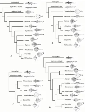
The Batoidea contain over half of all extant species of chondrichthyans, with about 630 spe-cies spread between up to 23 families. The Batoidea is now considered to form a sister group to all living sharks ([2–8], although some studies previously placed them as derived sharks [9,10]; Fig. 1A), with these forming the Neoselachii (or Elasmobranchii [11]), a clade that excludes many fossil‘shark’groups. While the monophyly of the Batoidea is not in doubt, phylogenetic relationships within the group are uncertain. There has been marked discrepancy in the relative positions of batoid clades in studies based on different phylogenetic methods, with recent mo-lecular analyses either largely based on sharks (e.g., [2,8]) or lacking members of all major batoid clades [6]. Morphological evidence has been used to suggest Torpediniformes are the most basal batoids (summarized by [10]), as have earlier molecular analyses [12], while more recent molecular analyses resolve the Torpediniformes as more derived than the Rajidae and a sister group to the Platyrhinidae [5] or Myliobatiformes [6]. These batoid phylogenies are sum-marized inFig. 1.
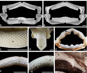
Despite the constraints of a variably flattened body and enlarged pectoral fins, the Batoidea are morphologically and taxonomically diverse. This high morphological diversity is reflected in their dentitions (e.g. [11,13–15] and refs therein), with some genera possessing hundreds of minute functional teeth all locked together in a pavement (such as inPristisandGlaucostegus; Fig. 2C), while in others the number of teeth in the pavement is reduced to one extremely en-larged symphyseal tooth file (Aetobatus;Fig. 2D). Teeth of many taxa of batoids are flat and wide (Myliobatissp.;Fig. 3D), adapted for some degree of durophagy, although more slender and narrow teeth, suitable for predation through grasping active prey, are present in some spe-cies. Other taxa, such as members of the Mobulidae (Myliobatiformes), have teeth that are re-duced in size and which may be non-functional, at least as far as feeding is concerned, due to a planktivorous diet.
Fig 1. Phylogeny of the Batoidea and selected outgroups.Four selected phylogenies showing the varying topologies and the differing positions of clades such as the Torpediniformes. A. After [10] with batoids as derived sharks. B. After [7] with Torpediniformes in a basal position. C. After [6] with the Torpediniformes as a sister group to the Myliobatiformes, but phylogenetic analysis did not include the Rajidae. D. After [5] with polyphyly of the‘rhinobatids’and Torpediniformes as a sister group to the Platyrhinidae. Profile images not to scale, slightly modified from FAO publications under a Creative Commons Attribution-Noncommercial 3.0 Unported License. Species studied are: Rajidae;Amblyraja doellojurdoi,Amblyraja frerichsi,Atlantoraja castelnaui,Bathyraja griseocauda,Bathyraja scaphiops,Dipturus batis,Dipturus binoculata,Dipturus chilensis,Leucoraja circularis,Leucoraja erinacea,Leucoraja naevus,Psammobatis normani,Psammobatis rudis,Raja brachyura,Raja clavata,Raja microocellata,Raja undulata,Rioraja agassizi,Sympterygia acuta. Platyrhinidae;Platyrhinidis triseriata. Torpediformes:Discopyge tschudii,Narcinesp.,Torpedo puelcha. Zapteryx:Zapteryx brevirostris. Pristidae:Anoxypristis cuspidata,Pristis perotetti. Glaucostegus:
Glaucostegus typus. Rhynchobatus:Rhynchobatus djiddensiss.s.,Rhynchobatusex. gr.djiddensis. Rhina:
Rhina ancylostoma. Rhinobatos:Rhinobatos horkelii,Trygonorrhina fasciata. Dasyatidae:Dasyatis brevis,
Dasyatis?macrophthalma,Himantura uarnak,Himanturasp. 1.,Himanturasp. 2.,Neotrygon kuhlii,
Pastinachus sephen,“Taeniura”lymma,Taeniura meyeni. Myliobatidae and other derived Myliobatiformes:
Aetobatusex. gr.narinari,Aetomylaeus maculatus,Myliobatis aquila,Myliobatis australis,Myliobatis californica,Myliobatis goodeis.s., Myliobatis spp.,Rhinoptera javanica,Mobulasp.
at the symphysis, with initiation of the symphyseal tooth proposed as the starting point for tim-ing and placement of teeth in dentition patterntim-ing [1]. Our hypothesis is that batoids, as in sharks, possess symphyseal teeth (e.g.,Fig. 2, st) initiating the pattern of the dentition in the earliest stages of ontogeny and that this state is primitive and conserved among chondrichth-yans. Other dentition patterns to be investigated in batoids, as potentially indicative of the gen-eral chondrichthyan condition, include whether the adult number of teeth are present in the earliest ontogenetic stages, and how the pattern unfolds in developmental time (e.g., how do se-quential files and rows develop relative to one another), and how aspects of crown and root morphology develop. In this way, we can begin to better understand characteristics of the chondrichthyan dentition and its organization and structural patterning, ultimately for com-parison to other gnathostome taxa.
Materials and Methods
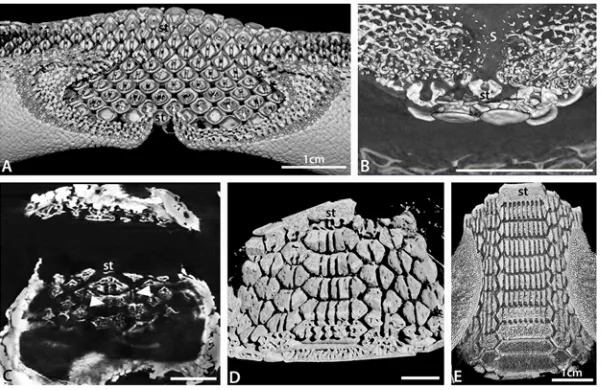
[image:5.612.202.500.76.326.2]To assess development in the Batoidea, dentitions of a range of modern batoids were studied. Dried and prepared dentitions of over 45 batoid species, including examples of all major clades, were studied (Fig. 1). Additional specimens of adults and late stage embryos of a number of species were selected for more detailed study. Late stage embryos (defined as being from within the mother or egg but having at least some developed teeth) ofRhinobatos horkelii,Myliobatis Fig 2. Morphological variation in batoid dentitions.A, B. Labial and lingual views, volume rendered scan of the jaws of an adult femaleRaja clavata(BMNH 2015.1.25.1). C. Symphyseal region of the lower dentition of the‘rhinobatid’Glaucostegus typus(BMNH 2015.1.25.3), showing alternate row pattern and massive numbers of small teeth. D. Lower dentition of the myliobatidAetobatusex. gr.narinari(BMNH 2015.1.25.4), in which only enlarged symphyseal teeth are present. E. Whole, articulated jaw ofRhina ancylostoma(BMNH 2015.1.25.5) showing convoluted pattern of the teeth and a region of malformed teeth (black arrow, see also
Fig. 3Din [1]). F. Upper jaw of a femaleRaja clavata, showing homodont dentition of low crowned teeth. G. Upper jaw of a maleRaja clavata(BMNH 2015.1.25.2), showing a heterodont dentition with tall cusped teeth. H. Upper dentition of a young female of the dasyatidNeotrygon kuhlii(BMNH 2015.1.25.6) with enlarged ‘caniniform’teeth, in S+9 position on the jaw. In this and other figures, symphyseal teeth are labelled‘st’, the jaw symphysis as‘S’. All scale bars are 1cm unless marked otherwise.
sp.,Discopyge tschudiiandRaja clavatarepresenting the Rhinobatidae (s.s.), Myliobatidae, Narcinidae (Torpediniformes) and Rajidae (s.s.) respectively (Fig. 1) were studied by x-CT. In addition, neonate specimens (free swimming but with prominent umbilical scars) ofMyliobatis
sp.,Rioraja agassiziiandBathyrajasp. were studied by the same method. Embryos of seven shark taxa were also studied for comparison. It should be noted that the taxonomy of S.W. At-lanticMyliobatisis in a state of flux, and the specimens here belong to eitherM.freminvilliior an unnamed taxon similar toM.goodei. All figured specimens are accessioned to the Natural History Museum, London; accession numbers are given in the figure captions the first time each specimen is figured. Specimens of a range of ontogenetic stages ofRaja clavataand Leu-coraja erinaceawere also cleared and stained for optical study.
[image:6.612.202.499.78.286.2]Thornback Ray (Raja clavata) embryos were kindly donated by the Native Marine Centre, UK (www.nativemarinecentre.com) and reared at the University of Sheffield marine aquarium (14°C), Department of Animal and Plant Sciences, until the required stage of development. Embryos were then removed from their egg cases and lethally anaesthetized in MS-222 (Tri-cane) before fixation in 4% paraformaldehyde for paraffin sectioning and further analysis. Lit-tle Skate (Leucoraja erinacea) embryos were kindly donated by Andrew Gillis, Dalhousie University (Canada), and sourced from Woods Hole Marine Biological Laboratories (MA, USA). These early embryos ofL.erinaceawere cleared and stained following standard proto-cols (CS; Alizarin red and Alcian Blue [16]) then dissected and mounted as upper and lower jaws to show sites of first tooth formation. All animals were culled under the Animals
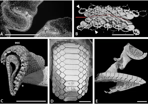
Fig 3. Tooth arrangement in dentition of batoids.A. Histological DAPI (DNA) stained section through upper (left) and lower (right) jaws of an embryonicRaja clavata. Teeth (T1) developing within the dental lamina (*). B. Volume rendered lingual view (from inside oral cavity) of the developing teeth of an embryo of
Discopyge tschudii(BMNH 2015.1.25.7) showing progressive mineralization of the teeth on the upper and lower jaws (occlusal surface indicated by red line). C. Volume rendered segment of the lower jaw of a female adultRaja clavata, with a vertical section view through tooth files showing migration of teeth from the newest in the dental lamina (*), to oldest beyond the occlusal surface (occ), as well as the close alternate packing of the teeth. D. Volume rendered occlusal view of the lower dentition of an adultMyliobatissp. (BMNH 2015.1.25.10) showing enlarged symphyseal teeth and interlocking of alternate rows of tooth files S+1–3. E. Volume rendered longitudinal (rostral to caudal) section view through both jaws of an adultMyliobatissp. showing close interlocking of the teeth and different degrees of curvature of the occlusal surfaces of upper and lower jaws. Scale bars = 1mm (A, B) or 1cm (C-E).
(Scientific Procedures) Act 1986 at the University of Sheffield; no manipulation of the animals was performed prior to their culling.
Specimens were studied using x-CT (X-Tek HMX ST CT scanner, Image and Analysis Cen-tre, Natural History Museum; scans volume rendered with VGStudiomax (http://www. volumegraphics.com/en/products/vgstudio-max.html), Avizo (http://www.vsg3d.com/avizo/ overview) and Drishti (http://sf.anu.edu.au/Vizlab/drishti); Micro x-CT at Dental Institute, King’s College, London; GE Locus SP, CT Tech scanner with Microview, creating volumes with voxel sizes 6.5μm to reveal structural pattern order of the developing teeth. ForRaja cla-vata, we used soft tissue contrast-enhancing stains [17] with 4μm resolution of tooth germs, using X-Radia, with Xray scintillators, and Zeiss optical lenses, in the Department of Theoreti-cal Biology, University of Vienna [17].
In certain figures, images have been combined in Adobe Photoshop to improve clarity; as well teeth have been false colored in Photoshop or Aviso to identify individual tooth rows. Data (including images and rendered files) will be made available athttp://chondrichthyes. myspecies.info/.
Terminology used follows [13]. It should be noted that the terminology used for description of chondrichthyan teeth is commonly the same as that used for the teeth of other gnathostomes (such as‘root’and‘cusp’). In the absence of a chondrichthyan-specific terminology, this is standard practice (e.g. [13]) and based on morphological and/or functional similarities and does not imply direct homology.
Nomenclature used to describe jaw position of the teeth has been inconsistent. For instance, in batoids the jaw is often almost straight and perpendicular to the body axis so the terms ante-rior and posteante-rior are not appropriate. Here, the terms proximal and distal are used to refer to jaw position, where proximal is closest to the neurocranium. As the jaw hinge is closest to the neurocranial attachment for the suspensorium, proximal is taken as a position closest to the hinge between the jaw cartilages, while distal refers to positions closer to the jaw symphysis. La-bial and lingual refer to the outside and inside of the jaw, respectively, and occlusal is used to describe the functional jaw surface. Terminology related to dentition patterning follows [1,11, 18]. The dental lamina is a double, oral epithelium that invaginates deep into the jaw to form successional teeth, organized into developmental timed sets (tooth files, or families), formed from its aboral aspect (away from the oral epithelium) and continuous along the jaws, always within the concavity of the lingual surface of the jaw cartilages [1,18,19].
Batoid dental diversity
lingual extension of the tooth base and the close, alternate crown shapes as hexagons form a rigid grinding surface (Fig. 3D, E). In these taxa, lower teeth may be retained for some time, into the post-occlusal stage, forming part of a labially extended tooth plate.
[image:8.612.199.500.76.375.2]Many batoid dentitions show a weak monognathic heterodonty [13], with teeth gradually changing shape between the symphysis and proximal extremities of the jaw. In contrast to monognathic heterodonty, dignathic heterodonty, with different tooth morphologies present on both the upper and lower jaws, is uncommon in most batoids. In taxa where the number of tooth files is very reduced, such as inMyliobatis, the symphyseal teeth may be greatly enlarged relative to other teeth resulting in a disjunct heterodonty (Fig. 3D). In some batoid taxa (such asRhinaandPastinachus), jaw cartilages have a strongly undulose profile and as a result the shape of teeth varies with both the position relative to high and low points along the jaw and
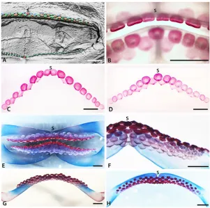
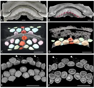
Fig 4. Tooth addition in early ontogeny of rajid and rhinobatid batoids.A. Volume rendered dentition of an embryo ofRhinobatos horkelli(BMNH 2015.1.25.13) showing on the upper jaw, the alternate positions of the first formed teeth across the entire width of the jaw. B-D, cleared, Alizarin Red stained preparation of an embryo ofRaja clavata. The length of theR.clavataspecimen is uncertain sue to it being dissected prior to mounting but is within a late stage of development prior to hatching. The first teeth are well mineralized (Alizarin positive for calcium), relative to the second tooth row (less strong Alizarin Red), in alternate positions. Two parasymphyseal teeth are present next to the jaw symphysis (S in B, C) in the upper first tooth row; in the lower jaw, a symphyseal tooth is present (S in D-F, H) in the first tooth row. B. Labial view of both jaws of the same specimen, showing the relative positions of the first row teeth of upper and lower jaws. C-D. Separated upper and lower jaws respectively. E, F. Cleared and stained lower and upper jaw dentitions of
Leucoraja erinacea, total length 107mm. F. Portion of the lower dentition ofLeucoraja erinaceashowing alternate rows of teeth with progressive mineralization (degree of Alizarin Red uptake) in the successive tooth rows. G. Lower dentition ofLeucoraja erinaceain lingual view. H. Labial view of the lower dentition of
Leucoraja erinaceashowing that the first tooth row of 10 positions contains a symphyseal tooth, as in that of
distance between the symphysis and proximal jaw extremities (Fig. 2E). In some other batoids, small‘caniniform’teeth are present within the upper jaw (Fig. 2F). In most cases the change in tooth shape along the jaw is still gradual, as a graded tooth size (e.g.,Fig. 2E).
[image:9.612.199.500.76.359.2]Sexually dimorphic dentitions are known within a large proportion of batoids (e.g. [23]), and are probably present in most batoid species. Where sexual dimorphism is present, the teeth of adult males are more cuspate than those of adult females and juveniles (Fig. 2A, B, F, G, H). While this heterodonty may be slight in some taxa, in others, including many species of the Rajidae and Dasyatidae, the differences may be extremely pronounced. In some species there may also be seasonal variation in the degree of heterodonty, with more cuspate teeth de-veloping in males in the breeding season, presumably used for grasping the female during mat-ing [24].
Fig 5. Tooth addition in early ontogeny ofRaja clavata.A-D. Volume rendered and segmented developing teeth inRaja clavataembryos (either VG Studio Max, Drishti, or Avizo). A-B. Embryo 85mm TL, volume rendered upper jaw showing developing teeth as tooth germs under the skin labial to the numerous papillary projections and the symphysis of the palatoquadrate cartilages (S). B. Upper jaw with false colour to highlight the sub-epithelial tooth germs. C-F. Embryo 104mm TL (C, D, stained with iodine and potassium, I2M. Density volume rendered, E, F, unstained). C. Labial view of segmented developing tooth germs in the upper and lower jaws (Avizo); symphyseal tooth (st, red) present in initial row of both jaws, (high density of the connective tissue enhanced at symphyseal junction between cartilages). D. Higher magnification of upper jaw, of lingually rotated jaw to show symphyseal junction (high density tissue) and 3rdrow teeth with one at the symphysis (S). E-F. Volume rendered images of the developing teeth (Drishti). Note that the lateral edges of the images are the limits of the render and not the full extent of the teeth. In both jaws there is a symphyseal tooth (st) in the first row and third row of the lower jaw, but the tooth positions may be shifted with growth and not regular. In the upper jaw there is a symphyseal tooth in the first tooth row. Teeth in second row are developing at later times (arrow heads, start of mineralization) relative to the symphyseal tooth Note in E, cusps have a different orientation in 1stand 2ndrows as tooth germs change their developmental positions. F. Lingual view, tooth roots have not started to form and the pulp cavity is open. Scale bars = 500μm.
Results
Batoid dentitions are described from their developmental characters, for the initiation of each tooth that either, shows tooth addition to a new proximal position on the jaw, or the addition of successor teeth in already established loci for each jaw position. These characters are regulat-ed by genes with differential timing for the initiation of new tooth sets proximally and for new successor teeth in jaw positions set up both early, and late in development. Our observations test a proposed model (19) for relative timing of sets ordered by addition from the symphyseal locus, either side in a sequence proximally (laterally), with those of successor teeth proposed to follow this genetically embedded, timed sequence. Developmental characters concerning crown and root development at these developmental times are also described.
Tooth set initiation and successional addition
Early embryonic stages of tooth initiation. Embryonic stages ofRaja clavataand
Leucor-aja erinaceawere used to describe the earliest stages of tooth development. The first tooth germs inRaja clavata(prior to mineralization) at the morphogenetic stage are formed from in-teraction between dental epithelium and condensed mesenchyme (Fig. 3A,‘T1’). Their devel-opment starts superficially in the oral epithelium, when a thickened sheet of epithelial cells (odontogenic band) invaginates, forming the dental lamina (Fig. 3A, asterisk). This epithelium is rich in proliferative cells (data not shown here), as observed from PCNA immunohistochem-istry of theR.clavataembryonic dentition at T1-stage (Fig. 3A, T1). The dental lamina contin-ues to proliferate, extending deeply into the mesenchyme of the jaw, restricting provision of the odontogenic cells that initiate continuous tooth addition throughout life to this part of the dental lamina.
Tooth germs of the first tooth row develop along the jaw (Raja clavata,Fig. 5A, B), and when these have begun to mineralize (Figs.4B-Dand5E, F), the next alternate row of tooth germs forms, with lightly mineralized crowns and a lingual cusp that is first to mineralize (Figs. 4C, Dand5E, F, arrowheads). All have flat crowns as in the adult female morphology, with a pulpal opening that closes gradually prior to root development (Fig. 5F); root mineralization occurs well after the formation of the tooth crown, a developmental pattern also seen in all subsequent teeth.
InDiscopygeonce the sixth successive generation of teeth has started to mineralize (seen in crown outline,Fig. 3B, arrow heads), the first teeth have changed their orientation to move into the occlusal position with the upper jaw. These early tooth sets in both jaws are tightly packed, with reciprocal occlusal curves (concave in upper, convex in lower,Fig. 3B). However, these first generations of teeth are probably non-functional (as these embryonic ones are pres-ent in embryos confined to the egg case).
Similarly, inLeucoraja erinacea(Fig. 4E, F; arrow, st4) the latest full row of successor teeth formed in proximal order, with limited uptake of the Alizarin Red (less calcium), is starting from the symphyseal one, then the next row to form, with alternate teeth, has left and right parasymphyseal teeth first (Fig. 4F, T1-4). This Alizarin Red gradation approximates to differ-ential timing of tooth development in each set and along the row, is equated with mineraliza-tion levels of xCT density volume renders (Fig. 5E, Farrow heads). InLeucorajathe addition of new proximal tooth sets at the jaw margin is probably no longer occurring at this stage
(Fig. 4E, F, 15 sets in each half) when approximately the 10throw of successional teeth is form-ing at the lform-ingual edge.
Symphyseal tooth initiation and continuous proximal file addition. The adult
Embryos ofDiscopyge tschudiihave incompletely mineralized jaw cartilages, and as a result the last formed teeth are directly visualized in rendered micro-CT scans (Figs.3B, arrowheads, 6A-Eand7B). The first tooth row in the embryo of both upper and lower jaws is identified as the most labial row on the jaw; this row comprises a pair of teeth, one either side of the jaw symphysis (Fig. 6A-E, blue teeth); there is no evidence of an initial symphyseal tooth, and the early developmental stage studied suggests that this tooth would be seen if present (i.e., not at a stage where it would have been biologically lost). The parasymphyseal teeth are low and discoi-dal, with only an incipient cusp, differing from teeth in adults or free-swimming juveniles (Fig. 6A-F). The second tooth row comprises three teeth, a symphyseal tooth (st) and two teeth
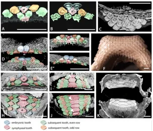
Fig 6. Tooth addition in early ontogeny of batoids.A-E. Volume rendered, segmented, or false colour images of the early dentitions of the torpediformDiscopyge tschudii(A-C BMNH 2015.1.25.7, D-E BMNH 2015.1.25.8). A. Occlusal surface (labial), with density rendered transparent cartilage, lower jaw, segmented colour for teeth, first and second symphyseal teeth (S, for clarity all teeth in the rows are not shown). However, all tooth rows are shown in same specimen inFig. 3B, and in false colour images D, E. B. Visceral surface of the same teeth shown in A, to show developing tooth roots. C. Volume rendered view of the upper and lower dentitions ofDiscopyge tschudii, with the lower jaw cartilages digitally dissected to show the bases of the lower teeth. Note the abnormal symphyseal tooth with a labial expansion protruding between the first pair of parasymphyseal teeth. D. Volume rendered upper and lower jaws in labial view, with parasymphyseal pair of teeth (blue) in the apparent first row but subsequent, succeeding row with teeth in alternate positions does have a symphyseal tooth. E’and E”are from D after rotation of image with lingual and labial views of the lower jaw to show progressive increase in tooth numbers in successive rows, (symphyseal tooth is red). F. Photomacrograph of the lower dentition of an adultDiscopyge tschudii(BMNH 2015.1.25.9) demonstrating the multitude of tooth files gained by gradual proximal addition of teeth during ontogeny (occlusal view). G, upper, H, lower, jaws of an embryo ofMyliobatissp. (BMNH 2015.1.25.11), volume rendered to show occlusal surface of the dentition with the first formed pair of teeth as parasymphyseal (blue) in both jaws; the second row has a larger symphyseal tooth (red) and subsequent teeth in alternating rows of three and four teeth. I, J, volume rendered upper and lower dentition of a neonate ofMyliobatissp. (BMNH 2015.1.25.12), occlusal surface showing the fixed number of teeth in each row (S+3) and the gradual enlargement of all through ontogeny (occlusal view). The‘tongue and groove’tooth locking is indicated by white arrows. K. Volume rendered jaws of I, J, showing mineralized crowns at occlusal surface compared with younger lingual teeth with less mineralization. False colour coding is used in the dentitions A, B, D, E, G-J as inFig. 4. All scale bars are 1mm.
either side of this, forming files that alternate with those of the first two teeth. At this stage well-differentiated tooth morphology is present, with a small lingual cusp and two root lobes (sensu [13]) with splayed roots separated by a wide groove (Fig. 6B,C). Subsequent tooth rows alternate in a similar pattern to the first two, with rows bearing a symphyseal tooth alternating with those lacking this tooth but having a pair of parasymphyseal teeth. In the third tooth row, a tooth additional to those in previous rows is added proximally each side (further from the symphysis). Each successive tooth row observed has an additional pair of proximal teeth pres-ent, so that the number of tooth files along the jaw (added proximally) increases rapidly through the early stages of ontogeny and tooth development. Dentition of adultDiscopygeis comprised of tooth rows containing a large number of teeth (Fig. 6F), so it is probable that this tooth addition continues throughout life continuously as new proximal sets.
Symphyseal tooth initiation and limited proximal file addition. Whereas adult
denti-tions ofDiscopygecontain large numbers of small teeth, those ofMyliobatisdiffer in having rel-atively few, very large, teeth even in the adult (Fig. 3D). The symphyseal teeth are especially enlarged, and can comprise over half of the width of the entire dentition. Symphyseal teeth have a large number of root lobes, with the smaller parasymphyseal and proximal teeth also commonly possessing more than two root lobes, particularly in older individuals (Fig. 7D, E). The teeth of embryos ofMyliobatisare only slightly mineralized, and show little x-ray density contrast relative to the jaw cartilages, as a result they are more difficult to isolate (Figs.6G, H and7C).
[image:12.612.201.502.77.272.2]Tooth development is identical in upper and lower dentitions ofMyliobatis, with two rudi-mentary parasymphyseal teeth as the first row (Fig. 6G, H); as withDiscopygeabove, these are the first-developing teeth in the embryo, with a symphyseal tooth being absent. These teeth are oval and very low (comparable to those in theDiscopyge), and appear to have a flared root
Fig 7. Development of batoid tooth crown size co-ordinated with roots.A. Composite volume render of the basal surface of the lower dentition of a femaleRaja clavatashowing the bilobed tooth roots typical of batoids. B. Composite volume render of an embryo ofDiscopyge tschudiiwhere the initial upper teeth show the presence of bilobed roots. C. Render of a basal section through the lower teeth of an embryo ofMyliobatis
sp. showing the presence of one or two, poorly developed grooves within the teeth (white arrows). D. Composite volume render of the basal surface of the lower dentition of a neonate specimen ofMyliobatissp. showing up to seven grooves in the roots of the symphyseal teeth and fewer in the other teeth. Many of the grooves are to a greater or lesser extent roofed over. E. Composite volume render of the basal surface of the lower dentition of an adult specimen ofMyliobatissp. showing multiple, well developed, grooves in the roots and in the top row their relationship to the crown. All scale bars are 1mm except where indicated.
considerably wider at the base than the crown (Figs.6G, Hand7C). The second tooth row comprises three teeth, all of which are considerably larger than those in the first row (Fig. 6G, H). These comprise a symphyseal tooth and a pair of more proximal teeth alternating with the teeth in the first row, identical to those inDiscopyge(Fig. 6A-E). The two more proximal teeth are rounded, but the symphyseal tooth is somewhat angular and wider than deep, giving an overall‘diamond’shaped occlusal profile (st,Fig. 6G, H). The third tooth row lacks a symphy-seal tooth and comprises just four tooth files. The teeth are separated and not in contact with each other, and to some extent, angular. No further proximal tooth files are added with succes-sive rows, such that the dentition comprises an alternate series of three teeth, including an en-larged symphyseal tooth, alternating with four teeth. In each successive tooth row, the teeth are relatively larger and more angular, with the symphyseal tooth becoming considerably larger than the other teeth. The roots of these early teeth are very poorly differentiated, and the grooves separating the roots are very shallow. Two grooves (and hence three root lobes) are present on the symphyseal and parasymphyseal teeth, and a single groove separating two roots lobes in more proximal teeth (Fig. 7C).
The dentition of a neonate specimen of the same species ofMyliobatisshows the transition in tooth morphology from embryo to adult (Fig. 6I-K). The most labial tooth row consists of a symphyseal tooth and one tooth on either side suggesting that the first row of two parasymph-seal teeth have been lost, and the general shape and size of the most labial teeth is similar to teeth in the early stages of development in the embryos, including those in the symphysial file. The dentition of this specimen retains the same general pattern seen in the embryo, and also seen in the adult (Fig. 3D). As noted above, rows of three teeth with an enlarged symphyseal tooth alternate with rows of four, not enlarged, but more uniform teeth. All teeth are polygonal and closely packed. In vertical section, the teeth possess the flange and groove system that lock the teeth together in the adult dentition (Fig. 3E). Sequential tooth rows show a marked in-crease in the width of the dentition, via inin-crease of the width of the symphyseal teeth (see sec-tion on root development, below). The mode of tooth mineralizasec-tion is not clearly evident in the embryonic specimens.
Simultaneous symphyseal to proximal tooth initiation addition. In contrast to
Mylioba-tisandDiscopyge, the early dental development inRhinobatosinitially occurs across the entire width of the jaw rather than being restricted to the symphysis (Fig. 4A). The first formed teeth are seen only in the upper jaw; in the lower jaw these are concealed behind later teeth due to shrinkage of the specimen before examination. These teeth are extremely small and have not yet moved into a position close to where they would be shed, so any earlier teeth would have been visible if they had been present. The first tooth row extends across the entire width of the lower jaw, clearly alternating with the teeth of the second row. There is a symphyseal tooth at the centre of the first tooth row (red tooth, symphysis, S,Fig. 4A), although slippage of the teeth as the jaw became dehydrated cannot be eliminated. The total number of teeth along each row appears to be similar to the conspecific adult, indicating that there is no subsequent proxi-mal addition of tooth files, again differing fromMyliobatisandDiscopyge.
Variable tooth initiation pattern. Upper and lower jaws of the two species of Rajidae have a different arrangement of the position of teeth in the initial rows of each although in both, these are followed by a second row, with teeth in alternate positions. InLeucoraja erina-ceasymphyseal teeth are present in the first row of both upper and lower dentitions
(Fig. 4B, D). Lingual to these regularly spaced teeth, the second row teeth alternate in positions between those in the initial row.
The other specimens ofRaja clavatashow symphyseal teeth in the first row forming on the upper jaw, as well as the lower jaw, but the arrangement of these teeth relative to the jaw carti-lage symphyses is not as clear as in other taxa. One specimen, representing the smallest of the three examined, has an upper symphyseal tooth germ but this is slightly offset relative to the symphysis between the jaw cartilages (Fig. 5A, B). A second, larger, specimen shows developing tooth germs, mineralizing tooth crowns and connective tissue at the symphysis (Fig. 5C, D; soft tissues stained), or mineralizing teeth alone (Fig. 5E, F; unstained). A symphyseal tooth is pres-ent in both jaws, associated with the jaw symphyses (S), but both teeth are again slightly offset from the jaw symphysis. Second row teeth on the upper jaw show differing degrees of minerali-zation in left-right positions relative to the symphyseal tooth (Fig. 5E, F, st). Whilst this denti-tion initially appears to lack a symphyseal tooth in the first upper row, as the teeth are paired with some in the second row. Closer inspection, however, suggests that the mineralization of the teeth in the second row is irregular, and nor synchronous in adjacent teeth. As a result, it is not possible to use mineralized teeth in the second tooth row to assess tooth position. Addi-tional complexity is seen in the lower dentition of the same specimen, where teeth are extreme-ly close packed and cannot readiextreme-ly be assigned to their correct positions (Fig. 6E-F). Lingual to these regularly spaced teeth, the second row teeth alternate in positions between those in the initial row. In both of the rajid taxa studied, a number of teeth are present in the first formed tooth rows, approximating the number in adults of the same species; with 5–8 teeth in the first half row ofRaja clavataas opposed to about 15 teeth in each half row in the adult (10–12; Figs. 2A, B,4E-Hand5A, B). Thus, development of teeth along the row is more rapid than the initi-ation of subsequent rows of successor teeth (Figs.4and5A, B).
Changes in crown morphology
InDiscopyge, the first teeth have a flat, discoidal crown (Fig. 6A-E) over a root with well-devel-oped lobes (Fig. 7B). There is some degree of labial-lingual asymmetry in the crown, with an in-cipient cusp at the lingual edge (Figs.3Band6A, D, E). The initial teeth are closely spaced along the row, and the margins of the teeth are in contact. By the second tooth row, a more adult-like tooth morphology is already established (Fig. 6E, F), with a well-developed cusp and a flared lingual-lateral crown edge. Subsequent teeth have a more elongate cusp and concave la-bial crown face, but in other respects are similar in shape to the teeth in the second row.
The first teeth ofRajaandLeucorajaare very similar in overall form to those ofDiscopyge, being flat and oval, with a cusp the first to mineralize, but with far less root development (Figs. 4,5). Incipient cusps are present in the earliest teeth of both genera, in the adult position (lin-gual), but cusps remain small (Figs.2G,4and5). This is consistent with the lack of well-formed cusps in the juveniles and mature females ofRaja clavata(Fig. 2A, B), in contrast to the cuspate teeth of mature males (Fig. 2D, E).
locking mechanism seen inMyliobatisdentitions (Fig. 6I,J). In the third row, all teeth are some-what angular, and have a relatively well-developed ridge and groove on the crown edges. Even at this point, however, the teeth are widely spaced. It is therefore evident that the‘tongue and groove’tooth locking mechanism appears earlier in ontogeny than the close packing of the teeth that it facilitates.
The first teeth inRhinobatosare small and globular and show little structure (Fig. 4A). Sub-sequent teeth are not clearly seen, but are wider than deep and have a poorly developed occlusal bulge The tooth morphology is close to that seen in juveniles and adult females of the species; adult males have more strongly cuspate teeth.
Changes in root morphology
In the majority of batoids, as in sharks, the tooth has a root comprising two root lobes separat-ed by a groove (Fig. 7A, B), which may be closed over in some sharks and some extinct batoids [25]. While teeth of some batoids possess multiple root lobes (e.g., Myliobatidae, discussed below), this remains rare within most clades. WithinDiscopygethe root morphology of the first formed teeth is very similar to that in adult teeth, demonstrating that the template for tooth root morphology must be set within the tooth germ at a very early ontogenetic stage (Fig. 7B). By comparison, the root development inRajaandLeucorajais delayed relative to the development of the tooth crown, with no roots observed in the embryonic teeth studied (e.g. Fig. 5E-F). This suggests that whilst the morphology of the roots of fully formed teeth of Disco-pygeandRajaare similar, the relative timing of root development is very different.
Within the Myliobatidae and Rhinopteridae multiple root lobes are present on the teeth. Enlarged symphyseal teeth inMyliobatismay have up to 30 root lobes with smaller proximal teeth having up to five root lobes; over 50 root lobes may be present on the upper symphyseal teeth ofAetobatus. Our observations suggest there is an increase in the number of root lobes through ontogeny in these taxa (Fig. 7C-E). Teeth present in embryos ofMyliobatissp. have poorly differentiated roots, with rendered images suggesting a similar histology of the root and interior part of the crown. The base of the root is flat, and grooves are present only as shallow excavations. Despite this, sections through the basal part of the root clearly show that only one or two grooves are present in teeth, and if two are present, one is larger than the other
(Fig. 7C). In the teeth of a neonate of the same species, multiple root lobes are present
(Fig. 7D). Up to four grooves are present in the roots of symphyseal teeth, two in parasymphy-seal teeth and one in other teeth (Fig. 7B). Some of these grooves are partly or almost complete-ly closed over and the spacing across the width of the tooth is irregular. In the adult dentition, up to nine grooves are present in symphyseal teeth, three in parasymphyseal teeth and two in more proximal teeth (Fig. 7E). These are all open and evenly spaced across the tooth. It is there-fore evident that the increase of tooth width is directly related to the number of grooves present in the teeth. In addition, the degree of homogeneity of the roots within the teeth increases through ontogeny.
Discussion
Construction of the initial chondrichthyan dentition is highly conserved, especially with the formation of the pattern and the process of lamina-initiated tooth replacement. A better under-standing of how the dentition is built through development in chondrichthyan clades such as the Batoidea will allow general characteristics of the structural pattern of the chondrichthyan dentition to be defined. This understanding is crucial before broader comparisons to other major extant groups such as the bony fishes (Osteichthyes) and fossil groups such as the phylo-genetically basal‘Placodermi’can be undertaken. Recent phylogenetic analyses [26–28] have resolved fossil groups such as the‘Acanthodii’as paraphyletic, with some or all‘acanthodian’ taxa resolved as stem-group chondrichthyans. These new analyses further complicate any as-sessment of the basal chondrichthyan dentition. Understanding the developmental basis of temporal and spatial order of sequential tooth addition to dentitions within the modern chon-drichthyans is, therefore, critical to the interpretation of tooth acquisition and formation of the functional dentition in basal gnathostomes.
Tooth morphology
[image:16.612.201.500.452.654.2]In shark dentitions achievement of adult morphology occurs well after initial tooth develop-ment [1,14,18,29], and adult tooth shape emerges over many rounds of tooth replacement, a pattern also seen in osteichthyan fishes [28]. However, in the embryonic ray the first sets of teeth (in rows 1 and 2; Figs.3A, B,4,5and6) already show a broad flattened morphology, with a single low cusp representing the adult dental morphology (e.g., inDiscopyge, femaleRaja). This compares with sharks that possess an initial set of teeth that are only lightly mineralized, very small, and with two accessory cusps,‘simple tooth shards setting up’the tooth files to start the process that shapes the successor teeth toward the adult phenotype [18]. However, in sharks this‘shard-like’mineralized structure is the site of the first cusp (tallest on the crown morphology), also linked to first gene expression with the specific probe for sonic hedgehog, and second expression related to position of lateral cups [18,19]. Among batoids,Myliobatisis
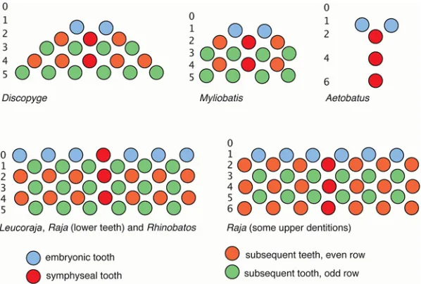
Fig 8. Summary of dental development patterns in the Batoidea.The dentitions ofDiscopyge,Myliobatis,
Aetobatus,Leucoraja/RajaandRhinobatosare compared, with time order for initiation of tooth rows indicated, proposed as homologous patterns (jaw positions, blue 2,4,6: green 1,3,5. Colour is the same as in Figs.4and6.
more comparable to the shark condition, in that the first teeth differ from those of the adult in terms of size and shape, particularly symphyseally. Root development is also advanced in
MyliobatisandDiscopyge(present and developing in the embryo), but delayed in taxa such as
Raja.
Dentition development
Within the different batoid taxa studied there are several distinct modes of tooth order addi-tion, involving the symphyseal/parasymphysial teeth as the putative initiators of tooth pattern-ing along the jaw [19], as well as rates of proximal tooth addition along the jaw relative to the addition of successive teeth more lingually.
[image:17.612.200.499.323.672.2]The presence of a symphyseal tooth in the first tooth row is variable within the Batoidea. Garman [30] was one of the first to illustrate the presence of two parasymphysial teeth in the first tooth row in embryonicRhinopteraandAetobatusdentition, with a symphyseal tooth first appearing in the second row even though symphyseal teeth dominate the dentition in adults (see Figs.2Dand8). Our observations have shown that a first row symphyseal tooth is also ab-sent inDiscopygeandMyliobatisas well as in the upper jaw ofRaja clavata, but present in taxa such asRhinobatos,Leucorajaand in the lower jaw ofRaja clavata. It is evident (from current
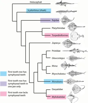
Fig 9. Phylogeny of the Batoidea and selected outgroups with positions of dental development type. Tooth development type as seen in different batoid clades (seeFig. 1E).
information) that the presence of a morphological symphyseal tooth in the first row is not fun-damental to the development of a batoid dentition. However, as teeth always form initially at, or alongside, the symphysis before bilateral proximal addition, it is probable that focused gene expression at the symphysis is critical to tooth development even if the symphyseal tooth itself is absent. Studies of the tooth development in the sharkScyliorhinushave shown that initial tooth development starts at the jaw symphysis, as indicated by the expression of the gene sonic hedgehog [22]. We suggest this symphyseal-driven dentition pattern is shared between sharks + rays (Elasmobranchii). This model will be tested in ongoing molecular
developmental studies.
InScyliorhinus, consecutive teeth form along the first tooth row away from the symphysis prior to the initiation of the teeth in alternate positions of the second row, so that the first tooth row is complete before the second is initiated [20]. Despite this, tooth initiation inScyliorhinus
is not simultaneous along the jaws. Teeth initially form near the symphysis, then in a closely timed developmental sequence from this position, proximally along the jaw. This alternate pat-tern of tooth addition is seen in all batoids, with the rate of propagation away from the sym-physis relative to the rate of development of successive tooth addition (within files) being variable among taxa. The proximal addition of tooth files inRhinobatos, as inScyliorhinus, is more rapid than those added as successive teeth, i.e. before the alternate rows are initiated. In the Rajidae the proximal propagation is still rapid, but somewhat slower than development of successive tooth rows, so that the initial tooth row contains less than the full complement of teeth (Fig. 5versusFig. 2A, B). InDiscopygeandMyliobatis, the rate of proximal addition is very slow relative to the generation of successive tooth rows, and as a result only one additional tooth is added proximally each side (Fig. 6). It is therefore evident (seeFig. 9) that slowing of the positional tooth addition inDiscopygeandMyliobatisis convergently derived. There does appear to be a general reduction of proximal tooth addition in the group as a whole, while maintaining the alternate pattern of row addition (proximo-distal) characteristic of the Neose-lachii [1].
Conclusions
Our observations on embryonic and post-embryonic dentitions of the chondrichthyan group Batoidea suggest that despite substantial diversity there are shared batoid characters requiring further comparison to outgroup taxa (e.g., sharks), including the presence of an alternate denti-tion. Further evidence regarding genetic regulation of a symphyseal region in patterning the initial and subsequent tooth rows in the Batoidea will depend on pending molecular develop-mental data. Similarly, to determine relative rates of proximal tooth addition versus addition of successive tooth rows will require complete series of developmental data from all significant groups. These comparisons will allow us to establish the phylogentically basal condition for elasmobranchs. Similarities to the shark dentition (e.g., early presence of initiator symphysial region) indicate shared characters for the Elasmobranchii as a whole. Some of these characters are more problematic, for example, whether the alternate dentition characteristic for the Batoi-dea is plesiomorphic or derived for Elasmobranchii is still to be determined [1] and requires data on selected chondrichthyan taxa showing a dentition composed of single tooth files (e.g.,
Torpediformes + Myliobatidae is recovered in some of these analyses (Fig. 1C[6]), and would be characterized by this reduction in proximal tooth addition.
Further, we will be able to test how these characters may be controlled in development, for example, with genetic factors that might restrict later timed tooth families along the jaw and initial activator/inhibitor patterning gene interactions (i.e. Hedgehog, Ectodysplasin and Wnt signaling molecules) emerging from the first embryonic morphogenetic stages (odontogenic band) that would allow symphyseal tooth sites to expand and create one large-sized tooth to achieve specialized dentitions at the extremes of morphological diversity (i.e.Myliobatis, Aeto-batus). Because at least three chondrichthyans are now considered‘model’organisms for devel-opmental studies (Scyliorhinus canicula,Raja clavata,Leucoraja erinacea), it is now possible to study the evolution and development of dental and general diversity in these chondrichthyan clades that have generated extreme food processing modules, including crushing pavement dentitions, to a more gripping/tearing dentition.
The hypothesis that a symphyseal-driven dentition pattern is shared between sharks + rays (Elasmobranchii) is fully supported by data given here, and is a distinction between? Chon-drichthyes and Osteichthyes, the latter have a different initiation site for each dentate bone [19]. We intend to test this hypothesis with data on the genetic regulatory basis of the dentition pattern in forthcoming papers. Ultimately it will allow the general vertebrate condition for a dentition at the origins of jaws to be proposed, and the fossil types in stem vertebrates, assessed and analysed for these characters.
Acknowledgments
We thank the Natural Environmental Research Council (NERC grants NE/K01434X1, NE/ K014595/1, NE/K0122071/1) for financial support. We would like to thank Dan Sykes, Farah Ahmed and Rebecca Summerfield (NHM) and Chris Healy (Craniofacial Development and Stem Cell Biology, KCL) for producing the mCT scan. We would also like to thank Native Ma-rine Centre and Andrew Gillis for donations of embryos and Alicia of Zidona Shells for assis-tance in obtaining Uruguayan material.
Author Contributions
Conceived and designed the experiments: CJU ZJ MMS GJF. Performed the experiments: CJU ZJ MW LJR GJF MMS BM. Analyzed the data: CJU ZJ MW LJR GJF MMS. Contributed re-agents/materials/analysis tools: CJU ZJ MW LJR GJF MMS BM. Wrote the paper: CJU ZJ MW LJR GJF MMS.
References
1. Smith MM, Johanson Z, Underwood C, Diekwisch T (2013) Pattern formation in development of chon-drichthyan dentitions: a review of an evolutionary model. Hist Biol 25: 1–16.
2. Douady CJ, Dosay M, Shivji MS, Stanhope MJ (2003) Molecular phylogenetic evidence refuting the hy-pothesis of Batoidea (rays and skates) as derived sharks. Mol Phylogenet Evol 26:215–221. PMID:
12565032
3. Winchell CJ, Martin AP, Mallatt J (2004) Phylogeny of elasmobranchs based on LSU and SSU ribosom-al RNA genes. Mol Phylogenet Evol 31:214–224. PMID:15019621
4. Vélez-Zuazo X, Agnarsson I (2011) Shark tales: A molecular species-level phylogeny of sharks (Sela-chimorpha, Chondrichthyes). Mol Phylogenet Evol 58:207–217. doi:10.1016/j.ympev.2010.11.018
PMID:21129490
6. Pavan-Kumar A, Gireesh-Babu P, Babu PP, Jaiswar AK, Hari Krishna V, Prasasd KP, et al. (2013) Mo-lecular phylogeny of elasmobranchs inferred from mitochondrial and nuclear markers. Mol Biol Rep 41:447–457. doi:10.1007/s11033-013-2879-6PMID:24293104
7. McEachran JD, Aschliman N (2004) Phylogeny of Batoidea. Pp 79–113 in Carrier JC, Musick JA, Heithaus MR eds. Biology of Sharks and their Relatives. CMC Press, Boca Raton.
8. Naylor JPG, Ryburn JA, Fedrigo O, Lopez JA (2005) Phylogenetic relationships among the major line-ages of modern elasmobranchs. Pp 1–25 in Hamlett WC ed. Reproductive Biology and Phylogeny of Chondrichthyes: Sharks, Batoids and Chimaeras. Science Publishers, Enfield, NH.
9. Shirai S (1996) Phylogenetic interrelationships of neoselachians (Chondrichthyes: Euselachii). Pp. 9–34inStiassny MLJ, Parenti LR, Johnson GD eds. Interrelationships of fishes. Academic Press, San Diego.
10. McEachran JD, Dunn KA, Miyake T (1996) Interrelationships of the batoid fishes (Chondrichthyes: Batoidei). Pp. 63–84inStiassny MLJ, Parenti LR, Johnson G D eds. Interrelationships of fishes. Aca-demic Press, San Diego.
11. Maisey JG (2012) What is an‘elasmobranch'? The impact of palaeontology in understanding elasmo-branch phylogeny and evolution. J Fish Biol 80: 918–951. doi:10.1111/j.1095-8649.2012.03245.x
PMID:22497368
12. Rocco LI, Liguori D, Costagliola MA, Morescalchi F, Tinti F, Stingo V (2007) Molecular and karyological aspects of Batoidea (Chondrichthyes, Elasmobranchi) phylogeny. Gene 389:80–86. PMID:17098380
13. Cappetta H (2012) ChondrichthyesMesozoic and Cenozoic Elasmobranchii: Teeth. Volume 3E. Pp. 1– 512. Verlag Dr. Friedrich Pfeil, München, Germany.
14. Peyer B (1968) Comparative Odontology. Pp. 1–347. The University of Chicago Press, Chicago, USA. 15. Owen R (1840–1845). Odontography. Pp. 1–655. Hippolyte Bailliere, London.
16. Taylor WR, van Dyke GC (1985) Revised procedures for staining and clearing small fishes and other vertebrates for bone and cartilage study. Cybium 9:107–119.
17. Metscher BD (2009) MicroCT for comparative morphology: simple staining methods allow high-contrast 3D imaging of diverse non-mineralized tissues. BMC Physiol 9:11. doi:10.1186/1472-6793-9-11
PMID:19545439
18. Reif W- E (1978) Shark dentitions: Morphogenetic processes and evolution. Neues Jarb Geol Palaontol 157:107–115.
19. Smith MM (2003) Vertebrate dentitions at the origin of jaws. Evol. Dev. 5: 394–413. PMID:12823456
20. Smith MM, Fraser GJ, Chaplin N, Hobbs C, Graham A (2009) Pattern formation in development of chondrichthyan dentitions: a review of an evolutionary model. Proc. R. Soc. B 2009 276: 1225–1233. doi:10.1098/rspb.2008.1526PMID:19141424
21. Reif W-E (1982) Evolution of dermal skeleton and dentition in vertebrates: the odontode-regulation the-ory. Evol Biol 15: 287–368.
22. Smith MM, Fraser GJ, Chaplin N, Hobbs C, Graham A (2009) Reiterative pattern of sonic hedgehog ex-pression in the catshark dentition reveals a phylogenetic template for jawed vertebrates. Proc Roy Soc B Biol Sci 276:1225–1233.
23. Jose JL (2007) Morphological variation and sexual dimorphism in the California Skate,Raja inornata
Jordan and Gilbert, 1881 from the Gulf of California, Mexico. Zootaxa 1545:1–16.
24. Kajiura SM, Tricas TC (1996) Seasonal dynamics of dental sexual dimorphism in the Atlantic stingray
Dasyatis sabina. Jour Exp Biol 199: 2297–2306. PMID:9320215
25. Underwood CJ, Ward D (2004) Neoselachian sharks and rays from the British Bathonian (Middle Juras-sic). Palaeontology 47: 447–501.
26. Brazeau MD (2009) The braincase and jaws of a Devonian 'acanthodian' and modern gnathostome ori-gins. Nature 457:305–308. doi:10.1038/nature07436PMID:19148098
27. Davis SP, Finarelli JA, Coates MI (2012)Acanthodesand shark-like conditions in the last common an-cestor of modern gnathostomes. Nature 486:247–250. doi:10.1038/nature11080PMID:22699617
28. Zhu M, Yu X, Ahlberg PE, Choo B, Lu J, Qiao T, et al. (2013) A Silurian placoderm with osteichthyan-like marginal jaw bones. Nature 502: 188–193. doi:10.1038/nature12617PMID:24067611
29. Fraser GJ, Bloomquist RF, Streelman JT (2013) Common developmental pathways link tooth shape to regeneration. Dev Biol 377:399–414. doi:10.1016/j.ydbio.2013.02.007PMID:23422830
30. Garman S (1913) The Plagiostomia. Mem Mus Comp Zool 36:1–515.