A STUDY ON ACTIVE VERSUS EXPECTANT MANAGEMENT AND PERINATAL OUTCOME OF PRETERM PREMATURE RUPTURE
OF MEMBRANES BETWEEN 32-37 WEEKS OF PREGNANCY
Dissertation submitted to
The Tamil Nadu Dr. MGR University
Chennai
In partial fulfillment of the regulations
For the award of the degree of
M.S.
OBSTETRICS AND GYNAECOLOGY
MADRAS MEDICAL COLLEGE CHENNAI
CERTIFICATE
This is to certify that the dissertation entitled “A STUDY ON ACTIVE
VERSUS EXPECTANT MANAGEMENT AND PERINATAL
OUTCOME OF PRETERM PREMATURE RUPTURE OF
MEMBRANES BETWEEN 32-37 WEEKS OF PREGNANCY”
submitted by Dr.Vijayalakshmi N in the Institute of Social Obstetrics, Govt Kasturba Gandhi hospital (Madras Medical College) Triplicane ,
Chennai, in partial fulfillment of the university rules and regulations for
award of MS degree in Obstetrics and Gynaecology under my guidance and
supervision during the academic year2011-2014.
DEAN DIRECTOR
Prof. DR.V.KANAGASABAI M.D Prof. DR.S.DILSHATH.M.D., DGO.
Rajiv Gandhi Govt. general hospital Institute of Social Obstetrics
Madras Medial College Govt. Kasturba Gandhi hospital Chennai-3 Madras Medical College
Chennai–3
GUIDE
Prof. DR. S. BABY VASUMATHI,M.D., DGO.
Professor
DECLARATION
I solemnly declare that this dissertation entitled “A STUDY ON ACTIVE
VERSUS EXPECTANT MANAGEMENT AND PERINATAL
OUTCOME OF PRETERM PREMATURE RUPTURE OF
MEMBRANES BETWEEN 32-37 WEEKS OF PREGNANCY” was
done by me at The Institute Of Social Obstetrics, Govt Kasturba Gandhi
Hospital, Madras Medical College during 2011-2014 under the guidance
and supervision of, Prof. Dr. S. BABY VASUMATHI MD. DGO.
This dissertation is submitted to the TamilNadu Dr. M.G.R. Medical
University towards the partial fulfillment of requirements for the award of
M.S. Degree in Obstetrics and Gynaecology.
Place: Chennai Signature of Candidate
Date:
DR. VIJAYALAKSHMI N
MS OG, Post Graduate Student
Institute Of Social Obstetrics, Govt. Kasturba Gandhi Hospital Chennai-3.
GUIDE
Prof. DR.S. BABY VASUMATHY.M.D., DGO.
Institute Of Social Obstetrics,
ACKNOWLEDGEMENT
I would like to thank Prof.Dr.V. KANAGASABAI, MD;Dean, Madras Medical College for having permitted me to do this dissertation work.
I would like to express my deep gratitude and regards to, Prof.Dr.S.DILSHATH,MD,DGO; Director , Institute of Social obstetrics and Govt. Kasturba Gandhi hospital, for her keen acumen and suggestions.
I am deeply indebted to my guide, Prof. Dr. S.BABY VASUMATHI MD, DGO; Professor, Institute of Social obstetrics and Govt. Kasturba Gandhi hospital, for her valuable guidance, interest and encouragement in her study. I take this opportunity to express my deep sense of gratitude and humble regards for her timely guidance, suggestion and constant inspiration which enabled me to complete this dissertation.
I would like to thank all my Assistant Professors for their support.
I thank all my patients for their co-operation & hence for success of this study.
ABBREVIATIONS
FHR - Fetal Heart Rate
FLM -Fetal Lung Maturity
GA -Gestational Age
GBS -Group B Streptococcus
HMD -Hyaline Membrane Disease
IVH -Intra Ventricular haemorrhage
LN -Labour Natural
LMP -Last Menstrual Period
LSCS - Lower Segment Caesarean Section
MMP -Matrix MetalloProteinaeses
NEC - NectrotisingEnterocolitis
NICU -Neanatal Intensive Care Unit
PROM -Premature Rupture Of Membranes
PPROM -Preterm Premature Rupture Of Membranes
RDS -Respiratory Distress Syndrome
ROM -Rupture of Membranes
ROS -Reactive Oxygen Species
TIMP -Tissue Inhibitor MetalloProteinaeses
A Study on Active Versus Expectant Management and Perinatal Outcome of Preterm Premature Rupture of Membranes Between 32-37 Weeks of
Pregnancy
ABSTRACT
INTRODUCTION:
PPROM is defined as a rupture of the amniotic membranes before 37 weeks of gestation and before the onset of labour. PPROM is one of the high risk factor leading to approximately one third of preterm births and it complicates about 3% of pregnancies. It is associated with many neonatal and maternal complications including neonatal sepsis, hyaline membrane disease (HMD), placental abruption, and eventually fetal death.
OBJECTIVES:
1)To study active versus expectant management in preterm premature rupture of membranes (PPROM) between 32-37 weeks of pregnancy.2)To estimate the prevalence and identify the risk factors of preterm premature rupture of membranes. 3) To study the perinatal outcome of preterm premature rupture of membranes.
MATERIALS AND METHODS:
weeks (37 weeks) with confirmed ROM, Singleton pregnancy, primi and multigravida in the age group between 15-35 years were randomly allocated to active and expectant management groups. The admission, management procedures and events during delivery and puerperium and neonatal outcome were studied.
RESULTS:
The incidence of PPROM was 3.56%. It was high in 34-36 weeks of gestation. The mean MRO duration during admission was 14.91 hours, admission to delivery interval 15.81 hours. The incidence of LSCS in active management is 32.12 % whereas in expectant group is 16.9%.The duration of mother hospitalization and post-operative complications like fever, abruption placenta were not statistically associated with active and expectant management (p>0.05). A statistically significant (p=0.007) differentiation in neonatal hospitalization, RDS were noted in both groups. Admission delivery interval was significant in both 32-34 as well as 34-36 weeks preterm PPROM.
CONCLUSION:
between 32-34 weeks is safer for mother and fetus in pregnancies complicated by PPROM.
KEY WORDS:
CONTENTS
S.No.
Title
Page no.
1. INTRODUCTION 1
2. REVIEW OF LITERATURE 5
3. AIM OF THE STUDY 34
4. MATERIALS AND METHODS 35
5. OBSERVATION AND ANALYSIS 41
6. DISCUSSION 66
7. SUMMARY 71
8. CONCLUSION 75
9. BIBLIOGRAPHY 76
10 ANNEXURES
I-Proforma i-iii
1
Introduction
Pregnancy is considered a unique, physiologically normal episode in a
women‟s life. While most pregnancies and births are uneventful, all
pregnancies are at risk. Around 15% of all pregnant women develop a
potentially life-threatening complication which in turn require a major
obstetrical intervention to survive.1
Labour is a naturally occurring phenomenon which usually starts on its own.
Labour is defined as the spontaneous onset of regular painful uterine
contractions associated with the progressive effacement and dilatation of the
cervix and descent of the presenting part, with or without a „show‟ or ruptured
membrane.2
Preterm Labour (PTL) is defined by World Health Organization (WHO) as
the onset of labour after the period of viability that is after 28 weeks of
gestation and before 37 completed weeks or 259 days of pregnancy .It is
estimated 15 million preterm births occur worldwide. Pre-term birth is
associated with significant perinatal morbidity and mortality rates. About 35%
of preterm birth follows preterm pre-labour rupture of membrane. The early
detection of preterm labour or preterm rupture of membranes in traditional
antenatal care is problematic because symptoms or signs may vary only a
little from the normal physiological symptoms and signs of pregnancy.3
2
More than 1 in 10 of the world‟s babies born in 2010 were born prematurely,
making an estimated 15 million preterm births (defined as before 37 weeks of
gestation), of which more than 1 million died as a result of their prematurity.
Preterm birth is divided into several categories, based on weeks of gestational
age:
1) Extremely preterm (<28 weeks)
2) Very preterm (28 to <32 weeks)
3) Moderate to late preterm (32 to <37 weeks).
Moderate preterm birth may be further split to focus on late preterm
birth (34 - <37 completed weeks).
Preterm birth is a syndrome with a variety of causes which can be classified
into two broad subtypes:
(1) Spontaneous preterm birth (spontaneous onset of labour or following
pre-labour premature rupture of membranes (PPROM)) and
(2) Provider-initiated preterm birth (defined as induction of labour or elective
caesarean birth before 37 completed weeks of gestation for maternal or fetal
indications (both “urgent” and “discretionary”), or other non-medical reasons.
Around 60% of preterm births in the world occur in Africa and South Asia,
and it is truly a global problem. India had 3 519 100 preterm birth in 2010.4
Spontaneous rupture of membranes usually coincides with labour. Membrane
rupture at term without spontaneous uterine contractions complicates
3
contractions before the spontaneous onset of labour, with or without ruptured
membranes. So an orderly and systematic approach to labour management
results in better maternal and perinatal outcomes.5
Premature rupture of membranes (PROM) refers to the loss of integrity of
membranes before onset of labour, with resulting leakage of amniotic fluid
and establishment of communication between the amniotic cavity and the
endo cervical canal and vagina. PROM occurs in approximately 5–10 % of all
pregnancies, out of which around 80 % occur at term (term PROM).6
PPROM is defined as a rupture of the amniotic membranes before 37 weeks
of gestation and before the onset of labour. PPROM is one of the high risk
factor leading to approximately one third of preterm births and it complicates
about 3% of pregnancies. It is associated with many neonatal and maternal
complications including neonatal sepsis, hyaline membrane disease (HMD),
placental abruption, and eventually fetal death. The risk of fetal death in
PPROM is 1 to 2%. In addition, PPROM puts the mother at risk for infection
(chorioamnionitis) and premature delivery, and increases the risk of lower
segment Caesarean section delivery.7
Preterm pre-labour rupture of the membranes (PPROM) is an important
clinical problem and the management option creates a dilemma for the
4
(Expectant line of management) may lead to an increase in infectious disease
for both mother and child, whereas on the other hand induction of labour
(Active line of management) leads to preterm birth with an increase in
neonatal morbidity and a possible rise in the number of instrumental
deliveries.8
The aim of this study is to systematically compare the induction of labour and
expectant management in case of preterm premature rupture of membranes
between 32 and 37 weeks in terms of neonatal sepsis and RDS, maternal
5
Review of Literature
Definition
ROM: Spontaneous rupture of membranes (ROM) is a normal component of
labour and delivery.
PROM: Premature rupture of membranes (PROM) refers to rupture of the
fetal membranes prior to the onset of labour from 37 to 42 weeks of gestation.
Since it ruptures before the onset of labour it is also referred to as pre-labour
rupture of membranes. PROM can occur either at term or preterm
(<37weeks). Prolonged PROM refers to PROM greater than 24 hours, and is
associated with an increased risk of ascending infection.
PPROM: Preterm PROM defined as premature rupture of membranes
occurring prior to 37 weeks of gestation.9
Latent period- Time from rupture of the membranes up to delivery.
Latent
-interval
Time from rupture of the membranes to the beginning of the
active phase of labour.10
Incidence
The incidence of PPROM is
6
1.0% between 26 and 34 weeks
1.5% between 34 and 37 weeks.
It accounts for about one fourth of all cases of ruptured membranes. PPROM
is responsible for close to 40% of preterm births.11
Structure of Fetal Membranes
In humans the foetal membranes are composed of the amnion and the chorion.
The amnion is the innermost of the two human fetal membranes and, as such,
it is in contact with the contents of the amniotic sac which includes the
amniotic fluid, the foetus and the umbilical cord. The chorionic membrane,
which is attached to the outer surface of the amniotic membrane, separates the
amnion from the decidua and the uterus .12
Structure of Amnion
The Amnion is derived from ectoderm and it is composed of two layers –
inner and outer layer. The inner layer lies near to amniotic cavity and outer
layer lies near to myometrium of uterus. The amnion is composed of five
layers of cells and measures around thickness of 0.02 to o.5 cm. It is
avascular and nerveless. The cells are cuboidal to columnar in shape and
undergo squamous cell metaplasia at areas of mechanical stress. The amnion
has got a single layer of epithelial cells which is strengthened by the cells‟
7
basement membrane composed of type IV and V collagens that attach to a
collagenous extracellular matrix consisting predominantly of type I and type
III collagen, reticular fibrils and fibroblasts.
Structure of Chorion
The Chorion is derived from mesoderm that originates from the
trophoblastic mass. The trophoblastic villi undergo atrophy as the embryo and
gestational sac grow away from the implantation site towards the opposite
wall of the intrauterine cavity. The cells are arranged in 2-10 layers and are
polygonal in shape. The thickness of Chorion is 0.4 mm. In contrast to the
amnion the Chorion is vascular, and it carries the nutrients in its vessels. The
amnion receives its nutrients from chorion by the process of diffusion.
Etiopathogenesis
PPROM is a multifactorial in nature. The fetal membranes are composed of
the amnion and chorion which are bound together by different layers of
extracellular matrix. This matrix is the key factor for maintaining the
elasticity and tensile strength of fetal membranes. The tensile strength of the
membranes which in turn acts as a physical and functional boundary for the
8
elasticity and tensile strength of the fetal membranes is at its maximum; hence
any process that interferes or weakens the elasticity and tensile strength of the
matrix metallo proteinases (MMPs) increases the risk of PPROM.
Risk Factors
-Infection of the woman‟s genital tract (nonspecific vaginosis,
Trichomonasvaginalis, Mycoplasma hominis, Chlamydia trachomatis,
Neisseria gonnorhoe, streptococci of group B (GBS), other sexually
transmittable diseases(STD)
-PPROM in previous pregnancies (21%) 14
- Premature uterine activity
- Multiple pregnancies
- Antepartum haemorrhage
- Incompetence of cervix
- Polyhydramnios
- Placenta praevia and other placental disorders
- Congenital anamolies of uterus
- Condition after interventions on the cervix (conization, cerclage)
- Coitus
- Low socio-economic status related to poor nutrition
- Cigarette smoking
9
- Maternal vitamin and mineral deficiencies.15
Premature rupture of membranes is multifactorial in nature. In any patient
with PPROM, one or more pathophysiologic processes may be evident. The
most common risk factor for PPROM is infection. 16 The commonest germs
associated in complicated cases of PPROM are: Chlamydia, Mycoplasma,
group B Streptococcus.10
Choriodecidual infection or inflammation appears to play an important role in
etiology of PPROM, particularly at early gestational ages. In PPROM women
the membrane collagen content has been decreased with increasing
gestational age. In support of this, there is an increase in amniotic fluid matrix
metalloproteases (1, 8, and 9) as well as a decrease in tissue inhibitors of
matrix metalloproteases (1 and 2) have been identified among women with
preterm PROM.16
Collagen is produced by fibroblasts and degraded by a family of enzymes
known as matrix metalloproteinases (MMPs). During the process of labour,
the membrane strength weakens in response to an up-regulation of matrix
metalloproteinase-9. The action of MMPs is normally controlled by
10
disruption of the balance between MMP and TIMP activity, which is the final
event that results in collagen degradation and eventual membrane rupture.
Menon et al showed that following infection there will be increase in local
inflammatory mediators such as tumour necrosis factor-alpha (TNF- α) and
interleukins-1, 6, and 8 and which in turn up-regulate MMPs and inhibit
TIMPS leading to degradation of collagen and eventual membrane rupture.17
An another study by Mercer et al shows the association of decreased
membrane collagen content in preterm PROM and with increasing
gestational age. The same study shows the presence of increased amniotic
fluid matrix metalloproteases (1, 8, and 9) decreased tissue inhibitors of
matrix metalloproteases (1 and 2) in women with preterm PROM.18
The risk factors of PPROM acts through different pathways that up-regulate
the inflammatory process. Infection is the major risk factor that leads to
recruitment of activated neutrophils and macrophages. These activated cells
have the capacity to kill bacteria by releasing reactive oxygen species (ROS)
that destroy the bacterial cell wall. The ROS released and hypochlorous acid
is also capable of damaging the fetal membrane directly and acts as a signal
for the up-regulation of MMPs.
Smoking and cocaine abuse generate ROS which induces tissue damage and
11
manifested as vaginal bleeding stimulates inflammation and membrane
damage by at least three different pathways. First, the iron released from the
lysed erythrocytes will act as a catalyst to generate the hydroxyl radical, a
potent and short- lived ROS. Second, thrombin in the clot directly enhances
decidual cell production of MMP-3.19 Finally, platelets in the clot stimulate
the release of chemoattractants, via the CD-40 ligand system, that recruit
inflammatory cells to the site of bleeding. 20
Micronutrients and infection and inflammation during pregnancy:
Preterm PROM has been attributed to the effects of matrix-degrading
enzymes on the fetalmembranes, and reduction-oxidation status may affect
the activity of matrixmetalloproteinase 9, an enzyme responsible for
membrane rupture. Studies showed the dose response relationship between
the plasma ascorbic acid concentration and prevalence of premature rupture of
membranes, in patients with poor nutritional status.21
Vascular lesions of the placental bed have been described in patients with
PPROM, including failure of normal physiologic transformation of the
decidual segment of the spiral arteries, thrombosis, and atherosis. Studies
have shown an increased incidence of maternal vascular lesions in patients
with preterm premature rupture of membranes than normal pregnancies
12
There are studies showing, relaxin as one of the component in the mechanism
of membrane rupture. Laboratory experimentation has shown the relaxin
induced collagenase activity when incubated with membranes in vitro.23
Bogic et al study shows the overexpression of relaxin gene in the membranes
PPROM women when compared with those from women in preterm labour
with intact membranes or from women not in labour. 24 Some studies have
indicated that the relaxin mediated pathway of PPROM is independent of
infection.25
Age
The incidence of PPROM is more in younger age group. It is 43.2% in 26–30
years age and 23.3% in > 30 years group. 26It was seen to be common among
patients who were young (15–25 years) 58.8%.27
Diagnosis of PPROM
The diagnosis of PPROM requires a thorough history, physical examination,
and selected laboratory studies. Patients often report a sudden gush of fluid
with continuous leakage .The history of patient alone has the sensitivity of
13 Speculum Examination
Rupture of membranes was assessed clinically with a sterile speculum
examination and visualising the passage of amniotic fluid through the cervical
os and pooling of the amniotic fluid in the posterior fornix of the vagina. If
there is no pooling of fluid in vagina the patient is asked to perform valsava
maneuver such as coughing or fundal pressure is given to evaluate the leakage
of fluid from the cervical os. Whenever preterm PROM is suspected, it is
always important to avoid performing a digital cervical examination because
such examinations have associated with increase morbidity. 28
If rupture of membranes cannot be determined by a Speculum examination,
other tests like the nitrazine paper test and the fern test may be performed to
diagnose PPROM. The combination of the patients history, speculum
examination, the nitrazine test, and the fern test for evaluating a patient with
symptoms suggestive of PPROM yields a sensitivity of 93.1 %(Gold et al).29
Nitrazine Test
The pH of vagina during pregnancy 4.5 to 5.5 and pH of amniotic fluid is 7 to
7.5. pH of vaginal secretions would rise to 6.0 when it is contaminated by
escaping amniotic fluid causing the nitrazine paper to turn from yellow to
14
yellow (pH 5.0-5.5) when the membranes are intact. The nitrazine test may
give false positive results if contaminated with semen, blood, some lubricants,
or if a vaginal infection is present. The nitrazine test has got 16.2% false
positive and 12.7% false negative results. Like nitrazine paper, litmus paper is
also used to detect changes in vaginal pH. When vagina is bathed with
amniotic fluid red litmus paper turns into blue colour.
Fern Test
Ferning occurs due to drying of salts that present in amniotic fluid. This test is
done by collecting fluid from posterior fornix or sidewalls of vagina and
allowed to dry on a glass slide for 10 minutes the microscopic appearance of
ferning or arborisation pattern indicates positive test. In 1944 this ferning or
arborisation was used for the first time to diagnose PPROM with sensitivity
of 96-99% and specificity of 98-99% 10. A false positive result may obtain
due to presence of cervical mucous and vaginal blood.28 The fern test gives
4.4% false positive and 4.8% false negative results .30
Fetal Fibronectin
Fetal fibro nectin is an extra cellular glycoprotein secreted by chorionic tissue
at maternal and fetal interface and it is present in large quantities in amniotic
fluid. It can be detected in ectocervix of vagina by ELISA with an FDC-6
15
sensitivity and the specificity of fetal fibronectin in diagnosing PROM were
94.5 and 89.1 % .31 In multiparas, a positive cervico vaginal fetal fibronectin
test was also associated with PPROM. Nulliparas with a positive fetal
fibronectin and a short cervix had a 16.7% risk of preterm birth because of
PPROM, whereas multiparas with a previous history of PPROM, a short
cervix, and a positive fetal fibronectin had a risk of 25 % in PPROM.32
Dye Test
It is an USG guided invasive test, mainly used for PPROM. The main
indication of this test is in women with clinical history consistent with
PPROM and negative nitrazine and fern test. This test consists of
intraamniotic injection of 1 ml of indigo carmine diluted with 9 ml of distilled
water. A tampon is placed in vagina and examined visually after 30 minutes.
The presence of blue discolouration in tampon is diagnostic of PPROM.
Methylene blue dye is not used now a days because of the risk associated with
hyperbilirubinemia and haemolytic anaemia in infants.28, 33
Amnisure
PAMG -1(Placental microglobulin -1) is a protein secreted by cells of
decidual part of placenta. This protein is present in amniotic fluid after
16
PAMG-1 by immunochromatographic method. This is a highly diagnostic test
with 99 % of sensitivity and 100% of specificity .34
Ultrasound
Ultrasound examination showing oligohydramnios is also used to help
confirm the diagnosis of PROM. It also helps in detecting the position of the
fetus, presenting part, placental location, estimated fetal weight, and presence
of any anomalies.28 It is also useful in assessing fetal biometry for estimation
of gestational age, cervical length, funnelling or dilatation of cervix.
Other tests in PPROM includes
1) Detection of alpha fetoprotein in amniotic fluid
2) Detection of fetal cells in amniotic fluid (Nile blue sulphate test)
3) Microscopic detection of lanuga hair and vernix caseosa in amniotic
fluid
Complications of PPROM
I-Neonatal Complications
17 Neonatal Complications
Prematurity
It is the most common complication of PPROM. In 80% of women with
PPROM delivery occurs within 7 days leading to high perinatal morbidity and
mortality.35
Hyaline Membrane Disease
It is the most important threat when the baby is delivered before 37 weeks of
gestation. The incidence of RDS is estimated to decrease from 15% at 34
weeks to below 1% at 37 weeks' gestation.36, 37 The incidence of RDS was
22.5% in 33 weeks and 5.8% in 34 weeks. It was relatively low after 34
weeks; it still affects neonates up to 36 weeks with incidence of 10.4% in 35
weeks and 1.5% in 36 weeks. The incidence of respiratory distress was nearly
4.5- fold higher in the preterm patients than in the term patients.38
Infection
Fetal infection is the major complication in mid trimester PPROM. Studies on
very low birth weight infants has shown that neonates born with infection are
associated with increased incidence of sepsis.39 The incidence of sepsis
increases when expectant management is advocated. In case the child is born
18
7.5% in case of expectant management.36 E.coli is the commonest organism
responsible for neonatal sepsis .The incidence of sepsis is 36.4% at 24 weeks,
24.4% at 27-28weeks, 1.6% at31-32 weeks and 0.8% 33-34weeks.
Neurological Damage
In 6-12% of PPROM infants hypoxia, inflammation and prematurity
contributes to neurological damage (Yoon et al 1999).40
Pulmonary Hypoplasia
This occurs when PROM occurs before 26 weeks and the latent period is
prolonged for more than 5 weeks. In PPROM the pressure gradient between
the amniotic cavity and alveoli is altered. As a result there is a loss of fetal
lung fluid into the amniotic cavity, leading to pulmonary hypoplasia. The
incidence of pulmonary hypoplasia is 50% at 19 weeks, 10% at 25 weeks and
rare after 26 weeks.41
Cerebral Palsy
It is a long term sequelae of PPROM especially in patients complicated with
chorioamnionitis, intra ventricular haemorrhage, intrapartum fetal acidosis
19 Musculoskeletal Deformities
Facial and skeletal deformities can occur due to prolonged PROM.
Deformities in prolonged PROM are due to severe oligohydramnios. Like
pulmonary hypoplasia, most of these cases occur in PPROM before 26 weeks
and after a latency period of 5 weeks or more.43
Maternal Complications
Acute chorioamnionitis
The incidence of Chorioamnionitis in all pregnancies is 0.5% to 1% and
PPROM patients are 0.5% to 71%. The incidence of chorioamnionitis in
PPROM increases with decreasing gestational age and with the duration of
membrane rupture.38
Diagnosis of chorioamnionitis is based on the clinical presentation
-Maternal fever > 38° C with any 2 of the following:
- Maternal tachycardia (> 100 bpm)
-Fetal tachycardia (>160 bpm)
- Uterine tenderness
- Offensive vaginal discharge
- Increased white cell count (> 15 x 109 / L)
20
- Histological examination of placenta and membranes with evidence of acute
inflammation may confirm the diagnosis after birth.44
The incidence of chorioamnionitis is 58.6% in patients with PROM before
28 weeks whereas the incidence PROM occurring after 36 weeks is less
than 10%45 .The reason for high incidence of acute chorioamnionitis and
neonatal infection in PPROM are due to decreased antibacterial activity of
amniotic fluid46,47 . In early pregnancy, the amniotic fluid antibacterial activity
is low and it increases with gestational age. Also the immature immunological
system of the fetus limits the preterm infants from fighting against infection.
Acute chorioamnionitis may present at the time of admission or it may
develop during the latency period in women do not have signs of infection at
the time of admission. In these cases, the incidence of infection is related to
the duration of latency period. Butchers (1964) found that 1.7% of his patients
with PROM developed fever within 24 hours, 7.5% between 25-48hours and
8.6% beyond 48 hours. The incidence of histologic chorioamnionitis at 12
hours after rupture of the membranes is 10%, after 24 hours is 30%, after 48
hours is 45%, and after 72 hours is 48%.14 Ghidini et al., have found that there
is no increase in histologic chorioamnionitis with the increase in the duration
of latency period.48 Another factor that predisposes to chorioamniotic
infection is internal fetal monitoring. Newton et al determined by logistic
regression analysis that the chance of developing chorioamnionitis in patients
21
20%. This probability was increased to 40% if the latency period was more
than 20 hours and internal fetal monitoring lasted for 12 or more hours.
Subclinical chorioamnionitis
The bacteriologic studies on amniotic fluid by Romero et al has shown
that 40% of patients with PPROM during admission are infected ,but only a
few patients will develop signs and symptoms of overt infection . Most of the
times, uterine contractions are the only symptom of chorioamniotic infection.
Other signs of subclinical infections are an absence of respiratory movement
in biophysical profile and a change from a reactive to nonreactive pattern in
Non Stress Test. It can be detected by the elevated C-reactive protein in
blood samples of PPROM patients. Studies have shown that estimation of
C-reactive protein was superior to cervical swab culture, placental culture, urine
culture, and histology in detecting subclinical infection in cases of PROM.49
Placental Separation
The incidence of abruptio placentae is approximately 6% in patients with
PPROM which is significantly higher than the 1 in 150 found in patient with
intact membranes.50 Abruption usually occurs in PPROM when there is
prolonged and severe oligohydramnios. It usually presents as preterm labour
with mild to moderate vaginal bleeding. Fetal demise or disseminated
intravascular coagulation due to abruption occurs rarely. The cause for
22
surface area, leading to placental detachment. Placental abruption occurs in
upto 50% of PPROM prior to 20 weeks gestation (Fortunato et al) .42
Postpartum Endometritis
Postpartum endometritis mainly occurs in mid trimester PPROM. It is more
common in patients who develop chorioamnionitis and are delivered by
caesarean section. The incidence has been reported to be between 15-60%,
whereas the reported incidence of postpartum maternal sepsis lies between 0 -
3%.51
Role of Corticosteroids
Corticosteroids should be given in cases with preterm PROM between 24 and
32 weeks‟ gestation to reduce the risk of intraventricular hemorrhage,
respiratory distress syndrome, and necrotizing enterocolitis .28 The National
Institutes of Health recommends administration of corticosteroids before 30 to
32 weeks‟ gestation, assuming fetal viability and with no evidence of
intra-amniotic infection. Use of corticosteroids between 32 and 34 weeks still
remains controversial. Corticosteroids administration after 34 weeks of
gestation is not recommended. It can be administered after 34 weeks if there
23
Current ACOG recommendations 2007
A single course of antenatal corticosteroids is recommended for women with
PROM before 32 weeks' gestation to reduce the risks of
1) Respiratory distress syndrome
2) Perinatal mortality
3) Other morbidities.52
A single course of corticosteroids is recommended for pregnant women 24-34
weeks' gestations that are at risk of preterm delivery within 7 days. If
pulmonary immaturity is documented, corticosteroid treatment at 32-33
weeks of completed gestation may be beneficial. Corticosteroid is not
recommended before 28 weeks of gestation as there is no sufficient data. A
single rescue course of antenatal corticosteroids may be considered if the
antecedent treatment was given more than 2 weeks prior, the gestational age
is less than 32 6/7 weeks, and the woman is judged by the clinician to be
likely to give birth within the next week. However, repeated administrations
of more than two courses are not recommended.
Role of Antibiotics
The role of prophylactic antibiotics for patients with PPROM is to reduce the
risk of neonatal infections and to prolong the latency period. A few
randomized control studies have shown that there is a prolongation of the
24
endometritis and neonatal sepsis.54,55 Prolonging the latency period is
important because FLM improves with increasing gestational age, resulting in
reduction in the hospital stay for the new born. It has been found that each day
of intrauterine life in preterm fetus will reduce 2-3 days of neonate stay in
NICU after birth. The National Institute of Child Health and Human
Development trial 25 shows an use of intravenous combination of ampicillin
2 grams and erythromycin 250 mg every six hours for first 48 hours,
followed by amoxicillin 250 mg and erythromycin 333 mg every eight hours
for the next five days. Co-amoxiclav is not recommended for women with
PPROM because of concerns about necrotizing enterocolitis.
NIH Maternal Fetal Collaborative Group and the Oracle I Randomised Trial
show that the incidence of severe IVH, pneumonia, neonatal sepsis, and
necrotizing enterocolitis are reduced with the use of ampicillin or
erythromycin. There is no effect of antibiotics in respiratory distress
syndrome. There is no evidence to recommend regarding the duration of
antibiotic therapy. It is important that antibiotics given should be effective
against GBS and E.coli. Azithromycin can be given if chlamydia is present in
culture and Rocephin is added if N.gonorrhoea is present. The most common
regimen used is cefazolin 2 g IV every 8 hrs for 48 hrs followed by oral
cephalexin 250 mg for 5 days. Recent studies suggested that the results are
25 Tocolytics in PPROM
PPROM is one of the major causes of preterm deliveries and perinatal
morbidity; hence the use of tocolysis may be appealing to the obstetrician
.However the use of tocolysis in PPROM cases remains controversial. There
have been several randomized controlled studies regarding the use of oral
tocolytics in PPROM, intravenous tocolytics, and short term and long term
tocolysis 58.But all these studies have failed to show decreased perinatal
morbidity or improvement in neonatal outcome. However, tocolysis can be
useful in women with contractions during admission who may deliver before
the glucocorticoid administration. Aggressive tocolysis after PPROM does
not prolong the pregnancy or reduce neonatal mortality more than a limited
treatment for a few days.59 There is no clear first line tocolytic drug. The
choice of drug should be individualised and is based on the maternal
condition, potential side effects of drug and gestational age of the patient.
(ACOG 2003)
Management
The initial evaluation of PPROM should include a detailed history taking, a
sterile speculum examination to confirm the diagnosis of rupture of
membranes. Cervical cultures for Chlamydia trachomatis and Neisseria
26
obtained. The other laboratory investigations like complete blood count
including the total number of white blood cells and differential count and
estimation of C - reactive protein should be done. Maternal vital signs should
be monitored and continuous fetal monitoring is also done to find out the fetal
status. Ultrasonography should be done to establish the gestational age, fetal
presentation, fetal weight and amniotic fluid index. The duration of latency
period, the mode of management of patient and the maternal and fetal
prognosis are dependent on gestational age at the time onset of PROM.
Therefore, an accurate assessment of gestational age is an important tool in
initial evaluation of PPROM patients. An ultrasound examination done during
the first trimester of pregnancy is extremely accurate in the estimation of
gestational age.60 Likewise, a gestational age derived from the second
trimester ultrasound does not differ by more than 1 week from the
gestational age based on last menstrual period. If the ultrasound derived and
the LMP derived estimations of gestational age differ by more than1 week,
the gestational age derived by ultrasound will be more accurate and it should
be taken for clinical management.61 It is important to consider that the
decrease in amniotic fluid in case of PPROM affects the accuracy of
ultrasound measurements and it leads to underestimation of the gestational
age.62
Speculum examination is used to assess the cervical dilatation instead of
27
PPROM decreases the latency period and increases the chances of infections
without providing any additional useful clinical information.53 It has been
found that digital examination of cervix will cause an average decrease of
nine days in the latent period.28
In certain conditions of PPROM, immediate delivery of the fetus is indicated.
These conditions include
1) Chorioamnionitis
2) Advanced labour
3) Fetal distress
4) Placental abruption
5) Non reassuring fetal surveillance.
If there is documented fetal lung maturity either by collecting vaginal fluid or
amniocentesis, delivery should be initiated. In a non-cephalic presentation of
fetus with advanced cervical dilatation (3 cm or more), the risk of cord
prolapse is more and the risk may outweigh the benefits of conservative
management and immediate delivery should be considered.
After the initial evaluation of the mother and fetus, if both are found to be
clinically stable, expectant management of PPROM can be considered to
improve fetal outcome. The most common maternal risk associated with
expectant management of PPROM is infection. This includes
chorioamnionitis (13-60%), endometritis (2-13%), sepsis (< 1%), and
28
(4-12%) and retained placenta or postpartum hemorrhage requiring uterine
curettage (12%) can also occur.53
The management of PPROM depends on the gestational age at the time of its
occurrence. This is due to the difference in the incidence of fetal/ neonatal and
maternal complications at different gestational age.
PPROM at 36 Weeks
Women with PROM occurring after 36 completed weeks should be delivered.
After 36 weeks there is only a little gain by conservative management when
the pregnancy has advanced to the stage at which the pulmonary maturity of
fetus is complete or almost complete and there is minimal incidence of
Respiratory Distress syndrome.63
PPROM between 32 to 36 Weeks
Approximately 50% of the fetuses of women with PPROM between 32 and
36 weeks of gestation will have adequate lung maturity. Spinnato et al found
that there is no difference in neonatal outcome between expected management
and immediate delivery in 47 patients with premature rupture of membranes
before 36 weeks with documented foetal lung maturity. However, they
demonstrated an increased risk of maternal infection when expectant
management was applied. 36.64 Mercer et al (1993) did a randomised trial in
29
chorioamnionitis, prolonged maternal and neonatal hospitalization and
prolonged antimicrobial therapy in the neonates of women with expectant
group. 65
The management of women in PPROM between 32 and 36 weeks is matter of
discussion among experts. There are several studies favouring immediate
induction.65, 66 Expectant management with steroids and antibiotics decreases
the incidence of RDS, which is the most common neonatal morbidity in this
group. 16,67
Hence the care of these patients should be individualized. Immediate delivery
by induction may be the best options in certain conditions like
chorioamnionitis, oligohydromnios, non-reassuring fetal cardiac activity,
patients in active labour and transverse lie. In women with conservative
management should be hospitalised until delivery. Intravenous antibiotics
should be given for 48-72 hours followed by oral treatment for 5-7days. Daily
electronic fetal monitoring should be done. Patients should be assessed for
fever, maternal or fetal tachycardia, foul smelling discharge and uterine
tenderness. The role of glucocorticoids in preventing RDS in PPROM woman
between 32-36 weeks is controversial. Similarly incidence of IVH is rare after
32 weeks.
The main benefit of the conservative management is prolonging pregnancy
30
prematurity, but the benefit must be balanced with the risks of conservative
management, like clinical chorioamnionitis. Many studies have demonstrated
the advantages in conservative management for gestations of less than 34
weeks, whereas the management of pregnancies complicated by PPROM
between 34 and 37 weeks continues to be a contentious issue .66 Proponents
for delivery at 34 weeks, argue that because of the lack of significant neonatal
benefit in prolonging the pregnancy until 37 weeks, early delivery is justified
to reduce the risk of chorioamnionitis. .
A Cochrane review for Women with PPROM prior to 37 Weeks of gestation
was published in 2010 (Buchanan, 2010), concludes that there is not sufficient
evidence to guide clinical practice regarding the benefits and harms of
immediate delivery compared with expectant management.19
PPROM between 24 and 32 weeks
Delivery before 32 weeks in PPROM patients is associated with severe
neonatal morbidity and mortality. The predominant risk factor is RDS usually
due to HMD, affecting 30-100%. Other common morbidities associated are
sepsis, affecting from 10- 50%; Intra Ventricular Haemorrhage affecting
about 5-50%,
Chronic lung disease affecting around 2-80% and necrotizing enterocolitis
31
age at the time of birth and will be more frequent and severe it occurs in
pregnancies less than 28 weeks.
The aim of the management of women with PPROM between 24 and 32
weeks is prolonging the latency period, preventing the incidence of RDS and
IVH, and preventing the fetal/neonatal and maternal infectious morbidity and
mortality. These things can be achieved by the use of antibiotics, steroids and
tocolytic agents. Contraindications to conservative therapy include
chorioamnionitis, non-reassuring fetal testing and abruptio placentae.
Women with PPROM between 24 and 32 weeks should be hospitalised and
remain as in patients until delivery. These patients in conservative
management should be on bed rest with adequate facilities.
PPROM before 24 Weeks
The survival rate of newborn that is born before 24 weeks is very low (Iess
than 20-25%).The perinatal mortality in PPROM before 24 weeks is very
high (60-90 %). About 48%, 67%, and 83% of these patients in this group
will deliver within 3 days, 1 week, and 2 weeks respectively. (Moretti and
sibai et al). Approximately 50% of mothers will develop choriooamnionitis,
50% of mothers will be delivered by caesarean section, and abruption occurs
in 6.8% of patients. 16% of the surviving new born will have a severe long
32
period for 2 weeks or more. Some of the patients have prolonged latency
period for several weeks after PPROM without any evidence of infection with
little or no liquor amnii. They are associated with high risk for fetal
musculoskeletal deformities and pulmonary hypoplasia. Deformities usually
appear when the PPROM prolongs to 4 or more weeks.
If the pregnancy is less than 24 weeks the fetal morbidities should be dicussed
with the mother and family and the mother should be given the choice of
induction of labour and conservative management. If the patient chooses
termination of pregnancy it should be made clear that there is only 10-20%
probability that the fetus will be born alive. If the mother does not want to go
for termination and chooses expectant management, she will be treated with
antibiotics, tocolytics and glucocorticoids. If the patient is less than 24 weeks
and if the maternal and fetal conditions are satisfactory she may be sent home
and readmitted in hospital after 24 weeks for further expectant management.
Surgical Approaches to the Treatment of PPROM
The exact site of rupture of membranes can be visualized endoscopically.68 In
spontaneous rupture the site is usually located above the internal cervical os
whereas in traumatic rupture, following amniocentesis, or fetal surgery, the
33
membranes has clean, sharp edges which become irregular with the passage
of time. Various experimental approaches have been used to seal the rupture
site. Earlier there were attempts made by using fibrin glue which is prepared
from mixing thrombin with cryoprecipitate. Amniopatch was created by
successive intra-amniotic injections of platelets and cryoprecipitate for
traumatic rupture.69This method is not useful in patients with spontaneous
rupture. It may cause sudden fetal death in some cases due to the release of
toxic substances by the activated platelets. Trans cervical application of
commercial fibrin tissue sealant made up of thrombin and cryoprecipitate has
been tried.70 Gelatin sponge embolization is also used in the surgical
34
Aim of the study
To study active versus expectant management in preterm premature
rupture of membranes (PPROM) between 32-37 weeks of pregnancy.
To estimate the prevalence and identify the risk factors of preterm
premature rupture of membranes
To study the perinatal outcome of preterm premature rupture of
MATERIALS
AND
35
Materials and Methods
This study was conducted in Institute of Social Obstetrics, Govt. Kasturba
Gandhi Hospital for Women and Children under Madras Medical College,
Chennai for the period of one year from December 2012 to November2013.
Institutional Ethics Committee approval was obtained from Madras Medical
College for this study.
Study Method
This was a prospective study which was carried out among pregnant women
came with preterm premature rupture of membrane from 32 weeks to 36
weeks 6 days(37 weeks) of gestational age. Sample size was calculated to 108
by using 7.72%72 prevalence of PPROM with 5% precision.
Sampling Frame
The study participants of 32 weeks to 36 weeks 6 days were divided in to two
groups
1. 32-34 weeks completed gestational age group
2. 34-36 weeks completed gestational age group
For presentation purpose 32-34, 34-36 and 32-36 were used instead of
additional six days in 34 and 36 gestational age. All the study participants
36
management group. The pregnancy outcomes of above two groups were
studied. The study groups were given in below diagram.
Inclusion criteria
Pregnant Women with Gestational age between 32-36 weeks 6 days with
Singleton pregnancy
Primi and multigravida
Previous LSCS
Age group between 15-35 years
Confirmed cases of leaking
Exclusion criteria
Multiple pregnancies
Features of chorioamnionitis
Meconium stained liquor
Severe oligohydromnios
Study Participants-32-36 weeks (108)
32-34 weeks A 34-36 weeks B
Expectant Management
Group II
Active Management
Group I
Expectant Management
Group II Active
37
Active labour
Non reassuring fetal heart rate in CTG
Major congenital anomalies
Medical or obstetric complications indicating prompt delivery
The study participants with history of PPROM were admitted and PPROM
was confirmed by sterile speculum examination, nitrazine test/fern test.
The gestational age was ascertained by LMP and first trimester dating
ultrasound. If there is disparity of more than 7 days between the two then
gestational age was assumed as per USG. And maternal temperature, pulse,
blood pressure and fetal heart rate were recorded.
Investigations
All the baseline investigations like Hb, blood sugar, blood grouping and
typing, HIV, VDRL and urine albumin and sugar were done. High vaginal
swab was taken at the time of admission for culture and sensitivity. A sterile
speculum examination was done to assess the bishop‟s score initially. Then
further digital examinations were strictly prohibited
Expectant Management
The patients who were managed expectantly were observed in labour room.
The fetal conditions were monitered by continuous external fetal heart
38
labour, non-reassuring fetal conditions and absence of infection these patients
were transferred to antepartum room where periodic assessment of maternal
and fetal conditions was done. Modified biophysical profile was done daily
till delivery. Delivery was either by spontaneous onset of labour or
termination of pregnancy due to development of chorioamnionitis,
non-reassuring fetal status in non-stress test and development of severe
oligohydramnios. Termination is done by oxytocin induction and caesarean
section was done for obstetric indication.
Active Management
In this group, labour was induced either by intra cervical instillation of PgE2
gel or continuous infusion of oxytocin depending on the bishops score. A
continuous intravenous infusion of oxytocin starting with a dose of 5mIU/min
and doubling of dose is done till delivery of the patient. 0.5 mg of PgE2 gel is
kept intra-cervically and bishop‟s score was assessed after 6 hours and then
labour was accelerated by continuous oxytocin infusion.
In both the groups progress of labour was monitored carefully by partogram
and Caesarean section was done only for obstetric indications.
All the patients in both the groups irrespective of duration of rupture of
membranes, will be given intravenous Ampicillin 2 gm 8 hourly for first 48
39
patient goes into labour and delivers to reduce the infections. And single
course of corticosteroid where given in 32-34 weeks group.
In puerperium, all patients will be followed clinically and investigated for
evidence of infection (endometritis). Clinical parameters considered for
maternal morbidity were fever, tachycardia, abdominal tenderness, foul
smelling lochia, sub involution of uterus, and evaluation of stitch line. And
other maternal outcome was recorded.
All the neonates were examined by paediatrician. Neonatal morbidity was
considered in cases of neonatal septicaemia, convulsions, or with birth
asphyxia and death. The neonatal sepsis was confirmed by blood culture and
sensitivity. Respiratory Distress Syndrome (RDS) was defined for our study
as early onset of tachypnea, retractions, and oxygen requirement for 24hrs or
mechanical ventilation with radiographic confirmation.
All the information was collected in pre-tested questionnaire. All the study
participants were assessed until they get discharged. The study was conducted
after getting consent from the study participants.
Statistical analysis
All the data were entered in Microsoft Excel. Statistical analysis was done
using SPSS version 12. Proportion, nonparametric test and independent
40
vaginal delivery, LSCS, Maternal morbidity, Neonatal morbidity, mean
hospital stay between two groups.
Benefit to the participants
Close monitoring of all preterm premature rupture of membranes patient‟s
management and identify best method of management of preterm premature
OBSERVATION
AND
41
Observation and Analysis
The study of management of Pre-term Premature rupture of membrane
management was conducted in Madras Medical College- Govt. Kasturba
Gandhi Institute of Social Obstetrics, Triplicane, Chennai from the period of
December 2012 to November 2013 of one year duration.
There was 7656 admission of pregnant women for delivery purpose during
the study period. Out of the total admission in this category in the hospital
381 were presented with pre-term premature rupture of membrane. And 273
cases were excluded and 108 cases were included in our study. The selection
[image:55.595.92.518.419.727.2]process of study participants was given in Figure1.
Figure 1. Study participants in the study
Total Admissions for delivery
7656
Total PPROM cases
381
Excluded PPROM cases
273 ---
Total selected PPROM cases for study
108
Total Put in Expectant Management
53
Total put up in Active management
42
The incidence of PPROM in the study was 3.56% during study period.
[image:56.595.121.550.219.451.2]Socio demographic indicators of study participants:
Figure 2. Number of study participants
The majority of the study participants were in the age group of 20-25 years
(57%). And 26% were in the age group of 25-30 years, 5% in 30-35%, 12% in
15-20%.
Table 1.Age distribution and educational status of study participants (n=108)
Age group (yrs)
<6th
Std %
6th to 10th Std
% 10th to
12th Std % Degree % Total 15-20 3 23.1 5 38.5 3 23.1 2 23.1 13
20-25 22 35.5 25 40.3 11 17.7 4 6.5 62
25-30 10 35.7 12 42.9 5 17.9 1 3.6 28
30-35 3 60.0 1 20.0 1 20.0 0 0.0 5
[image:56.595.86.498.616.730.2]43
Most of the study participants (39.8%) were studied 6th to 10th standard. And
35.2% were studied only up to 6th std. Only 6.5% of the study participants
[image:57.595.109.508.215.325.2]studied up to degree.
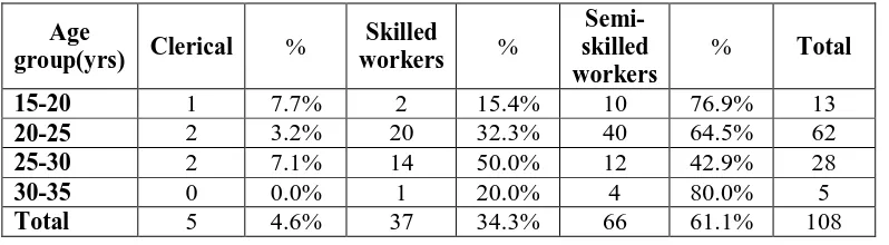
Table 2.Age distribution and socioeconomic status of study participants (n=108)
Age
group(yrs) Clerical %
Skilled
workers %
Semi-skilled workers
% Total 15-20 1 7.7% 2 15.4% 10 76.9% 13
20-25 2 3.2% 20 32.3% 40 64.5% 62
25-30 2 7.1% 14 50.0% 12 42.9% 28
30-35 0 0.0% 1 20.0% 4 80.0% 5
Total 5 4.6% 37 34.3% 66 61.1% 108
Among the study participants 61.1% were semi-skilled workers, 34.3% were
skilled workers and 4.6% clerical workers.
Table 3.Age distribution and socioeconomic status of study participants (n=108)
Age group(yrs)
Obstetric
formula 0 1 2 3 4 5 15-20
Gravida 0 12 1 0 0 0
Para 12 1 0 0 0 0
Live 12 1 0 0 0 0
Abortion 13 0 0 0 0 0
20-25
Gravida 0 44 12 2 3 1
Para 48 10 4 0 0 0
Live 50 8 4 0 0 0
Abortion 54 7 0 0 1 0
25-30
Gravida 0 11 12 4 1 0
Para 13 14 1 0 0 0
Live 15 12 1 0 0 0
Abortion 23 4 0 1 0 0
30-35
Gravida 0 2 1 1 0 1
Para 2 2 1 0 0 0
Live 2 3 0 0 0 0
[image:57.595.111.504.470.711.2]44
Figure 3 and Table 2 shows the distribution of parity among study
participants. Out of the total participants 69 presented with G1, 26 with G2, 7
with G3, 4 with G4, and 2 with G5. Para 1 , Live1, Abortion1 were 27,24 and 12
[image:58.595.132.499.243.468.2]respectively.
[image:58.595.164.451.525.673.2]Figure 3. Distribution of gravity among study participants
Table 4.Descriptive statistics of study participants profile
Item
Minimum Maximum Mean
Std. Deviation Age(yrs) 18 34 24.31 3.38
Height(cms) 150 170 158.29 5.52
Weight(kg) 49 75 62.90 7.78
Gravida 1 5 1.56 0.91
Para 0 2 0.36 0.59
Live 0 2 0.31 0.56
Abortion 0 4 0.19 0.59
Gestational
Age(weeks) 32 36 34.70 1.23
The mean age of study participants was 24.31 years (SD: 3.38) with 95%
45
of study participants was 34.7(SD: 1.23& 95% CI: 34.47-34.94). The mean
height was 158.29 cms, weight 62.90 kgs.
[image:59.595.139.504.222.445.2]Membrane Rupture:
Figure 4. Age group and pre-term PROM
Figure 4. Shows the distribution of gestational week and age group of study
participants. The maximum number of PPROM occured in the age group of
20-25 years with 17 pregnant mother with early PPROM (32-34weeks) and
45 with late PPROM (34-36weeks). In 15-20 years, 12 mothers presented
46
Figure 5. Pre-term PROM
The pie diagram in figure 5 shows among study participants of 80.6 %( 87) in
[image:60.595.158.456.498.675.2]late PPROM and 19.4 %( 21) in early PPROM.
Table 5.Details of Membrane Rupture outside
MRO Minimum Maximum Mean
Std. Deviation MRO at
admission(hrs) 1 77 14.91 13.828 Admission
Delivery
interval(hrs) 2 128 20.42 22.915
MRO-Delivery intervel(hrs)
4 135 35.32 20.718
Table 5. and Figure 6. Shows the membrane rupture duration at the time of
47
MRO to delivery. The mean MRO during admission was 14.91 hours,
admission to delivery interval 15.81 hours and MRO to delivery interval
[image:61.595.105.509.215.431.2]30.72 hours.
[image:61.595.108.508.489.569.2]Figure 6. Details of Membrane Rupture and delivery outcome
Table 6. Details of MRO at admission
Gestational
age/Duration <6hrs(%)
6-12hrs(%)
12-24hrs(%)
>24
hrs(%) Total(%) p value 32-34 8(38.10) 6(4.76) 1(4.76) 6(28.57) 21(100)
0.14
34-36 30(34.48) 20(28.74) 25(28.74) 12(13.79) 87(100)
Total 38(35.19) 26(24.07) 26(24.07) 18(16.67) 108(100)
Table 5 shows 35.19% of pregnant mothers presented with less than six hours
of MRO with 38.1% in 32-34 weeks and 34.48% in 34-36 weeks of gestation.
The admission after 24 hours was 16.67%. The MRO duration and the
48
Table 7. Details of admission to delivery duration and pre-term group
Preterm Group/Admis sion Delivery
interval <6hrs (%)
6-12hrs (%) 12-24hrs (%) 24-48hrs (%) >48hrs (%) Total
(%) p value 32-34 2(9.50) 2(9.50) 4(19.06) 9(42.90) 4(19.06) 21(100)
0.007
34-36 31(35.60) 23(26.40 9(10.30) 18(10.30) 6(6.90) 87(100)
Total 33(30.6) 25(23.1) 13(12.03) 27(25) 10(9.25) 108(100)
Totally 23.1% of MRO clients delivered in 6-12 hrs of admission and 9.5% in
32-34 weeks of gestation and 26.4% in 34-36 weeks. And 35.6% were
delivered within six hours of admission in 34-36 weeks and 9.5% in 32-34
weeks. Only 10.2% were delivered after 48 hours out of that 6.9% delivered
in 34-36 weeks and 23.8% in 32-34 weeks. The difference of delivery
duration between these two groups was statistically significant (p<0.007)
Table 8. Details of MRO to delivery
Preterm group/MRO-Delivery
group(hrs) <12 (%)
12-24 (%)
24-48
(%) >48 (%)
Total (%)
p value 32-34 1(4.76) 3(14.29) 7(33.33) 10(47.62) 21(100)
0.001
34-36 14(16.09) 32(36.78) 31(35.63) 10(11.49) 87(100)
Total 15(13.89) 35(32.41) 38(35.19) 20(18.52) 108(100)
In the gestational age group of 32-34 weeks, 4.76% deliveries were conducted
from MRO to delivery duration of <12 hrs. But 16.09% were conducted in
34-36 weeks. And 47.62% of total deliveries conducted in 32-34 weeks at
[image:62.595.118.498.471.569.2]49
significant difference between early and late pre-term PROM group
(p<0.001).
[image:63.595.119.485.214.446.2]Active and Expectant management:
Figure 7. Management protocol for PPROM patients
Among total study participants 50.9 %( 55) were given active management
and 49.1 %( 53) given expectant management for PPROM.
The height of 155-160 cms was high (44.00%) in active management while it
was high in 150-155 cms in case of active management group (39.62%). The
weight was high in 60-70kgs group both the management groups (44%). The
age group of 20-25 years was high in expectant management (53.23%) and
30-35 yrs group was high (80%) in active management while compare both
the group. But in both the groups in the age group of 20-25 was high in