Copyright © 2004, American Society for Microbiology. All Rights Reserved.
Impaired Processing and Presentation of Cytotoxic-T-Lymphocyte
(CTL) Epitopes Are Major Escape Mechanisms from CTL
Immune Pressure in Human Immunodeficiency Virus
Type 1 Infection
Yoshiyuki Yokomaku,
1Hideka Miura,
1Hiroko Tomiyama,
2Ai Kawana-Tachikawa,
3Masafumi Takiguchi,
2Asato Kojima,
4Yoshiyuki Nagai,
1† Aikichi Iwamoto,
3Zene Matsuda,
1and Koya Ariyoshi
1*
AIDS Research Center1and Department of Pathology,4National Institute of Infectious Diseases, and Department of Infectious
Diseases, Institute of Medical Science, University of Tokyo,3Tokyo, and Center for AIDS Research, Kumamoto University,
Kumamoto,2Japan
Received 21 April 2003/Accepted 7 October 2003
Investigating escape mechanisms of human immunodeficiency virus type 1 (HIV-1) from cytotoxic T lym-phocytes (CTLs) is essential for understanding the pathogenesis of HIV-1 infection and developing effective vaccines. To study the processing and presentation of known CTL epitopes, we prepared Epstein-Barr
virus-transformed B cells that endogenously express thegaggene of six field isolates by adopting anenv/nef-deletion
HIV-1 vector pseudotyped with vesicular stomatitis virus G protein and then tested them for the recognition by Gag epitope-specific CTL lines or clones. We observed that two field variants, SLFNTVAVL and SVYNTV
ATL, of an Aⴱ0201-restricted Gag CTL epitope SLYNTVATL, and three field variants, KYRLKHLVW,
QYRLKHIVW, and RYRLKHLVW, of an A24-restricted Gag CTL epitope KYKLKHIVW escaped from being killed by the CTL lines, despite the fact that they were recognized when the synthetic peptides corresponding to these variant sequences were exogenously loaded onto the target cells. Thus, their escape is likely due to the changes that occur during the processing and presentation of epitopes in the infected cells. Mutations responsible for this mode of escape were located within the epitope regions rather than the flanking regions, and such mutations did not influence the virus replication. The results suggest that the impaired antigen processing and presentation often occur in HIV-1 field isolates and thus are one of the major mechanisms that enable HIV-1 to escape from CTL recognition. We emphasize the importance of testing HIV-1 variants in an endogenous expression system.
Accumulated evidence has indicated a critical role of cyto-toxic T lymphocytes (CTLs) in controlling human immunode-ficiency virus (HIV) replication during acute and chronic in-fection (16). Eliciting HIV type 1 (HIV-1)-specific CTLs has been thought to be crucial for effective HIV/AIDS vaccines (15). However, despite the presence of CTLs, the majority of HIV-1-infected cases eventually progress to AIDS, probably as a consequence of the emergence of escape mutants from CTLs (8, 20). Among immunized monkeys, which developed strong cellular immune responses against HIV-1, eventual vaccine failure occurs by viral escape from CTLs (2). Thus, investigat-ing the mechanisms of CTL failure to control the virus is essential to understanding the pathogenesis of HIV-1 infection and to develop HIV/AIDS vaccines.
The high rate of HIV-1 replication in vivo indicates that HIV-1 has tremendous ability to mutate swiftly (9, 30) and to make a dynamic adaptation to host-immune environments (3, 14, 18, 21, 31). Several mutations have been described in CTL epitopes in HIV-1-infected individuals, which result in either a lack of
bind-ing to the MHC class I molecule or nonrecognition by T-cell receptor (TCR) (3, 8, 12, 20, 21). Consequently, the virus escapes from CTL recognition. There are other mutations that do not lead to either escape effects (12); very little is known about the influence of these mutations on CTL recognition. CTL antigens are processed and presented on the cell surface in a very complex manner. Peptides are cleaved from endogenously synthesized proteins by proteasome in the cytoplasm and transported into the endoplasmic reticulum by the transporter of antigen presentation. Amino-terminal extended peptides are trimmed to the right size of peptides by aminopeptidases, which exist in both the cytoplasm and the endoplasmic reticulum (23). These steps have various degrees of substrate sequence specificity (17). The generated pep-tides should have sufficient affinity to bind to a major histocom-patibility complex (MHC) class I molecule in the presence of various other peptides derived from host proteins and to maintain the stability of peptide-MHC complexes until they are presented on the cell surface (28). Thus, it is plausible that some amino acid substitutions in the epitope and its flanking regions have a signif-icant influence on antigen processing and presentation. In the present study, we hypothesized that such mutations often enable HIV-1 to escape from CTL recognition.
Conventionally, the intracellular HIV-1 antigen processing and presentation has been studied with recombinant vaccinia viruses expressing an HIV-1 gene (3, 4, 11, 20, 26). Several
* Corresponding author. Mailing address: AIDS Research Center, National Institute of Infectious Diseases, 4-7-1 Gakuen, Musashimu-rayama, Tokyo, 208-0011 Japan. Phone: 561-0771. Fax: 81-42-561-7746. E-mail: ariyoshi@nih.go.jp
†Present address: Toyama Institute of Health, Toyama, 939-0363 Japan.
1324
on November 8, 2019 by guest
http://jvi.asm.org/
studies have addressed this issue in the context of HIV-in-fected T cells (4, 29, 32, 33). Most studies, however, have only evaluated a single or a few laboratory-established strains. The CTL recognition of HIV-1 clinical isolates has been evaluated, in most cases, by exogenously applying synthetic variant pep-tides to the cell surface to replace MHC-bound peppep-tides (8, 12, 20, 21). Very little is known about how the antigenic products of HIV-1 clinical isolates are processed and presented in the infected cells. To address this issue, we prepared CTL target cells that endogenously express the gag gene derived from HIV-1 clinical isolates by adopting anenv/nef-deletion HIV-1-based vector pseudotyped with vesicular stomatitis virus pro-tein G (VSV-G) propro-teins. Here, we show evidence that HIV-1 escapes from CTL recognition often via the impairment of antigen processing and presentation.
MATERIALS AND METHODS
Subjects.Peripheral blood mononuclear cells (PBMC) were collected from five HIV-1-infected individuals from the HIV clinic affiliated with the Institute of Medical Science, University of Tokyo. Two individuals (IMS1 and IMS2) had no therapy; one individual (IMS6) was off drugs but had received treatment (zidovu-dine alone) 2 years prior to blood sampling; two individuals (IMS4 and IMS7) had received therapy (zidovudine-lamivudine-indinavir and stavudine-lamivu-dine-nelfinavir, respectively) but for less than 3 months. CD4 count, viral load, and HLA type of the recruited individuals are shown in Table 1. HLA class I typing was initially performed by serology. Subtyping of HLA-A2 was done by a PCR–sequence-specific primer method (Dynal Classic SSP HLA-A2; Dynal A.S., Oslo, Norway).
Isolation and cloning of full-lengthgag.Full-lengthgagwas amplified from
proviral DNA extracted from the PBMC by nested PCR withPfuDNA
poly-merase (Strategene, La Jolla, Calif.) and oligonucleotides specific for HIV-1 long terminal repeat (LTR) and reverse transcription (RT) regions. Four
oligonucle-otides were mixed as outer primers: the sense primers 1U5AS-S (5⬘-ACTCTG
GTADCTAGAGATCCCTCA-3⬘; the position in HXB2 being 578 to 601) and
TAR-2 (5⬘-TGAGCCTGGGAGCTCTCTGGCT-3⬘; 478-499) and the antisense
primers RT7A-A (5⬘-TATGTTGAYAGGTGTAGGTC-3⬘; 2485 to 2504) and
RT18A-A (5⬘-CTACYARTACTGTACCTATAG-3⬘; 2464 to 2484). Two
oligo-nucleotides were used as inner primers: the sense primer TPBS1-S (5⬘-AAAA
TCTCTAGCAGTGGCGCCCGAACAGG-3⬘; the position in HXB2 being 622
to 650) and the antisense inner primer PRO6A (5⬘-ACTGTATCATCTGCTCC
TGTRTCTAA-3⬘; 2322 to 2347). The thermocycling conditions were 95°C for
45 s, 50°C for 45 s, and 72°C for 210 s (30 cycles) and 72°C for 7 min for both primary and secondary PCR. The PCR products were purified by using spin columns (QIAquick PCR purification kit; Qiagen, Santa Clarita, Calif.) and cloned into PT7Blue3 vector by using a commercial cloning kit (Perfectly Blunt cloning kit; Novagen, Dedham, Mass.). Two to three clones for each individual were sequenced by an automated sequencer (ABI Prism 377 automated DNA sequencer; Perkin-Elmer, Norwalk, Conn.) with BigDye terminators (PE
Ap-plied Biosystems, Foster City, Calif.). The sequences ofgagclones that were used
in the present study are available under GenBank Accession numbers as follows: AB074049 (IMS1-28), AB074050 (IMS1-29), AB074052 (IMS2-5), AB074058 (IMS4-24), AB074061 (IMS6-34), and AB074064 (IMS7-11).
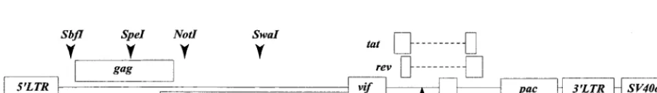
Construction of HIV-1 vector.The design of HIV-1 vector, pCTLpac, is shown in Fig. 1. The backbone of the vector is derived from an infectious molecular
clone, HXB2Ecogpt (22), which lacks the function ofvpr,vpu, andnefgenes. We
deleted a 1.5-kb portion from theenv-coding region but kept the function of Rev
responsive element, Tat, and Rev. Thenefgene was replaced with the puromycin
N-acetyltransferase (pac) gene (pPUR; BD Biosciences Clontech, Palo Alto,
Calif.) by usingXhoI and ClaI sites where theClaI site was introduced by
site-directed mutagenesis.SbfI andSwaI sites were introduced by site-directed
mutagenesis in the upstream of thegag(nucleotide 788) and in thepol
(nucle-otide 3717), respectively. The fragment fromSpeI in the gag (nucleotide 1507) to
theSwaIwas then replaced with that of a previously published vector, pHXB2cv
(25), which has aNotI site but lacks anSbfI site in thepolgene. Consequently,
the final construct carries the singleSbfI site (nucleotide 788) and theNotI site
(nucleotide 2275) that corresponds to the 10th codon of protease. These sites
were used for incorporating thegagclones derived from clinical isolates into the
pCTLpac vector. We confirmed that the expected variant sequences were in-serted in the vector by sequencing.
Generation of VSV-G pseudotype virus.Subconfluent COS7 cells in 25-cm2T
flasks (Becton Dickinson, Lincoln Park, N.J.) were cotransfected with 4g of
pCTLpac and 2g of pVSVG (BD Biosciences Clontech), which expresses
VSV-G protein, by lipofection (FuGENE6; Roche Molecular Biochemicals, Mannheim, Germany) and then incubated for 48 to 60 h. The supernatant, which contains pseudotype viruses carrying the HIV-1 vector with VSV-G envelope
proteins, was harvested, filtered through a 0.45-m (pore-size) Millex filter
(Millipore, Bedford, Mass.), and used as pseudotype virus stocks, some of which
were stored at⫺80°C before use. The amount of p24 antigen in the stocks was
measured by p24 antigen capture enzyme-linked immunosorbent assay (ELISA; RETRO-TEK; Zeptometrix Corp., Buffalo, N.Y.). The range of the p24 antigen yield was 40 to 100 ng/ml.
Preparation of target cells by using VSV-G pseudotyped HIV-1 vector. Ep-stein-Barr virus-transformed B-lymphocyte lines (B-LCLs) were infected with pseudotype virus stocks for 6 h at 37°C. The medium was then replaced with fresh RPMI 1640 (Sigma-Aldrich, St. Louis, Mo.) supplemented with 10% fetal bovine serum (R10; HyClone, Logan, Utah), and the cells were incubated for an
addi-tional 36 h. Subsequently, 0.5g of puromycin (BD Biosciences Clontech)/ml
was added to the R10 medium to select transduced cells. The culture was maintained until the number of transduced cells became sufficient for CTL
experiments. When 106B-LCLs were infected with 1 ml of pseudotype virus
stocks, the transduction efficiency was 20 to 30%. Usually, more than 107
trans-duced cells were generated within 2 weeks and used as CTL target cells. To standardize the expression level of Gag protein in target cells, we
quanti-fied the amount of extracellular p24 antigen that 106cells per ml of target cells
had produced in 24 h. The supernatant was harvested before (supernatant A) and after (supernatant B) the 24 h of culture for the measurement of p24 antigen by p24 antigen capture ELISA (Zeptometrix Corp.). The level of p24 antigen production was defined by the difference in the concentration of p24 antigen between supernatants A and B. If the target cells produced p24 antigen that was
⬎1 ng/ml in 24 h, they were used for CTL experiments, since the specific percent
[image:2.603.42.282.81.164.2]lysis did not significantly differ among target cells producing Gag protein above
FIG. 1. Structure of pCTLpac. A 1.5-kbp portion ofenvwas deleted (Œ). PuromycinN-acetyltransferase gene (pac) was inserted in thenef
[image:2.603.53.531.637.705.2]region. The locations of restriction enzyme sites are indicated (). RRE, Rev responsive element.
TABLE 1. Characteristics of five HIV-1-infected donorsa
Donor HLA type count/CD4l Virus load(copies/
ml)
No. of isolated
clones
A B Cw
IMS1 *0201/2402 52/75 3 286 ⬍400 3
IMS2 *0201/31 27/5101 2 797 ⬍400 2
IMS4 *0207/2402 46/52 1 448 ⬍400 2
IMS6 2402/26 7/5101 7 368 3.6⫻105 3
IMS7 1/⫺ 37/⫺ 6 544 1.3⫻103 3
aHLA alleles, CD4 count, viral load, and the number of isolated clones from
each donor’s sample are shown.
on November 8, 2019 by guest
http://jvi.asm.org/
this level (data not shown). We also investigated the level and pattern of protein
expression ofgagvariants by Western blot analysis, as previously described (25).
51Cr release experiments with HLA-class I-mismatched target cells in parallel
with HLA-class I matched target cells of different donors confirmed that these target cells were recognized by CTLs in an HLA-restricted manner (data not shown). Repeated experiments showed that specific lysis of blank controls was
equivalent to that of cells expressinggagvariants that are known to escape either
from TCR recognition or MHC binding. Some examples appear in the Results section below: specific lysis against IMS2-5 (Fig. 3a), IMS4-24 (Fig. 3c), IMS6-34 (Fig. 3e), and HXB2-wild (Fig. 5b). Thus, we regarded the blank control as a negative control.
Preparation of target cells by using recombinant vaccinia viruses. Recombi-nant vaccinia viruses used in the experiment shown in Fig. 3b were made as previously described (10). HLA-matched B-LCLs were infected with recombi-nant vaccinia viruses at a multiplicity of infection of 3:1 overnight before being
tested in a51Cr release assay.
Effector cells.Peptide-specific CTL lines were induced from PBMC of HIV-1-infected donors. Half of the PBMC were stimulated with phytohemagglutinin
(2g/ml) for 24 h and then pulsed with corresponding peptides at 100M for 1 h
and irradiated before being added to the other half of the PBMC. A total of 3⫻
105cells in each well of a 96-well U-bottom plate, with at least 10 wells for each
sample, were cultured in R10; 10% Lymphocult T (Biotest, Dreieich, Germany) was added to the medium on day 3 of culture. The CTL lines were maintained by adding fresh R10 medium containing 10% Lymphocult T every 3 to 4 days and splitting the well accordingly. Assays were performed on day 14 to 28 of culture.
Synthetic peptides.Peptides were manufactured at the Takara Shuzo Co., Ltd.
(Shiga, Japan). The purity of peptides was⬎99% as determined by high-pressure
liquid chromatography, and the identity of peptides was confirmed by matrix-assisted laser desorption ionization–mass spectrometry. Lyophilized peptides were dissolved in dimethyl sulfoxide and diluted in phosphate-buffered saline to make a stock concentration (2 mM). Further dilution was made in RPMI 1640 to
make working concentrations of 200M for the induction of CTLs and of 20M
for the preparation of target cells.
51Cr release assay.In 96-well U-bottom plates, target cells were divided into
aliquots at 5,000 per well. Effector cells were added to target cells at different
effector/target (E:T) ratios. The amount of51Cr release in the culture
superna-tants was quantified after 6 h of incubation, and the percent specific lysis was
determined by using the following formula: [(E⫺M)/(D⫺M)]⫻100, whereE
is the experimental51Cr release,Mis the51Cr released in the presence of culture
medium (which ranged between 15 and 25% of total release), andDis the total
51Cr released in the presence of 5% Triton X-100 detergent. The results were
regarded as positive when recognition of the HIV target was⬎10% above the
control. The SD50is the peptide concentration giving 50% of maximal specific
lysis of target cells pulsed with 10M synthetic peptide (28).
Replication kinetics assay.Subconfluent 293T cells in Falcon 25-cm2T flasks
(Becton Dickinson) were transfected by lipofection (Roche Molecular
Biochemi-cals), with 2g of HXB2cv replication-competent HIV-1 plasmids, in which
various mutations were introduced. After 60 h of culture, the supernatant was
harvested, filtered through a 0.45-m-pore-size filter, and used as mutant virus
stocks. Two million Jurkat cells or eight million H9 cells were infected with an equivalent of 40 ng of p24 antigen of mutant viruses in 2 ml of R10 for 1 h. Cells were washed three times with 10 ml of R10, resuspended with 5 ml of R10, and
cultured in a 12.5-cm2T flask at 37°C in 5% CO
2. Every 2 or 3 days, 1.5 ml of
supernatant was harvested and replaced with fresh R10. The concentration of p24 was measured by using a p24 ELISA kit (Zeptometrix Corp.).
This study was approved by the Ethics Committee of the University of Tokyo.
RESULTS
Full-length gag clones of field isolates. We used 6 of 13
full-lengthgagclones that were isolated from the five infected individuals (Fig. 2). All of the clones did not have any stop codons. In the present study, we focused on the processing and presentation of three CTL epitopes: the HLA-Aⴱ 0201-re-stricted epitope SLYNTVATL, the A24-re0201-re-stricted epitope KYKLKHIVW in p17 matrix protein (MA), and the HLA-Bⴱ5101-restricted epitope NANPDCKTI in p24 capsid protein (CA) (11, 26, 27). Amino acid sequences within the three epitope regions and the N- and C-terminal 15-amino-acid res-idues flanking each epitope were analyzed; the six clones were
selected to maximize the diversity of amino acid sequences in the epitopes and its flanking regions.
The Aⴱ0201-restricted epitope and its flanking regions were highly variable. However, we did not observe a previously rec-ognized variation in the flanking region, Arg (R) to Lys (K) at position 76 in our clones (4). In contrast, the Bⴱ5101-restricted epitope and its flanking regions were conserved except for clones IMS2-5 and IMS4-24. In the A24-restricted epitope and its flanking regions, variations were seen almost exclusively within the epitope region with two exceptions, a Lys (K)-to-Ser (S) mutation at position 26 (K26S) in clone IMS4-24 and an Arg (R)-to-Lys (K) at position 15 in clone IMS2-5. The Lys (K)-to-Arg (R) mutation at position 30 within the A24-re-stricted epitope was seen more frequently than any other se-quences; none of the 13 clones had the wild-type sequence of KYKLKHIVW. We incorporated the six gag clones into the HIV-1 vector withenvand nefdeleted, pCTLpac (Fig. 1), to make target cells expressinggaggenes of these filed isolates.
CTL recognition of target cells endogenously expressinggag
genes of clinical isolates. We generated Aⴱ0201-restricted
SLYNTVATL (wild type) epitope-specific oligoclonal CTL lines from one HIV-1-infected individual (IMS1) with Aⴱ0201 and used the lines to test the killing of the six different gag
clones expressed on Aⴱ0201-matched B-LCLs by a conven-tional 51Cr release assay. The Aⴱ0201-restricted CTLs
effi-ciently recognized target cells expressinggagclones IMS1-29, IMS1-28, and IMS6-34, which encode either wild type or the SLYNTIATL sequence in the CTL epitope region. In contrast, the same CTLs did not recognize cells expressing gagclones IMS2-5, IMS4-24, and IMS7-11, which encode SLYNLVATL, SLFNTVAVL, and SVYNTVATL, respectively, indicating that these clones escaped from Aⴱ0201-restricted CTL recog-nition (Fig. 3a).
CTL recognition of IMS1-29 and IMS6-34 was also tested with recombinant vaccinia viruses expressing the gaggene of these variants in parallel with the VSV-G-pseudotyped HIV-1 vectors. The HIV-1 vector method demonstrated the CTL killing as well or slightly better than the vaccinia method did (Fig. 3b).
We used three Bⴱ5101-restricted NANPDCKTI -specific CTL clones to test the CTL recognition of five representative
gagclones. The CTL clones recognized fourgagclones, which convey the wild-type Bⴱ5101-restricted epitope sequence; they also recognized IMS2-5 that had a substitution in the flanking region. None of the clones recognized the IMS4-24 clone, which had the variant sequence NSNPDCKNI in the epitope region (Fig. 3c).
A24-restricted KYKLKHIVW (wild type) specific-CTL lines did not recognize synthetic peptides of the most common se-quence, KYRLKHIVW (3R mutant type), and the other variant, RYRLKHIVW (Fig. 3d). These two variants were shown to bind to the Aⴱ2402 MHC class I molecule in a binding assay (data not shown). We screened eight A24-positive individuals for the pres-ence of CTL activities against the 3R mutant epitopes and found one individual who carried CTLs recognizing the 3R mutant peptide. A24-restricted 3R mutant-reactive CTL lines were in-duced from this A24-positive individual and used for the remain-ing experiments. The 3R mutant-reactive CTL lines recognized target cells expressing IMS1-29 and IMS4-24gagclones, both of which carry the 3R mutant sequence, but did not recognize any
on November 8, 2019 by guest
http://jvi.asm.org/
other target cells expressing different variants (Fig. 3e). Interest-ingly, IMS4-24 with Lys (K)-to-Ser (S) mutation at position 26 outside the epitope region was less well recognized than IMS1-29. We consistently observed this phenomenon in repeated experi-ments (data not shown).
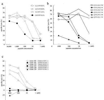
CTL recognition of exogenously loaded variant peptides.To
investigate whether the above findings of escape phenomenon from CTL killing were due to either loss of peptide binding to the MHC class I molecule or to the lack of TCR recognition, we prepared synthetic peptides that represented the variant epitopes and tested them for cross-recognition of the peptides in peptide titration assays by using the same CTL lines or clones that were used in experiments described for Fig. 3. To our surprise, Aⴱ0201-restricted CTL lines recognized the pep-tides of two Aⴱ0201-restricted CTL epitope variants, SVYNT VATL and SLFNTVAVL, which were not recognized by the CTLs when expressed endogenously. They recognized the SLFNTVAVL peptide less efficiently, with an SD50of⬎100
nM (Fig. 4a). Target cells pulsed with SLYNLVATL peptide representing clone IMS 2-5 were not cross-recognized even at a saturated concentration (10M) (data not shown).
We also obtained similar discordant results in experiments of A24-restricted CTL epitope variants. A24-restricted 3R mu-tant-specific CTL lines recognized peptides of three variant s—KYRLKHLVW, RYRLKHLVW, and QYRLKHIVW—
that were not recognized by the CTLs when they were ex-pressed endogenously. In fact, the CTLs recognized QYRLKHIVW peptide even better than the 3R mutant pep-tide but did not cross-recognize the QYKLKHIVW peppep-tide (Fig. 4b).
We tested one Bⴱ5101 variant peptide, NSNPDCKNI, in a peptide titration assay. This variant was not cross-recognized by any of the CTL clones even at a high concentration (1M) (Fig. 4c). The two amino acid mutations in this epitope coin-cided with two anchor residues to the MHC biding, suggesting that the lack of recognition of this variant was likely due to loss of peptide binding.
Mutations responsible for impairing the epitope processing
and presentation.The discrepancies seen above between the
[image:4.603.110.474.65.394.2]CTL recognition of endogenously expressed and exogenously loaded antigen indicate that some mutations have caused the impairment of epitope processing and presentation. To locate specific variations that were responsible for the poor recogni-tion of endogenously expressed HIV-1 gag variants, we con-structed four different target vectors: an HXB2gagsequence with Aⴱ0201-restricted epitope variations (SLFNTVAVL [HXB2-3F8V] or SVYNTVATL [HXB2-2V]) and IMS4-24-or IMS7-11-derivedgag sequence with the wild-type Aⴱ0201 epitope sequence (IMS4-24-wild or IMS7-11-wild, respec-tively). The replacement of the variant epitope region with the
FIG. 2. Sequence variation in three CTL epitopes and their flanking regions. The amino acid sequences of sixgagclones are shown. The reference sequence is derived from HXB2, and the differences are indicated. The numbering is done according to the HIV sequence database, Los Alamos National Laboratory, Los Alamos, N.Mex. The CTL epitope regions are boxed.
on November 8, 2019 by guest
http://jvi.asm.org/
FIG. 3. (a) Specific lysis of Aⴱ0201-matched B-LCLs (HLA-Aⴱ0201/⫺and HLA-Bⴱ5101/⫺) producing Gag proteins of clinical isolates. Peptide target cells were pulsed with the Aⴱ0201 wild-type peptide, SLYNTVATL (10M). Aⴱ0201-restricted SLYNTVATL -specific CTL lines were induced from a single donor (IMS1). The E:T ratio was 10:1. This experiment was repeated, with a different B-LCLs (HLA-Aⴱ0201/31 and HLA-B27/ⴱ5101), giving the same pattern of recognition (data not shown). (b) Specific lysis of Aⴱ0201-matched B-LCLs (HLA-Aⴱ0201/⫺and HLA-Bⴱ5101/⫺) expressinggagclones of two clinical isolates with the VSV-G-pseudotyped HIV-1 vector versus recombinant vaccinia viruses. Recombinant vaccinia virus expressing the human CD4 gene was used as a vaccinia virus control (1). The effector and peptide target cells were prepared as described for panel a. (c) Specific lysis of Bⴱ5101-matched B-LCLs (HLA-Aⴱ0201/⫺and HLA-Bⴱ5101/⫺) producing the Gag proteins of five clones. Three Bⴱ5101-restricted NANPDCKTI -specific CTL clones were used as effector cells at an E:T ratio of 2:1 (23). The peptide target was pulsed with the B51 wild-type peptide NANPDCKI (1M). (d) Specific lysis of A24-matched B-LCLs (HLA-A24/⫺and HLA-B46/52) pulsed with the peptides KYKLKHIVW, KYRLKHIVW, and RYRLKHIVW at 10M. A24-restricted, KYKLKHIVW -specific CTL lines were induced from one A24-positive donor. (e) Specific lysis of A24-matched B-LCLs (HLA-A24/⫺ and HLA-B46/52) producing variant Gag proteins. A24-restricted KYRLKHIVW (3R)-specific CTL lines were induced from another A24-positive donor. The peptide target was pulsed with 3R mutant type peptide (10M). The E:T ratio was 20:1. The lysis of target cells without any peptide pulsing is shown as a blank control.
on November 8, 2019 by guest
http://jvi.asm.org/
[image:5.603.100.485.80.581.2]wild-type epitope sequence restored CTL recognition of the escape variants, whereas replacement of the wild-type epitope with the two variant epitopes resulted in no CTL recognition of HXB2 Gag (Fig. 5a). The levels and patterns of Gag protein expression in target cells were analyzed by Western blot ex-periments (Fig. 5b). The expression levels of p55 Gag precur-sor and p24 CA did not significantly differ between the mutants and the wild type. The p17 MA band was not clear in HXB2-2V, IMS7-11-wild, and IMS4-24-wild, but the appearance of this band did not correlate with CTL killing. These results indicate that amino acid substitutions within the Aⴱ 0201-stricted epitope region, rather than those in the flanking re-gions, have caused the inhibition of CTL recognition in our endogenous expression system.
To further investigate the effect of amino acid substitutions within the A24-restricted epitope on antigen processing and pre-sentation, we introduced various point mutations into the wild-type HXB2 vector, pCTLpac, and tested them for the recognition by A24-restricted 3R mutant-reactive CTL lines. The
A24-re-stricted 3R mutant-specific CTLs did not cross-recognize the wild-type peptide and the wild-type HXB2 vector but did recog-nize HXB2 with a 3R mutation (HXB2-1R). The substitution of Lys (K) with Arg (R) at position 28 (HXB2-1R3R) did not affect the A24-restricted 3R mutant-specific CTL recognition, but a Lys (K)-to-Gln (Q) substitution at position 28 (HXB2-1Q3R) or an Ile (I)-to-Leu (L) substitution at position 34 (HXB2-3R7L) re-sulted in the escape from CTL killing (Fig. 5c).
Replication kinetics of HIV-1 mutant viruses.We analyzed
the replication kinetics of recombinant viruses carrying muta-tions that have affected the epitope processing and presenta-tion by infecting H9 or Jurkat cells. All mutants were found to replicate to equivalent levels, suggesting that these mutations do not have a significant influence on HIV-1 replication (Fig. 6).
DISCUSSION
[image:6.603.97.506.65.455.2]The present study focused on three Gag CTL epitopes re-stricted by three common HLA alleles in Japanese people (24).
FIG. 4. Peptide titration assays. (a) Specific lysis of Aⴱ0201-matched B-LCLs pulsed with Aⴱ0201 variant peptides by Aⴱ0201-restricted CTLs at an E:T ratio of 20:1. (b) Specific lysis of A24-matched B-LCLs pulsed with 3R and its variant peptides by A24-restricted 3R mutant reactive CTLs at an E:T ratio of 20:1. (c) Specific lysis of B51-matched B-LCLs pulsed with B51 variant peptides by B51-restricted CTL clones at an E:T ratio of 2:1. The same effector and target cells were used as for Fig. 3. The percent lysis of the blank control has been subtracted.
on November 8, 2019 by guest
http://jvi.asm.org/
The Gag protein is most commonly targeted by CTL-inducing HIV/AIDS vaccines (15). In our endogenous expression system, three Aⴱ0201-restricted epitope variants and one Bⴱ 5101-re-stricted epitope variant escaped from the wild-type CTL recog-nition, and four A24-restricted epitope variants escaped from the A24-restricted 3R mutant-reactive CTL recognition. Intriguingly, two Aⴱ0201-restricted variants and three A24-restricted variants escaped from CTL killing when thegagclones were expressed endogenously in the target cells by the HIV-1 vector, despite the fact that the synthetic variant peptides were well recognized by the CTLs when loaded onto the MHC class I molecule exog-enously. The peptide titration experiments have revealed that the strength of these variant peptides’ recognition was almost equiv-alent to that of the Aⴱ0201-restricted wild-type peptide or the
A24-restricted 3R mutant peptide. The results were not likely due to differences in the pattern of Gag protein expression, as shown in the Western blot experiments. All target cells were confirmed to express a sufficient level of Gag protein by p24 antigen pro-duction. Therefore, we believe that the escape mechanism of these variants resides in the antigen processing and presentation, as has been observed in a mouse model with murine leukemia virus infection (19). The observation of such phenomenon in two epitopes restricted by different alleles implies that this finding is not unique to a particular epitope-MHC pair.
Since all variants investigated here were derived from clin-ical samples and those mutations did not affect the virus rep-lication, our observations are relevant for discussing what may be going on in HIV-infected individuals. Our results indicate
FIG. 5. (a) Specific lysis of Aⴱ0201-matched B-LCLs (HLA-Aⴱ0201/⫺and HLA-Bⴱ5101/⫺) that endogenously express chimericgagclones bearing the variant CTL epitopes SLFNTVAVL and SVYNTVATL in the frame of HXB2gag(HXB2-3F8V and HXB2-2V, respectively) or bearing the wild-type epitope in the frame of IMS7-11 and IMS4-24 Gag (IMS7-11-wild and IMS4-24-wild, respectively). Aⴱ0201-restricted SLYNTVATL CTL lines were induced from the same donor as for Fig. 3. Specific lysis of target cells expressing HXB2, IMS7-11, or IMS4-24gag clones and being pulsed with the Aⴱ0201 wild-type peptide (10M) is shown in parallel. The E:T ratio was 20:1. (b) Levels and patterns of HIV-1 protein expression in target cells used in the experiments described for panel a. The Western blot was reacted with the serum from an HIV-1-infected individual. (c) Specific lysis of A24-matched B-LCLs (HLA A24/⫺and HLA-B46/52 or HLA-A24/26 and HLA-B51/52) that expressgagclones with various point mutations. Point mutations were inserted into the A24-restricted CTL epitope region in the frame of wild-type HXB2 Gag (HXB2-wild): amino acid substitutions of Lys to Arg at position 30 (HXB2-3R) with Lys to Arg at position 28 (HXB2-1R3R), Ile to Lue at position 34 (HXB2-3R7L), or Lys to Gln at position 28 (HXB2-1Q3R). Peptide target cells were pulsed with either the KYRLKHIVW (3R) or the RYRLKHIVW (1R3R) mutant peptide at 10M. The effector cells were A24-restricted 3R mutant-specific CTL lines from the same donor as in the Fig. 3e experiment. The E:T ratio was 20:1.
on November 8, 2019 by guest
http://jvi.asm.org/
[image:7.603.108.484.67.440.2]that the impaired antigen processing and presentation often occurs in HIV-1 field isolates and thus is one of the major mechanisms that enable HIV-1 to escape from the CTL rec-ognition. To understand further the significance of this escape mechanism, it is important to evaluate an accumulation of such escape variants in infected hosts in a longitudinal study or at a population level. A previous report using a vaccinia virus ex-pression system did not reveal that any mutations in the Aⴱ0201-restricted p17 epitope of HIV-1 and its flanking region altered the processing and presentation of its variant epitope (4). However, that study did not investigate Aⴱ0201-restricted
2V and 3F8V variants, which we found affected epitope pro-cessing and presentation.
Experiments with chimeric genes, as well as point mutations, showed that escapes from epitope processing and presentation were mostly attributable to mutations within the epitope re-gions rather than its flanking rere-gions. In the present study, we demonstrated that point mutations of Lys (L) to Gln (Q) at position 28 and of Ile to Leu at position 34 drastically impaired the processing and presentation of the A24-restricted CTL epitope. Moreover, the experiment with HXB2 clone carrying IMS 7-11 variant of Aⴱ0201-restricted CTL epitope indicates that a substitution of Leu (L) to Val (V) at position 78 was responsible for the impaired processing and presentation of the epitope. These mutations in the epitope region may have induced a proteasome cleavage site within the epitope (19). On the other hand, we observed that the variations in the 15 amino acids up- and downstream of the epitope did not affect CTL recognition. An exception was a Lys (L)-to-Ser (S) substitution (⫺2S) at position 26, which is only two amino acids adjacent to the N terminus of the A24-restricted epitope. However, this⫺2S substitution did not void the A24-restricted 3R mutant-reactive CTL recognition completely. One possible explanation is that the ⫺2S substitution shifted the optimal proteasome cleavage site, resulting in the generation of a larger peptide, which has a lower affinity to the MHC class I molecule.
We have first attempted to investigate the antigen process-ing and presentation by the conventional recombinant vaccinia virus method for all variants before we established this VSV-G-pseudotyped HIV-1 vector method. Soon, we realized that preparing recombinant vaccinia viruses was much more labo-rious and time-consuming. Early experiments of comparing two methods by using the first available recombinant vacinia viruses concluded that the HIV-1 vector method demonstrated CTL killing better than did the recombinant vaccinia virus method (Fig. 3b). In the recombinant vaccinia virus expression system, the massive production of vaccinia virus proteins inev-itably takes place, along with the expression of an HIV-1 gene and sometimes causes a high background lysis. The expression manner and the production ratio to non-HIV proteins may also influence antigen processing and presentation (27, 34). Thus, we thought that the antigen processing and presentation in the HIV-1 vector expression system is more physiological than the recombinant vaccinia virus expression system and that continuing vaccinia virus experiments would not be signifi-cantly beneficial to address the issue of antigen processing and presentation. Nevertheless, there remains a concern that there might be a potential difference in the antigen processing and presentation between immortalized B cells that were used here and primary CD4⫹T cells (32, 33). Perhaps it is important to
reevaluate the interaction of CTLs and these variants in ex-periments with variant HIV-1-infected T cells. Our HIV-1 vec-tor carries neither thenefgene nor thevpugene, which signif-icantly affect antigen presentation by downregulating MHC class I cell surface expression (5, 13). From this point of view, one might expect that more variants would escape from the CTL recognition in the actual HIV-1 infection than what is shown in our experiments. However, we think that our system is suited to identify a specific association between a certain mutation and the escape from antigen processing and presen-tation. To prove the existence of this mode of escape
mecha-FIG. 6. Replication of HIV-1 clones with mutations that impaired the processing and presentation of Aⴱ0201 or A24 CTL epitopes in H9 (a) and Jurkat (b) cells. The kinetics of each recombinant virus repli-cation were monitored as the production of p24 antigen by p24 ELISA. Symbols:E, wild-type;䊐, A24-3R;■, A24-K26S⫹3R;‚, A24-3R7L; Œ, A24-1Q3R;F, Aⴱ0201-3F8V;〫, Aⴱ0201-2V;⫻, mock.
on November 8, 2019 by guest
http://jvi.asm.org/
nism, we may need a new system that can directly detect a trace of specific epitopes that are eluted from MHC class I mole-cules of HIV-1 antigen-producing cells.
Although the structure analysis of MHC class I molecules and its binding motif has facilitated the prediction of CTL epitopes from the primary amino acid sequence data of HIV-1 (6, 11, 26), it remains difficult to envisage the efficiency of epitope processing and presentation. Enormous diversity real-ized in HIV-1 field isolates causes a further complexity (7). Our data emphasize the importance of testing HIV-1 variants in an endogenous expression system. Detailed analysis of epitope processing and presentation among HIV-1 field iso-lates, particularly of non-B subtypes circulating in the vaccine trial fields, is essential, since such information allows us to forecast which virus may elude the immunity elicited by vac-cines, thus providing a clue for a rational design for effective HIV/AIDS vaccines.
ACKNOWLEDGMENTS
We thank the donors in this study for their participation, Sachiko Tateishi for assistance, and Kunito Yoshiike for critical reading of the manuscript.
This study was supported by the Japanese Ministry of Health, Labor, and Welfare and the Japan Health Science Foundation.
REFERENCES
1. Aoki, N., T. Shioda, H. Satoh, and H. Shibuta.1991. Syncytium formation of human and non-human cells by recombinant vaccinia viruses carrying the
HIVenvgene and human CD4 gene. AIDS5:871–875.
2. Barouch, D. H., J. Kunstman, M. J. Kuroda, J. E. Schmitz, S. Santra, F. W. Peyerl, G. R. Krivulka, K. Beaudry, M. A. Lifton, D. A. Gorgone, D. C. Montefiori, M. G. Lewis, S. M. Wolinsky, and N. L. Letvin.2002. Eventual AIDS vaccine failure in a rhesus monkey by viral escape from cytotoxic T
lymphocytes. Nature415:335–339.
3. Borrow, P., H. Lewicki, X. Wei, M. S. Horwitz, N. Peffer, H. Meyers, J. A. Nelson, J. E. Gairin, B. H. Hahn, M. B. Oldstone, and G. M. Shaw.1997. Antiviral pressure exerted by HIV-1-specific cytotoxic T lymphocytes (CTLs) during primary infection demonstrated by rapid selection of CTL escape
virus. Nat. Med.3:205–211.
4. Brander, C., O. O. Yang, N. G. Jones, Y. Lee, P. Goulder, R. P. Johnson, A. Trocha, D. Colbert, C. Hay, S. Buchbinder, C. C. Bergmann, H. J. Zweerink, S. Wolinsky, W. A. Blattner, S. A. Kalams, and B. D. Walker.1999. Efficient
processing of the immunodominant, HLA-Aⴱ0201-restricted human
immu-nodeficiency virus type 1 cytotoxic-T-lymphocyte epitope despite multiple
variations in the epitope flanking sequences. J. Virol.73:10191–10198.
5. Collins, K. L., B. K. Chen, S. A. Kalams, B. D. Walker, and D. Baltimore.
1998. HIV-1 Nef protein protects infected primary cells against killing by
cytotoxic T lymphocytes. Nature391:397–401.
6. Falk, K., O. Rotzschke, S. Stevanovic, G. Jung, and H. G. Rammensee.1991. Allele-specific motifs revealed by sequencing of self-peptides eluted from
MHC molecules. Nature351:290–296.
7. Gaschen, B., J. Taylor, K. Yusim, B. Foley, F. Gao, D. Lang, V. Novitsky, B. Haynes, B. H. Hahn, T. Bhattacharya, and B. Korber.2002. Diversity
con-siderations in HIV-1 vaccine selection. Science296:2354–2360.
8. Goulder, P. J., R. E. Phillips, R. A. Colbert, S. McAdam, G. Ogg, M. A. Nowak, P. Giangrande, G. Luzzi, B. Morgan, A. Edwards, A. J. McMichael, and S. Rowland-Jones.1997. Late escape from an immunodominant cyto-toxic T-lymphocyte response associated with progression to AIDS. Nat. Med.
3:212–217.
9. Ho, D. D., A. U. Neumann, A. S. Perelson, W. Chen, J. M. Leonard, and M. Markowitz.1995. Rapid turnover of plasma virions and CD4 lymphocytes in
HIV-1 infection. Nature373:123–126.
10. Hoshikawa, N., A. Kojima, A. Yasuda, E. Takayashiki, S. Masuko, J. Chiba, T. Sata, and T. Kurata.1991. Role of the gag and pol genes of human immunodeficiency virus in the morphogenesis and maturation of retrovirus-like particles expressed by recombinant vaccinia virus: an ultrastructural
study. J. Gen. Virol.72:2509–2517.
11. Ikeda-Moore, Y., H. Tomiyama, M. Ibe, S. Oka, K. Miwa, Y. Kaneko, and M. Takiguchi.1998. Identification of a novel HLA-A24-restricted cytotoxic
T-lymphocyte epitope derived from HIV-1 Gag protein. AIDS12:2073–2074.
12. Kawana, A., H. Tomiyama, M. Takiguchi, T. Shioda, T. Nakamura, and A. Iwamoto.1999. Accumuation of specific amino acid substitutions in HLA-B35-restricted human immunodeficiency virus type 1 cytotoxic T lymphocyte
epitopes. AIDS Res. Hum. Retrovir.15:1099–1107.
13. Kerkau, T., I. Bacik, J. R. Bennink, J. W. Yewdell, T. Hunig, A. Schimpl, and U. Schubert.1997. The human immunodeficiency virus type 1 (HIV-1) Vpu protein interferes with an early step in the biosynthesis of major
histocom-patibility complex (MHC) class I molecules. J. Exp. Med.185:1295–1305.
14. Koenig, S., A. J. Conley, Y. A. Brewah, G. M. Jones, S. Leath, L. J. Boots, V. Davey, G. Pantaleo, J. F. Demarest, C. Carter, et al.1995. Transfer of HIV-1-specific cytotoxic T lymphocytes to an AIDS patient leads to selection for mutant HIV variants and subsequent disease progression. Nat. Med.
1:330–336.
15. McMichael, A. J., and T. Hanke.2003. HIV vaccines 1983–2003. Nat. Med.
9:874–880.
16. McMichael, A. J., and S. L. Rowland-Jones.2001. Cellular immune
re-sponses to HIV. Nature410:980–987.
17. Momburg, F., and G. J. Hammerling.1998. Generation and TAP-mediated transport of peptides for major histocompatibility complex class I molecules.
Adv. Immunol.68:191–256.
18. Moore, C. B., M. John, I. R. James, F. T. Christiansen, C. S. Witt, and S. A. Mallal.2002. Evidence of HIV-1 adaptation to HLA-restricted immune
responses at a population level. Science296:1439–1443.
19. Ossendorp, F., M. Eggers, A. Neisig, T. Ruppert, M. Groettrup, A. Sijts, E. Mengede, P. M. Kloetzel, J. Neefjes, U. Koszinowski, and C. Melief.1996. A single residue exchange within a viral CTL epitope alters
proteasome-medi-ated degradation resulting in lack of antigen presentation. Immunity5:115–
124.
20. Phillips, R. E., S. Rowland-Jones, D. F. Nixon, F. M. Gotch, J. P. Edwards, A. O. Ogunlesi, J. G. Elvin, J. A. Rothbard, C. R. Bangham, C. R. Rizza, et
al.1991. Human immunodeficiency virus genetic variation that can escape
cytotoxic T-cell recognition. Nature354:453–459.
21. Price, D. A., P. J. Goulder, P. Klenerman, A. K. Sewell, P. J. Easterbrook, M. Troop, C. R. Bangham, and R. E. Phillips.1997. Positive selection of HIV-1 cytotoxic T lymphocyte escape variants during primary infection. Proc. Natl.
Acad. Sci. USA94:1890–1895.
22. Ratner, L., A. Fisher, L. L. Jagodzinski, H. Mitsuya, R. S. Liou, R. C. Gallo, and F. Wong-Staal.1987. Complete nucleotide sequences of functional
clones of the AIDS virus. AIDS Res. Hum. Retrovir.3:57–69.
23. Rock, K. L., and A. L. Goldberg.1999. Degradation of cell proteins and the
generation of MHC class I-presented peptides. Annu. Rev. Immunol.17:
739–779.
24. Saito, S., S. Ota, E. Yamada, H. Inoko, and M. Ota.2000. Allele frequencyes and haplotypic associations defined by allelic DNA typing at HLA class I and
class II loci in the Japanese population. Tissue Antigens56:522–529.
25. Sugiura, W., Z. Matsuda, Y. Yokomaku, K. Hertogs, B. Larder, T. Oishi, A. Okano, T. Shiino, M. Tatsumi, M. Matsuda, H. Abumi, N. Takata, S. Shira-hata, K. Yamada, H. Yoshikura, and Y. Nagai.2002. Interference between D30N and L90M in selection and development of protease inhibitor-resis-tant human immunodeficiency virus type 1. Antimicrob. Agents Chemother.
46:708–715.
26. Tomiyama, H., T. Sakaguchi, K. Miwa, S. Oka, A. Iwamoto, Y. Kaneko, and M. Takiguchi.1999. Identification of multiple HIV-1 CTL epitopes
pre-sented by HLA-Bⴱ5101 molecules. Hum. Immunol.60:177–186.
27. Tsomides, T. J., A. Aldovini, R. P. Johnson, B. D. Walker, R. A. Young, and H. N. Eisen.1994. Naturally processed viral peptides recognized by cytotoxic T lymphocytes on cells chronically infected by human immunodeficiency
virus type 1. J. Exp. Med.180:1283–1293.
28. Tsomides, T. J., B. D. Walker, and H. N. Eisen.1991. An optimal viral
peptide recognized by CD8⫹T cells binds very tightly to the restricting class
I major histocompatibility complex protein on intact cells but not to the
purified class I protein. Proc. Natl. Acad. Sci. USA88:11276–11280.
29. Van Baalen, C. A., M. Schutten, R. C. Huisman, P. H. Boers, R. A. Gruters, and A. D. Osterhaus.1998. Kinetics of antiviral activity by human immuno-deficiency virus type 1-specific cytotoxic T lymphocytes (CTL) and rapid
selection of CTL escape virus in vitro. J. Virol.72:6851–6857.
30. Wei, X., S. K. Ghosh, M. E. Taylor, V. A. Johnson, E. A. Emini, P. Deutsch, J. D. Lifson, S. Bonhoeffer, M. A. Nowak, B. H. Hahn, et al.1995. Viral
dynamics in human immunodeficiency virus type 1 infection. Nature373:
117–122.
31. Wei, X., J. M. Decker, S. Wang, H Hui, J. C. Kappes, X. Wu, J. F. Salazar-Gonzalez, M. G. Salazar, J. M. Kilby, M. S. Saag, N. L. Komarova, M. A. Nowak, B. H. Hahn, P. D. Kwong, and G. M. Shaw.2003. Antibody
neutral-ization and escape by HIV-1. Nature422:307–312.
32. Yang, O. O., S. A. Kalams, M. Rosenzweig, A. Trocha, N. Jones, M. Koziel, B. D. Walker, and R. P. Johnson.1996. Efficient lysis of human immunode-ficiency virus type 1-infected cells by cytotoxic T lymphocytes. J. Virol.
70:5799–5806.
33. Yang, O. O., S. A. Kalams, A. Trocha, H. Cao, A. Luster, R. P. Johnson, and B. D. Walker.1997. Suppression of human immunodeficiency virus type 1
replication by CD8⫹cells: evidence for HLA class I-restricted triggering of
cytolytic and noncytolytic mechanisms. J. Virol.71:3120–3128.
34. Yewdell, J. W., and J. R. Bennink.1999. Immunodominance in major histo-compatibility complex class I-restricted T lymphocyte responses. Annu. Rev.
Immunol.17:51–88.