Synergy testing between sulbactam and meropenem/ colistin in
MDR Acinetobacter baumannii-calcoaceticus complex isolated
from ventilator associated pneumonia- a pilot study
Dissertation submitted as part of fulfilment for the M.D.
(Branch-IV Microbiology) Degree examination of the Tamil Nadu
CERTIFICATE
This is to certify that the dissertation entitled, “Synergy testing between sulbactam
and meropenem/ colistin in MDR Acinetobacter baumannii-calcoaceticus complex isolated from ventilator associated pneumonia- a pilot study” is the bonafide work of Dr. Lydia Jennifer. S toward the M.D (Branch – IV Microbiology) Degree
examination of the Tamil Nadu Dr. M. G. R . Medical University, to be conducted in
April-2016.
Dr. Shalini Anandan Dr. V. Balaji
Guide Professor and Head
Associate Professor Department of Clinical Microbiology
Department of Clinical Microbiology Christian Medical College
Christian Medical College Vellore - 632004
Vellore – 632004
Principal
Christian Medical College
DECLARATION
I hereby declare that this M.D Dissertation entitled “Synergy testing between
sulbactam and meropenem/ colistin in MDR Acinetobacter baumannii-calcoaceticus
complex isolated from ventilator associated pneumonia- a pilot study” is the bonafide
work done by me under the guidance of Dr. Shalini Anandan, Associate Professor,
Department of Clinical Microbiology, Christian Medical College, Vellore. This work
has not been submitted to any other university in part or full.
Dr. Lydia Jennifer. S
Department of Clinical Microbiology
Christian Medical College
Vellore.
Acknowledgement
I profoundly thank my guide Dr Shalini Anandan, Associate Professor from the
Department of Clinical Microbiology, who mentored me with all aspects of the study
with exceptional amount of patience, perseverance, consideration and composure.
I am grateful to Dr. V. Balaji, professor and Head of Department of Clinical
Microbiology, for his immense motivation, and also for allowing me to do the study
in the Department of Clinical Microbiology.
I would like to thank Dr. Shakti Laishram, Assistant Professor from the Department
of Clinical Microbiology, for standardizing the procedures and protocol of the study.
Being passionate about the subject, she was a constant stream of inspiration for me.
Much obliged to Dr. J. V. Peter from the Department of Medical ICU, Dr.
Subramani, Dr. Shoma V Rao and Dr Vasudha Bhaskar Rao from the Department of
Surgery ICU for providing the clinical details of the patients.
I thank associate research officers, Ms. S. Baby Abirami, Ms. P. Agila Kumari,
Ms. M. Elakkiya, Ms. B. Priyanka, Mr. R. Raji, Ms. G. Priya, Ms. T. Bhuvaneshwari,
Ms. R. Venda, Mrs. R. Dhanabagayam, Mrs. K. Durga, Ms. I. Julie Selvi,
Mrs. Y. Violet Rani, Mrs. Divya Kumaran, and Mrs. Dhivya from the Department of
Clinical Microbiology, for helping me out with the procedures, compilation of data
and analysis.
I also would like to acknowledge Dr. Jeyaseelan L for helping me with the statistical
I am indebted to my dear parents, beloved sister, friends and seniors for being a
ceaseless source of care, concern and encouragement.
I thank the Institutional Review Board for funding the study.
Above all I thank almighty God for filling me with His grace, wisdom, resilience and
1 Contents
1. Introduction ... 2
2. Aim and objectives ... 7
3. Review of literature ... 8
4. Materials and methods ... 49
5. Results ... 72
6. Discussion ... 88
7. Limitation of the study ... 92
8. Conclusion and recommendation ... 93
9. Bibliography ... 94
2 1. Introduction
A.baumannii- calcoaceticus complexhas surfaced as a major nosocomial pathogen. This
has been made possible by the environmental adaptability of these organisms and the
development of resistance to most drugs (1)
Among the nosocomial infections, these organisms are a major cause of blood stream
infections and ventilators associated pneumonia (VAP) and are affiliated with increased
mortality and morbidity.
As reported from reviews dating back to the 1970s (2), hospital acquired pneumonia is
still the most common infection caused by this organism. In majority of the institutions,
greater numbers of A. baumannii- calcoaceticus complexisolates are from the respiratory
tracts of hospitalized patients. Community-acquired pneumonia due to A. baumannii-
calcoaceticus complexhas been elucidated in tropical zone of Australia and Asia (3, 4,
5). It is distinguished by a fulminant clinical course, secondary bloodstream infection,
and a high mortality rate of 40 to 60%. In a study conducted on nosocomial bloodstream
infection in Brazil (June 2007- March 2010), Acinetobacter species accounted for 11.4%,
out of which, 55.9% of Acinetobacter species were resistant to imipenem. The crude
mortality rate in ICU patients and patients with monomicrobial blood stream infections
were 65.5% and 52.1% respectively (6). In another study, crude mortality from A.
baumannii- calcoaceticus complexbloodstream infection was 34.0% to 43.4% in the ICU
3
calcoaceticus complex, had the third highest crude mortality rate in the ICU, exceeded
by P. aeruginosa and Candida sp. infections (7). Acinetobacter sp. accounts for VAP as
high as 30.4% of ventilator associated pneumonia (8). A. baumannii - calcoaceticus
complexis infrequently associated with cause skin/soft tissue infections, UTI, meningitis
and endocarditis.
The antibiotics that are used to treat infections caused by A. baumannii are carbapenems,
polymyxins E and B, sulbactam, piperacillin/tazobactam, tigecycline and
aminoglycosides. Due to the development of resistance to the above mentioned
antibiotics; carbapenems (imipenem, meropenem, doripenem) have become a cornerstone
in the treatment for A. baumannii- calcoaceticus complex (9). Resistance to these
carbapenem has been mediated by the production of carbapenemases, expression of
efflux pumps and porin channel loss.
Rate of carbapenem resistance is as high as 74 to 90% resistance among hospital
acquired infections and 94.3 % resistance in ventilator associated pneumonia (8).
Combination antibiotic therapy is an approach frequently used in the treatment of such
MDR strains. This strategy strives to achieve synergy, especially against MDR strains.
To defend this clinical practice, in vitro combinations of antibiotics have long been
examined using checkerboard methods, to-kill assays, and E-tests (10) . The
time-kill assay identifies synergy at a much considerable frequency when compared to the
other tests described. Moreover, it also measures the bactericidal activity of the drug
4
bactericidal activity (primary end point), which is a more significant and definite
pharmacological indicator than synergy (secondary end point) (11). Even though reliable
results are obtained by the checkerboard assay, it needs to be supplemented by time kill
assay for confirmation (12).
Need for in-vitro techniques for measuring antibiotic synergism
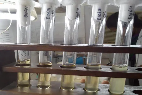
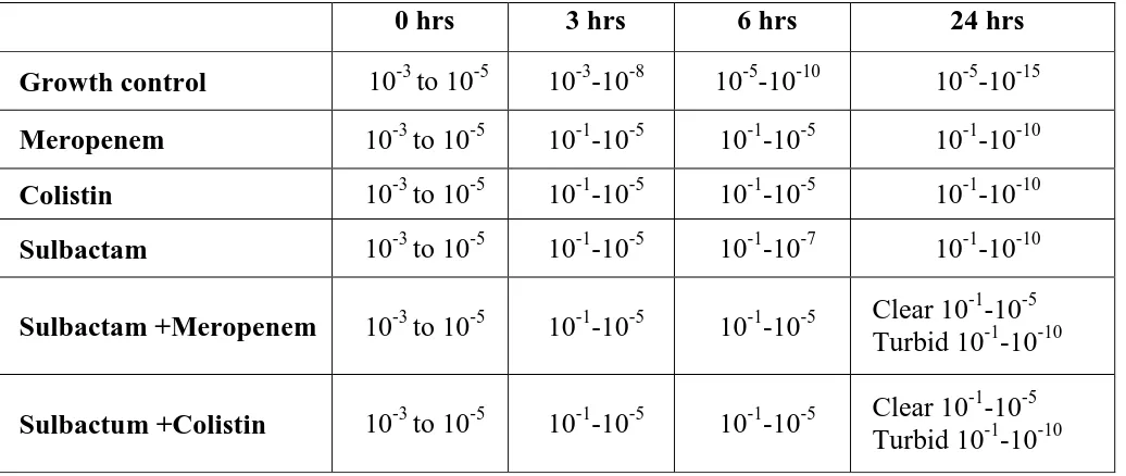
Time kill assay can be used to study the dynamics of synergism or antagonism for a
combination of antimicrobial agents by determining the number of viable bacteria
remaining over time after exposure to each individual agent and various combinations.
Time kill assay data can assess both the rate and extent of killing. Synergism in time kill
assay is usually interpreted as a ≥2-log10 CFU/ml decrease with the combination
compared with the most active single agent. Antagonism is usually described as a
≥2-log10 CFU/ml increase with the combination compared to the most active single agent
(13).
In this proposed research scheme we will be determining baseline MICs for sulbactam
tested against Acinetobacter baumannii- calcoaceticus complexby broth micro
dilution. Of the β-lactamase inhibitors, sulbactam due to its intrinsic activity, is
considered as most efficacious and most investigated agent in the context of
Acinetobacter infection. Sulbactam resembles aminopenicillins pharmacologically. It
5
unavailability of pure sulbactam in many countries, it has been formulated as
ampicillin/sulbactam (2 : 1 ratio), although the two agents are not synergetic. (14). Sulbactam, alone or in combination, shows significant activity against A. baumannii-
calcoaceticus complex(15). Most data on sulbactam therapy in humans come from
retrospective analyses or case series. Cure rates of 80–90% have been reported by
several authors in both bacteraemic and non-bacteraemic patients (13, 16, 17). Studies
assessing the activity of sulbactam alone compared to its combination with a β-lactam
clearly demonstrate the intrinsic activity of the agent rather than its ability to inhibit
β-lactamase enzyme(13,14, 17).
Recent observations have shown that combinations of sulbactam with aminoglycosides,
rifampicin, and azithromycin have demonstrated synergy against imipenem- susceptible
strains (19, 20).
Few studies have shown variable synergism between sulbactam and meropenem and
other drugs in in-vitro studies using different methodologies. All these studies have been
done on a small number of isolates, and in particular time-kill assays have not been used
in large scale to determine synergy, while it has been used only to confirm the synergism
seen in other assay like the checkerboard assay (21,22). Another combination being
studied is between carbapenem and colistin/polymyxin B for treatment of A. baumannii -
calcoaceticus complex (23).
In the present study we aim to determine synergism using the reference method of
time-kill assay between sulbactam and meropenem/colistin for Acinetobacter baumannii-
6
assay, which in turn will help in understanding the effective drug combinations and
7 2. Aim and objectives
Aim of the study
Synergy testing between sulbactam and meropenem/colistin in
MDR Acinetobacter baumannii-calcoaceticus complex isolated from ventilator associated
pneumonia- a pilot study
Hypothesis
Isolates of MDR Acinetobacter baumannii-calcoaceticus complex isolated from
ventilator associated pneumonia are susceptible to combination antimicrobials like
sulbactam –meropenem and sulbactam –colistin
Objectives
a) To find susceptibility profile for the isolates of Acinetobacter baumannii-calcoaceticus
complex resistant to meropenem
b) To determine the minimum inhibitory concentration (MIC) of meropenem, sulbactam and colistin by broth micro dilution technique
c) To determine if synergy is seen between sulbactam and meropenem/ colistin for these isolates by using Time Kill assay at a sub-inhibitory concentration and checkerboard
assay
8 3. Review of literature
Intricate history of the Genus Acinetobacter
Historically, rediscoveries of this omnipresent organism has led to the creation of
diverse genera, which in turn gave rise to waves of taxonomic confusion. It was in the
early 20th century, 1911, a Dutch microbiologist, Beijerinck reported an ubiquitous
microorganism and named it as Micrococcus calcoaceticus. This soil organism was
isolated by cultivating in calcium acetate- containing minimal medium (24). As the years
went by, organism resembling Micrococcus calcoaceticus were elucidated. These were
designated to atleast 15 diverse genera and species, along with Diplococcus mucosus,
Micrococcus calcoaceticus, Alcaligenes haemolysans, Mima polymorpha, Moraxella
lwoffi, Herellea vaginicola, Bacterium anitratum, Moraxella lwoffi var. glucidolytica,
Neisseria winogradskyi, Achromobacter anitratus, and Achromobacter mucosus (24).
Using nutritional properties, Brisou and Prévot in 1954 named the genus Acinetobacter.
They divaricated non-motile from motile organism within the genus Achromobacter (24).
Based on nutritional properties Baumann et al. established that various species described
above belonged to a single genus Acinetobacter. In addition to this, they concluded that
sub-classification into various species was not possible based on phenotypic
characteristics (24).
Based on the above studies the Sub-committee on the Taxonomy of Moraxella and Allied
Bacteria in 1971 evidently accepted the genus Acinetobacter (24). The genus
9
Acinetobacter calcoaceticus was documented in the 1974 edition of Bergey’s Manual of
Systematic Bacteriology (24).The species Acinetobacter calcoaceticus was in-turn
subdivided into two subspecies, subsp. anitratus (previously known as Herellea
vaginicola) and subsp. lwoffii ( previously known as Mima polymorpha), however this
proposal was not approved by taxonomists. It was only in the recent years that an
accepted taxonomic proposal has materialized. Yet, separation of various species within
the genus is still being explored.
Current taxonomy
Gram-negative, strictly aerobic, non-fermenting, non-fastidious, non-motile,
catalase-positive, oxidase-negative, cocco-bacilli with a DNA guanine-cytosine (G- C) content of
39% to 47% are incorporated into the genus Acinetobacter. Based on 16S rRNA studies
and rRNA-DNA hybridization assay a proposal was put forth, that, members of the genus
should be classified in the family Moraxellaceae within the order Gammaproteobacteria.
This family includes Moraxella, Acinetobacter, Psychrobacter, and related organisms
(24). Several studies were performed using DNA-DNA hybridization assay to describe
the genomic species. Of these studies, notable research were done by Bouvet and
Grimont in 1986, Nishimura et al. in 1987, Tjernberg and Ursing in 1989 (25). Currently
there are 27 named species and nine genomic species in the genus Acinetobacter (26). A.
baumannii-calcoaceticus complex is composed of four species,namely, A. calcoaceticus
10
A. baumannii (genomic species 2), Acinetobacter pittii (previously named as
Acinetobacter genomic species 3), and Acinetobacter nosocomialis (formerly named as
Acinetobacter genomic species 13 TU). It is difficult to differentiate between the
individual species, as they possess similar phenotypic characteristics; hence they are
referred to as A. baumannii-calcoaceticus complex (27). A.calcoaceticus is an
environmental species isolated from soil, rarely involved in causing clinical disease,
however other species in the A. baumannii-calcoaceticus complex have been
incriminated in causing community –acquired and hospital acquired infections (27).
Scientific classification of A. baumannii-calcoaceticus complex
Domain: Bacteria
Kingdom: Eubacteria
Phylum: Proteobacteria
Class: Gammaproteobacteria
Order: Pseudomonadales
Family: Moraxellaceae
Genus: Acinetobacter
Species: Acinetobacter baumannii- calcoaceticus complex
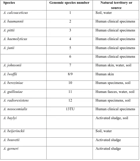
Species : Table.3.1 summarizes various species and genomics species in the genus
Acinetobacter along with their habitat and source from which they were commonly
11 Table.3.1 Various species of genus Acinetobacter along with their habitat .Adapted from Visca et al. 2011(28)
Species Genomic species number Natural territory or source
A. calcoaceticus 1 Soil, water
A. baumannii 2 Human clinical specimens
A. pittii 3 Human clinical specimens
A. haemolyticus 4 Human clinical specimens
A. junii 5 Human clinical specimens
6 Human clinical specimens
A. johnsonii 7 Human skin, water, soil
A. lwoffii 8/9 Human skin
A. bereziniae 10 Human specimens, soil
A. guillouiae 11 Human faeces, water, soil
A. radioresistens 12 Human specimens, soil
A. nosocomialis 13TU Human clinical specimens
A. baylyi Activated sludge, soil
A. beijerinckii Soil, water
A. bouvetii Activated sludge
12 Species Genomic species number Natural territory or
source
A. grimontii Activated sludge
A. gyllenbergii Human specimens
A. parvus Humans and animals
A. schindleri Human specimens
A. soli Soil
A. tandoii Activated sludge, soil
A. tjernbergiae Activated sludge
A. towneri Activated sludge
A. ursingii Human specimens
A. venetianus Marine water
13BJ, 14TU Human specimens
14BJ Human specimens
15 BJ Human specimens
15TU Human specimens
16 Human specimens
17 Human specimens, soil
Between 1 and 3 Human clinical specimens
13
Transformation assay of Juni is a method frequently used to identify Acinetobacter to the
genus level. This method exploits the ability of the transformable tryptophan auxotroph,
mutant A. baylyi strain BD413 trpE27, to be transformed to a wild type phenotype by
crude DNA of any Acinetobacter species (27). For the identification of human skin-
derived Acinetobacter up-to species level, 28 available phenotypic tests have been
reliable to an extent of 95.6% (27). Yet, these phenotypic tests do not identify the
recently discovered genomic strains of Acinetobacter.
Following molecular diagnostic methods have been used for the identification of
Acinetobacter to the species level to differentiate their ecology, epidemiology, and
pathogenicity (27, 28):
Amplified 16S rRNA gene restriction analysis (ARDRA)
High- resolution fingerprint analysis by amplified fragment length
polymorphism (AFLP)
Ribotyping
tRNA spacer fingerprinting
Restriction analysis of the 16S- 23S rRNA intergenic spacer sequence
Sequence analysis of the 16S- 23S rRNA gene spacer region
Sequencing of the rpoB (RNA polymerase beta-subunit) gene and its flanking
spacers (29)
14
Detection of a gene called bla OXA-51 – like carbapenemase (which is intrinsic to this
species), PCR- electrospray ionization mass spectrometry, a novel multiple-locus variable
number tandem-repeat analysis (MLVA) assay are the molecular methods used for
identification of A. baumannii (24) .
To differentiate between A. baumannii and Acinetobacter genomic species 13TU based
on their individual characteristic gryB gene, a PCR method has been elucidated by
Higgins et al. (24, 25). Currently these molecular methods are performed only in
specialized reference laboratory.
Manual and semi-automated commercial identification systems, like API 20NE, Vitek 2,
Phoenix, and MicroScan WalkAway systems are available for Acinetobacter species
identification. However, the specificity and reproducibility of these systems for species
identification are questioned due to their restricted database content (24).
Clinically significant speciesof the genus Acinetobacter
It has been noticed that A.baumannii is the most important genomic species accountable
for community and hospital acquired infections. Acinetobacter baumannii-calcoaceticus
complex are incriminated in 80% of all the clinical infections caused by Acinetobacter
species (30).
In a study that was conducted for over a period of twelve months, strains of
Acinetobacter were collected from 12 different hospitals. It was observed that, out of 584
15
72.9%. Of these A. baumannii isolates, majority of the isolates were from respiratory
tract specimens (42.9%), 19% from blood cultures, 12.8% from central venous lines, and
15.5% isolates from wound swabs. Of the rest 158 samples, Acinetobacter genomic
species 3 accounted for 9.4%, 4.9% of A, johnsonii, 3.5% of A. lwoffii, 1.88% of A. junii,
1.5% of A. haemolyticus, 1.5% of genomic species 10, 1.35% of genomic species 11,
12,and 6. About 2.7% of isolate could not be identified (31).
One of the Acinetobacter species, A. lwoffii, usually found as commensal of human skin,
oropharynx, and perineum was associated with bacteremia in immune-compromised
catheterized patients (32) . Even though this catheter-related bacteremia was caused by
multidrug resistant A. lwoffii, with the removal of the catheter, patients responded to
antibiotic therapy. In immune-compromised patients, A. lwoffii is a low risk bacterium
when compared to high mortality rate associated A. baumannii.
A normal commensal of the skin, A. johnsonii (genomic species 7) has also been
documented in causingcatheter-related bloodstream infections. These infections had
benign clinical course, and had an effective response to therapy (33).
A. pittii (formerly known as Acinetobacter genomic species 3) and A. nosocomialis
(formerly known as Acinetobacter genomic species 13TU) have also been noted to cause
hospital acquired infections (25).
A. junii has been known as an unusual cause of disease in humans. Yet few case reports
16
patient with underlying malignancy, bacteremia in preterm infants and pediatric
oncologic patients have been reported (34, 35) . During an outbreak of infection due to
A. junii , aerators and heparin solution served as reservoir of the organism reinforcing the
fact that these organisms are contaminants from environment and lapses in infection
control practices results in hospital acquired infections (36). Another study conducted in
Taiwan showed that A. junii was associated with hospital-acquired and healthcare
facility-related infections with 55.8 % of patients having impaired immunity (37).
Though several Acinetobacter species have been isolated from various clinical samples,
the clinical importance is still debatable, as these are usually viewed as contaminants
from the environment. Analysis of the cause of infection is based on repeated isolation of
the same strain from the similar source with the same antibiogram pattern.
Epidemiology
Members of the genus Acinetobacterare considered to be found everywhere in the
environment and these organisms can be isolated from all soil and surface water samples
in enrichment culture. There exists a common misunderstanding that A. baumannii are
also ubiquitous in nature (24). Yet, little knowledge exists about the natural habitat of
organisms belonging to the genus Acinetobacter.
―Can the presence of pathogenic organism in animals, inanimate objects, environment or
human skin be a source of infection to the patients belonging to a high risk group?‖ is a
17
Colonization of human skin, pharynx and gastrointestinal tract by
A. baumannii
In reality, few members of the genus Acinetobacter do not have environment as their
natural habitat. In a study that was conducted in Europe to understand the distribution of
Acinetobacter species on human skin, approximately 43% of healthy volunteers and 75%
of patients were colonized with Acinetobacter species. Acinetobacter lwoffii (47%) was
the most commonly isolated species, followed by A. johnsonii (21%), A. radioresistens
(12%), and DNA group 3 (11%). Augmentation in the colonization rates were observed
during the hospital stay of the patients (38). In a similar epidemiological survey based
study, Acinetobacter species were found in 40% healthy individuals, with Acinetobacter
lwoffii being the most common (61%), followed by genospecies 15BJ (12.5%) and 8% of
Acinetobacter radioresistens. It was also observed that the colonization rates were
maximum in forearm (51%), followed by forehead (47%) and toe web (34%) (39). In
both the studies, Acinetobacter baumannii that predominates in hospital-acquired
infections were found to be in a meager percentage of 0.5 -1 on the human skin.
On analyzing the various infections that were notified to Nosocomial Infections
Surveillance System, it was found that seasonal variation existed in the infections caused
by A. baumannii, with the infection rate being higher in late summer months compared
to the lower rates in early winter months (40) . In a study conducted in Hong Kong, a
seasonal difference in the skin colonization by Acinetobacter specieswas observed. In
18
and new nurses. However, during the winter season colonization rates dropped to 32%
(41). Of the Acinetobacter species, thegenomic species 3 (A. pittii) (36%) was the most
commonly isolated species, succeeded by Acinetobacter genomic species 13TU (15%),
Acinetobacter genomic species 15TU (6%). The nosocomial pathogen A. baumannii
accounted for only 4% (41).
Climatic conditions related variation in skin colonization may have an impact on the
seasonal discrepancy of infections by A. baumannii. Various evidence have pointed
towards the association between infection by Gram negative bacteria and atmospheric
temperature. For every 10° F hike in the temperature, monthly rates of infections caused
by A. baumannii increased to 17% (42). It has been postulated that increase in
temperature may boost the growth of bacteria in the environment, which can, in turn,
augment the colonization of humans. Another reason could be due to increase in the
virulence of Gram negative bacteria with the elevation of environmental temperature,
brought about by regulation of lipid A moiety of lipopolysaccharide (43).
To summarize, the skin colonization rates of A.baumannii in Europe is far less (1%)
when compared to a Southeast Asian city Hong Kong (4%). This difference could be due
to differences in climatic conditions of temperate and tropical zones.
Apart from human skin colonization by A.baumannii, there have been studies carried out
on throat and fecal colonization. Pharyngeal colonization was 10% in community
inhabitants of Australia with regular practice of alcohol consumption in excessive levels.
19
post micro-aspiration in alcoholic subjects. Hence excess alcohol consumption is one of
the important risk factor for Community-acquired Acinetobacter pneumonia (44).
In healthy humans, normal flora prevents colonization of the digestive tract by
A.baumannii; however similar scenario was not seen in patients admitted in intensive
care units with severe illness. Fecal colonization by multidrug resistant A.baumannii was
found to be 41% in patients admitted to an ICU in Spain. The frequency at which the
clinical infections occurred was higher in patients with multidrug resistant A.baumannii
colonization when compared to non-colonized individuals (45).
Animals, vegetables and soil as reservoirs for A.baumannii
Possibilities of animal reservoirs of A.baumannii have been explored. A.baumannii
infections like abscesses and wound infections have been described in horses and pets.
These animals could probably be a source for the transmission of organism to other
animals, humans or environment. In Scotland, colonization rate of A.baumannii was 1.2%
in farm animals like pigs and cattle (46).
A.baumannii has been isolated from arthropods like human body lice and head lice. In
one of the study, 22% of body lice recovered from homeless people in France showed the
presence of A.baumannii (24). Of the head lice sampled from an elementary school,
A.baumannii was found in 33% of lice. In another study, where data was compiled from
20
A.baumannii DNA was about 3% to 58% (46). The clinical significance of the presence
of A.baumannii in head lice and body liceis yet to be ascertained.
Investigators searched for the presence of A.baumannii in vegetables, found that out of
177 raw vegetables, 17% were positive for Acinetobacter species and of those 30
vegetables, A.baumannii accounted for about 27% (47). In a similar study conducted in
Hong Kong a higher rate of 55% of vegetables positive for Acinetobacter species was
found. However, A.baumannii was isolated from only a single sample (46).
Soil samples in Hong Kong were investigated for the presence of Acinetobacter species.
The prevalence rate of Acinetobacter species was 36% with 23% of A.baumannii and
27% of Acinetobacter genomic species 3. A.baumannii have also been isolated from soil
containing petroleum hydrocarbons, they help in degradation of diesel fuel (46) .
To conclude, colonization of human skin, pharynx and digestive tract by A.baumannii
could be a major source of infection to those patients with risk factors. In spite of the
existing data regarding the possible reservoir of A.baumannii, environmental source and
human reservoir still remains elusive.
MDR and PDR A. baumannii
In the literature we find various criteria being used to define multi-drug resistant and
pan-drug resistant A. baumannii. Hence no standard definition exists for describing
21
According to European Centre for Disease Prevention and Control (ECDC) and the
Centers for Disease Control and Prevention (CDC), MDRAB was defined as
A. baumannii resistant to at least 1 agent in ≥3 antimicrobial categories [aminoglycosides
(gentamicin, tobramycin, amikacin, netilmicin), antipseudomonal carbapenems
(imipenem, meropenem, doripenem), antipseudomonal fluoroquinolones (ciprofloxacin,
levofloxacin), antipseudomonal penicillins +beta-lactamase inhibitors
(piperacillin-tazobactam, ticarcillin- clavulinic acid), extended-spectrum cephalosporins (cefotaxime,
ceftriaxone, ceftazidime, cefipime), folate pathway inhibitor
(trimethoprim-sulphamethoxazol), penicillins +beta-lactamase inhibitor (ampicillin-sulbactam),
polymyxins (colistin and polymixin B), tetracyclines (tetracycline, doxycycline,
minocycline)](48) .
Extensive drug resistance is defined as resistance to at least 1 agent in all but 2 or fewer
of the above mentioned antimicrobial categories.
Pan-drug resistant Acinetobacter baumannii was defined as A. baumannii with additional
antimicrobial resistance in all drug classes (49).
Mechanism of antimicrobial resistance
Compared to other bacteria, A. baumannii has the ability to develop antibiotic resistance
very rapidly (25). In the early 1970‘s, Acinetobacter species were susceptible to
gentamicin, minocycline, nalidixic acid, ampicillin, or carbenicillin, singly or in a
combination. It was around 1975 resistance to all groups of antibiotics, especially the first
22
against the third and fourth generation cephalosporins, fluoroquinolones, semi synthetic
aminoglycosides, and carbapenems was observed. All of these isolates were suceptible to
imipenem. In the late 1980s and 1990s, isolates of imipenem resistant A. baumannii
emerged, which restricted the available therapeutic options. By the end of 1990s,
carbapenems were the only group of drug that could be used against severe infections
caused by imipenem resistant strains (30).
Of all the modes of chromosomal gene transfer, conjugation is important for exchange of
antibiotic resistant genes among the members of genus Acinetobacter. The mechanism of
resistance can be either intrinsic or procured by plasmids, integrons and transposons (25).
The mechanisms of antimicrobial resistance in A. baumannii are classified as follows
(30):
1. Antimicrobial-degrading enzymes
2. Reduction in the antimicrobial agent‘s approach to bacterial targets by crippling the
outer membrane permeability. This could be due to the loss or reduced expression
of porins, and over expression of efflux pumps
3. Mutations that results in alteration of targets or cellular functions
Antimicrobial-degrading enzymes
Acinetobacter-derived cephalosporinases (ADCs),
Non-inducible chromosomal AmpC cephalosporinases are present in all strains of
A. baumannii. AmpC gene is under the control of an upstream IS (Insertional sequence)
23
expression of AmpC cephalosporinases and resistance to extended spectrum
cephalosporin; however, these enzymes do not act on cefepime, carbapenems (27,50).
Acquisition of carbapenem resistance through the procurement of B and D
class carbapenemases
Class D (OXA) carbapenem-hydrolyzing serine oxacillinases are beta-lactamases
conferring resistance to carbapenems in A. baumannii. This includes intrinsic OXA-51/69
and clusters of acquired CHDLs (carbapenem-hydrolyzing class D β-lactamases) like
OXA-23, OXA-24/40, OXA-58, and OXA-143.
Ambler Class B extended-spectrum β-lactamases cleave all β-lactam except the
monobactam and aztreonam. Among the MBLs (Metallo-β-lactamases) described, IMP,
VIM, SIM, and NDM are found in A. baumannii. Ambler class A extended-spectrum
β-lactamases (ESBLs) like blaTEM, blaSHV, and blaCTX-M are also produced from
A. baumannii strains (51).
Efflux pumps expression
Efflux pump are proteins required for removal of chemical molecules harmful to the
cytoplasmic membrane and the bacterial cellular constituents. Efflux pump do play a role
in antibiotic resistance by expelling β-lactams, quinolones, and aminoglycosides. In
A. baumannii, RND family of efflux pumps are involved in extrusion of antimicrobial
agents. Presence of AdeABC efflux pump in A. baumannii confers resistance to
kanamycin, gentamicin, tobramycin, netilmicin, amikacin, erythromycin, tetracycline,
chloramphenicol, trimethoprim, sparfloxacin, ofloxacin, perfloxacin, norfloxacin,
24
provide high-level resistance to chloramphenicol, clindamycin, fluoroquinolones, and
trimethoprim, along with decreased susceptibility to tetracycline–tigecycline and
sulfonamides (44, 46).
AbeM, an efflux pump of the MATE family have been described to confer resistance to
aminoglycosides, fluoroquinolones, chloramphenicol, and trimethoprim.
Tetracycline efflux systems (Tet) belongs to the MFS (the major facilitator superfamily)
efflux pumps family. TetA and TetB are known to provide resistance to tetracycline.
Among these two, TetB is frequently associated with multi-drug resistant A. baumannii
(50).
The activity of efflux pump results in small amount of increase in MIC. These efflux
pumps can contribute to a high level resistance for an antimicrobial agent only in the
presence of an additional resistance mechanism.
Porin channel deficiency
Porin channels and outer membrane proteins are necessary for transportation of
antimicrobial agents into the periplasmic space of the cell. For carbapenem resistance,
few studies have shown reduction in the expression of 47-, 44-, and 37-kDa OMPs along
with increased expression of the class C cephalosporinases. Another reason for
carbapenem resistance in A. baumannii could be due to loss of CarO porin channel, a
heat-modifiable 29-kDa OMP (50).
Target modification by mutation
Alteration in the antimicrobial‘s key target is one of the versatile modes of resistance an
25
like acetyltransferases, nucleotidyltransferases, and phosphotransferases modifies
hydroxyl or amino groups present in aminoglycosides. This decreases the affinity of
aminoglycosides for the target site (50). High-level resistance against gentamicin,
tobramycin, and amikacin by 16S rRNA methylation determined by armA has also been
elucidated in A. baumannii (50).
Point mutation leading to amino acid substitutions in the quinolone-resistance
determining regions (QRDR) of DNA gyrase and DNA topoisomerase IV results in
resistance to fluoroquinolone in A. baumannii. Mutation in gyrA that codes for
topoisomerases II (DNA gyrase) seems to be more significant when compared to
mutation in parC coding for topoisomerase IV. The reason for this is, gyrA mutation are
seen in both high and low level flouroquinolone resistant A. baumannii isolates, whereas,
mutation in parC that addresses high level flouroquinolone resistant is always associated
with gyrA mutation (50).
To summarize the resistance mechanism in A. baumannii to various class of
antimicrobials.
a) Development of resistance to β- lactams can be either by enzymatic or non
enzymatic mechanism (24).
Enzymatic mechanisms include production of β-lactamases like TEM,
26 Non enzymatic mechanism includes,
Presence of Outer Membrane Proteins (OMPs) like CarO (29 kDa),47-,
44-, 37-kDa OMPs, 22-, 33-kDa OMPs, HMP-AB (heat-modifiable
protein), 33- to 36-kDa OMPs, 43-kDa OMP and OmpW
Presence of efflux pumps like AdeABC
Alteration in penicillin-binding proteins
a) A. baumannii develops resistance to aminoglycosides by the following mechanism
(24):
Production of aminoglycoside- modifying enzymes like acetyltransferases,
nucleotidyitransferases, phosphotransferases
Target binding site alteration by ribosomal (16S rRNA) methylation
Expression of efflux pumps (AdeABC, AdeM)
b) Resistances to quinolones are due to (24):
Modification of target binding site by mutation in the genes of gyrA and parC
which codes for DNA gyrase and DNA topoisomerase respectively
Expression of efflux pumps (AdeABC, AdeM)
c) For tetracyclines and glycylcyclines (24):
Expression of tetracycline-specific efflux pumps, Tet(A), Tet(B)
Presence of ribosomal protection Tet(M)
Multidrug efflux AdeABC, a resistance-nodulation-division family pump
27
A. baumannii has an exceptional capacity like another non-fermenting bacilli
Pseudomonas aeruginosa to accumulate multiple resistant mechanism, resulting in
multidrug and pan drug resistant strains.
Global burden of MDR A. baumannii infections
In the recent years investigators have noticed a steep rise in the rates of MDR
A. baumannii associatedinfections in European and American countries. The spread of
multidrug resistant A. baumannii strain from Southern to Northern European countries
has been observed. This international movement of resistant clones across the countries
and continents has been attributed to airline travel. Hospital outbreaks have been
described in England, France, Germany, Italy, Spain and Netherlands (24). In a
surveillance based study conducted from 2002-2004 in 48 European hospitals on
meropenem and broad spectrum antimicrobial susceptibility rates, it was found that
73.1% and 69.8% of A. baumannii strains were susceptible to meropenem and imipenem
respectively. In addition, falling susceptibility rates of gentamicin (51.9%,), ciprofloxacin
(40.5%,), and ceftazidime (38.1%) was observed (53).
In 1991 and 1992 ,the use of imipenem escalated after an outbreak of ESBL-producing
Klebsiella pneumoniae associated infection in a New York City hospital, later on, an
outbreak of carbapenem-resistant A. baumannii was observed (24). This could be due to
28
In SENTRY antimicrobial surveillance programme, isolates of A. baumannii were
collected from across the world showed resistance rate of more than 25% for imipenem
and meropenem (54).
In a surveillance based study on A. baumannii, isolates that were collected from 76
centers during 2004-2005 in the United States showed 60.2% susceptibility rates to
imipenem. This study also presented the rate of MDR A. baumannii, which was about
29.3% (55).
Surveillance on spread of multi-drug resistant Acinetobacter species in Latin American
medical centers conducted by SENTRY Antimicrobial Surveillance Program found that,
MDR Acinetobacter species were isolated more from intensive care units than other units
or wards (56). Surveillance study carried out on A. baumannii in South Africa showed
resistance rates of around 33% for imipenem and 32% for meropenem (57).
ANSORP‘s (Asian Network for Surveillance of Resistant Pathogens) surveillance study
describes high rates of resistance for imipenem (67.3%) in Acinetobacter species isolated
from VAP cases in South-East Asian countries. Malaysia, Thailand, India and China
showed imipenem resistance rate of 86.7%, 81.4%, 85.7%, and 58.9% respectively. This
study also elaborates on the list of organism most frequently isolated in VAP; with
Acinetobacter species (36.5%) occupying the first place followed by P. aeruginosa
29 Indian scenario
In Christian Medical college, Vellore, the prevalence rate of carbapenem resistant
Acinetobacter species from endotracheal aspirates was found to be 14% (60).A study
conducted in one of the tertiary care hospital in Haryana showed 30.4% of A. baumannii
related ventilator associated pneumonia, 35.2% of catheter associated blood stream
infections, 12.5% surgical site infections and 2.94% catheter associated urinary tract
infections. Furthermore, an alarming carbapenem resistance rate of 90% was observed in
these isolates (8).
In another study from North India, Acinetobacter species were isolated from a variety of
samples from different units and wards. 20 % of Acinetobacter species were resistant to
meropenem and 87% were resistant to third-generation cephalosporins, aminoglycosides
and quinolones. Of the total Acinetobacter species, pathogenic A. baumannii contributed
to about 92.14% (61). In a similar study that was organized in Madhya Pradesh, 3.6% of
Acinetobacter species isolated from ICUs were MDR and 9.6% isolates produced
carbapenemase enzyme (62). Goel et al. have described meropenem resistance rate of
25.6% in Acinetobacter species isolated from transtracheal or bronchial aspirates in ICU
patients (63) .
Shareek et al. from Chennai noticed a high carbapenem resistant rate (75%) in
30
Looking at the existing evidence in India, we can infer that the prevalence rates of MDR
A. baumannii varies widely from one geographical area to another depending on the
usage of broad spectrum antimicrobial agents and the presence of risk factors.
Immune response
The role of neither innate nor acquired immune response required for protection against
A. baumannii is clearly described. The role of neutrophils in building up resistance
against A. baumannii relatedsystemic infections has been understood by the use of
mouse models (65). NADPH phagocyte oxidase also plays an imminent part in neutrophil
mediated defense against A. baumannii. The NADPH phagocyte oxidase reduces the
local bacterial load, which further gives protection against dissemination in the host
system (66).
Role played by Toll-like receptor (TLR) signaling pathway have been studied in mouse
pneumonia model. TLR4 signaling controls the bacterial load, cytokine/ chemokine
response, and the rate at which lung inflammation proceeds. Lipopolysaccharide of
A. baumannii stimulates the release of proinflammatory cytokines like interleukin-8 and
tumor necrosis factor alpha via TLR4 signaling pathway (24).
Human β-defensins (hBDs) are antimicrobial peptides produced from epithelial cells
having antimicrobial and immunomodulatory activity on A. baumannii. Additionally
31
and chemoattract T cells, immature dendritic cells, B cells, neutrophils, and
macrophage (67).
The involvement of humoral immunity has been explored for A. baumannii infections.
Antibodies that develop against outer membrane proteins are found to have neutralizing
effects. For A. baumannii infection in mouse model, Outer Membrane Complex vaccine
was developed using multiple outer membrane antigens. This vaccine showed reduction
in post infection bacterial loads, post infection proinflammatory cytokine levels in serum,
and it also protected the mice from infection (68).
Mouse derived monoclonal antibodies developed against OMPs have shown to possess
bactericidal and opsonizing properties. These antibodies also inhibited the iron uptake
intervened by acinetobactin in iron deficient environment (24).
Outer membrane vesicles have been used as vaccine in sepsis mouse model. In this study
the antibodies that were produced had the capability to reduce bacterial load. Immunized
mice had lower serum levels of the pro-inflammatory cytokines like IL-6 and IL-1β (69).
Pathogenesis of A. baumannii infections
Risk factors for MDR A. baumannii colonization or infection
For colonization or infections caused byMDR A. baumannii inhospital settings, various
risk factors have been described. High APACHE II score indicating underlying severity
32
catheterization, mechanical ventilation, presence of gastric tubes, receipt of blood
products, contaminated parenteral solutions, enteral feeding.
Previous use of broad spectrum antimicrobial therapy (like carbapenems,
fluoroquinolones, third generation cephalosporins, aminoglycosides) can result in
disturbing the normal flora, which, in turn leads to empowerment of resistant
micro-organism like Acinetobacter species (25).
Duration of any procedure performed on the patient also influences on the acquisition of
infection by MDR A. baumannii. Likewise,circumstances of hospitalization like length
of stay in the intensive care units, high work load, admission to wards with a high density
of infected and/or colonized patients plays an important role during an outbreak in the
hospital (70).
For community-acquired infections caused by A. baumannii, the risk factors include male
gender, age > 45 years, cigarette smoking, alcoholism, diabetes mellitus, chronic
obstructive airway disease, aboriginal ethnic back-ground (24).
Ingredients contributing to the virulence of A. baumannii
Acinetobacter species are usually considered as a low virulent organism. Several studies
are conducted to understand the epidemiology, clinical features and antimicrobial
resistance mechanism in A. baumannii, yet, little is known about the virulence and
33
The following are few of the factors that have been found to contribute to the pathogenic
potential of A. baumannii (25).
Capsule composed of L-rhamnose, D-glucose, D-glucuronic acid, and D-mannose
is an important determinant which protects against phagocytosis in Acinetobacter
baumannii and other Acinetobacter species.
Acinetobacter species have the capability to adhere to biotic surface like human
epithelial cells due to the presence of fimbriae, capsular polysaccharide and OmpA.
They do produce enzymes like phospholipases which can have detrimental effects
on tissue lipids. Phospholipase D contributes for resistance to human serum,
invasion of epithelial cell and pathogenesis, on the other hand, phospholipase C is
necessary for enhancing the bacterial toxicity on host‘s epithelial cells (27).
Like other gram-negative bacteria, lipopolysaccharides present in the cell wall and
lipid A are toxic to the host. Septicemia seen in severe Acinetobacter infection
could be attributed to the presence of endotoxin.
Presence of epithelial cell apoptosis inducer OmpA enhances the virulence of
A. baumannii. After binding of OmpA to epithelium and mitochondria, dysfunction
of mitochondria occurs with the release of pro-apoptotic factors like cytochrome c
and apoptosis-inducing factor, causing chromosomal DNA degradation and
apoptosis of host‘s epithelial cells (27). Furthermore, resistance against the killing
potential of human serum is seen due to impediment of host complement C3
34 In A. baumannii, formation of biofilms on abiotic surface like glass and
polysterene plays an important role in colonization of hospital equipment,
indwelling medical devices such as urinary catheters, central venous catheters,
endotracheal tubes and implants (71). Development of biofilms is facilitated by the
production of exopolysaccharide. Which, in turn, is under the regulation of
CsuA/BABCDE pilus usher-chaperone assembly system (72). Poly-β-(1-6)-N
-acetyl glucosamine (PNAG) forms a dominant element of biofilms. Apart from
helping in cell to cell or cell to surface adhesion and supporting the dynamic
integrity of biofilms, poly-β-(1-6)-N-acetyl glucosamine is known to give
protection against host‘s innate immune response (50).
Outer membrane vesicles (OMVs): These are vesicles produced by outer
membrane of the bacteria. They are made up of outer membrane proteins and
periplasmic proteins, phospholipids, lipopolysaccharide, and DNA or RNA. OMVs
transport virulence factors to the interior of host cells, prevent the
phagosome-maturation in macrophages and protect the bacteria from the host‘s immunity. They
are also involved in horizontal gene transfer and quantum sensorum (24, 47).
In order to persist in the host, bacteria require iron. However, iron is not easily
available to the organism as it is bound to either heme, lactoferrin, or transferrin.
To overcome this limitation, a high-affinity iron chelator called siderophore
acinetobactin is produced by A. baumannii to acquire iron from an iron-deficient
35
It has been realized that, no single virulence factor of this bacteria has proved to be lethal
to the host, but a combination of several virulence determinants acting together like a
team during infection process.
Clinical manifestation of A. baumannii
Wide spectrum of illness is caused by multi-drug resistant A. baumannii. These include
hospital acquired pneumonia, community-acquired pneumonia, catheter related
bloodstream infections, urinary tract infections related to urinary catheters, meningitis,
traumatic battlefield and other wound infections. In the literature we do find case reports
of A. baumannii associated endocarditis, osteomyelitis, corneal perforation and infection
related with peritoneal dialysis (24).
The role of A. baumannii in health care associated infections was described lucidly by the
survey conducted by the National Healthcare Safety Network (NHSN). A. baumannii
contributed to 8.4% of ventilator-associated pneumonia, 2.2% of central line-associated
bloodstream infection, 1.2% of catheter-associated urinary tract infection, 0.6% of
surgical site infection. Apart from this, the resistant rates to carbapenems in central line–
associated bloodstream infection, catheter-associated urinary tract infection,
ventilator-associated pneumonia and surgical site infection were 29.2%, 25.6%, 36.1%, and 30.6%
36
Routes of transmission
Transmission of infection could be by contact with contaminated surfaces, or exposure in
the environment (by colonized medical equipment). During an outbreak, the bacteria are
known to enter through open wounds, catheters, hands of the hospital staff, respiratory
therapy equipment, food (including hospital food), tap water, infusion pumps, mattresses,
pillows, bed curtains, and blankets in vicinity of infected patients. In addition, soap
dispensers, humidifiers , fomites like bed rails, stainless steel trolleys, door handles,
telephone handles, tabletops, hospital sink traps, hospital floor have been implicated as
the source of MDR A. baumannii infection in the hospital environment (30).
Hospital acquired pneumonia
There has been a sharp increase in outbreaks of A. baumannii associated nosocomial
pneumonia. In a surveillance based study conducted in China (2010-2014) on health
care-associated infections in ICU‘s of a tertiary care hospital, the rate of HAI was 10.64%.
Among the device associated infections, ventilator associated pneumonia was commonly
seen with an incidence rate of 9.561 per 1,000 mechanical ventilator days. A. baumannii
was the leading cause of VAP (29.39%), followed by Pseudomonas aeruginosa and
Klebsiella pneumoniae accounting for 15.76% and 14.55% respectively. Moreover,
A. baumannii was responsible for maximum number of device associated infections
37
Similar results were seen from a retrospective surveillance study executed in Thailand on
VAP (2008-2012), the most frequently isolated organism was A. baumannii (38.7%) with
a mortality rate of 41.4%. Resistance rates to carbapenem were 30.6%. Risk factors for
mortality include age of the patient, infections with multi-drug resistant organism, high
acute physiology and chronic health evaluation II score (APACHE II) and inappropriate
antibiotic therapy (76).
A. baumannii have also been incriminated to be an important causative agent of
nosocomial pneumonia in non-invasive ventilation (NIV) patients. In an observational
study (2012-2014) that was carried out on nosocomial pneumonia in respiratory intensive
care unit, 81% of nosocomial pneumonia in NIV patients (incidence rate of 4.5 per 1000
NIV-day) was due to A. baumannii, followed by Pseudomonas aeruginosa (6.25%),
methicillin-resistant Staphylococcus aureus (6.25%), methicillin-sensitive
Staphylococcus aureus and Klebsiella pneumoniae (6.25%) (77).
In patients with A. baumannii infections, attributable mortality has ranged from 7.8% to
23% in hospital and from 10% to 43% in intensive care. Likewise, statistically significant
higher mortality rates were found in patients with A. baumannii infections (78).
Community acquired pneumonia
A. baumannii associated community acquired pneumonia (CAP) has been elucidated in
tropical and subtropical zones of Australia and Asia (Hong Kong, Singapore, Taiwan,
South Korea), with infections predominantly occurring in rainy season (79). This
38
acquired pneumonia is its fulminant clinical course and high mortality rate of 40 to 64%.
Complications like septic shock, acute respiratory distress syndrome, disseminated
intravascular coagulation and early death are often seen with CAP. Alcoholism, tobacco
use, diabetes mellitus, renal failure, underlying pulmonary disease (COPD, asthma),
malignancy are risk factors that have been described for CAP (5).
Blood stream infections
Increase association of A. baumannii with blood stream infections has been noted in the
literature, patients admitted in intensive care with multiple co-morbid conditions.
Formation of biofilms in the peripheral or central line catheters are a valuable niche for
the organism to set up infection.
In a nationwide surveillance study conducted by SCOPE in United States (surveillance
and control of pathogens of epidemiologic importance) on blood stream infections,
A. baumannii was accountable for 0.6 blood stream infections per 10,000 admissions,
taking the 10th place of most common organism causing monomicrobial nosocomial
bloodstream infections. Crude mortality ranged from 34.0% to 43.4% for ICU patients,
and 16.3% for patients admitted in the wards other than ICU (7).
Comparable mortality rates of A. baumannii associated sepsis (40%) were observed from
a prospective study conducted in an Indian tertiary hospital. A. baumannii contributed to
39
Meningitis
A. baumannii’s increasinginvolvement in causing nosocomial post neurosurgical
(elective surgery, external shunt and emergency surgery due to trauma) meningitis is
documented in the literature. The mortality rate has been described to be high from one of
the studies conducted in Brazil (72.7%). This study also described a high rate of
imipenem resistance of up to 40% (81).
Battle field casualties and natural calamities
Skin and soft tissue infections like cellulitis and necrotizing fasciitis by A. baumannii
have been illustrated in patients with traumatic war related injuries in Iraq and
Afghanistan. Yet, when compared to other health care associated infections, these
infections are uncommon (82). A study on patients wounded in Iraq war illuminates on
the most common organism isolated from the skin and soft tissue infections. Here,
Acinetobacter species accounted for 36% of wound isolates and 41% of bloodstream
isolates, followed by Escherichia coli (14%) and Pseudomonas species (14%) (83).
Studies have also shown osteomyelitis and burn infection due to Acinetobacter species
(84).
The occurrence of Acinetobacter infections during natural disasters have been detailed in
a study on Southeast Asia tsunami (2004) related soft tissue injuries and fractures, where,
multi-drug resistant Acinetobacter was responsible for 18% of wound infections (85).
40
Marmara earthquake, notably,A. baumannii was the most frequently isolated organism,
accounting for 31.2% wound infections (86).
A. baumannii’s association with outbreaks in the hospital is due to its propensity to
rapidly gather multiple resistant genes from the environment, ability to tolerate
desiccation and disinfectants, along with its capability to form bio-films on implants and
inert surfaces.
Antibiotic treatment of A. baumannii infections
Carbapenems
In spite of scarcity of randomized clinical trial on treatment regimens, carbapenems are a
critical group of antimicrobial agents available for treating MDR A. baumannii related
serious infections. Evidence for the effect of carbapenem (imipenem, meropenem, or
doripenem) on MDR A. baumannii is available from in vitro, animal, and observational
studies. For carbapenem susceptible MDR A. baumannii associated severe infections,
carbapenems are the drugs of choice due to their exceptional bactericidal activity and
resistance to many β-lactamases (87). Increase in the rate of carbapenem resistance all
across the globe has cornered clinicians and microbiologists. This, in turn has forced their
attention towards antimicrobial agents like polymyxins and tigecycline. Susceptibility
rates to carbapenems have ranged in between 32% to 90% (87).Studies have suggested
that susceptibility testing for imipenem cannot be extrapolated to susceptibility testing of
41
however, β-lactamases confer resistance against imipenem. There has been evidence for
variability in potency among carbapenems, with imipenem possessing more potency that
meropenem (88).
Sulbactam
Sulbactam is a valuable β-lactamase inhibitor known to carry an inherent bactericidal
activity against A. baumannii. Commercially available as combination with ampicillin
and cefoperazone. For its action on Acinetobacter species, the companionship of
ampicillin with sulbactam has neither provided additional activity nor synergy to
sulbactam (88).
Efficacy of sulbactam in severe A. baumannii infections has been documented in several
studies. On the institution of ampicillin-sulbactam therapy, clinically favorable response
was seen in seriously ill patients infected with A. baumannii (16). However, there are
studies that don‘t show statistically significant differences in clinical outcome and
mortality rates between sulbactam and combination based therapy (carbapenem,
fluoroquinolones, amikacin, cephalosporins ,piperacillin, piperacillin -tazobactam and
aztreonam) (89).
Systematic review and meta- analysis have deduced that, efficacy of sulbactam-based
therapies for A. baumannii infections could not be distinguished from the efficacy of
non-sulbactam-based therapies, they have also suggested the need for more randomized
42
There has been an increase in resistance rates to sulbactam in A. baumannii. A 10 year
surveillance conducted by the Taiwan Surveillance of Antimicrobial Resistance (TSAR)
program on sulbactam resistance have found the rates of resistance to range from 56.1%
to 64.1% (91).
Sulbactam is an active against A. baumannii when ISAba1 is absent. Few possible
explanations given for development of resistance to sulbactam are amino acid
substitutions in β-lactamases due to mutation and alteration of penicillin-binding proteins
(PBPs). These mutants acquire the ability to have increased half maximal inhibitory
concentrations (IC50s), decreased affinity, and slower activation rate constants for
sulbactam (51).
Polymyxins
Clinicians have resurrected the use of polymyxin B and polymyxin E (colistin) for
carbapenem resistant A. baumannii infections. Being a cationic detergent they cause
bacterial cell death by disturbance in cell membrane with subsequent increase in cell
permeability. Polymyxins are bactericidal and their effect is concentration dependent.
Most considerable side effects which make clinicians cautious towards the use of
polymyxins are nephrotoxicity, neurotoxicity, and pulmonary toxic effects, hence their
use is restricted to infections caused by multi-drug resistant gram negative bacilli (30).
There are discrepancies regarding dosage and units of measurement that are described by
43
pharmacodynamics, and toxicodynamics properties of colistin, encouraging clinical
response rates ranging from 56%to 61% has been observed for VAP caused by
multidrug-resistant A. baumannii (88). Studies have also described colistin‘s poor lung
and CNS distribution, with clinical efficacy differing in various infections.
Resistance to colistin has been reported; this could be due to loss of lipopolysaccharide or
mutations and increased expression of pmrA and pmrB genes. The resistance rates have
been noted to be less than 12% in Asia, 7% in Europe, 2.1% in USA and 7.1% South
Africa (92).
Tigecycline
Tigecycline belongs to a new class of antimicrobial agents called as glycylclines. A semi
synthetic product derived from minocycline, bacteriostatic in nature. Acts by inhibiting
30S ribosomal subunit of the bacteria. It has a broad spectrum of activity, efficient
against MDR nosocomial pathogens like methicillin-resistant Staphylococcus aureus
(MRSA), vancomycin-resistant enterococci (VRE), pan-drug resistant
Acinetobacter species and the Enterobacteriaceae (93). Tigecycline has capability to
overcome tetracycline specific resistant mechanisms mediated by tet (A-E) and tet (K)
efflux pumps. On the other side of coin, clinical evidence on the efficacy of tigecycline
44
Aminoglycosides
On susceptible isolates, aminoglycosides like amikacin and tobramycin are approved for
infections caused by carbapenem resistant A. baumannii. These are used along with
another effective antimicrobial agent. Nephrotoxicity limits the lengthy treatment courses
(88).
Tetracyclines
Use of intravenous infusion of minocycline and doxycycline for Acinetobacter infections
has been authorized by FDA. Like aminoglycosides, these are administered in
combination with another agent (87). Monotherapy is not advocated in VAP as
tetracyclines are bacteriostatic. Evidence for efficacy is limited in the literature. In one of
the retrospective study, 6 out of 7 patients who received minocycline or doxycycline for
carbapenem resistant A. baumannii associated VAP responded to treatment (94).
Reasoning for antibiotic combinations
Rationale behind the combination therapy for multi-drug resistant organism is to achieve
synergy, this, in turn can boost the efficacy of treatment. Reduction in mortality rate,
broadening of antimicrobial spectrum, and minimizing the emergence of resistance
during the course of therapy has been few additional postulated benefits. The downside of
combination regimens are the increasing chances of drug related allergic reactions or
toxicity, super infections, selection of resistant strains, and financial burden to the patient
45
Extensive researches have been conducted on understanding combination therapy for
A. baumannii. Currently evidences on synergy or indifference in combination regimens
are available from uncontrolled case series, animal models, and in vitro studies. A dire
deficiency in controlled clinical trials has resulted in questioning the actual advantage of
combination therapy over monotherapy (96).
The method by which synergism is brought about in combination therapy is unclear.
However, when colistin is used in combination regimen, disruption of the cell
membrane‘s integrity occurs by its detergent effect, thereby increasing cell membrane
permeability for other antimicrobial agent. Furthermore, this can overcome acquired
resistance that has developed due to porin loss (95).
In vitro techniques for measuring antibiotic synergism
The in-vitro techniques included in our study for the purposes of measuring antibiotic
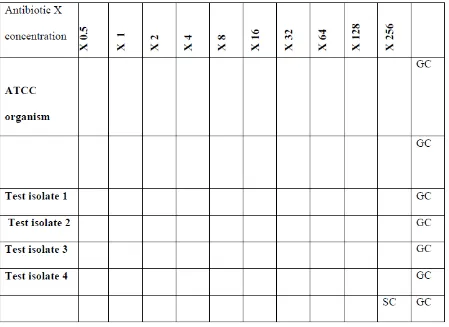
synergism are time kill assay and checkerboard assay. Other in-vitro tests available for
detection of synergism are E-test, in-vitro pharmacodynamic model, critical inhibitor
concentration, double disc synergy, paper strip diffusion, multiple combination
bactericidal test, overlay inoculums susceptibility disc method and serum bactericidal
titre. In-vivo assay applied for understanding synergism are animal model simulating
