A Thesis in General Surgery
COMPARITIVE STUDY OF ONLAY AND
PREPERITONEAL MESH REPAIR IN THE
MANAGEMENT OF VENTRAL HERNIA
Submitted in partial fulfilment of the
Requirements for the Degree of
M.S General Surgery
(Branch I)
Kilpauk Medical College
The Tamilnadu Dr. M.G.R Medical University
Chennai
DECLARATION BY THE CANDIDATE
I hereby declare that this dissertation titled “COMPARITIVE STUDY OF
ONLAY AND PREPERITONEAL MESH REPAIR IN THE MANAGEMENT
OF VENTRAL HERNIA” is a bonafide and genuine research work carried out by
me under the guidance of Dr. S.Balakrishnan, M.S., Professor, Department of
General Surgery, Kilpauk Medical College, Chennai.
This dissertation is submitted to THE TAMIL NADU DR. M.G.R. MEDICAL
UNIVERSITY, CHENNAI in partial fulfillment of the requirements for the degree of
M.S. General Surgery examination to be held in April 2016.
Date :
CERTIFICATE BY THE GUIDE
This is to certify that the dissertation titled “COMPARITIVE STUDY OF ONLAY AND PREPERITONEAL MESH REPAIR IN THE MANAGEMENT OF VENTRAL HERNIA ” is a bonafide research work done by Dr. Durai Raj. M, Post Graduate in M.S. General Surgery, Kilpauk Medical College, Chennai under my direct guidance and supervision in my
satisfaction, in partial fulfillment of the requirements for the degree of M.S. General Surgery.
Date : Dr. S.Balakrishnan M.S.,
Place: Professor,
Department of General Surgery, Kilpauk Medical College,
ENDORSEMENT BY THE HOD AND
HEAD OF THE INSTITUTION
This is to certify that the dissertation titled “COMPARITIVE STUDY OF ONLAY AND PREPERITONEAL MESH REPAIR IN THE MANAGEMENT OF VENTRAL HERNIA ” is a bonafide research work done by Dr.Durai Raj. M,
Post Graduate in M.S. General Surgery, Kilpauk Medical College, Chennai
under the guidance of Dr.S.Balakrishnan M.S., Professor, Department of General Surgery, Kilpauk Medical College, Chennai.
Dr.P.N.Shanmugasundaram M.S., Dr.R. Narayana Babu M.D., DCH
Professor and Head, Dean,
Department of General Surgery, Kilpauk Medical College Kilpauk Medical College, Chennai-10 Chennai-10
Date:
Date:
ACKNOWLEDGEMENT
My sincere thanks to Prof. Dr. R. Narayana Babu, M.D., DCH., Dean,
Kilpauk Medical College and Hospital for allowing me to conduct this studyin
the Department of General Surgery, Government Royapettah Hospital,
Chennai.
I am extremely grateful to Dr.P.N.Shanmugasundaram, M.S, Professor
and Head Of the Department of General Surgery, Government Kilpauk
Medical college for his encouragement and permission in granting unrestricted
access to utilizing the resources of the Department.
I thank my mentor and guide Dr.S. Balakrishnan, M.S, Professor of
General Surgery, Government Royapettah Hospital for her valuable guidance
during the tenure of my course.
I thank my Professors Dr. R.Kannan, Dr. V.Ramalakshmi, Dr. V.Chitra
and Dr. R.A.Pandiyaraj, for their support and guidance.
I also acknowledge my assistant professors Dr.S. Dharmarajan and
Dr.M. Manikandan for their valuable support and timely help rendered to
complete this study.
I thank my colleagues Dr.lokeshwari.K, Dr. Jeena Josephin,
I would like to thank the entire medical and paramedical staff of the
Department of General Surgery.
My utmost thanks to all my patients who cooperated to complete my
dissertation. Without their help it would have been impossible for me to
complete this study.
I thank my family for their great help and support.
Last but not the least, I thank God for being the prime force in guiding
LIST OF ABBREVIATIONS
C/O COMPLIANTS OF
DM DIABETES MELLITUS
HT HYPERTENTION
COPD CHRONIC OBSRUCTIVE PULMONARY DISEASE
PS PREVIOUS SURGERY
DIAG DIAGNOSIS
USG ULTRASONOGRAM
SOD SIZE OF DEFECT
W I WOUND INFECTION
RECCUR RECCURENCE
DOS DURATION OF STAY IN HOSPITAL
IP NO IN PATIENT NUMBER
F FEMALE
M MALE
S SWELLING
S+P SWELLING AND PAIN
UM UMBLICAL HERNIA
PM PARA UMBLICAL HERNIA
IH INCISIONAL HERNIA
C CONFIRMED
IL ILEUM
JJ JEJUNUM
OM OMENTUM
OL ONLAY MESH REPAIR
PPM PREPERITONEAL MESH REPAIR
ABSTRACT
BRIEF RESUME OF INTENDED WORK
Ventral hernia in the anterior abdominal wall includes both spontaneous
and, most commonly, incisional hernias after an abdominal operation. . Hernia
recurrence is distressing to patient and embarrassing to surgeons. Mesh repair can be
pre- peritoneal or onlay. Controversy exists among the surgeons regarding the use of
type of either meshoplasty, due to differences in ease in performing the surgery,
time of surgery, complications occurring in the post operative period and the
recurrence.Only few institution do preperitoneal mesh repair due to the need of
skilled surgeon, so we are comparing onlay and preperitoneal mesh repair.
AIMS AND OBJECTIVES OF THE STUDY
To compare outcome of onlay and preperitoneal mesh repair in the
management of ventral hernia.
Materials and methods
METHOD OF COLLECTION OF DATA
Patient admitted with ventral hernia are included in the study with details of
cases,clinical examination and symptoms are included in the study after confirming
the diagnosis by ultrasonography and are divided randomly into onlay and
to study the outcome reccurence.
PERIOD OF STUDY : November 2014 to April 2015
TYPE OF STUDY : Randomized control study.
SOURCE OF DATA :
Patient diagnosed as ventral hernia in department of general surgery,
Royapettah hospital and kilpauk medical college hospital. 50 of them are to be
selected on basis of non probability (purposive) sampling method.
INCLUSION CRITERIA :
Patient with ventral hernia including
• Umbilical hernia,
• Paraumblical hernia,
• Epigastric hernia
• Incisional hernia.
EXCLUSION CRITERIA :
Patient admitted with
• Groin hernia ,
• Divarication of recti,
• Recurrent hernia,
• Patient medically unfit for surgery,
CONCLUSION :
By analyzing the outcome of seroma, wound infection, flap necrosis and
TABLE OF CONTENTS
S.No CONTENTS Page No.
1 INTRODUCTION 1
2 OBJECTIVES OF THE STUDY 3
3 REVIEW OF LITERATURE 4
4 MATERIALS AND METHODS 36
5 RESULTS 37
6 DISCUSSION 69
7 CONCLUSION 75
8 SUMMARY 76
9 BIBLIOGRAPHY 78
10 ANNEXURE
• PROFORMA
• CONSENT FORM
• MASTER CHART
83
86
LIST OF TABLES
S.NO PARTICULARS PAGE NO.
1 AGE DISTRIBUTION 38
2 SEX DISTRIBUTION 40
3 SYMPTOMS 41
4 DIABETES MELLITUS 43
5 HYPERTENSION 44
6 CHRONIC OBSTRUCTIVE PULMONARY
DISEASE
45
7 PREVIOUS SURGERY 46
8 DIAGNOSIS 47
9 ULTRASONOGRAM 49
LIST OF TABLES
S.NO PARTICULARS PAGE NO.
11 SEROMA 52
12 WOUND INFECTION 53
13 FLAP NECROSIS 55
14 RECURRENCE 56
15 THE VENTRAL HERNIAS WITH RESPECT TO
NUMBERS AND PERCENTAGE
59
16 TYPE OF MESH REPAIR 59
17 RECURRENCE 60
18 PERCENTAGE OF VENTRAL HERNIA IN
FEMALE
70
19 MEAN HOSPITAL STAY 73
LIST OF FIGURES
S.NO PARTICULARS PAGE NO.
1 SHOWS AGE DISTRIBUTION 39
2 SHOWS SEX DISTRIBUTION 40
3 SHOWS SYMPTOMS 42
4 DIABETES MELLITUS 43
5 HYPERTENTION 44
6 CHRONIC OBSTRUCTIVE PULMONARY DISEASE 45
7 PREVIOUS SURGERY 46
8 DIAGNOSIS 48
9 ULTRASONOGRAM 49
10 CONTANT OF SAC 51
11 SEROMA 53
12 WOUND INFECTION 54
13 FLAP NECROSIS 56
14 RECURRENCE 57
1
INTRODUCTION
As a result of in man’s erect posture, his anterior abdominal wall is the
site of a variety of hernias. Most of these hernias protrude through the
abdominal wall to form obvious palpable swellings
Protrusion of an abdominal viscus or its parts through the anterior
abdominal wall occurring at any site other than groin is known as ventral
hernia.. It includes incisional hernias, umbilical hernia, epigastric hernias
paraumbilical hernias and spigelian hernias respectively.1
The patient seeks medical advice for swelling, acute pain, discomfort,
associated gastrointestinal symptoms or cosmetic symptoms, diagnosis can be
achieved with ease by clinical examination and by ultrasound scanning.
A number of predisposing factors have been identified that may be
related to specific patient characteristics, an underlying pathologic process, or
iatrogenic factors. From the surgeons perspective, repair of hernias is common
procedure. There are various surgical techniques for the hernia repair.
All Incisional hernias are unique in that they are the only abdominal
wall hernias that are considered to be iatrogenic.
For many years, high recurrence rate is associated in the repair of incisional
hernia. In more recent years, the introduction of synthetic prosthetic materials has
provided the opportunity to perform a tension free repair, thereby reducing the
2
Midline hernia occurring through linea alba abutting superiorly or
inferiorly on the umbilicus is called as “PARAUMBILICAL HERNIA”. They
are generally acquired lesions. If the defect is small it can be repaired surgically.
But large hernias with wide openings are difficult to repair surgically and should
be treated with synthetic mesh repair.
Epigastric hernia protrude through linea alba above the umbilicus.
Approximately 5% of the populations have epigastric hernias. After diagnosis of
an epigastric hernia, there is no reason to wait for repair, the chances for
incarcerations are high and surgery remains the only permanent cure.
Most of the spigelian hernias are acquired and requires surgery as the chances of
3
OBJECTIVES OF THE STUDY
The main objectives and aims of this study is to:
1. To study the anatomical, etiological, clinicopathological, factors leading to
ventral hernias.
2. To study the different techniques of repair of ventral hernia with special
emphasis on pre- peritoneal mesh repair and onlay mesh repair and their
4
REVIEW OF LITRETURE
2, 3,4,5,6The word Hernia is derived from the Greek word (Hernias, bud)
meaning an offshoot, a budding or bulge. The Latin word Hernia means
rupture or tear. Hernia was recognized about 1000 years ago. Probably the
reason for this is the upright position which man has assumed during the
revolutionary process. Hernia was treated by several ways with the available
simple measures like bandages, ointment, poultices and localized concoctions.
Cutting and countering operations were common in India, China and Japan long
before Hippocrates..
Astley Cooper discovered the Transversalis Fascia and pointed out that
this layer was the main barrier to herniation.
Lucas Championnere apparently was one of the first to use the overlapping fascia
technique in 1891.
Arroyo and coworkers in Spain performed one of the very few randomized
clinical trials with 200 patients. Their results showed a clear distinction between
the success of using mesh repair and primary suture. The latter resulted in a
recurrence rate of 11% while after using a tension free mesh repair is amounted
to only 1%.
INCISIONAL HERNIA.
Witzel (1900), Goepel (1900), Barlett (1903) and McGavin (1909)
advocated the use of Silver wire filigree. Koontz and Throckmorton (1948) used
5
Fascia Lata grafts used in the form of strips of sheets have been reported.
Shortly the advent of synthetic Plastic sheets and the polyvinyl alcohol sponge
were used.
The Modern era of prosthetic hernia repair began in 1958 when Usher
reported his experiment with Polyamide mesh. Later braided polyester mesh,
polypropylene mesh and expanded polytetrafluoroethylene (ePTFE) were
introduced which revolutionized the surgery for post-operative Hernia.
HISTORY OF SURGICAL MESHES
Artificial material was introduced in 1889 by Witzel who used a mesh
of silver wire for abdominal wall hernias.
In 1959, Usher et al. reported the successful implantation of surgical
meshes at first in 13 dogs and after ward inpatients with abdominal wall hernias.
Busse in 1901 even used meshes made of gold wire.
In 1940, Ogilvie published the use of cloth meshes to treat contaminated
gunshot wounds with defects of the abdominal wall.
6
HISTORIC OVERVIEW OF MESH REPAIR5
No. Event Introduction
1 Polyester mesh Wolsten Holme Arch Surg., 1956, 73,
2 Polypropylene mesh Usher Arch. Surg. 1962;84;325
3 GPRVS Stoppa et al., 1973 (72)
4 Trans-inguinal preperitoneal
prostheses
Rives et al., chirurgie, 1973; 99:564.
5 Subfascial prosthesis Lichtenstein and Schulman, 1986(44)
6 Preperitoneal prosthesis by
extraperitoneal access
Nyhus et al., An. Surg., 1988; 208:733.
Wantz, Surg., 1989;169:408
7 Mesh plug Rutkow/Robbins Surgery, 1993; 114:3.
8 Plug Laparoscopy Shultz et al., clin. Laser Mon., 1990;8:103
9 Intraperitoneal onlay mesh
prosthesis (IPOM)
Transabdominal preperitoneal
prosthesis (TAPP)
Shultz et al., clin. Laser Mon., 1990;8:103
Corbitt, Surg. Laparos Endose, 1991;
1:23.
10 TEPP Ferzil etal., laparosendcsc, Surg.,
1992;2:281
McKerna Laws, Surg. Endosc, 1993;7:26.
PARAUMBILICAL HERNIA
Celsus in the first century A.D used an elastic suture7 in the treatment
of umbilical hernias.
Willian J Mayo8.9, on Aug 4th 1898 delivered his classical paper, remarks
7
overlapping fascia for repair of umbilical hernia.
In 1979 Usher described a technique of repair using Marlex Mesh.
EPIGASTRIC HERNIA
Epigastric hernias were first described in 1285.
The term epigastric hernia was introduced by Leveille in 1812. The first
successful operation on this hernia was reported by Maunnior in 1802. Ulrike
Muschaweck in 2003 concludes using a Mesh plug in an epigastric hernia has
advantages over the commonly used methods.
EMBRYOLOGY 10.11.12.13
The abdominal wall begins to develop quite early in the embryo, but it
does not achieve its definitive structure until the umbilical cord separates from
fetus at birth. Most of the abdominal wall forms during closure of the midgut
and reduction in relative size of the body stalk.
The primitive wall is somatopleure (ectoderm and mesoderm without
muscle, blood vessels, or nerves). The somatopleure of the abdomen is
secondarily invaded by mesoderm from the myotomes that developed on either
side of the vertebral column. This mesodermal mass (hypomere) migrates
ventrally and laterally as a sheet, and the edges differentiate while still widely
separated from each other into the right and left rectus abdominis muscles.
The final opposition of these muscles in the anterior midline closes the body wall.
Before the primordial of the rectus muscles fuse anteriorly, the mesoderm from
the hypomere splits into three layers that can be recognized by the seventh
8
abdominis muscle, the middle sheet becomes the internal oblique muscle and
aponeurosis. Dorsally, the superior and inferior posterior serratus muscles develop
from the superficial layer of the hypomere.
Approximation of the two rectus abdominis muscles in the midline
proceeds from both caudal and cranial ends and is complete by the 12th week,
except at the umbilicus. The final closure of the umbilical ring awaits the
separation of the cord at birth, but the ring may remain open, in which case an
9
ANATOMY
14,15,16,17,18ANTERIOR ABDOMINAL WALL
The abdominal wall is a complex musculoaponeurotic structure. It is
bounded by the flare of the costal margins and the xiphoid process of the
sternum above and by the iliac crests, inguinal ligaments and pubis below.
The structures that comprise the anterior abdominal wall are skin, subcutaneous
tissue, superficial fascia, antero-lateral muscles of the abdomen, together with
their enveloping fascial sheaths and aponerosis, transversalis
fascia,extraperitoneal adipose and areolar tissue and parietal peritoneum.
The linea alba, a tendinous raphe in the midline divides the anterior
abdominal wall into two parts.
The umbilicus lies in the anterior median line, at the level of the disc
between third and fourth lumbar vertebrae.
I. SUPERFICIAL FASCIA
The fascia contains fat, cutaneous nerves, cutaneous vessels and superficial
lymphatics below the level of umbilicus fascia is divided into a superficial
fatty layer (fascia of camper) and a deep membranous layer(fascia of scarpa).
Most part of fascia is a single layer that contain variable amount of fat.
II. CUTANEOUS NERVES
Skin of anterior abdominal wall is supplied by the lower six thoracic
10
III. CUTANEOUS ARTERIES AND VEINS
Anterior cutaneous arteries are branches of superior and inferior
epigastric artery and accompany the anterior cutaneous nerves. Lateral
cutaneous arteries are branches of the lower intercostals arteries and accompany
the lateral cutaneous nerves. Superficial epigastric, superficial external pudendal,
superficial circumflex iliac artery arise from the femoral artery and supply the
skin of the lower part of abdomen. The venous drainage is by superficial
epigastric, superficial external pudendal, superficial circumflex iliac vein which
drains into femoral vein.
IV. SUPERFICIAL LYMPHATICS
Above the level of the umbilicus, the lymphatics run upwards to drain
into the axillary lymph nodes. Below the level of umbilicus they run downwards
to drain into superficial inguinal lymph nodes and pay respect to the watershed
line.
V. MUSCLES OF THE ANTERIOR ABDOMINAL WALL
1. EXTERNAL OBLIQUE [OBLIQUUS EXTERNUS ABDOMINIS]
This muscle is largest and thickest of the flat abdominal muscles. Its broad
origin includes the last eight ribs. Those from lower two ribs are attached to outer
lip of anterior segment of iliac crest. The upper and middle fibres gives way to
flat, strong aponeurosis at about the midclavicular line, and it inserts medially into
the linea alba. The aponeurosis passes anterior to the sheath of the rectus
abdominis and with care, it can be dissected from it. In general, the fascicles
pass from the superolateral to inferomedial. Thus, the direction of force
11
Nerve supply: Ventral Rami of the lower six thoracic spinal nerve.
2. INTERNAL OBLIQUE [OBLIQUUS INTERNUS ABDOMINIS]
It originates from the last five ribs, the thoracolumbar fascia, the
intermediate lip of the iliac crest and the lateral half of the inguinal ligament. Its
fibres course opposite the direction of those of external oblique. It gives way to a
flat aponeurosis medially, which splits to enclose the rectus muscle. The
aponeurosis reunites medial to the rectus and inserts into the linea alba. The
posterior lamina ends below in a free curved margin called Arcuate line midway
between umbilicus and symphysis. The fibers that arise from the lateral half
of the inguinal ligament pursue a downward course and insert into os pubis
between symphysis and the tubercle. Some of the lower fibres are pulled into
the scrotum by the testis as it passes through the abdominal wall and called the
cremastric muscles of the spermatic cord.
Nerve supply: Ventral rami of lower six thoracic and first lumbar spinal nerves.
3. TRANSVERSUS ABDOMINIS MUSCLE
It is the smallest of the three flat muscles and originate from lower five
ribs, the thoracolumbar fascia, the internal lip of iliac crest, and the lateral third of
the inguinal ligament. The direction of its fibres is transverse and they give way
to a flat aponeurosis that inserts into the linea alba. The aponeurosis passes
behind the rectus sheath in its upper two-third. The fibres that originate from
inguinal ligament pass downward to insert os pubis, as do the fibers of the
internal oblique. Occasionally, the lower fibres of both muscles inserts by
means of a common tendon called conjoined tendon.
12
NOTE:
The neurovascular plane of the abdominal wall lies between the
internal oblique and transverses abdominis.
The spigelian fascia is the aponeurotic part of transverses abdominis
muscle between the medial border of its muscular part and the insertion of the
aponeurosis into the posterior rectus sheath.
4. RECTUS ABDOMINIS
It is a long strap like muscle which arise by two tendinous heads. The
lateral head arise from the lateral part of pubic crest. The medial head from the
anterior pubic ligament. The fibres run vertically upwards and inserted into
xiphoid process, seventh, sixth, fifth costal cartilages.
Nerve supply: Ventral rami of lower six or seven thoracic spinal nerves.
5. CREMASTER
The muscle is fully developed only in the male. In female it is represented
by few fibres only. Along with the intervening connective tissue, the muscle
loops form a sac like cremastric fascia around spermatic cord deep to external
spermatic fascia.
Nerve supply: Genital branch of genitofemoral nerve derived from first and
second lumbar spinal nerves.
6. PYRAMIDALIS
It is rudimentary in human beings. This is a small triangular muscle arising
from anterior surface of body of pubis. Fibers pass upwards and medially to be
13
Nerve supply: Subcostal nerve which is the ventral ramus of the 12th thoracic
spinal nerve.
III. DEEP ARTERIES AND VEINS OF ANTERIOR
ABDOMINAL WALL
The anterior abdominal wall is supplied by superior epigastric and
musculophrenic artery above, inferior epigastric and deep circumflex iliac artery
below, small branches of lower two or three posterior intercostal, subcostal and
lumbar arteries, superficial epigastric, circumflex iliac artery. The venous
drainage is by superior epigastric and musculophrenic vein below, inferior
epigastric and deep circumflex iliac vein below.
IV. DEEP NERVES OF THE ANTERIOR ABDOMINAL WALL
The anterior abdominal wall is supplied by lower and six thoracic
nerves and by first lumbar nerve through its iliohypogastric and ilioinguinal
branches.
V. FUNCTIONS OF ANTERIOR ABDOMINAL WALL MUSCLES
The abdominal muscles provide a firm but elastic support for the
abdominal viscera against gravity. This is chiefly due to the tone of the oblique
muscles, especially the internal oblique.
The oblique muscles assisted by the transverses, can compress the
abdominal viscera and this help in all expulsive acts, like micturition, defecation,
parturition, vomiting.
The external oblique can markedly depress and compress the lower part
14
shouting. Flexion of the lumbar spine is brought about mainly by the rectus
abdominis. Lateral flexion of the trunk is done by one sided contraction of the
oblique muscles. Rotation of trunk is by action of external oblique with opposite
internal oblique.
VI. RECTUS SHEATH
This is an aponeurotic sheath covering the rectus-abdominis muscle.
Above the costal margin anterior wall is formed by external oblique aponeurosis,
posterior wall is deficient. Between the costal margin and the arcuate line
anterior wall is formed by external oblique aponeurosis and anterior lamina of
the aponeurosis of the internal oblique, posterior wall is formed by posterior
lamina of the aponeurosis of the internal oblique and aponeurosis of the
transverse muscle. Below the arcuate line anterior wall is formed by aponeurosis
of all the three flat muscles. The aponeurosis of the transverses and internal
oblique are fused, but external oblique aponeurosis remains separate. Posterior
wall is deficient.
VII. LINEA ALBA
The linea alba is a tendinous raphe formed by interlacing of the fibres
of the three aponeurosis forming the rectus sheath. It extends from the xiphoid
process to the public symphysis. Above the umbilicus it is about 1 cm wide, but
below the umbilicus it is narrow and difficult to define. It is so called because it is
15
VIII. FASCIA TRANSVERSALIS
This fascia lines the inner surface of the transverses abdominis muscle. It is
more properly should be called the endoabdominal fascia because it is a
continuous lining of the abdominal cavity and is considered to be the strongest
layer of the abdominal wall.
Deep inguinal ring is an oval opening in the fascia transversalis. Anteriorly,
it is adherent to the linea alba above the umbilicus. Posteriorly, it merges with the
anterior layer of the thoraco lumbar fascia and is continuous with the renal fascia.
Superiorly, it is continuous with the diaphragmatic fascia. Inferiorly, it is attached
to the inner lip of the iliac crest and to the lateral half of the inguinal ligament. At
both these places it is continuous with the fascia iliaca. Medially it is attached to
public tubercle, the pubic crest and the pectineal line. Part of it is prolonged into
the thigh as the anterior wall of the femoral sheath.
IX. CONJOINT TENDON
It is formed from lower fibres of internal oblique and lower part of
aponeurosis of transverse abdominis. It is attached to pubic crest and pectineal
line. It descends behind the superficial inguinal ring and acts to strengthen the
medial portion of the posterior wall of the inguinal canal.
X. INGUINAL LIGAMENT
It is the thick, in rolled lower border of the aponeurosis of external oblique
and stretches from anterior superior iliac spine to the pubic tubercle. Its grooved
16
XI. EXTRAPERITONEAL ADIPOSE AND CONNECTIVE TISSUE
LAYER
It contains adipose tissue, inferior epigastric artery and vein and four
fetal structures, medial umbilical ligaments (obliterated umbilical artery),
obliterated urachus (median umbilical ligament), ligamentum teres (obliterated
umbilical vein).
XII. PARIETAL PERITONEUM
It is the inner most layer. It is thin layer of dense irregular connective
tissues and this is covered on the inside by layer of simple sequences
mesothelium. The peripheral membrane is innervated from above downwards is
a sequational manner by spinal nerves T7-L1. The peritoneum provides little
strength in wound closure, but it apart protection from infection if it remains
unviolated.
INCIDENCE
POST-OPERATIVE VENTRAL ABDOMINAL HERNIA 2
In the best centers, the incidence of post-operative hernia has been at least
10% as shown by long term follow up studies. Where less emphasis is placed
on the niceties of abdominal wound closure, the incidence is much higher.
Recent studies show that about 2/3 appear with in first five years and that at least
another third appear 5 to 10 years after the operation. As longer and more
accurate follow up studies are done, it will probably be shown that with ageing
and weakening of the tissues, post-operative hernias may appear even more than
17
PARAUMBILICAL HERNIA
Estimates of the incidence of umbilical hernia at birth vary greatly. In
Caucasian infants, they range between 10-30%. In children of African descent,
it may be several times greater. Children with raised intrabdominal pressure
owing to ascites, COPD, or ventriculoperitoneal shunt, also tend to develop an
umbilical hernia.
The incidence of paraumbilical hernia in the adult is unknown19. It is
more common in the female, with a female to male ratio of 3:1, middle aged,
obese, multiparous females are prone to develop significant paraumbilical hernia,
as are individuals with ascites, usually secondary to cirrhosis of the liver. In
addition, as Mayo suggested in 1899, the old, cachectic and feeble are subject to
umbilical hernia and likely to develop complications.
EPIGASTIC HERNIA 2
The frequency of epigastric hernia in the general population is estimated to
be about 5%. It is more common in early adulthood and middle age. This hernia
is three times more common in men than in women. Upto 20% of Epigastric
hernias may be multiple, but usually one is dominate. Epigastric hernias
18
ETIOLOGY
INCISIONAL HERNIA2
Many factors, singly, or in various combinations, may cause failure of the
wound to heal satisfactorily and may lead to the development of a post-operative
hernia.The common causes are poor surgical technique and sepsis. The causes are
explained below;
1. POOR SURGICAL TECHNIQUE
a) Non-anatomic incisions
Vertical pararectus incision made in rectus sheath along lateral border ,
which destroys nerve and vascular supply to the tissues medial to incision,
causing them to atrophy.
b) Layered closures
These are followed by a greater incidence of post-operative hernias
than are wounds closed by the single layer mass closure technique. This may
be owing to the fact that many more sutures are used; which are closely placed,
and because insufficiently sized bites of each thin layer are taken.
c) Inappropriate suture material
Wounds closed with non-absorbable suture material are followed by lower
incidence of post-operative hernias than wounds closed with absorbable material.
The ideal suture material for abdominal closure, especially of midline incision,
in monofilament stainless steel wire used in the form of interrupted mass
closure, taking large bites of the musculoaponeurotic layers from the abdominal
19
monofilament polyamide or polypropylene as a single thread or, preferably in the
form of a commercially available loop.
d) Suturing Technique
Small sutures take only a small amount of tissues close to the cut edge of
the incision. In vertical abdominal incision at; or near, the midline, these sutures
pull in the line of fibers of the aponeurotic muscles and since they are so closed
to the incision, easily cut out of the tissues. A small, tightly tied suture causes
ischemia and necrosis of the tissues it contain and also of an area on each side of
the suture. When these small, tightly tied sutures are placed close to each other,
their ischemic areas merge and thus, cause necrosis of a strip of tissue all along
the the incision edge, which separates, together with the sutures, from the rest of
the abdominal wall, leading to failure of the wound
e) Tension
The lateral pull of the abdominal wall muscles against the suture
reduces tension, which tends to pull them in the opposite direction, creates an
area of pressure necrosis where the suture meets the tissue
2. Sepsis
It is the second major cause of early wound failure. It may range from
frank acute cellulites, with fasciitis and necrosis of the tissues on each side of the
incision, to low grade chronic sepsis around sutures such as silk.
The infection causes inflammation and edema of the tissues, which
becomes soft and weakened so that the sutures tear the tissues and pull out
20
3. Drainage tubes
The tissue planes along the track of the drain are not sutured, an open and
weak passage is present through all layers of the wound through which a hernia
may develop if drain tube are brought out through operation wound.
Drain allows for two way traffic of secretions outwards and organisms
inwards. The irritation caused by drain causes edema or softening and tearing of
the tissues and cutting out of the sutures.
4 . Obesity
Obesity is associated with high percentage of port-operative hernias as
well as with recurrence following repair of these hernias. Cutting through
large masses of fat and the increased retraction needed may raise the
infection rate in these patients and lead to recurrence. Tissues infiltrated with fat
may not be able to hold the sutures, especially since the excess of intra and
extra abdominal accumulation of many kg of fat may add enormous tension
on the sutures. Obese patients tend to develop post-operative complications
such as paralytic ileus, atelectasis, pneumonia and deep vein thrombosis that
may increases the risk of incisional hernia.
5. General Condition
The general condition of the patient influences the rate of post-operative
ventral hernia. The factors include age, generalized wasting, malnutrition and
starvation, hypoproteinemia (especially hypoalbuminemia); avitaminosis
(especially vitamin C), malignant disease, anemia, jaundice, Diabetes mellitus,
chronic renal failure, liver failure, ascites, prolonged steroid therapy,
21
6. Post-operative Complications
These include prolonged post-operative paralytic ileus, intestinal
obstruction with abdominal distension, chronic obstructive pulmonary disease,
pulmonary collapse, bronchopneumonia, emphysema and asthma which increase
the incidence of post-operative hernias.
7. Types of Operation
These include Laparotomy for generalized or localized peritonitis in
patients with perforated peptic ulcer, appendicitis, diverticulitis and acute
pancreatitis. Operation for intra- abdominal malignant disease, inflammatory
bowel disease, re-operation through the original wound, especially within the
first six months after the initial procedures have tendency to be followed by
hernia.
8. Tissue Failure
Hernia develops in what apparently is a perfectly healed wound that
has functioned satisfactorily for five, ten or even more year and after operation
and is presumably the result of the failure of the collagen in the scar. Rodriques
has recently shown a decrease in OXYTALAN FIBERS and an increase in the
amorphous substance of the elastic fibers as a function of age and may be
responsible for alterations in the resistance of the transversals fascia and
abdominal wall scar tissue. Ageing and weakening of the tissue and increased
intra-abdominal pressure associated with chronic cough, constipation and
22
PARAUMBILICAL HERNIA
Etiological factors can be divided into congenital and acquired factors.
1. Congenital20
2. Acquired
a) Predisposing factors
1. Faulty umbilical cord ligation20.
Umbilical cord ligation > 4--5cm from the abdominal wall may give rise to
development of hernia.
2. Umbilical sepis- weakness umbilical area
3. Increased intrabdominal pressure, due to chronic cough, constipation,
straining while passing urine, ascites
4. Direct trauma.
b) Contributing factors7,19
1. Low birth weight
2. Race
3. Sex: Female: Male=3:1
4. Family history: Familial history contributes but no generic pattern of
inheritance has been seen.
5. Age: more common in children< 2yrs and elderly people.
6. Obesity
7. Multiparty due to stretching and weakening of
anterior abdominal wall musculoaponeurotic layer.
23
cretinism, meningomyelocele, hurler’s syndrome, and amourotic family idiocy
may be associated with umbilical hernia. May be associated with cholelithiasis,
abdominal malignancies, collagen disease, hemorrhoids, varicose veins, and
cystocele.
EPIGASTRIC HERNIA2,5
The cause of epigastric hernia is unknown, but since it occurs even in new
born children, it is assumed to be the result of a structural congenital weakness of
the line alba between xiphoid process and the umbilicus. It is possibly owing to
a lack of fibers at the midline decussation, which allows preperitoneal fat to be
herniated between the gaps. The fact that it is common between 20 and 50
years of age probably reflects a balance between a congenital defect and a rise
of intra-abdominal pressure, adiposity, and weakening of the muscles in adults.
It is more frequent in people with a wide linea alba2
Epigastric hernia is generally considered an acquired lesion, probably
related to excessive strain on the anterior abdominal wall aponeurosis.
Moschowitz emphasized the importance of blood vessels perforating the
linea alba and prolongation of the transversalis fascia at this point.
Askar’s studies also demonstrated that fibers originating from the
diaphragm traverse the upper midline aponeurosis posteriorly and join the
fibers of the posterior rectus sheath and middle tendinous intersection. They
attach to the linea alba at a site midway between the xiphoid and the umbilicus.
Uncoordinated vigorous, synchronous contraction of the diaphragm and upper
24
traction on the diaphragm and lateral traction on the tendinous intersection
would be maximal at this point of attachment midway between the xiphoid and
the umbilicus, the most common site of Epigastric Hernia.
DIVARICATION OF RECTI21
This is seen principally in elderly multiparous patients and also who
has undergone repeated midline abdominal operations, in which linea alba may
stretch.
CLINICAL MANIFESTATIONS
I. INCISIONAL HERNIA2
The patient’s complain of an unsightly bulge in the operation scar as
well as of pain and discomfort. They often suffer from a heavy, sickening,
dragging sensation aggravated by coughing and straining. In large dependent
hernias, areas of skin may undergo pressure ischemic necrosis and may ulcerate,
and rarely, the hernia may rupture. If the hernia strangulates, the symptoms of
intestinal obstruction and ischemic bowel will supervene. There is often a
history of repeated mild attacks of intestinal obstruction manifesting as colicky
pains and vomiting. Intertrigo may develop in the deep crease between the
hernia and the abdominal wall and the skin may become moist, infected and
odorous. Obese patients with large pendulous hernias are practically
immobilized and find life almost unbearable
II. PARAUMBILICAL HERNIA7,19,23
It usually develops in middle and old age and it is commonly found in case
of obese females. The consistency is firm when it contains omentum and soft
25
III. EPIGASTRIC HERNIA2
The usual epigastric hernia is symptom less.It usually present with a
small round swelling in the midline between xiphisternum and umbilicus. They
are often irreducible, sometimes multiple. In obese patients the typical smooth,
rounded, slightly tender lump may be lost in the depths of subcutaneous fat.
IV. DIVARICATION OF RECTI22
When the patient strains, a gap can be seen between the recti abdominis
through which the abdominal contents bulge. When the abdomen is relaxed the
fingers can be introduced between the recti.
DIAGNOSTIC IMAGING IN THE EVALUATION AND MANAGEMENT5
I. SONOGRAPHY
Sonography is indicated primarily in patients with palpable masses within
deep layers of the abdominal wall.. In patients with hernia, a measurement of the
defect can be done.
Incisional Hernia
Sonography shows the typical hernial pattern with a fascial gap and
protruding hernial sac. After mesh repair for hernia, a recurrence can occur at
the edge of the mesh which can be seen sonographically.
In comparison with CT or herniography, the ultrasonography is time as
26
Epigastric Hernia
The hernia is visualized by a characteristic midline fascial defect.
Predictive Value
Sensitive Specificity Positive test Negative test
Epigastric Hernia 100% 100% 100% 100%
Divarication of Rectus Abdominis
Can be clearly visualized by sonography and the resulting herniation in
abdominal wall.
II. COMPUTED TOMOGRAPHY
Computed tomography is an excellent method of evaluating the
abdominal wall and its relations to the abdominal viscerae. Lesions can be easily
identified, owing to their different density.
There are several reports in the literature concerning the primary
diagnosis of spigelian hernia by CT which can elegantly demonstrate.
CT allows exact evaluation of the volume and content of giant hernias. CT
is also used to differentiate postoperative findings such as haematoma, abscess,
or recurrence of hernia after laparoscopic repair of ventral hernia.
III. MAGNETIC RESONANCE IMAGING
Compared to CT, MRI offers the advantage of direct multiplane imaging
without ionizing radiation and the use of contrast agents. A relative merit of MRI
27
IV. HERNIOGRAPHY
Herniography has a very low complication rate, disadvantage is accidental
colonic puncture which is less than 1%, contrast allergy, and irradiation to pelvic
region.
With the techniques now available, there is no indication for
herniography, even if the complication rate is low because it is invasive.
The order of recommendations for the evaluation of abdominal wall hernias is as
follows:
1. CLINICAL HISTORY
2. CLINICAL EXAMINATION
3. SONOGRAPHY
4. CT/MRI
OPERATIVE MANAGEMENT
PRE-OPERATIVE PREPARATION
1. Optimal skin hygiene.
2. Weight reduction for obese patient.
3. To stop smoking.
4. The repair of a large postoperative ventral hernia should be delayed for
atleast one year after the operation that caused the hernia or after a previous
attempt at repair.
5. Wait for atleast one year after all infection and sinuses have healed.
6. Associated cardiovascular, respiratory, renal conditions, Diabetes Mellitus,
28
treated. The operation is usually elective and must be delayed until the
patient is in an optimal state.
7. Perioperative antibiotics are used more liberally.
8. The patient is investigated for coexisting abdominal pathology so that it can be
dealt with at the same operation.
INDICATIONS
1. Pain and discomfort.
2. Large hernias with small openings.
3. A history of recurrent attacks of subacute obstruction, incarceration,
irreducibility and strangulation,
4. For cosmetic reasons for a large and unsightly hernia.
GENERAL PRINCIPLES IN REPAIR OF VENTRAL HERNIAS
1) Spinal and epidural anaesthesia gives excellent relaxation with minimal
respiratory depression.
2) Hemostasis should be as careful and as effective as possible.
3) Permanent suture material should be used for the repair.
4) The choice of incision is governed by the orientation of the defect.
5) Healthy fascia must be isolated.
6) Closure of the sac is done in one layer, incorporating both fascia and
peritoneum after opening the sac, freeing all adhesions, reducing the
viscera and exploring the abdomen.
29
OPERATIVE METHODS FOR REPAIR OF VENTRAL HERNIA2,5
The three basic methods are
I. PRIMARY SUTURE OR EDGE TO EDGE CLOSURE
According to Bonnet’s (writes in the fifth edition of Nyhus and
Condon’s Hernia) a fascial defect should be repaired by primary suture with
non-absorbable suture and edge-to-edge closure.
The hernial sac is dissected, opened and all adherent omentum and loops
of bowel are freed by dissection and mouth of the sac defined. The sac and its
peritoneal lining, scar tissue, and old suture material are excised upto the edge of
the defect to expose the normal tissues of the linea alba.
The fascial defect is sutered with monofilament polypropylene with
interrupted sutures transversely or vertically. Full thickness bites or sutures taken
on the abdominal wall along lower and upper margins of the hernia.
II. SHOELACE DARN REPAIR
This method is based on the understanding of the functional anatomy of the
abdominal wall. Skin,subcutaneous tissue and fat are dissected from the
hernial sac and rectus sheath. The new linea alba is now constructed, using a
vertical strip 1-1.5 cm wide split off the medial edge of each anterior rectus
sheath about 1 cm or more from medial edge to confirm the presence of rectus
muscle. Two steps are sewn together. Next, suture started at the top end of the
incision in the rectus sheaths from inside the sheath and passes out on that side,
returning inside through opposite corner and slipping through the loop. The flat
30
prolene suture passing end- to-end in front of the rectus abdominis muscle,
between art edges of external rectus sheath and through strong new midline
anchor for the whole length of the hernia, in the manner of shoelace tightening
of boot.
In obese patients panniculectomy and abdominoplasty are combined with
repair of hernia.
A vacuum drain is placed on either side and brought out through separate
stab.
The operation is entirely extraperitoneal. The post-operative recovery is
smooth and rapid. Complications are few and usually minor. They are infection
and the recurrence rate 2%.
II. NUTTALL PROCEDURE
For midline defects in the lower abdomen, this procedure has been
reported to be quite effective. The lower aspect of the rectus abdominal muscle
and its enveloping fascia are mobilized off the pubis and approximated to the
contralateral bone. This manoeuvre provides anterior rectus sheath coverage for
the lower midline defect.
V. CATTELL’S OPERATION
The sac is dissected and opened. The viscera are reduced after freeing the
contents form the sac. The peritoneum is repaired. The edges of the abdominal
wall are approximated and are sutured with non-absorbable suture material
layer by layer. After the surrounding aponeurosis has been sutured an incision
31
are sutured over the previous line of suture and the lateral margins are now
sutured over this medical layer of suture. This is continued till the edges of the
healthy aponeurosis are brought together firmly.
VI. KEEL OPERATION21
The hernial sac is not opened but pushed back into the abdomen. With
non-absorbable suture it is pleated so that it projects into the abdominal cavity. A few
layers of sutures are applied one after the other till the healthy margin of the
muscles and aponeurosis are brought close. Now the margins of the healthy
muscles and aponeurosis are sutured to each other. As in this operation the
hernial sac is pushed into the peritoneal cavity in a pattern which on cross section
looks like the ‘keel’ of a ship, this operation is called keel operation.
MAYO’S DOUBLE BREASTING22,23
The Mayo double breasting technique is most c o m m o n surgical
techniques routinely performed in most hospitals.
PROSTHETIC MESH REPAIR
CHOICE OF MATERIAL
The ideal mesh is one that is cheap and universally available, is easily cut
to the required shape, is flexible, slightly elastic and pleasant to handle. It
should be practically indestructible and capable of being rapidly fixed and
incorporated by human tissues. It must be inert and elicit little tissue reaction. It
must be sterilisable and non-carcinogenic.
32
today the most commonly used material for repair of all types of hernia.
a) POLYPROPYLENE MESH (MARLEX, PROLENE)
This is currently most widely used prosthetic material in hernial repair. It is
formed of knitted monofilament plastic fibers and has minimal elasticity or stretch
capacity. Prolene elicits an intense reaction in tissue.The disadvantages are
visceral adhesions, erosion into the bowel/skin causing enterocutaneous fistula/
sinus formation, erosion of mesh into urinary bladder.
Sterilization: gamma radiation; after removal from its package, the mesh can be
resterilised by autoclaving for three times only.
b) PTFE (Teflon, Gore-Tex)
It is supplied as a felted sheet in which fibers randomly interlace. It is
used for vascular prosthesis. It is strong, pliable, soft, smooth and slippery to
touch, biologically inert and causes little tissue reaction. It is costly.
c) POLYESTER MESH (DACRON) MERSILENE
It is multifilament knitted mesh. It is cheap, freely available, light, supple,
has a pleasant, soft feel and is strong and elastic. It excites greater tissue
inflammatory reaction than prolene. It tears easily.
d) FASCIA LATA
It is harvested from lateral aspect of the thigh. It is strong and flexible
although minimally elastic. The use has been abandoned.The other prosthetic
meshes tried are polyglycolic mesh, polyglactic mesh, metal meshes and
33
INDICATIONS FOR MESH REPAIR
The indications are:
a. Repair of recurrent incisional hernias: successful repair of recurrent hernias
in patients, whose musculature is of poor quality and weak and flabby,
fascial coverings are thin and weak, requires prosthestic material.
b. In primary repair of massive hernia in which tissues are deficient and
repair without tension cannot be accomplished readily by conventional
techniques of direct suturing. The employment of a bridging prosthesis in
a massive incisional hernia will enable the surgeon to avoid excessive
tension in wound closure and the hazards of increased intra-abdominal
pressure.
c. When continued presence of forces tending to disrupt in the future are
reasonably predicable. There are certain conditions which present a
relatively high risk of recurrence unless prosthetic materials are used.
They are chronic cough, increased intra-abdominal pressure from obesity
and massive incisional hernias.
d. Losses of essential fascial segments by severe trauma, radical resection of
malignant tumours involving the abdominal wall may sometimes require
prosthetic materials for effective closure.
Hesselink et al. have shown that any ventral/incisional hernia greater than 4
34
TYPES OF MESH REPAIR.
Onlay Technique
Polypropylene mesh is used in this technique.mesh is placed over the
sutured anterior rectus sheath and sutured after closure of fascial defect.
Procedure
After managing hernial sac and its contents as described in Mayo’s
repair,anterior rectus sheath aponeurosis is approximated by using polypropylene
suture and prosthetic mesh is placed above the aponeurosis and fixed by using
polypropylene suture material. Suction drain placed .
The potential advantage of this repair keeps the mesh separated from the
abdominal contents by full abdominal muscle fascial wall thickness.
Disadvantages of this repair include, s e r o m a f o r m a t i o n d u e
t o l a r g e s u b c u t a n e o u s d i s s e c t i o n , under tension repair, wound infection
and mesh infection. No studies available to accurately state recurrence rates with
this repair.
Inlay Mesh Repair
After the hernia sac and its contents is reduced mesh is fixed after closing
the peritoneum is closed Mesh is closed with anterior rectus sheath . Suction drain
tube kept and all layer closed .
Intraperitoneal Underlay Mesh Repair
The open technique done by opening the hernial sac and dissecting the
content and bowel from the abdominal wall and the mesh placed intraperitoneally
35
of the abdominal wall the non-adhesive surface of mesh facing against the
abdominal contents.
VII. PREPERITONEAL MESH REPAIR (RIVES-STOPPA
TECHNIQUE, RETRORECTUS MESH REPAIR)
Another promising technique is the Rives-stoppa procedure developed for
the repair of ventral hernias. Prosthetic material which is used to close the defect
in a so called sublay technique. The prosthesis is placed between the posterior
rectus sheath and rectus abdominis muscle. Above the umbilicus, hernial
dissection is performed above the posterior rectus sheath and underneath the
rectus muscle and below the umbilicus, due to the lack of a posterior rectus
sheath dissection to be done in the preperitoneal space. A large piece of
polypropylene mesh is placed in the space created, and fixed to muscle layer
36
MATERIALS AND METHODS
50 patients presenting with ventral hernia admitted to Royapettah hospital
and kilpauk medical college hospital were preoperatively assessed clinically and
by ultrasonography to confirm the diagnosis.25 patients each underwent
pre-peritoneal and onlay mesh repair after obtaining consent and satisfying the
inclusion & exclusion criteria.
Statistical significance was confirmed using software SPSS, version 20.0.
INCLUSION CRITERIA: All patients presenting with anterior abdominal wall
hernias:
a. Umbilical hernias
b. Epigastric Hernias
c. Paraumbilical Hernias
d. Incisional Hernias.
EXCLUSION CRITERIA:
a) Groin Hernia
b) Divarication of Recti
c) Recurrent hernia
d) Patients medically not fit for surgery
37
RESULTS
RED MARKED VALUES ARE P VALUE Concept of P value
• If the P value is 0.000 to 0.010 then denoted by ** it imply Significant at
1 level (Highly Significant )
• If the P value is 0.011 to 0.050 then denoted by * it imply Significant at 5
level (Significant )
• If the P value is 0.051 to 1.000 then do not put star it imply Not Significant
at 5 level (Not Significant)
Note:
If the P value is .000 then put it as <0.001**
38
Age Distribution
The total number of cases studied was 50.The study showed that the
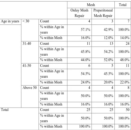
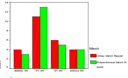
[image:60.595.88.523.222.652.2]maximum number of patients were in 3rd decade of life (48.0%).
Table 1 shows age disribution
Mesh Total
Onlay Mesh
Repair
Preperitoneal
Mesh Repair
Age in years < 30 Count 4 3 7
% within Age in
years 57.1% 42.9% 100.0%
% within Mesh 16.0% 12.0% 14.0%
31-40 Count 11 13 24
% within Age in
years 45.8% 54.2% 100.0%
% within Mesh 44.0% 52.0% 48.0%
41-50 Count 6 5 11
% within Age in
years 54.5% 45.5% 100.0%
% within Mesh 24.0% 20.0% 22.0%
Above 50 Count 4 4 8
% within Age in
years 50.0% 50.0% 100.0%
% within Mesh 16.0% 16.0% 16.0%
Total Count 25 25 50
% within Age in
years 50.0% 50.0% 100.0%
39 Age in years
Above 50 41-50
31-40 Below 30
C
ount
14
12
10
8
6
4
2
0
Mesh
Onlay Mesh Repair
[image:61.595.94.505.271.524.2]Preperitoneal Mesh R epair
40
Sex
Female Male
C
ount
18
16
14
12
10
8
6
Mesh
Onlay Mesh Repair
Preperitoneal Mesh R epair
Sex distribution
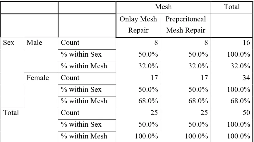
[image:62.595.86.520.189.431.2]In a total of 50 cases, 34 patients were females and 16 patients were males.
Table 2 shows sex distribution
Mesh Total
Onlay Mesh Repair
Preperitoneal
Mesh Repair
Sex Male Count 8 8 16
% within Sex 50.0% 50.0% 100.0%
% within Mesh 32.0% 32.0% 32.0%
Female Count 17 17 34
% within Sex 50.0% 50.0% 100.0%
% within Mesh 68.0% 68.0% 68.0%
Total Count 25 25 50
% within Sex 50.0% 50.0% 100.0%
[image:62.595.112.490.476.778.2]% within Mesh 100.0% 100.0% 100.0%
41
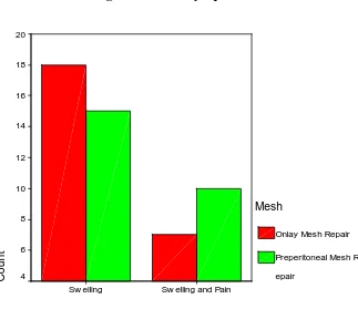
Symptoms
In total 50 cases, 33 patient has only swelling and 17 patient has both
swelling and pain.
SL NO. SYMPTOMS NO. OF PATIENTS PERCENTAGE
1 Swelling 33 66
2 Swelling & Pain 17 34
Tables 3 shows symptoms
Mesh Total
Onlay Mesh Repair
Preperitoneal Mesh Repair
Compliants Swelling Count 18 15 33
% within
Compliants 54.5% 45.5% 100.0%
% within Mesh 72.0% 60.0% 66.0%
Swelling
and Pain
Count
7 10 17
% within
Compliants 41.2% 58.8% 100.0%
% within Mesh 28.0% 40.0% 34.0%
Total Count 25 25 50
% within
Compliants 50.0% 50.0% 100.0%
42 Compliants
Sw elling and Pain Sw elling
C
ount
20
18
16
14
12
10
8
6
4
Mesh
Onlay Mesh Repair
Preperitoneal Mesh R
[image:64.595.146.469.100.379.2]epair
43 Diabetes Mellitus
No Yes
C
ount
20
18
16
14
12
10
8
6
4
Mesh
Onlay Mesh Repair
Preperitoneal Mesh R
[image:65.595.85.529.113.379.2]epair
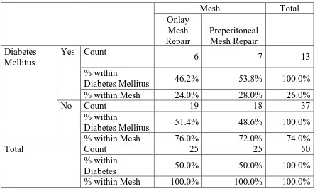
Table 4 Diabetes Mellitus * Mesh
Mesh Total
Onlay Mesh Repair
Preperitoneal
Mesh Repair
Diabetes Mellitus
Yes Count
6 7 13
% within
Diabetes Mellitus 46.2% 53.8% 100.0%
% within Mesh 24.0% 28.0% 26.0%
No Count 19 18 37
% within
Diabetes Mellitus 51.4% 48.6% 100.0%
% within Mesh 76.0% 72.0% 74.0%
Total Count 25 25 50
% within
Diabetes 50.0% 50.0% 100.0%
[image:65.595.139.468.435.699.2]% within Mesh 100.0% 100.0% 100.0%
44
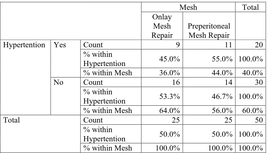
Table 5 Hypertention * Mesh
Mesh Total
Onlay Mesh Repair
Preperitoneal
Mesh Repair
Hypertention Yes Count 9 11 20
% within
Hypertention 45.0% 55.0% 100.0%
% within Mesh 36.0% 44.0% 40.0%
No Count 16 14 30
% within
Hypertention 53.3% 46.7% 100.0%
% within Mesh 64.0% 56.0% 60.0%
Total Count 25 25 50
% within
Hypertention 50.0% 50.0% 100.0%
[image:66.595.132.479.399.731.2]% within Mesh 100.0% 100.0% 100.0%
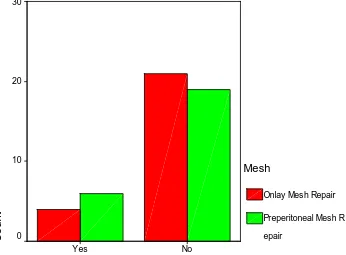
Figure 5 Hypertention
Hypertention
No Yes
C
ount
18
16
14
12
10
8
Mesh
Onlay Mesh Repair
45
Chronic Obsructive Pulmonary Disease
No Yes
C
ount
30
20
10
0
Mesh
Onlay Mesh Repair
[image:67.595.88.522.140.425.2]Preperitoneal Mesh R epair
Table 6 Chronic Obsructive Pulmonary Disease * Mesh
Mesh Total
Onlay Mesh Repair
Preperitoneal
Mesh Repair
COPD Yes Count 4 6 10
% within COPD
40.0% 60.0% 100.0%
% within Mesh 16.0% 24.0% 20.0%
No Count 21 19 40
% within COPD
52.5% 47.5% 100.0%
% within Mesh 84.0% 76.0% 80.0%
Total Count 25 25 50
% within COPD
50.0% 50.0% 100.0%
% within Mesh 100.0% 100.0% 100.0%
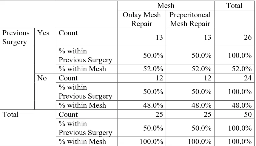
[image:67.595.143.489.485.740.2]46 Previous Surgery
No Yes
C
ount
13.2
13.0
12.8
12.6
12.4
12.2
12.0
11.8
Mesh
Onlay Mesh Repair
Preperitoneal Mesh R
[image:68.595.81.501.128.726.2]epair
Table 7 Previous Surgery * Mesh
Mesh Total
Onlay Mesh Repair
Preperitoneal
Mesh Repair
Previous Surgery
Yes Count
13 13 26
% within
Previous Surgery 50.0% 50.0% 100.0%
% within Mesh 52.0% 52.0% 52.0%
No Count 12 12 24
% within
Previous Surgery 50.0% 50.0% 100.0%
% within Mesh 48.0% 48.0% 48.0%
Total Count 25 25 50
% within
Previous Surgery 50.0% 50.0% 100.0%
% within Mesh 100.0% 100.0% 100.0%
Figure 7 Previous Surgery
[image:68.595.87.516.130.376.2]47
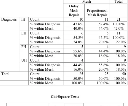
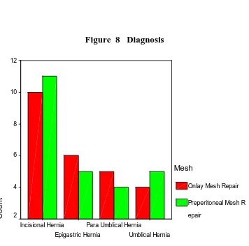
Table 8 Diagnosis * Mesh
In total 50 cases, 21 cases diagnosed as incissional hernias, 11 cases as
epigastric hernia, 9cases as paraumblical hernia and 9 cases as umbilical hernia.
Mesh Total
Onlay Mesh Repair
Preperitoneal Mesh Repair
Diagnosis IH Count 10 11 21
% within Diagnosis 47.6% 52.4% 100.0%
% within Mesh 40.0% 44.0% 42.0%
EH Count 6 5 11
% within Diagnosis 54.5% 45.5% 100.0%
% within Mesh 24.0% 20.0% 22.0%
PH Count 5 4 9
% within Diagnosis 55.6% 44.4% 100.0%
% within Mesh 20.0% 16.0% 18.0%
UH Count 4 5 9
% within Diagnosis 44.4% 55.6% 100.0%
% within Mesh 16.0% 20.0% 18.0%
Total Count 25 25 50
% within Diagnosis 50.0% 50.0% 100.0%
% within Mesh 100.0% 100.0% 100.0%
Chi-Square Tests
Value Df Asymp. Sig. (2-sided)
Pearson Chi-Square .361(a) 3 .948
Likelihood Ratio .361 3 .948
Linear-by-Linear
Association .000 1 1.000
No. of Valid Cases 50
A 4 cells (50.0%) have expected count less than 5. The minimum expected
48
Figure 8 Diagnosis
Diagnosis
Umblical Hernia Para Umblical Hernia
Epigastric Hernia Incisional Hernia
C
ount
12
10
8
6
4
2
Mesh
Onlay Mesh Repair
49
Table 9 Ultrasonogram * Mesh
Mesh Total
Onlay Preperitoneal
USG Confirmed Count 25 25 50
% within USG 50.0% 50.0% 100.0%
% within Mesh 100.0% 100.0% 100.0%
Total Count 25 25 50
% within USG 50.0% 50.0% 100.0%
[image:71.595.87.502.199.574.2]% within Mesh 100.0% 100.0% 100.0%
Figure 9 Ultrasonogram
Content * Mesh
Ultrasonogram
Conf irmed
C
ount
26
24
22
20
18
16
14
12
Mesh
Onlay Mesh Repair