A STUDY OF ANATOMY AND VARIATION
OF THE HUMAN MITRAL VALVE
APPARATUS IN AUTOPSY SPECIMENS
Dissertation submitted to
The Tamil Nadu Dr. M.G.R. Medical University, Chennai
in partial fulfillment of the requirements for the degree of
MCh Cardiothoracic Surgery
Branch II
CERTIFICATE
This is to certify that Dr.T.M.PONNUSAMI postgraduate student (2003 - 2006) in the
Department of cariothoracic surgery, Government General Hospital Chennai & Madras
Medical College, Chennai - 03, has done this Dissertation of "A STUDYOF NORMAL
ANATOMY AND VARIATIONS OF THE HUMAN MITRAL VALVE APPARATUS IN
AUTOPSY SPECIMENTS under my guidance and supervision in partial fulfillment of the
regulations laid down by TheTamil Nadu Dr.M.G.R. Medical University, Chennai, for MCh
cardiothoraci - Branch II examination to be held in August 2006.
Prof. Dr. K.HARSHAVARDHAN MCh.,
Prof & Head of the Department of Cardiothoracic Surgery,
Madras Medical College & Govt. General Hospital
Chennai - 03.
DEAN
Madras Medical College & Govt. General Hospital
ACKNOWLEDGEMENT
A great many people made this work possible. I thank our Dean Dr.
Kalavathypooniraivan, MD, Madras Medical College, Chennai for allwoing me to conduct this
study.
I would like to thank my advisor Prof. K.HARSHAVARDHAN., MCh., Head of the
Department for his guidance advice, encouragement, and enthusiasm.
I would like to thank retired Prof. SOLOMON VICTOR MCh. MRCP, FRCS, for his
guidance and helpful discussions. I would like to thank retired Prof. RAJANSANTHOSAM,
MCh, for the many useful comments made during this project. I acknowledge Prof.
T.S.Manohar, Prof. Dr.VISHWAKUMAR, Prof. VENKATACHALAPATHY and Prof.
VIJAYAN for the many useful comments they made during this project. In addition, I am
grateful to Dr.Raghupathy, Dr.Sukumar, Dr.Rajavenkatesh, Dr.Balanayagam, Dr.Sasankh,
Dr.Nagarajan for their guidance. Last but not the least I thank FORENSIC SCIENCE and
CONTENTS
SL.NO. CONTENTS PAGE NO
1 Certificate
2 Acknowledgements
3 Introduction & Review of Literature 1
4 Materials and Methods 4
5 Aim of the Study 5
6 Development of the Heart 6
7 Anatomy of Mitral Valve Proper 18
8. Conclusion and Tables 33
9 Pictures of Mitral valve Apparatus 39
10 Discussion and Conclusion 48
INTRODUCTION
One hundred human hearts from autopsies are studied to clarify controversies in the
literature about commissures, slits, chordae and leaflets of the mitral valve. This study revealed
that no 2 hearts are alike in the morphology of commissures, slits and scallops. We have
designed perpendiculars drawn from the annulus to the free edge at the shortest height of the
mitral valve on either side of the aortic leaflet as the anterolateral (AL) and posteromedial (PM)
commissural lines. These lines divide the mitral valve precisely into aortic and mural leaflets.
Slits in the leaflets are easily identified by the dipping in of the free edge into the cusp tissue.
There are no slits in the aortic leaflet. Ninety-eight hearts has slits in mural leaflet varied from
one to five. Unlike the relatively static and straight annulus of the aortic leaflet. The curved
annulus of the mural leaflet contracts and changes in contour during systole. Hence slits are
necessary in the mural leaflet to help it fold and adapt to the reduced orifice during systole and
unfold during diastole. In addition to this coarse adjustment of the mural leaflet, both leaflets
are pleated due to "hooding up" of the leaflet tissue between chordal attachments during
systole, providing the fine tunning to enable the leaflets to adapt themselves to the reduce
systolic orifice. The nodular appearance (rather than a straight ridge) of the line of apposition
of the leaflet is evidence of this pleating mechanism during valve closure. Slit lines, designed
as perpendiculars drawn from the summit of the slits to the annulus, are used arbitrarily to
divide the mural leaflet into 2 to 6 scallops. When slits are absent there are no scallops. The
slits and scallops are best serially numbered counterclockwise from the surgeon's view through
the atrium. The scallops immediately behind the commissural lines, customarily labeled as the
commissural line and the closet AL or PM slit line. All commissural scallops are seen in 81
hearts and PM commissural scallops in 76 hearts. There is no consistent fan-shaped chordal
pattern at the commissural lines or at the slits. However the chordae arising from each papillary
muscle group, in total, from a fan, reaching out to the corresponding adjacent halves of the 2
leaflets, restraining their splaying out in diastole and upward bulge during systole. These
findings are relevant to the pathogenesis of mitral valvular disease and reparative procedures.
The Central Mitral Plane passing through the middle of the aortic and mural leaflets
divides the chordopapillary support of the mitral value into anterolateral and posteromedial
halves. The papillary muscles of the mitral valve are studied in 100 human autopsy hearts
collected at random. the anterolateral papillary support has 1 belly in 67 hearts, 2 in 27, 3 in 4,
4 in 1, and 5 in 1 heart. Likewise, the postermedial papillary support has 1 muscle belly in 50
hearts, 2 in 36, 3 in 11, and 4 in 3. The single papillary muscle is conical, mammilated, flat
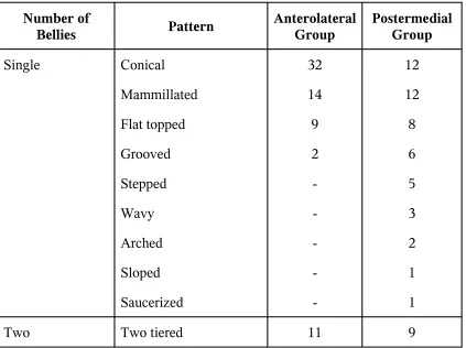
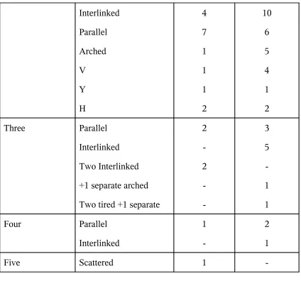
topped, grooved, stepped, wavy, arched, sloped or saucerized. When there are two bellies they
presented as two tiered, interlinked, parallel, arched, V, Y, or H configuration. Three papillary
muscles formed a parallel, interlinked arrangement with the third belly separately. When four
or five bellies existed, they are parallel or interlinked. In the anterolateral and posteromedial
group, the papillary muscle bellies are mostly intraluminal in 14% and 11%, mostly
intraluminal with the tip anchored in 19% and 28% equally sessile and intraluminal in 54.5%
and 41.5% mostly sessile in 12.5% and 19.5% respectively. In the anterolateral group 19% of
papillary muscle bellies arises from the upper third of the ventricle, 70.5% from middle third,
and 1.5% from lower third, the corresponding figures for postermedial group are 6% 92.5% and
in 14 to 72 chordale arises from the posteromedial papillary group ended in 12 to 89 leaflet
insertions. The chordae in each group are best considered in to as a fan. The configuration of
the fan is unique in each heart. Imaging techniques need to be refined to outline these variations
more precisely. the relevance of chordopapillary variations in rheumatic heart disease,
reparative procedures, papillary muscle dysfunction, mitral value prolapse, mitral valve
MATERIAL AND METHODS
One hundred normal human autopsy hearts collected at random are studied. The age and
sex of persons are not known. Looking through the left atrium, the mural leaflet is divided
between its anterolateral and posteromedial chordopapillary support. A few false chordae
connecting the two groups of papillary muscles are divided to splay the valve open with the
aortic leaflet in the middle, the anterolateral half of the mural leaflet with anterolateral group of
papillary muscles on the left, and the posteromedial half of mural leaflet with posteromedial
group of papillary muscles on the right.
The number of papillary muscle bellies in each varied from 1 to 5. When single, the
shape and size of the muscle belly varied. When there are 2 to 5 muscle bellies they form
various configurations.
The papillary muscles varied in the extent of protrusion into the ventricular cavity and
adherence to the ventricular wall. When mostly intraluminal, the tip of the muscle may be
anchored to the ventricular wall by a small mjuscle band. Commonly, papillary muscle are
related to the middle third of the ventricle.
The variations in number of chordae arising from each group of papillary muscle is
studied. The number of chordae inserting into the free edge of the leaflets in the corresponding
half of the valve is also noted.
In two hearts, the anterolateral papillary muscle remained muscular upto the annulus
also, the strut chordae to the aortic leaflet in two hearts had separate muscular bellies.
AIM OF STUDY
apparatus helps to preserve anatomical position of mitral valve in its natural position by
preserving all relations of mitral valve in repair of mitral valve instead of replacing with
mechanical valve.
The mechanical valve replacement is a long procedure with life long anticoagulant like
warfarin.
In view of, life long management of mechanical valve with anticoagulants it is
cumbersome for Indian Poor People. So, it is well advised to preserve mitral valve with repair
instead of replacement with mechanical valve. So by studying the normal anatomy of mitral
valve apparatus we can preserve the natural parts of mitral valve in its normal anatomical
DEVELOPMENT OF MITRAL VALVE
The atrioventricular valves are complex entities made up of the annulus, the leaflets, and
the supporting tension apparatus, the latter comprising the tendinuous cords and the papillary
muscles. The leaflets are hinged from the annulus, which is an integral part of the
atrioventricular junction. In the definitive heart, the tricuspid and mitral valves are separated by
septal structures, which are absent in hearts having a common atrioventrcular junction.
To understand the development of these valvular complexes, particularly the presence in
malformed hearts of a common atrioventricular valve, valvar formation must be examined in
the context of cardiac development as a whole. In addition to cardiac septation, interdependent
and mutually generative processes closely linked to the formation of the atrioventricular valves
include the mechanisms of connection of the right atrium to the right ventricule, and
incorporation of the subaortic outlet into the left ventricle, and incorporation of the subaortic
outlet into the left ventricle. In this review, we will seek to integrate these themes as we
describe how an initially unseptated and valveless tube is changed into a complex four
chambered organ, with the chambers and arterial trunks separated by a system of one-way
valves, concentrating attention of the atrioventricular valves. We will also speculate on how
normal development can be perturbed to produce some of the lesions seen in modern surgical
practice.
Primary Heart Tube
After the third week of gestation, a lumen forms within the primary endocardium of the
becomes converted into the initially valueless and inverted Y-shaped tube that initiates the
circulation. With time, this primordium will become the larger part of the left ventricle and part
of the atrial chambers. The future right ventricle and outflow tract, along with the remainder of
the left ventricle and the atriums, take their origin from a spatially distinct area, known as the
secondary heart field, with the cells migrating at a later state from the area to become integrated
within the arterial and venous poles of the developing heart tube.
The atrioventricular junction, the seat of the future atroiventricular valves, comes into
prominence following rightward looping of the heart tube, after the 25th day of gestation of
development. Looping occurs as the initial attachment of the tube to the body wall disappears,
this connection being the so called dorsal mesocardium. The atrial remnant of this structure
then serves as the site of entry of the pulmonary vein and the vestibular spine during the 7th
week. As we will see, the spine then plays a crucial role in atrial septation. Subsequent to
looping, the developing heart takes on the more characteristic three-dimensional appearance of
the mature organ. By the end of the 5th week, the developing ventricles have become visible as
pouches that balloon from the primary tube, with the primordium of the muscular ventricular
septum also being visible. At this stage of development, the primordial left ventricle supports
the developing right ventricle provides most of the muscular support for the developing
ventricular outflow tract. The lumen of the atrioventricular canal is largelyoccupied by two
large mesenchymal masses, the superior and inferior atrioventricular endocardial cushions.
Initially unfused, the cushions face each other within the canal, leaving slits on each side
between their edges and the lateral margins of the canal. These slits will eventually expand to
right-sided slit provides continuity between the developing right atrium and right ventricle
through the lumen of the primary heart tube, with the inner curve of the tube forming the roof
of this communication, which is called the primary interventricular foramen. Studies of human
embryos stained with an antibody revealed that the myocardium surrounding the foramen is
distinct from the remainder of the primary myocardium with part becoming transformed into
the atrioventricular node and bundle. It is expansion and remoulding within this region of
primary myocardium, known as the primary ring, that provides the substrate for formation of
first the right ventricular inlet, and then the tricuspid valve.
This right side of the atrioventricular canal expands across the developing muscular
ventricular septum as the superior and inferior cushions fuse within the lumen of the canal,
their rightward margins then becoming draped across the crest of the septum. Parts of the fused
cushions, nonetheless, remain to the left side of the septal crest, with their bulk protruding in to
the cavity of the left ventricle, where they will form the aortic leaflet of the mitral valve. The
parts spanning the crest of the developing muscular septum, which are the first parts to fuse,
will form the larger part of the membranous septum. This structure, along with contributions
from the outflow cushions, will eventually partition the aorta into the left ventricule.
Fate of the Primitive Atrioventricular Canal
Fusion of the atrioventricular endocardial cushions during the sixth week of
development divides the atrioventricular canal into the primordiums of the right and left
atrioventricular junctions, to which the developing leaflets of the mitral and tricuspid valves
beginnings of not only ventricular, but also atrial septation. The earliest indication of atrial
septation is the downgrowth of the primary septum, or "septum primum", from the atrial roof.
As it grows towards the cushions within the atrioventricular canal, this primary septum carries
a cap of mesenchyme on its leading edge. At the same time, contiguous with the right margin
of the dorsal mesocardial; connection, a further mass of mesenchyme grows into the heart at
the level of the base of the developing atrial septum. This latter structure is the so-called "
spina vestibuli", or vestibular spine, which is separated from the mesenchymal cap.
The tissue of the spine, together with the mesenchymal cap clothing the leading edge of
the primary atrial septum, merges with the atrial margins of the fused atrioventricular
endocardial cushions to close the primary atrial foramen, or "ostium primum". The vestibular
spine itself then muscularizes to form the thick base of the atrial septum. By this time, the
initial musculature of the atrioventricular canal is becoming incorporated into the now divided
atrioventricular junctions as the atrial vestibules. The final separation of the musculature from
the ventricular walls does not occur until much later in development, when the fibro-adipose
tissues of the atrioventricular grooves separates the atrial and ventricular muscular segments at
all sites other than the location of the bundle of His. The point of penetration of the bundle of
His marks the site within the septal components of the initial atrivoentricular canal
musculature.
Failure of fusion of the superior and inferior cushions is the process usually held
responsible for producing atrioventricular septal, or "canal", defects. Indeed, for many years
this group of malformations is labeled as "endocardial cushion defects". Recent research,
common atrioventricular junction, this feature being the halmark of the malformations. Failure
of this mesenchymal front to contribute to the base of the developing atrial septum permits the
primary foramen to remain patent and, at the same time, ensures persistence of the common
atrioventricular junction. When a valve is eventually formed in the setting of this common
junction, it bears scant morphologic resemblance to the normal mitral and tricuspid valves. This
is because the space between the bridging leaflets formed from the superior and inferior
atrioventrucular cushions in an integral part of the valvar orifice. The location of this space
reflects the arrangement seen very early during normal development. It is also case that,
subsequent to separation of the left atrioventricular junction, clefts of varying depth can be
found in the aortic leaflet of the otherwise normally formed mitral valve. Such clefts can also
be found when the mitral valve straddles through a ventricular septal defect opening to the
outlet of the right ventricle. Thus, although the zone of apposition found between the left
ventricular components of the bridging leaflets in a heart with an atrioventricular septal defect
with common atrioventricular junction, like the cleft of the aortic leaflet of an otherwise normal
mitral valve, exists because of failure of fusion of the atrioventricular endocardial cushions, it
is only the leftward tips of the cushions that have failed to fuse when there is an otherwise
normally structured mitral valve.
Development of the Mitral Valve
Formation of the normal mitral valve not only required division of the atrioventricular
canal, but also cannot proceed until the developing aorta becomes committed to the left
ventricle. In the definitive heart, almost always there is fibrous continuity between two of the
valve in continuity with the aortic root named the aortic leaflet, thus distinguishing it from the
mural leaflet, which is hinged from the parietal atrioventricular junction. As well will see, these
morphologic difference in the hinges of the leaflets reflect their development heritage.
The building blocks of the valvar leaflets are the endocardial cushios. Formation of the
definitive valve, therefore, requires reorientation of the newly separated left atrioventricular
junction, which expands in inferior direction. This inferior reorientation occurs concomitant
with incorporation of aorta into the outlet part of the left ventricle. Blood from the left ventricle
initially reaches the developing aorta through the primary interventricular foramen, with the
fused atrioventricular cushions forming the left ventricular border of the roof of this pathway.
As the outflow cushion fuse, and muscularize to separate the subpulmonary infundibulum from
the subaortic outlet, they also fuse with the crest of the muscular ventricular septum, thus
walling the developing aortic valve into left ventricle. The aortic root then occupies the space
that has appeared, concomitant with the expansion and reorientation of the left atrioventricular
junction, between the ventricular septum and the fused left ventricular components of the
atrioventricular cushions. This space forms a bay on the ventricular aspect of the
atrioventricular cushions. When the aorta is first walled into this newly created by within the
left ventricle, the myocardium of the inner heart curve continues to separate the developing
aortic valver leaflets from the cushions fusing to form the aortic leaflet of the mitral valve.
Only subsequent to the completion of septation does this muscle disappear, thus establishing
the definitive arrangement of fibrous continuity between the aortic leaflet of the mitral valve
and the noncoronary and left coronary leaflets of the aortic valve.
the ventricle, the two ends of this expanded crescent are associated with compacting columns in
the trabecular, or spongy, layer of the ventricular muscle. These columns, which will form the
papillary muscles, are positioned to support not only the ends of the lateral cushion, but also the
distal ends of the fused atrioventricular cushions. The layer of spongymyocardium that initially
supports the lateral cushion, however, will subsequently disappear. As it does so, the
endothelially derived lateral cushion itself becomes transformed into the mural leaflet of the
mitral valve. The liberated myoardium that initially joined the cushion to form papillary
muscles will also disappear with time, the myocardial, cells being replaced by fibrous tissue. A
similar process occurs at the interface between the edges of the developing aortic leaflet and the
tips of the papillary muscles, with fibrous tension apparatus eventually replacing the
myocardium.
Excessive or abnormal compactness of the trabecular layer of the developing ventricular
myocardium is responsible for producing the so-called "parachute" deformity of the valve,
either with a solitary papillary muscle supporting the entirety of the valvular complex, with a
leash of cords fanning out from this signal locus, or with incomplete formation of one of the
two papillary trabecular columns. Failure of formation of the tendinous cords from the original
myocardial primordiums results in the "hammock" or "arcade" lesions of the mitral valve, with
THE NORMAL MITRAL VALVE APPARATUS
The normal mitral valve apparatus is composed of various structures including the mitral
ring, the valve proper, the chordae tendineae, and the papillary muscles.
The Mitral Ring
The mitral (atrioventricular) ring or annulus in an important part of the skeleton of the
heart. It is an anteriorly incomplete, circular, and fibroelastic structure. It represents the
dividing line between the atrial and ventricular musculature from the eighth week of
intrauterine life and appears in the embryonic heart as a result of the crowding together of the
connective framework of the organ. The ring's upper border is the point of origin of the
ventricular muscular. It has recently been shown that the atrioventricular annulus decreases its
diameter during each systolic contraction. This event, which takes an effective part in the
closure of the normal mitral valve, is probably the result of the systolic contraction of the deep
bulbo and Sino Spiral muscles with the superficial bulb spiral muscle assisting in a minor role.
An impression is gained that the orifice is completely obstructed during the late stage of the
cardiac systole by the sphincter like action of the annulus.
TABLE - I
(100 HEARTS)
Structures Measured
MEN WOMEN
Height Breadth Height Range
Average Range Average Range Average Range Average Range
Aortic Leaflet 2.4 3.2-1.9 3.7 4.5-2.5 1.2 2.7-1.8 3.3 4.2-2.4
Ventricular Leaflet 1.4 2.5-1.0 3.3 4.1-2.5 1.2 2.4-0.8 3.0 3.6-2.3
Anterior Accessory
Leaflet 1.1 1.8-0.8 1.5 1.8-1.1 1.0 1.3-0.7 1.2 1.6-1.0
Posterior accessory leaflet
0.9 1.2-0.6 1.1 1.5-0.8 0.9 1.0-0.7 0.8 1.2-0.7
Anterior junctional tissue
0.8 1.2-0.6 1.7 2.4-0.7 0.7 1.1-0.6 1.5 2.1-0.7
Posterior junctional tissue
0.7 0.9-0.5 1.3 1.8-0.7 0.6 0.8-0.4 1.2 1.6-0.7
Circumference of valve
ring - - 10.0 11.5-8.5 - - 9.0 10.5-8.5
The outer aspect of the mitral annulus constitutes the deepest part of the atrioventricular
groove on the external surface of the heart. The mitral valve takes origin as a continuous veil
from the inner aspect of the ring and inserts around tee entire circumstance of the mitral orifice.
In this study we found that the circumstance of the mitral annulus is in average 10cm
(range: 8.5 to 11.5 cm) in the male specimens and 9 cm (range: 8 to 10.5 cm) in the female
specimen (Table 1). The readings obtained are very seldom found at variance with these
THE MITRAL VALVE PROPER
The mitral valve is represented by a continuous veil of valvular tissue attached as a muff
to the entire circumference of the atrioventricular annulus. Its free edge is split indentations,
none of which reaches the mitral annulus. These clefts divide the indentations, none of which
reaches the mitral annulus. These clefts divide the valvular tissue into two major leaflets which
are always present and into two minor accessory cusps not invariably represent. These minor
cusps, when present, are at the area of the junctional tissue.
The valve tissue consists of a fibroelastic texture covered on the atrial and ventricular
aspect by a coat of endocardium. The so-called fibrous skeleton which gives strength and shape
to the valve is furnished by a layer of connective tissue protruding from the inner aspect of the
mitral ring. Important contributions to the valve-supporting framework are the chordal
insertions. Rusted and his associates noted that the chordal insertions passes through the
substance of the leaflet for a considerable distance. This particular arrangement, as pointed out
by Mayo Clinic Group, is often seen on the transiluminated posterolateral leaflet, but more
often a fibrous band running along the margin of closure is seen in this leaflet. This fibrous
structure can be considered as the point of insertion of the chordae. Neither the junctional tissue
nor other additional valve leaflets show any such arrangements of their connective layers.
The two major cusps are attached to the ring in such a way as to be in anteromedical and
posterolateral locations. The anteromedial cusp has been referred to as the anteromedial, aortic,
septal or greater leaflet. The posterolateral has been designated as the posterlateral, ventricular
form right to left.
The Anteromedial or Aortic Leaflet
The aortic leaflet is the most important from the anatomical as well as the physiological
standpoint. Table-I shows the linear measurements of the various value leaflets. Table II lists
the surface measurements of the same structures, obtained by planimetric integration of the
removed valvular tissue. The aortic cusp is by far the largest. It exceeds every other leaflet in
height and breadth at the mitral ring. Its surface area gives a better idea of the importance of
[image:20.612.91.520.368.733.2]this cusp, because it is equal to almost one-half of all tissue surface area.
TABLE - II
Structures Measured MEN (8 Subjects) WOMEN (8 Subjects
Average Range Average Range
651 833-427 563 714-413
Ventricular leaflet 384 532-270 334 456-413
Anterior accessory leaflet 227 257-193 203 220-183
Posterior accessory leaflet 136 148-110 120 128-112
Anterior junctional tissue 171 200-129 144 171-125
Posterior junctional tissue 92 110-70 78 94-67
Valvular tissue area 1398 1770-1000 642 1518-970
The anteromedial cusp is roughly triangular in shape and, in our experience, its free edge
has never been found split.
It is the only dividing structure between the mitral and the aortic orifice. Directly inferior
to the aortic canal, it constitutes an integral part of the outflow tract of the left ventricle. Due to
this anatomical location, a considerable portion of its function is believed to be the direction of
the flow of blood toward the aorta. This has been described by Harken as the aortic baffle. In
other words, the leaflet apparently acts as a watershed which deflects the blood toward the
aorta in the ejection of the ventricular contraction.
Close anatomical observation of the normal heart as well as experimental investigation
of the valvular function would not be in favour of this view. The leaflet has the greatest
importance in the closure of the mitral valve, but it has no baffling action on the blood flow,
indeed, the blood driven forcefully from the inflow into the outflow tract has no other outlet but
the aortic orifice, since the mitral valve is, at this stage of the systole, hydraulically closed. The
physiological lines of progression upon which the blood is forced out into the aorta run directly
from the apex of the heart to the aortic canal. The contribution of the anteromedial leaflet to the
information of these lines of direction is certainly not greater than that of the interventricular
septum or the anterior left ventricular wall. The last mentioned two structures together with the
septal cusp constitute the walls of the outflow tract of the left ventricle. If a defect is produced
in the aortic leaflet, it is possible to do in experimental surgery, a great valvular incompetence
Likewise, a defect of the interventricular septum will permit on escape of blood into the right
ventricle. The two situations, the mitral reflux and the escape of blood through an
interventricular septal defect, are indeed dynamically analogous since both are governed
mainly by the intracavity pressure. On the other hand, the muscular fibers of the outflow tract
appear to play an important role in giving a definite direction to the blood flow. This fact is
demonstrated by the position of the left ventricle in the cases of prevailing hypertrophy of the
left ventricular outflow tract.
The Posterolateral or Ventricular Leaflet
The smaller ventricular leaflet is not as important as the aortic leaflet, but by no means
should it be disregarded in the mechanism of valve closure. This is confirmed by experimental
and clinical facts. The posterolateral cusp is roughly equal to one-third of all the valvular tissue
surface area. Its breath at the line of insertion is only a few millimeters less than that of the
aortic leaflet, while its height is almost one-half that of the anteromedial cusp (Table 1). The
contour is usually quadrangular with sloping sides and its free edge is often notched. However,
the notching is seldom deep enough to be mistaken as a cleft separating the poseterolateral
leaflet from an additional cusp. The importance of the ventricular leaflet in the normal mitral
valve to have been generally under estimated.
It was originally thought that the complex mechanism of the normal mitral valve
function could be simply described as an unpretentious or moderate systolic and diastolic
swinging movement of the septal leaflet. According to the see, the contraction of papillary
right half of the mitral ring an across the orifice, which would remain entirely hidden." At the
same time, the posterolateral ventricular wall "would march" toward the septal leaflet and come
into contact with its free margin. In diastole, the aortic leaflet would swing back opening the
valve. Consequently, the ventricular leaflet appeared to be a worthless structure as far as the
mechanism of the mitral valve closure was concerned. The perivalvular space was thought to
be entirely obliterated in systole by the posterior ventricular wall moving toward the aortic
cusp.
The posterior leaflet is sqeezed and butterssed by the posterolateral wall of the left
ventricle in systole. In diastole, the myocardium swings, away from the normal veli-like,
posterior mitral leaflet", mechanism of closure may well happen sometimes.
In the first place, the digital exploration of a normal human mitral valve in a mistaken
preoperative diagnosis of mitral stenosis, revealed a remarkable billowing effect of the
ventricular leaflet toward the left atrium. The ventricular leaflet could be forced out into the
atrium only by the blast of blood squeezed into the perivalvular space. The same could not be
produced by the ventricular wall being pushed against and butterssing the posterolateral leaflet.
Moreover, the digital pressure on the ballooned-out ventricular leaflet produces a feeling of
resilience like that of squeezing a fluid filed bag which would hardly be felt were the muscular
wall responsible for the bulging. In addition, this same maneuver causes an immediate mitral
reflux, which further suggests that the perivalvular space is not obliterated in systole.
Second, the chordae tendineae of third order, which constantly connect the ventricular
the heart wall buttresses the leaflet.
Commissural lines
The mitral leaflet tissue formed a continuous veil hanging down from the mitral annulus
except in 2 specimens. A perpendicular line from the annulus to the free edge was used to
measure the height of the leaflet tissue at a given point.
The aortic leaflet had maximum height in its middle. On either side, it is tapered. The
transitional lines at which the height of the leaflet tissue started increasing again are labeled as
AL and PM commissural lines. These passes through the highest point of the broad indentation
or concavity between the 2 leaflets. The height of the commissural lines varied from 2 to 8 mm.
The aortic leaflet is defined as the portion of the mitral curtain included between these 2
commissural lines anteriorly, hanging down from a relatively straight fibrous annulus shared
with the aortic valve. Posteriorly the rest of the curtain of leaflet tissue formed the mural
leaflet, hanging down from a curved annulus, related to the atrioventricular musculature.
Slits
Apart from tiny cremations between chordal insertions, the free edge of the leaflets may
exhibit deep indentations, which we have designated as slits. We prefer the term "Slit" rather
The aortic leaflet had no slits. Slits are seen in the mural leaflet in 98 hearts. The number
of slits varied : 0 in 2 hearts, 1 in 20, 2 in 56, 3 in 15, 4 in 6 and 5 in 1. As a surgeon would
view the valve through the atrium, these slits are serially numbered anticlockwise, from the AL
commissural line to the PM posteromedial in 76. The varied location of slits is noted.
Slit lines and scallops
The perpendicular drawn from the annulus to the deepest part of a slit is designated as
the slit line. Slit line subdivide the mural leaflet into 2 to 6 scallops. It is best to number these
serially. The leaflet tissue between the AL commissural line and closet AL slit line, when
present, is usually labeled as the AL commissural scallop. We saw this in 81 hearts.
Commissural and slit chordae
The chordae in relation to each commissural line, and the closet slit line, are traced
down to their origin and studied, All these chordae start as a single stem from their origin and
subdivided prior to insertion into the leaflet tissue either at the free edge, rough zone, or both.
Rarely there are 2 parallel chordae on either side of the commissural or slit line. The gross
appearances of the free edge chordae, in relation to the commissural or slit, varied Only 47% of
AL commissural chordae, 53% of PM commissural chordae. 39% of first AL slit chordae and
29% of last PM slit chordae were fan shaped. The length of these chordae span of attachment at
the free edge varied widely. The main stem usually inserts into the submit of the commissure or
slit.
When the commissural chordae are parallel with chordal insertion either side of the
to 5mm in 9 hearts. In 3 hearts the commissural scallops had basal chordae. The commissural
chordae had varied origins. The origins of slit chordae are found to be similarly varied.
Muscular Chordae
In 2 hearts the mitral veil of the leaflet tissue was interrupted at the location of the AL
commissural line by a muscular chorda which extended up to the annulus. The leaflet tissue is
attached to the sides of this muscular chorda close to the annulus.
Line of apposition
In order to observe nodular thickening at the line of apposition, the leaflet tissue adjacent
to commissural lines is transilluminated. Normally in both the leaflets, the line of apposition
consisted of a row of thickened nodules rather than a linear ridge. These are absent in the
scallops behind the commissural lines in 95 hearts and faintly observed in 5 hearts.
Discussion
Several tissues regarding the anatomy of the normal mitral value remain to be clarified.
Commissures
The free edge of the mitral skirt needs to have indentations, slits and pleats to allow it to
splay open, providing a large orifice during diastole, and to close neatly during systole. Two
constant, shallow, wide indentations divide the mitral value into an aortic leaflet and mural
leaflet. The junctions between the 21 leaflets commonly called commissures have been
Tissue joining the 2 leaflets;
Junctional zones of valvular tissue;
Point of attachment of mitral annuals to the fibrous trigones;
Indentations at either and of the aortic leaflet;
Angles at which the 2 leaflets meet;
Clefts separating anterior and posterior leaflets;
The space between identifiable components of the skirt of leaflet tissue;
Area of leaflets covered by typical commissure chordae have been considered as the
commissural area, which would include adjacent leaflet tissue. There has been dispute as to
whether the commissural is an anatomical reality or a pathologic entity. Papillary muscles and
chordal grooves have also been taken as guides to the commissures. We propose AL and PM
commissural lines as precise lines separating the 2 leaflets.
Slits and scallops of mural leaflet
The relatively straight intervalvular segment of the mitral annulus between the 2 fibrous
trigones does not alter in length during the systole. Hence the aortic leaflet hanging down from
this segment has no need to become folded. An un-slit aortic leaflet offers a smooth outflow
from the left ventricle.
In contrast, the C-shaped annulus of the mural leaflet, related to the atrioventricular
leaflet needs to have slits, to enable it to vary in size and contour. These slits divide the leaflet
into segments which vary in size and number and are unique in each heart. Instead of giving
names which may be confusing, it is best to number serially the segments or scallops,
commencing from scallop 1, behind the anterolateral commissural line. When there are no slits,
the mural leaflet is undivided, and its free border forms an uniform are.
The scallops just behind commissural lines have special pathological and surgical
significance. They have been labeled as commissural leaflets, accessory leaflets commissural
scallops or projections. The commissural scallops considered as separate leaflets of a
quadricuspid valve. Without AL or PM slits, there would be no separate demarcation of a
commissural scallop, which implies that such a demarcation is not essential for normal mitral
valve function.
The inconstant commissural scallops, varying in size and number, do not merit an
exclusive status. They are, when present, the first and last of the serially numbered scallops of
the mural cusp. This part of the mural leaflet tissue, whether it contains slits and scallops or
not, allows the central part of the mural leaflet to move well away from the aortic leaflet during
diastole. During systole, this segment does not appear to bear the brunt of force of closure of
the valve because it is usually devoid of nodular thickenings seen at the line of apposition of
the rest of the leaflets.
We noted lack of fibrous thickening of the junctional tissue. This indicates that at this
region, the leaflet tissue merely gets folded and gently plugs the two ends of the cresentric line
lines, an unique mechanism designed for closure of an asymmetrical value, with a curvilinear
closure line.
Commissural and slit chordae
Typical fan-shaped chordae help to locate the commissures and slits. The indentations
on either side of the anterior leaflet and slits in the mural leaflet are obvious. It is unnecessary
to depend on any chordae to locate them. Moreover, the chordae in relation to commissural and
slit lines are neither typical nor always fan-shaped. The configuration in each heart is unique.
Their primary function is to prevent the leaflet tissue on either side of the commissural
or slit lines from splaying apart too much during diastole. In fact all the chordae emanating
from the papillary muscles fan out like parachute strings giving a long rope for the leaflets to
the left atrium during systole. These basic functions are achieved with varying chordal
configurations.
Pleating of the leaflets
While intention between the 2 leaflets and slits in the mural leaflet provide a coarse
adjustment for closure of the reduced systolic orifice, it is the pleating of the leaflets like
pleating of a skirt, that provides precision for the closing mechanism. This is achieved by
upward bulging of the leaflet tissue in between the chordal insertions, causing multiple hoods
in the value leaflets, and pleating of the leaflet tissue.
In a study of 100 hearts, we found upward bulging or hooding of interchordal leaflet
does not occur, the line of apposition of the leaflets would be a fairly uniform, straight and
thickened ridge of tissue. Instead, invariably we see a row of nodular thickenings. The nodules
occur due to impact of apposition of the convex hoods. The pleats could be visuvalized during
surgery or in autopsy specimens. Obviously when slits are absent, pleating assumes greater
CONCLUSION
Detailed knowledge of the anatomy of slits and scallops of the mitral valve provides
improved understanding of mitral valve function, which aids the surgeon in understanding
TABLE
THE FREQUENCY OF ACCESSORY
MITRAL VALVE
CASES PERCENT
Mitral valve with anterior and posterior accessory
leaflets 20 20.9
Mitral valve with anterior accessory leaflet 30 28.6
Mitral valve with posterior accessory leaflet 10 11.5
Mitral valve without accessory leaflets 40 39.0
TOTAL 100 100.0 TYPES OF PAPILLARY MUSCLES IN 100 NORMAL HEARTS
Types of papillary muscles Cases Percent Cases Percent
Single 87 82.8 31 29.5
Double 15 14.3 57 54.3
Triple 3 2.9 12 11.4
[image:32.612.56.480.186.494.2]TABLE
TYPES OF INNER SURFACE OF PAPILLARY MUSCLES NORMAL HEARTS
Types of Papillary
Muscles Cases Percent Cases Percent Cases Percent Cases Percent
Single 84 80.0 3. 2.8 31 29.5 -
-Double 8 7.7 7 6.8 15 14.3 41 39.0
Triple 1 0.9 2 1.8 4 3.8 14 13.4
TABLE
Patterns of Papillary Muscles
Number of Bellies Pattern Anterolateral Group Postermedial Group Single Conical Mammillated Flat topped Grooved Stepped Wavy Arched Sloped Saucerized 32 14 9 2 -12 12 8 6 5 3 2 1 1
[image:33.612.59.482.403.719.2]Interlinked Parallel Arched V Y H 4 7 1 1 1 2 10 6 5 4 1 2 Three Parallel Interlinked Two Interlinked
+1 separate arched
Two tired +1 separate
2 -2 -3 5 -1 1 Four Parallel Interlinked 1 -2 1
Five Scattered 1
-TABLE
Extent of Protrusion of Papillary Muscles
Anterolateral
Group Posteromedial Group
Mostly Intraluminal 14% 11%
Mostly intraluminal with tip anchored 19% 28%
Equally sessile and intraluminal 54.5% 41.5%
[image:34.612.57.481.53.449.2]TABLE
Origin of Papillary Muscles
Site Anterolateral Posteromedial
Upper third 19% 6%
Middle Third 79.9% 92.5%
Lower Third 1.5% 1.5%
TABLE
Number of Chordae at Origin
Number of Chordae Anterolateral Group Posteromedial Group
1-3 - 1
4-6 21 31
7-9 51 31
10-12 20 22
13-16 6 10
17-20 1 2
21-23 1
-Range 4-22 2-18
TABLE
Number of Chordae at Origin
Number of Chordae Anterolateral Group Posteromedial Group
10-20 4 9
21-30 40 31
31-40 31 45
41-50 17 9
51-60 5 4
61-70 2 1
71-80 1 1
[image:36.612.56.477.121.393.2]MITRAL VALVE - C0MMISSURAL / SLIT LINES AND
SCALLOPS OF THE MURAL LEAFLET
DISCUSSION & INTERPRETATION
The mitral valve apparatus, including the papillary muscles, is as unique
to each individual as one's own finger prints.
The papillary muscles occupy either side of the mid-mitral line, passing
through the middle of the aortic and mural leaflects. The anterolateral papillary
muscle group provides chordal supports for the anterolateral halves of the
aortic and mural leaflets separated by the anterolateral commissural line. The
posteromedial halves of the two leaflets separted by the posteromedial
commissural line. The adjacent chordae from either group "shake hands" across
the mid-mitral line, two with a flimsy narrow band of leaflet tissue uniting the
chordae across the line on either side. It is of interest that a midtricuspid line
passing through the middle of the septal and "anterior" component of the mural
leaflet of the tricuspid valve also divides the chordopapillary support into two
(superior and inferior) groups. The direction of chordae in the groups is
different as in the mitral valve. The direction of chordae in the two groups is
different as in the mitral valve. The chordae band of leaflet tissue at the free
edge, as in the mitral valve. Similarity in divisibility of mitral and tricuspid
valves in this fashion is in consonance with the similarity of the basic bicuspid
design of these valves.
The anterolateral and posteromedial groups of chordae tendineae radiate
from either group like struts the "Commissural chordae" in relation to the
commissures and "cleft chordae" in relation to the clefts in the mural leaflet
have been described as fan shaped. We have shown that these commissural and
slit chordae are not necessarily fan shaped, and vary in configuration from
heart to heart. From the surgeon's angle it is advisable to consider the chordal
arrangement on either side in to as a fan. The fan forms various configurations,
depending upon the number and site of origin of the chordae, their pattern of
branching, and the number and mode of insertion into the leaflets. When there
is a single muscle belly the chordae arise from its upper edge and / or sides.
When there are multiple muscle bellies, chordae from each muscle belly radiate
to the corresponding segment of the leaflet in varied patterns. When there is a
two tiered arrangement of chordal support, the chordae to the commissural
region may arise from muscle bellies of various size and shape, or consist of
short direct chordae arising close to the leaflet edge. The variations would
influence the pathophysiological effects of various disorders. During surgery,
the chordae can be studied by traction on the concerned papillary muscle
group, or the middle of the aortic and mural leaflets can be splayed apart, using
long angled hooks and the chordal fan assessed on either side.
In essence, the reparative procedure adopted for mitral incompetence
should restore the chordal fan on either side. With this aim, it is necessary to
judge the type of repair required, and to access the length and number of
chordal substitutes and location of site for re-implantation of ruptured chordae
and papillary muscles.
In rheumatic mitral stenosis, thickening, shortening, fusion, and
eventually disappearance of the chordal fan impair the mechanism of fanning
out. There is fibrosis / fusion of the papillary muscles and leaflets. It is likely
that short chordae in relation to the commissural line emanating from the
papillary muscles reaching close to the free edge of the leaflet, are more likely
to be destroyed earlier by the rheumatic process.
Impairment of fanning out of the chordae results is failure of separation
of leaflets during diastole. Consequently, cobwebs of fibrinous strands and later
fibrorus tissue unite the two leaflets. Current techniques of closed, open, or
balloon mitral valvotomy merely disrupt the fibrous union and split fused
chordal pillars and papillary muscles lengthwise, and do not restore the normal
fan. Fortunately, such crude procedures widen the mitral orifice, improve the
mobility of the leaflets to varying extents, and are functionally adequate;
though residual stenosis, mitral incompetence, and restenosis would continue to
post problems. Ideally, one should restore to normalcy the papillary muscles,
chordal fan, and leaflet.
When there is left ventricular dilatation, the papillary muscles are
displaced, with alteration in the line of their long axis and resultant impairment
of closure of mitral valve. In mitral valve prolapse syndrome, the left ventricle
assumes various configurations, which is possibly dependent on the
architecture and location of papillary muscles, subject to pull by the prolapsing
leaflet.
In mitral valve replacement, retention of chordopapillary support is
being favoured to preserve optimum function of left ventricle. However, if the
native valve has too many chordae and papillary muscle bellies, these may
interfere with the function of the disc or ball, especially if they are mostly
intraluminal.
Replacement of mitral valve with mitral homograft has been tried and is
reemerging as a surgical alternative. Obviously, it is preferable to use
homograft mitral valves with single anterolateral and posteromedial papillary
muscle rather then multiple papillary muscles, which will be cumbersome to
fix. Location of the donor papillary muscles in the recipient heart needs to be
tailored, ensuring optimum fanning out of chordae to ensure its systolic and
diastolic function. Very long chordae in the homografts may be prone to
rupture. Very short chordae may favour early fibrous fusion between the
papillary muscle and the leaflet tissue. Of course, the transplanted valve is
devascularized and denervated and this would affect its function and durability.
The blood supply of the papillary muscle is less critical when tethered, rather
than when it is wholly protuberant into the ventricular cavity. It is advisable to
retain the patients own papillary muscle whenever possible, to preserve its
vascularity, inervation, and continuity with ventricular musculature, which
could improve the function of the homograft.
Homograft mitral valve has been used to replace the mitral valve. The
two groups of mitral papillary muscles are located in the non-septal wall, and
designed to draw the mobile aortic leaflet toward the relatively static mural
leaflet. In contrast, the two groups of papillary muscle of the tricuspid valve are
mostly scattered over the septum, drawing the more mobile mural leaflet
towards the less mobile septal leaflet. These factors should be considered while
expecting the leaflets and papillary muscles of the donor mitral valve to
function in the tricuspid location.
Papillary muscles are formed due to delamination of the ventricular
musculature. Later, they differentiate into muscle bellies and chordae.
Aberrations in this process would lead to persistence of muscle, to a varying
extent. When the muscle reaches upto the annulus, it should not be mistaken
for pathological fusion between papillary muscles and leaflet tissue, in which
condition Fibrotic changes would obvious.
Function of the mitral valve has been observed refinements in
echo-cardiography would make this possible in man.
BIBLIOGRAPHY
1. Wilcox BR, Anderson RH. Surgical anatomy of the heart. 2nd ed.
London: Gower Medical Publishing, 1992: 3.43.7.
2. Creech O, Ledbetter MK, Reemstsma K. Congential miral insufficiency
with cleft posterior leafter. Circulation 1962; 25:390-4.
3. Chiechi MA, Lees WM, Thomson R. Functional anatomy of the mitral
valve. 1J thomas surg 1956; 32: 378-98.
4. Rusted IE, Scheifley CH, Edwards JE. Studies of the mitral valve: 1.
Anatomif features of the normal mitral valve and associated structures,
Circulation 1952; 6: 825-31.
5. Davila JC, Plamer TE. The mitral valve: anatomy and pathtology for the
surgeon. Arch surg 1962; 84 : 174 - 98.
6. Du Plessis LA, Marchand P. The anatomy of the mitral valve and its
associates structures. Thorax 1964; 19 : 221-7.
7. Ranganthan N, Lam JHC, Wigle ED, Silver MD. Morphology of the
human mitral valve: II. The valve leaflets. circulation 1970;41:459-67.
8. Hollinshead Wh. The thorax, abdomen and pelvis. New York: Paul B.
Hoeber Inc. 1959: 126.
9. Anderson RH, Becker AE. The morphologically left ventricle. In:
Cardoac anatomy. An integrated text and colour atlas. London: Gower
Medical Publishing, 1980; 4.2 - 4.19.
10. Lam JHC, Ranganathan N, Wigle ED, Siler MD, Morphology of the
human mitral valve I. Chordae tendinae: a new classification.
Circualtion 1970; 41: 49-58.
11. Rusted IE, Scheifley CH, Edwards JE, Kirklin JW. Guides to the
commissures in operation upon the mitral valve. Proc Mayo Clin 1951;
26: 297 - 305.
12. Harken De, Ellis LB, Dexter L, Farrand RE, Dickson JF. The
responsibility of the physician in the selection of patients with mitral
stenosis for surgical treatment. Circulation 1952; 5: 349 - 62.
13. Victor S. Surgial Management of rheumatic heart disease. In: API text
book of medicine. 5th ed. G.S. Sainani, ed. Bombay: Association of
Physicians of Indian, 1992 : 443 - 8.
14. Becker AE De APM. A specturem of normality relevant to mitral valve
prolapse. Br Heart J 1979; 42 : 680-9.
15. Victor S. Commissures of the mitral valve: normal and variations, and
role in mitral valve stenosis, restenosis and repair. Fourth annual
conference of the Association of Thoracic and Cardiovascular Surgeons
of India and Second World Conderence on Open Heart Surgery.
Bombay, India June 1991 : 24.
16. Victor S, Nayak VM. Truly flexible D-shaped autogenous pericardial
ring formitral anuloplastry. An Thorac Surg 1993; 56 : 179-80.
17. Pomar JL, Mesters CA. Tricuspid valve replacement using a mital
homograft: surgical technique and results. J Heart Valve Dis. 1993; 2:
125-8.
18. Victor S, Nayak VM. tricuspid valve is bicuspid. J Heart valve Dis
1994; 3: 27-36.
19. Victor S, Nayak VM : Definition and function of commissures, slits and
scallops of the mitral valve: Analysis in 10 hearts. Asian Pacific J
Thorac Carvidovasc Sueg 1994; 3: 10-6.
20. Victor S, Nayak Vm : Closed mital commissurotomy: TEE probe
21. Victor S, Nayak : The tricuspid valve is biscupid J Heart Valve Dis
1994.
22. Victor S Nayak : Biscuspid evolution of the arterial and venous av vales
1994.
23. Lam JHC, ranganathan Morphology of the human mitral valve. I.,
Chordae tendinea : A new classification. Circulation 1970.
24. Victor S: Surgical Management or rheumatic heart disease. Fifth ed.
Association of physicians of Indian, Bombay, 1992.
25. Duran CMG, Gometza B, Sadd Valave rapair in rheumatic mitral
disease: An unsolved problem J Card Surg 1994.
26. Ranganathan N, Burch GE: Gross morphology and arteiral supply of the
papillary AM Heart J 1969.
27. Robert sWc, Cohen Ls: Left ventricular papillary muscles: Description
of the normal and a surgery of conditions causing them. Circulation
1972.
28. Scamardonis G, Yang SS Maranhas V, Et al: Left ventricular
abnormalities in prolapsed mitral leaftet 1973.