A STUDY ON SOLITARY NODULE OF THYROID
Dissertation submitted in partial fulfillment of the regulations required
for the award of
M.S. Degree in General Surgery
Branch I
The TamilNadu
Dr.M.G.R. Medical University
Chennai
CERTIFICATE
Certified that this is the Bonafide dissertation done by
Dr. N.V. Sathish
and
submitted in partial fulfillment of requirement for degree of
MASTER OF SURGERY
Branch 1 of
General Surgery of the Tamilnadu Dr.M.G.R. Medical University,
Chennai.
Unit Chief
Date:
Prof. & Head of the Department of Surgery
Date:
DECLARATION
I solemnly declare that the dissertation titled
“A STUDY OF
SOLITARY
NODULE OF THYROID”
was done by me at Coimbatore Medical College and
Hospital, Coimbatore during the period of September 2003 to September 2005 under the
guidance and supervision of
Prof.Dr. A.RAMAMOORTHY M.S.
The dissertation is submitted to the Tamilanadu Dr.M.G.R. Medical University
towards the partial fulfillment of the requirement for the award of
M.S DEGREE IN
GENERAL SURGERY BRANCH- I
Place:
ACKNOWLEDGEMENT
I wish to thank our dean Dr.T.P. KALANITHI M.D, for having permitted me to conduct the study in this hospital
I am ever grateful to Professor and Head of the Department of Surgery Prof.Dr. K.P. ARUNKUMAR.M.S. for his excellent expert advice,and help in preparing this dissertation
I am greatly indebted to my unit chief Prof.Dr.A.RAMAMOORTHI M.S for his excellent guidance and generous help in the preparation of this dissertation.Without his guidance and encouragement this work would not have been completed
I thank all the surgical unit chiefs Prof.Dr.R.PERUMAL RAJAN M.S, Prof.B.EASWARAN M.S,Prof.Dr.PREM THAMARAI SELVI M.S, Prof.Dr. G.S.RAMACHANDRAN M.S for permitting me to carry out this study in their respective units
I wish to extend my sincere thanks to Dr.R.VIMALA.M.D and Dr.M.MOORTHI. M.D for their guidance and assistance in this study.
I also thank all the Assistant Professors of the Department of Surgery for their guidance
CONTENTS
Page No
1. INTRODUCTION 2
2. AIM OF THE STUDY 4
3. MATERIALS AND METHODS 5
4. REVIEW OF LITERATURE
a. SURGICAL ANATOMY 6
b. PHYSIOLOGY 13
c. PATHOLOGY 19
d. CLINICAL PRESENTATION 29
e. INVESTIGATION 33
f. MANAGEMENT 40
5. OBSERVATION 46
6. DISCUSSION 53
7. CONCLUSION 61
8. BIBLIOGRAPHY
A STUDY ON SOLITARY THYROID NODULE
INTRODUCTION
Solitary thyroid nodule can be defined as a goitre which on clinical examination appears to be a single nodule in an otherwise normal thyroid gland1. Solitary thyroid nodule remains a
common clinical problem.
Virtually any disease of the thyroid can present as a solitary nodule. A solitary nodule has a high risk of being malignant (10-20%) than the multiple palpable nodules of a multinodular goitre (5%).
A solitary nodule may become cosmetically distressing to a patient and occasionally causes pressure symptoms. Less frequently, an autonomously hyperfunctioning single nodule may cause hyperthyroidism. However, in the greater proportion of patients the major concern relates to the potential of malignancy with in such a nodule.
Until recently, many clinicians have advised and practiced the routine surgical resection of all solitary thyroid nodules for definitive histological assessment. Thyroid surgery, even in experienced hands is associated with definite morbidity and should not be undertaken lightly. It is logical to propose a more selective surgical policy for a patient with a solitary thyroid nodule, advising operation only for those individuals in whom cancer has been diagnosed or suspected or who are otherwise at risk of their goitre.
The vast majority of thyroid nodules are benign and do not require removal. The physician or surgeon should be able to perform an accurate clinical assessment of any thyroid nodule, appreciate the risk factors for thyroid cancer, and be able to evaluate which patient would benefit from surgery.
AIM OF STUDY
1. To review the data regarding the prevalence of solitary thyroid nodule and analyse its distribution with respect to age, sex, etc.
2. Discussion of the clinical presentation and the significance of solitary thyroid nodule.
3. To evaluate the risk factors associated with occurrence of malignancy in a solitary nodule of thyroid.
4. To find out the percentage of malignancy in solitary nodule.
5. To study the clinicopatholgical correlation of solitary thyroid nodules
6. To study the correlation of FNAC and biopsy. To study the sensitivity and specificity of FNAC.
7. To provide a clinically applicable and cost-effective approach to the evaluation and management of solitary thyroid nodule.
MATERIALS AND METHODS
This is a prospective study of randomly selected patients with clinically palpable, solitary thyroid nodule diagnosed and treated at Coimbatore Medical College Hospital. Total duration of study was two years, from 2003 September to 2005 September.
node involvement and a routine systemic and general examination was done.
All patients were subjected to basic investigations like complete hemogram, Blood sugar, Blood urea, serum cholesterol, urine analysis, chest radiogram and radiogram of neck. Tissue diagnosis was obtained by fine needle aspiration cytology in all the patients.
Thyroid profile was done in selected patients with features of hyper or hypofunctioning of the gland. Radio isotope scan was not done since the facility was not available at our hospital. Ultrasound of neck and Computed Tomogram scan of skull and chest were done in selected cases. All operated specimens were subjected to Histopathological examination.
Preoperative and postoperative complications were analysed. Most cases were regularly followed up throughout the study period. All the observations were analysed and compared with other studies.
SURGICAL ANATOMY
The thyroid gland occupies an important position in the centre of the visceral compartment of the neck, lying astride the trachea, just above the thoracic inlet. It normally weighs about 25 g.
The gland consists of two symmetrical lobes, united in front of the second, third and fourth tracheal rings by an isthmus of gland tissue. The extent of the gland is from the thyroid cartilage to the 5th or 6th tracheal ring or from C3 to T12.
The right lobe is often larger than the left. The Thyroid gland is covered by fascia and the strap muscles, and more laterally, it is tucked under the diverging anterior borders of the sternomastoid muscles and adjacent to the lobes on the medial side, is the carotid sheath.
Because of its fascial attachments, the gland moves upwards with swallowing. The normal gland is impalpable - though can be felt in thin necks. It is soft and supple and the tracheal rings can be palpated through it.
THE MUSCULO FASCIAL COVERINGS
These muscles are applied to the anterior surface of the gland, but separated from it, by a loose condensation of fascia derived from the pretracheal fascia. This false capsule covers the gland which is enclosed by its diaphonous true capsule with its very rich blood supply, clearly visible just beneath its surface. The pretracheal fascia is attached above to the thyroid cartilage and cricoid cartilage and this suspension of the gland from the larynx is responsible for its movement with deglutition.
In the surgical approach to the thyroid gland, the musculo fascial envelope is incised down the midline, which is relatively avascular, and the space between the two capsules of the gland is entered. This loose plane is easily developed and the gland exposed by retracting the strap muscles.
The nerve supply of these muscles, the sternohyoid and sternothyroid, comes from Cervical roots 1, 2 and 3 via branches from the ansa cervicalis. This branches enter the muscle at its lateral border and on the deep surface and the muscles may be divided transversely to facilitate access to the gland, provided they are resutured, there does not appear to be any impairment of function.
BLOOD SUPPLY
Superior thyroid artery is the first branch from the anterior aspect of the external carotid artery, after giving off its sternomastoid and superior laryngeal branches, pierces the pretracheal fascia as a single vessel to reach the summit of the upper pole. It divides on the gland into an anterior branch that runs down to the isthmus and a posterior branch that runs down the back of the lobe and anastomoses with an ascending branch of the inferior thyroid artery from the lower pole.
The inferior thyroid artery, arises from the thyrocervical trunk, and passes behind the carotid sheath, and then runs transversely across the space between this and the Thyroid gland to enter the deep surface of the gland as several separate branches close to the tracheothyroid groove.
The recurrent laryngeal nerve lies normally behind this branches or may be related anteriorly or posteriorily to the branches.
The Thyroideima artery enters the lower part of the isthmus in 3 percent of individuals. It springs from the branchiocephalic trunk (or) direct from the arch of aorta.
The venous return from the upper pole follows superior thyroid artery. This superior thyroid vein, enters either internal jugular vein (or) common facial vein.
The middle thyroid vein, short and wide, passes from the middle of the lobe, directly into the internal jugular vein.
brachiocephalic veins.
LYMPHATIC DRAINAGE
The lymphatics follow the arteries. From the upper pole, they enter the antero superior group of deep cervical lymph nodes. From the lower pole, they pass with the inferior thyroid artery back to its point of origin from the subclavian behind the carotid sheath, into the posteroinferior group. A few pass downwards into pretracheal nodes, following the course of thyroidea ima artery.
NERVE SUPPLY
The gland receives its innervation from the sympathetic and parasympathetic divisions of the autonomic nervous system. The sympathetic fibres arise from the cervical ganglion and enter with blood vessels and are vasomotor in action. While the parasympathetic fibres are derived from the vagus and reach the gland via branches of the vagus and reach the gland via branches of the laryngeal nerves26.
The Thyroid gland’s relation to the recurrent laryngeal Nerve and to the external branch of superior laryngeal nerve is of major surgical significance.
The nerve was lateral to the trachea in 33% of cases on the right side and 22% on the left side. On one occasion on the right side, a direct recurrent laryngeal nerve was given off in the neck without looping around the subclavian artery. Sometimes the nerves can run anterolateral to trachea (Tr. 8%, Lt 6%), with a maximum risk of injury during surgery.
The inferior thyroid artery is often used as a landmark for demonstration of recurrent laryngeal nerve. In most of the cases, the recurrent laryngeal nerve passes posterior to the inferior thyroid artery or its branches. (60% of cases), in about 30% of cases, the nerves pass anterior to the artery and the rest, between the branches of the artery.
In 50% of cases, the nerve is embedded in the ligament of Beny, which is of importance, because traction on the gland will put the nerve on stretch and make it subject to section. Damage to the recurrent laryngeal nerve results in paresis or paralysis of the intrinsic musculature of the larynx on that side which results in vocal cord paralysis.
The non recurrent laryngeal nerves occurs in 1% of cases and almost invariably on the right side; the nerve from the vagus, runs directly to the gland along the superior thyroid pedicle, and may be at a risk, when this is transected.
External branch of superior laryngeal nerve innervates the cricothyroid muscle which is a tensor of the vocal cord. In most cases the superior laryngeal nerve lies adjacent to the
vascular pedicles of the superior poles of the Thyroid gland, requiring that the vessels to be ligated with care to avoid injury, which are individually ligated and divided low on the gland, dissected laterally to the cricothyroid muscle.
HISTOLOGY OF THYROID GLAND
The thyroid gland is enclosed by a dense connective tissue capsule. The lobes are divided into multiple lobules formed of follicles variable in size and shape. There is an average of about 3 x 10⁶ follicles in each gland, each measuring 30 micro m. Within the follicles is the clear viscid colloid. The follicles are lined by flattened cuboidal or columnar epithelium depending on glandular activity. Sparsely intermingled between the follicular cells and also within the interfollicular spaces are the parafollicular cells (the calcitonin secreting C cells). They are slightly larger than the follicular cells and have large nuclei and cytoplasmic granules. They are located in the upper poles of the thyroid lobes, reflecting their origin as neuroectodermal cells derived from the ultimobranchial bodies and part of the APUD series. The follicles are supported by a heavily vascularised connective tissue frame work.
EMBRYOLOGY
PHYSIOLOGY
Iodine Metabolism
The average daily requirement of Iodine is 0.1 mg. Iodine is converted to iodide in the stomach and duodenum and after absorption, distributed uniformly, actively transported into the thyroid, by an ATP - dependent process. The normal serum - thyroid 12 - ratio is about 1:5. But can be as high as 1:500.
Thyroid hormones T3 and T4 are bound to thyroglobulin within the colloid. Synthesis
within the thyroglobulin complex is controlled by several enzymes in distinct steps.
(i) Trapping of inorganic iodide ii) Oxidation of iodide to iodine
iii) Binding of iodine with tyrosine to form iodotyronine
iv) Coupling of monoiodtyronines and diodotyronines to form T3 and T4, rT3.
All the steps are accelerated by TSH acting through a specific membrane receptor, via the cAMP second messenger system. The coupling and oxidation of iodide are catalysed by a peroxidase enzyme, in the presence of H2O2.
When hormones are required the complex is reabsorbed in to the cell by endocytosis and thyroglobulin broken down by lysosomal action. This results in formation of T3, T4, rT3, MIT &
DIT; the MIT & DIT are deiodinated and 12 reused T3 & T4 enter the circulation and are
The circulating plasma levels of T3-T4 are in a ratio of 1:20, but T3 is less protein -
bound, So is the more potent hormone, with a half life of 1 day compared to about 7 days for T4. T3 is the more important physiological harmone. Approximately 85% of T3 is produced by
mono-deiodination of T4 in other tissues such as liver, muscle and Kidney.
Pituarty Thyroid Axis
Synthesis and liberation of thyroid hormones is controlled by TSH from anterior pituitary. Secretion of TSH depends on the level of circulating thyroid hormones and is modified in a classical negative feed back manner. Regulation of TSH secretion also results from the action of thyrotropin releasing hormone (TRH) produced in the hypothalamus.
Action of Thyroid Hormones
Thyroid hormones influence and speed up many metabolic processes in the body.
They are essential for normal growth, mental development and sexual maturation.
They also increase the sensitivity of the cardiovascular and central nervous systems to catecholamines and so influence cardiac output and heart rate. many of these actions are mediated by T3 which by binding to specific receptors in cell nuclei alters the expression of
some genes.
The thyroid hormones are deiodinated and the rest are excreted as glucoronide conjugates.
There are variety of tests available to assess the function of thyroid. No single test is diagnostic and therefore a combination of tests are indicated.
1. Measurement of Thyroid hormones in the serum
Total T4 and T3 represents total protein bound T4 an T3 and are not measurements of free
active thyroid hormones. Total T4 and T3 are influenced by the thyroxine binding proteins in the
serum. False high levels are seen in pregnancy and those who are taking oral contraceptive pills. False low values are seen in hypoproteinaemic states such as nephrotic syndrome. Drugs such as salicylates, penicillin compete with T3 and T4 for protein binding. So measurements of
free T3 and T4 by Radioimmunoassay is specific.
FT4 measurement helps in detecting early toxicity, when TT4 levels are normal. In
patients with end-organ resistance to T4 (Refetoffs sndrome). T4 levels are raised, but TSH
levels are usually normal. T3 levels may be low in patients with reduced peripheral conversion
of T4 to T3 due to starvation (low T3 syndrome) or due to drugs (eg. Propranolol). T3
thyrotoxicosis is a rare condition, in which levels of TT4 in the hyperthyroid patient are normal
and RATU is normal, but TT3 levels are raised. It is seen in patients with endemic goitres and
small solitary thyroid nodules.
2. Serum Protein Bound Iodine (PBI)
pregnancy, persons taking iodides, expectorants containing potassium iodide and in those taking oral contraceptive pills.
T3 Resin uptake:
Patients serum is incubated with radioactive T3 so that the latter becomes fixed to
unoccupied sites of Thyroid binding globulin. Naturally in hypethyrodism the unoccupied sites are low and in hypothyroidism the unoccupied sites are high. Then a secondary binder, a resin is added to the system, Resin uptake of T3 is more in thyrotoxicosis and low in hypothyroidism.
The test serves as indirect measurements of unbound T4. From this free Thyroxin index can be
calculated.
Free Thyroxine Index = Serum T4 x T3 uptake %26.
Measurement of Serum TSII:
Normal range is upto 0.15-4.2 micro u/mi. Levels over 40 micro u/mi are present in gross thyroid deficiency. The test is invaluable in the early detection of mild degrees of hypothyroidism seen after surgery for thyrotoxicosis or after radioiodine. Old RIA methods have been replaced with more sophisticated immunometric assays using monoclonal antibodies that target two separate sites on TSH molecule allowing reading with an accuracy down to 0.005 micro u/mi. Estimation of these low concentrations aids the distinction of hyperthryoidism from euthyroidism.
When thyroid hormones are high as in hyperthyroidism, TSH is suppressed and I.V. Injection of TRH does not result in rise of TSH. When thyroid hormones are normal or low, TRH injection increases TSH level. Serum TSH is estimated at the beginning of the test and again 20 minutes and 60 minutes after injection of 200 micro gram of TRR. In euthyroid TSH level increases just above the basal level. In hypothyroidism there is an exaggerated response. This test is infrequently used but it is useful if thyroid hormones and TSH levels are discrepant, in Grave’ diseases, hypothyroidism due to pituitary or hypothalamic disease.
Radioactive Iodine Uptake Test (RAIU)
RAIU indicate rate of thyroid hormone synthesis and release. 5-25 mci of radioiodine 1231 is given orally. Then after 24 hours thyroid content of 1231 is measured by a counter. It is measured after 24 hrs because it is convenient to the patient and also the value at 24 hrs is usually near its plateau. But in very severe hyperthyroidism measurement is taken earlier since the uptake and release is rapid.
Increased RAIU
Inference is increased hormone synthesis, causes
i) Hyperthyroidism (except T3 toxicosis and increased body iodide)
ii) Ablation in hormones synthesis e.g. ineffectively or inefficiently used iodine. iii) Acute or chronic iodine deficiency.
v) Compensatory increase in hormone synthesis after hormone loss e.g. Nephrosis, chronic diarrhoea, soya bean ingestion.
Decreased RAIU:
i) Hypothyroidism ii) Antithyroid agents
iii) Primary biosynthetic defects of hormone v) Hashimoto’s disease
v) Subacute thyroiditis
vi) Increased availability of iodine
PATHOLOGY
A single palpable nodule in otherwise impalpable thyroid gland is called solitary nodule of thyroid. A single discrete nodule in a palpable thyroid gland is called dominant nodule1.
The problem of solitary nodule of thyroid is that about 10-20 % of solitary nodule of thyroid is malignant and the physical signs of malignancy may not be evident in thyroid as noticed elsewhere.
Formation of nodules
A loss of co-ordination between iodine metabolism,. epithelial multiplication, thyroglobulin synthesis and coiloid endocytosis are important in the genesis of nodule.
haemorrhage and simulate malignancy. Many thyroid disorders, both benign and malignant may manifest as solitary nodule31.
CAUSES OF SOLITARY NODULE
A. CYST
Simple
Mixed - Cystic and Solid or Complex
B. PALPABLE NODULE OF TRUE MULTINODULAR GOITRE C. THYROIDITIS .
Hashimoto’s Sub acute D. Th4FECTION
Granulomatous disease Abscess
E. ADENOMA - 30% F. TOXIC ADENOMA G. MALIGNANCY
Carcinoma - Primary * Differentiated - Papillary - Follicular
- Medullary thyroid carcinoma * Undifferentiated
- Anaplastic - Metastatic Lymphoma
ADENOMA
Virtually all adenomas of thyroid present as a small discrete solitary nodule.
They occur most commonly in young and middle aged women. They rarely exceed 3 cm. Almost all adenomas are of follicular variety. Rare types are papillary cystadenoma and Hurthle cell adenoma.
The Differentiation of a nodule within a multinodular goitre from an adenoma is difficult not only clinically but also anatomically.
The morphologic criteria used to identify adenoma are.
1. Complete fibrous encapsulation.
2. A clear distinction between the architecture inside and outside the capsule. 3. Compression of the thyroid parenchyma around the adenoma.
4. Lack of multinodularity in the remaining gland.
Histological classification of Adenoma
Type-I
A. Embiyonal Adenoma
The follicles are premature, very cellular and arranged in the form of cords. B. Fetal Adenoma
C. SIMPLE ADENOMA
Composed of closely packed follicles of normal size.
D. Colloid Adenoma
Contains dilated follicles filled with colloid.
E. Hurthle Cell Adenoma
Composed of large granular cells identical to those encountered in various non - neoplastic thyroid lesions usually arranged in trabecular pattern.
Type II
A. Microfollicular B. Macrofollicular C. Atypical adenoma
Exhibits nuclear atypia, variability in cell morphology, including the presence of spindle shaped cells.
Follicular adenoma are differentiated from follicular carcinoma by the absence of capsular or vascular invasion. Thus careful sampling of capsule is required to exclude carcinoma.
administration of thyroid hormones.
Adenomatous goitre
Adenomatous goitre is usually multinodular but a few may present as solitary nodule. Most patients are euthyroid. Whether single or numerous, adenomatous nodules have similar appearances.
Typical features include
1. Nodularity created by islands of colloid filled or hyperplastic follicles. 2. Random irregular scarring.
3. Focal haemorrhages and haemosiderin deposition. 4. Focal calcification in areas of scarring
5. Microcyst formation.
Microscopically adenomatous nodules are composed of follicles of vaiying size. Some follicles are distended with colloid and lined by flattened epithelium, where as others are small and are more active appearing.
CARCINOMA
Papillary Carcinoma: (70%)
Common in adults and children. Responsible for 30% of the thyroid carcinoma occurring below 40 yrs. More common in women. It grows slowly. Metastasis to cervical lymphnodes are common. About 10-20% may present as only cervical lymphnode metastates.
Histology
Complicated branching tree like pattern of cells outlined by papilliferous axial fibrovascular stroma. Pale, empty nuclei (Orphan Annie eyed nuclei) and Psammoma bodies are present. Papillary carcinoma is subjected to the influence of pituitary T.S.H.
Follicular carcinoma (25%)
It is a well differentiated carcinoma of the thyroid but more aggressive than papillary carcinoma. More common in women. Peak incidence occurs in 5th and 6th decade.
Two types:
i) Encapsulated - less common ii) Invasive mass
Haemorrhages, cystic degeneration and necrosis are common. Microscopically picture is that of adeno carcinoma with considerable range in size and differentiation of glands. Blood spread occurs in 70% cases. Commonest sites are lungs, bones, brain etc. Regional lymphnodes are involved in only 5% of cases.
Medullary carcinoma
Derived from parafollicular cells (C cells). It is an APUDOMA. 80% occur sporadically usually in adults. 10-20% occur in children and teenagers with associated syndromes.
MEN lIb: MTC, Phaechromocytomas + Mucosal neuroma, Marfanoid features, ganglioneuromatosis.
90% of the patients secrete calcitonin. Less frequently histamine, prostaglandins, ACTH and serotonin are secreted. It may present as a single nodule
or multiple nodules. Sporadic forms occur in 5th - 6th decades. Often present in advanced forms. Familial type presents in second decade. Associated endocrine abnormalities bring the patient early. Diarrhoea is present in upto 30% of patients. Metastasis is usually to regional nodes (50%), lung, liver and bone. Medullaiy carcinoma is not TSH dependent. It does not take up radio iodine. Diagnosis of medullary carcinoma can be made by stimulating calcitonin secretion by Pentagastrin and calcium infusions.
Anaplastic carcinoma
Usually occurs in 7th and 8th decades of life, it is a rapidly growing, locally infiltrative tumour with very poor prognosis.
It spreads by lymphatics and by blood stream. Two histological types are small cell carcinoma and giant cell carcinoma. 1 year survival is about 20%.
Other tumours like lymphoma, sarcoma and secondaries also occur in thyroid. Secondary tumours usually arise from kidney, breast, colon, melanomas.
Thyroiditis
It is a an autoimmune thyroiditis. It is the commonest cause for goitrous hypothyroidism in places where iodine intake is adequate. It is a major cause for nonendemic goitre in children. The goitre is due to thyroid growth immunglobulins like auto antibodies to thyrotrophin receptors, follicular microsomes, thyroglobulin. Thyroid parenchyma is replaced by fibrous tissue because of the infiltration by lymphoid cells. So eventually hypothyroidism develops. Sometimes in the midcourse patient may develop thyrotoxicosis called ‘Hasitoxicosis’. More common in women at menopausal age. Usually both the lobes are involved. Nevertheless one lobe is larger than the other. It is lobulate and rubbery in consistency. It may be associated with pernicious anaemia, vitiligo, Rheumatoid arthritis etc.
Histology
Excessive replacement of parenchyma by lymphocytes, plasma cells, macrophages, lymphoid germinal centers. Follicular cells are transformed into eosinophilic granular cystoplasmic cells called Hurthle cells (or oncocytes or Askanazy cells). Diagnosis rests on measurement of serum auto antibodies by Radio Immuno Assay. It is positive in over 85% of cases. Lymphoma may develop in Hashimoto’s thyroiditis.
(ii) Subacute Thyroiditis (De Quervain’s Thyroiditis)
Histology:
Aggregation of macrophages admixed with multi nucleated giant cells; It is a self limiting condition.
Acute bacterial thyroiditis is rare. Commonest organisms are staphylococcus. (iii) Riedel’s Thyroiditis (Lignious Thyroiditis).
Aetiology is unknown. There is extensive fibrosing reaction that destroys more or less all the thyroid gland. The fibrous tissue may extend beyond the capsule and involve other structures in the neck. More common in females.
It is characterised by painless enlargement of thyroid, woody hard in consistency, asymmetrical, pressure symptoms may be present especially tracheal compression. It may be associated with retropentoneal fibrosis. Occasionally it may be associated with sclerosing cholangitis. About 25-50% of the patients are hypothyroid.
Toxic Adenoma:
This type produces hormones in sufficient quantity to give rise to hyperthyroidism. Thyroxine output is not controlled by TSH or LATS. It is an autonomous tumour. Patient has increased metabolic rate, loss of weight, intolerance to heat, tremors, fibrillations. Usually there is no exophthalmos. Radioiodine scan shows it to be a hot nodule.
Thyroid Cyst:
Causes:
i) Colloid degeneration (50%)
CLINICAL PRESENTATION
Patients with solitary nodule in thyroid may present for:
• Toxic symptoms (autonomous, hyper functioning nodule).
• Pressure symptoms - dyspnoea, dysphagia, hoarseness of voice and rarely superior venacaval obstruction.
• Metastatic disease in the neck lymph nodes • Distant metastasis.
• Cosmetic reasons.
CLINICAL RISK FACTORS
Age
The risk of cancer in a solitary thyroid nodule is more than 10% in adults. This risk is more in teenagers and after 60 years of age (30% - 40%).
Sex
Nodules in men are more likely to harbour malignancy than in women.
Growth patterns
IRRADIATION
The finding of solitary thyroid nodule in an individual with a history of external irradiation31
therapy over face, neck and chest should be regarded with a high degree of suspicion for the presence of malignancy.
FAMILY HISTORY
The presence of a solitory nodule in a patient with a family history of thyroid malignancy increases the risk for malignancy in the nodule.
PHYSICAL CHARACTERISTICS OF THYROID NODULE
1. Single Versus multiple:
Malignancy occurs in 10-20% of single thyroid nodules but in only 3-5% multi nodular goitre.
2. Consistency and fixation:
Hard consistency and the clinical impression of fixation implying invasion of adjacent structures also suggest malignancy. Indeed the majority of cancers occurring in single nodules are mobile and indistinguisable from benign lesions.
3. Recurrent nerve paralysis.
In the absence of previous neck surgery, it is virtually pathognomonic of malignancy.
4. Obstructive signs:
5. Lymphnodes
Associated palpable cervical lymph adenopathy points strongly to cancer 6. Distant metastasis.
CLINICO PATHOLOGICAL STAGING OF THYROID CARCINOMA; (DEGROOT) 13
I AUnilateral confined to thyroid B. Bilateral or multifocal
II. A. Unilateral Significant cervical nodes
B. Bilateral cervical or mediastinal lymphnodes
III. Local invasion with or without positive nodes
IV. Distant metastasis
TNM classification is separate for each type of thyroid carcinoma.
Definitions of• low risk and high risk for differentiated carcinoma as per lahey clinic.
• AGES scale : Age, Grade of Tumour, Extent of disease, Size of fumor
Low risk group
• Men of 40 years and younger, women of 50 years and younger without distant metastases.
• All older patients with intra thyroid papillary carcinoma or follicular carcinoma with minor capsular involvement, in association with tumors less than 5 cm in diameter and no distant metastases.
High risk group
• All patients with distant metastases
INVESTIGATION
Following investigation are required for evaluating a case of solitary thyroid nodule.
I. BIOCHEMICAL ASSESSMENT
Serum T3 T4 and TSH
In a hyper functioning nodule T3 and T4 will be high where as TSH will be low; but most
patients with thyroid nodules are euthyroid. Risk of malignancy in a toxic nodule is negligible. Post - opTSH after thyroxine treatment should be about 0.1 micro u/ml in low risk group and <
0.1 micro u/ml in high risk group.
Plasma Calcitonin
A raised level of plasma calcitonin strongly suggests medullary carcinoma. Serum Calcitonin can be measured after a challenge with calcium or pentagastrin; can also be used for post-operative follow-up in MTC.
Thyrologlobulin assay.
Mainly useful after total thyroidectomy for malignancy. A raised level is a marker of early recurrence. After total thyroidectomy, the levels should be <2 ng/ml when patient is on thyroxine and when not, it should be <3 ng/ml.
Serum levels of CEA, histaminase, CGRP and serotonin are elevated in cases of MTC. CEA can also be used for post op follow - up. Serum calcium levels should be observed to rule
24-h urinary values of VMA (Vanillyl mandelic acid), catecholamine and metanephrine should be done to rule out co-existent phaeochromocytoma.
These biochemical investigations were done only for few cases in this series.
II. STANDARD RADIOGRAPH
Routine chest radiogram was done for all patients planned for surgery to assess the respiratory system. Radiogram of neck also was taken, they showed the position of trachea, any compression over it and rarely calcification in the nodule
III. ISOTOPE SCANNING
With appropriate apparatus, isotopically labelled materials that are differentially accumulated by thyroid tissue can be detected and quantified. Radio Isotopes used are 99mTc
Pertechnetate, 131I, 125Iand 123 I
99MTC PERTECHNETATE4
Actively concentrated by the thyroid but unlike iodide undergoes negligible organic binding. Half life is six hours, so requires only single patient visit. Also it delivers very low irradiation to the thyroid tissue so it provides information about iodide-transport function of thyroid and not about organic binding and retention. The stay in the thyroid gland is brief and hence imaging done early. It is inappropriate for metastasis and substernal goitre.
Route - Single IV bolus and imaging performed 4-6 hour later. Apparatus used is scintillation camera.
125lodine:
sternal goitre or distant metastasis.
123Iodine:
Half life 13 hours . Radiation to the thyroid tissue is about 1% of that is delivered by 131I
since there is absence of beta radiation. So it is the ideal isotope.
131Iodine:
Half life 8 days . Useful to find out functioning metastatic lesions of thyroid carcinoma.
Tc-Pentavalent - dimercaptosuccinate Scanning (DMSA)
DMSA Scintigraphy scanning has been shown to be more accurate in localising medullary carcinoma than the more standard techniques such as USG, CT and MRI scanning. Tc-Pentavalent DMSA shows high concentration in lesions in about 80% of patients with medullary carcinoma thyroid.
Tc99m - Sestamibi Scan, MIBG (Meta- Iodo Benzyl - Guanidine) Scan and PET scan
can also be used in cases of recurrent / metastatic MTC. Tc99m -sestamibi scan also be used for
recurrent Hurthle cell cancer.
USE OF THYROID SCAN
To define areas of increased or decreased function from the reminder of the gland provided these areas are 1 cm or more in diameter. Better visualisation of small nodules can be achieved by oblique or lateral view along with antero --posterior view.
Though majority of cold nodules are not malignant, lack of function increases the chance of developing malignancy particularly if only one nodule is present. Conversely hot nodules are unlikely to be malignant.
Scans performed after exogenous thyroid hormone administration (suppression scans) can reveal autonomous nodules.
Thyroid scans are also useful in detecting retrosternal goitres and ectopic thyroid tissue in the neck or ovary.
The most important use is to trace metastasis from thyroid carcinoma. (differentiated type), which can be detected and treated by radioactive iodine in 75% of cases. Screening can be facilitated by removal of all thyroid tissue which is the most compelling of all arguments in favour of total thyroidectomy in differentiated thyroid malignancies, except Hurthle cell carcinomas. Scanning is more sensitive to detect pulmonary micro metastases than chest x-ray or CT scan.
Histologically radioisotope imaging has played a major role in the work up of thyroid nodules, however with the advent of fine needle aspiration cytology, this role has become less clear. Because thyroid scans add little in determining which nodules require surgical excision, they should no longer be a routine part of the evaluation of a solitary thyroid nodule.
IV. Ultrasonogram of thyroid
V. CT scan
Useful in assessing the extent of primary tumor and neck node metastases.. Also useful to detect pulmonary or brains metastasis. MRI can be used to differentiate suspicious recurrent tumours from scar tissue.
VI. Tissue Diagnosis
• Fine needle aspiration cytology • Core needle biposy
a. Fine Needle Aspiration Cytology (FNA C) 18
FNAC is the gold standard investigation for evaluation of solitary nodule and should be done for all cases. Its use in the recent years has resulted in a significant decrease in the number of thyroid surgeries being performed, while increasing the yield of malignant lesions of patients who have undergone operation.
ADVANTAGES
1.Most valuable in diagnosis
2.Safe, doesn’t require experience, and can be repeated several times. 3.Has given better selection of patients for surgical removal of nodules 4.Diagnostic accuracy - 96%.
5.No complications.
6.Is therapeutic in cases of simple thyroid cysts.
MATERIALS
1. Disposable hypodermic needle 2 1-23 size and length of 1-1.5 inches. 2. Disposable 20 cc syringe, pistol syringe holder (cameco) preferred 3. Swabs and spirit to clean.
5. Koplin jar for keeping smeared slides in the fixative i.e., isopropyl alcohol. 6. Transport box
7. Laboratory requisition form with full clinical details.
8. Stain - Haematoxylin & Eosin stain or May - Grounwald Giemsa stain.
TECHNIQUE
The lesion should be fixed between two fingers and needle placed in the centre of nodule for small lesions (1-2 cms) and at periphery for larger lesions (2-4 cms). With suction maintained several passes (usually 4-6) of the needle are made through the tissue preferably with a slight change of angle during each pass.
The plunger is released to equalise the pressure, the needle is withdrawn and the contents is expelled on a series of glass slides, smeared, air dried and stained.
DISADVANTAGES
1. More likelihood of insufficient material.
2. To differentiate follicular adenoma from follicular carcinoma.
b. Core Needle Biopsy:
It produces a small cylinder of tissue which is submitted to histopathological examination. Also useful in diagnosing follicular carcinoma, lymphoma and anaplastic carcinoma. This procedure has a high risk of haemorrhage and injury to adjacent structures, hence not routinely done.
VII. Indirect laryngyoscopy
surgery.
Selective venous Catheterisation
This is done for cases of suspicious recurrent / metastatic MTC. Hepatic and jugular veins are catheterised and a rise in calcitonin levels of sample, after pentagastrin stimulation is diagnostic.
GENETIC STUDIES
This is done in cases of MTC to look for RET protoncogene (Cur. 10) Point mutation and if positive family members
MANAGEMENT
Management opted for solitary thyroid nodule are 1. Surgical
2. Non-Surgical
SURGICAL
Indications:
1. All proven malignant nodules
2. All cytologically diagnosed follicular neoplasm
3. All lesions exhibiting an atypical pattern but non-diagnostic cellular pattern on cytology.
4. All papillary adenomas
5. Cystic lesion which recurred following aspiration; > 4 cm in size; complex in nature.
6. High suspicion of malignancy on clinical grounds even if cytology suggests benign disease and presence of high - risk factors.
7. Hyperfunctioning nodule resulting in hyperthyroidism. 9. Obstructive symptoms, actual or potential
The standard surgical procedure in a benign single nodule should be Hemithyroidectomy. The line of resection should be extended to the junction of the isthmus & the contralateral lobe.
Patients with toxic nodule may be treated by surgery. Ideal procedure is Hemithyroidectomy. Prior to surgery patient should be brought to euthyroid state with propranolol and carbimazole. Surgical treatment is safe, certain and without morbidity. Patients with toxic nodule over the age of 45 years can be treated with radioiodine.
In patients with differentiated thyroid carcinoma, there are two schools of thought, 1. HEMI THYROIDECTOMY
2. TOTAL or NEAR TOTAL THYROIDECTOMY
The Treatment Objectives in differentiated thyroid cancer are 1. Eradicate primary disease
2. Reduce the incidence of local / distant recurrence 3. Facilitate the treatment of metastasis.
4. Cure the maximum number of patients
5. Achieve all of the above with minimal morbidity
In case of minimal papillary carinoma, and minimally invasive follicular carcinoma and the rare encapsulated papillary carcinoma, hemi thyroidectomy is the treatment. In all other cases of differentiated thyroid cancers total thyroidectomy is the procedure of choice.
i) Multifocal disease
ii) decreased incidence of local recurrence
iii) reduced risk of anaplasia in any residual tissue.
iv)facilitation of diagnosing unsuspected metastatic disease by radioactive iodine scanning or treatment with I131.
V)Greater sensitivity of blood thyroglobulin levels to predict persistent / recurrent disease.
If total thyroidectomy is contemplated, it is not better than near total thyroidectomy in which 1 - 2 gms of normal thyroid tissue is preserved on the contralateral side to protect blood supply to one or more parathyroid glands.
If conservative surgery has been done TSH is suppressed by levothyroixine 0.2- 0.3 mg/ day. But TSH suppression is of doubtful value in Low risk patients.
In preparing patients for isotope scanning, T4 is stopped 8 weeks before and T3 is used
for the first 6 weeks and stopped only 2 weeks before so that the patient will not develop thyroid insufficiency. A low iodine diet is also recommended during those 2 weeks.
If lymphnodes are affected by secondary deposits in cases of papillary carcinoma or MTC and if a nodule of MTC is > 2 cm in size, modified neck dissection is performed on the
ipsilateral side. Routine central neck node dissection is done for Hurthle cell carcinoma and MTC and in cases of involved central nodes bilateral neck dissection should be done.
is TSH suppression with T3.
In patients with medullary carcinoma the treatment of choice is total thyroidectomy with central neck node dissection and T3 replacement therapy. Close relatives are screened by
estimating serum calcitonin in both basal and after pentagastrmn or calcium. If rise in serum calcitonin is observed, prophylactic thyroidectomy is done. Phaeochromocytoma should be excluded before surgeiy, Screening of family members with RET protooncogene point mutation has replaced the provocation tests.
In case of anaplastic carcinoma, total thyroidectomy with modified neck dissection is treatment of choice but in almost all patients it is not resectable. Isthmusectomy is done to decompress trachea and to obtain tissue for histology. External irradiation should be given in all cases for palliation of pain and dyspnea. Chemotherapy with adriamycin or adriamycin, chlorambucil and vincristine combination are advised.
In case of lymphoma, radical surgery is unnecessary. Once the diagnosis is established by biopsy, isthmusectomy is done for tracheal decompression. Lymphoma responds well to radio therapy.
THYROIDECTOMY PROCEDURE29
For Hemi thyroidectomy
1. Patient in supine position, neck extended with the help of a sandbag placed between the shoulders and rotation of the head is avoided by keeping the head on a ring.
3. Elevation of upper & lower flaps in the plane between platsyma and deep cervical fascia.
4. Vertically incise the deep cervical fascia in the midline.
5. Vertically split infrahyoid muscles. 6. Ligate and divide middle thyroid vein. 7. Ligate and divide superior thyroid pedicle
8. Inferior thyroid artery to be ligated incontinuity away from the gland.
9. The thyroid isthmus is clamped at the junction with contralateral lobe and divided.
For total tyroidectomy:
The same technique to be followed on contralateral side also. The parathyroids are carefully separated and left insitu.
NON SURGICAL MANAGEMENT
Thyroid surgery, even in experienced hands, is associated with definite morbidity and should not be undertaken lightly.
When the question of malignancy within a solitary thyroid nodule has been eliminated by FNAC and in the absence of obstructive symptom, it is reasonable to offer the patient a conservative line of management.
FNAC.
Suppressive dose of exogenous T4 to inhibit further growth of solitary thyroid nodule is
a controversial question.
ASTWOOD6 et al, Celani et al 1990, reported successful reduction in size of nodules
OBSERVATION
PREVALENCE OF SOLITARY THYROID. NODULE
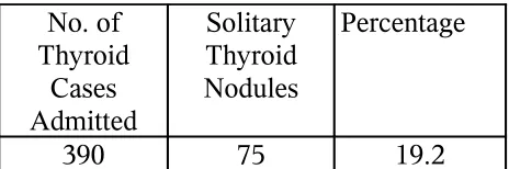
TABLE :1 No. of Thyroid Cases Admitted Solitary Thyroid Nodules Percentage
390 75 19.2
Solitary thyroid nodule represents thyroid pathology in about 19.2% of case.
Out of 75 patients, 65 were females and 10 were males. This gives a Male:Female ratio = 1:5. Solitary thyroid nodule is 5 times more common in women. In C.F.Nagori2222 series the male
[image:50.612.191.423.170.247.2]female ratio was 1:3.5
TABLE 2
SEX DISTRIBUTION
Sex No. of Patients
percentage Female 65 86.7%
Male 10 13.3%
This distribution is in accordance with most of the reported series in our country and else where.
TABLE 3
AGE DISTRIBUTION
Age In years No. of
Patients Percentage
Upto2O 8 11
21-30 27 36
31-40 22 29
41-50 14 18
51-60 2 3
6landAbove 1 1
In this study the youngest patient was 14 years old and the oldest was 65 years old. 80% of solitary nodules occurred during the age between 2 1-50 years. The highest incidence of 36% was recorded during the third decade of life.
TABLE 4
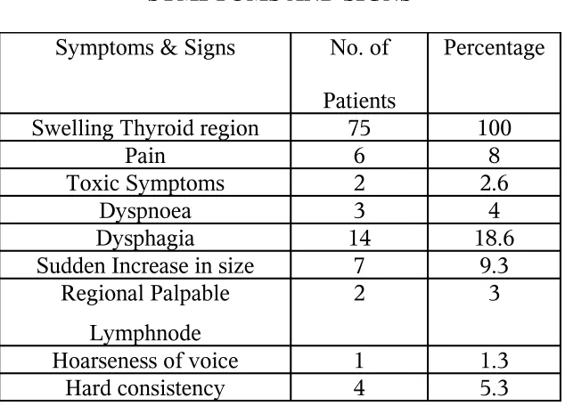
SYMPTOMS AND SIGNS
Symptoms & Signs No. of Patients
Percentage
Swelling Thyroid region 75 100
Pain 6 8
Toxic Symptoms 2 2.6
Dyspnoea 3 4
Dysphagia 14 18.6 Sudden Increase in size 7 9.3
Regional Palpable Lymphnode
2 3
[image:51.612.149.466.376.604.2]All the patients had only single palpable nodule. Toxic symptoms were present in 2 patients. Clinical evidence of obstruction to airway or of the great veins of neck by a solitary thyroid nodule is rare. The presence of stridor, respiratory wheeze; engorgement of neck veins should be interpreted with caution and the possible existence of a second pathology with in the mediastinum or lungs should be considered.
Seven patients complained of a sudden increase in size; while hoarseness of voice was present, only in one patient. On examination the only finding was hard consistency in 4 cases.
[image:52.612.191.422.413.510.2]In this study, 2 patients had regional lymphs node enlargement; which on FNAC proved to be secondary deposits from papillary carcinoma. 14 patients had difficulty in swallowing and 6 patients, pain over the swelling. 3 patients had difficulty in breathing, which was mostly uncharacteristic, neither exertional nor positional.
TABLE 5
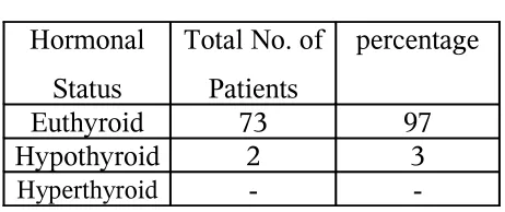
HORMONAL STATUS (CLINICAL)
Hormonal Status
Total No. of Patients
percentage Euthyroid 73 97 Hypothyroid 2 3
Hyperthyroid -
DIAGNOSTIC WORK UP
In this study the following investigations were done in included Urine analysis,. Blood urea, Blood sugar, serum radiograph of Neck, IDL scopy chest radiogram and fine needle aspiration cytology.
Ultrasound and CT Scan were done in few cases. Serum T3, T4 and TSH estimation were
done in two patients admitted with toxic symptoms and confirmed toxicity. USG was done in a few patients suspected to have thyroid cysts, while CT scan, was done in a patient with extensive neck secondaries.
Radio nucleotide scanning was not done for any of the patients due to the non-availability of the facility in our hospital.
Fine Needle Aspiration Cytology represents a reliable method of providing a tissue diagnosis and is the investigation of choice for solitary thyroid nodule.
[image:53.612.169.448.592.706.2]In this study FNAC was a very dependable and an easy investigation without complications.
TABLE 6
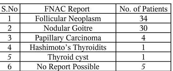
FNAC REPORT
S.No FNAC Report No. of Patients 1 Follicular Neoplasm 34 2 Nodular Goitre 30 3 Papillary Carcinoma 4 4 Hashimoto’s Thyroidits 1
Amongst the FNAC reports, follicular neoplasm was the commonest to be reported with the inability to identify vascular/capsular invasion. Adenomatous goitre, presenting as a solitary nodule was next commonest eventuality. 4 cases were reported as papillary carcinoma with two of them showing deposits in the neck nodes. No cases of MTC / Hurthel cell carcinoma were reported in our series. No report was possible in 5 cases.
TABLE 7
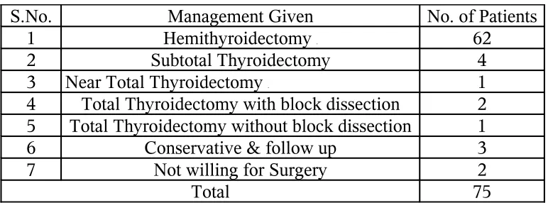
MANAGEMENT GIVEN
S.No. Management Given No. of Patients 1 Hemithyroidectomy . 62
2 Subtotal Thyroidectomy 4 3 Near Total Thyroidectomy . 1
4 Total Thyroidectomy with block dissection 2 5 Total Thyroidectomy without block dissection 1 6 Conservative & follow up 3 7 Not willing for Surgery 2
Total 75
[image:54.612.114.500.278.421.2]Provided there is no clinical suspicion of cancer and the cytology is again unequivocally benign, the individual is seen on an annual basis for re-examination and further FNAC (RUSSEL).
TABLE 8
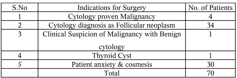
INDICATIONS FOR SURGERY
S.No Indications for Surgery No. of Patients 1 Cytology proven Malignancy 4
2 Cytology diagnosis as Follicular neoplasm 34 3 Clinical Suspicion of Malignancy with Benign
cytology
1
4 Thyroid Cyst 1
5 Patient anxiety & cosmesis 30
Total 70
This study includes two cases of solitary thyroid module with hyperthyroidism with benign cytology report. For these patients hemithyroidectomy was done after preparation with antithyroid drugs, on the assumption that the solitary nodule is the overactive tissue, since Radioisotope scan was not available to study the functional status of the nodule and these patients had no recurrence of thyrotoxicosis.
Total thyroidectomy was done for three patients of papillary carcinoma with neck node dissection (FND) in two patients, whereas neck node dissection was not done in 1 case.
Sub-total thyroidectomy was done for 4 patients who had nodules on contralateral lobe preoperatively.
Hemi-thyroidectomy, the standard surgical procedure for solitary thyroid nodule was done in 62 patients i.e., about 88% of patients who underwent surgery.
[image:56.612.121.492.321.502.2]Surgery was done in a patient with an inconclusive FNAC report, based on clinical suspicion of malignancy. All resected specimens were submitted for histopathological examination.
TABLE 9
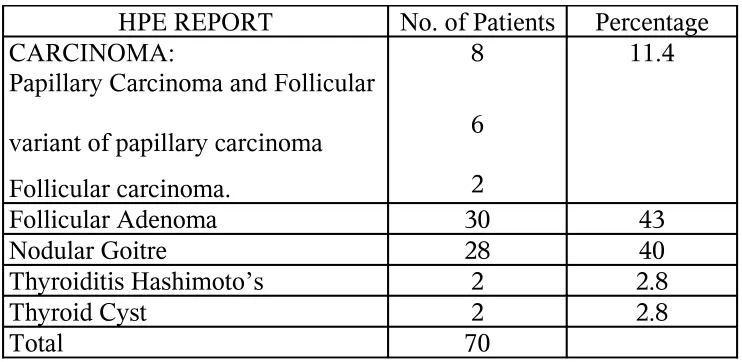
HISTOPATHOLOGICAL REPORT
HPE REPORT No. of Patients Percentage CARCINOMA:
Papillary Carcinoma and Follicular variant of papillary carcinoma Follicular carcinoma.
8 6 2
11.4
Follicular Adenoma 30 43
Nodular Goitre 28 40
Thyroiditis Hashimoto’s 2 2.8
Thyroid Cyst 2 2.8
Total 70
DISCUSSION
The Incidence of thyroid cancer in patients with a solitary thyroid nodule.
Study by Kendall & Condon l96917}
Pnarras et al 1972} 11-20% Study by Cohn F J Russel at Royal Victoria} Hospital, BELFAST., U.K.} 13%.
Study at Coimbatore Medical College Hospital Coimbatore 2003-2005 11.4%
This compares well with the other international studies.
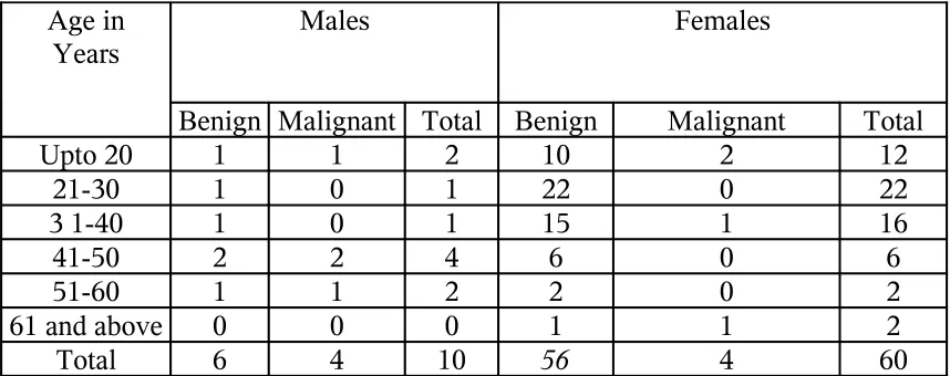
[image:57.612.92.521.514.684.2]In our study 40% of solitary nodule in males proved to be malignant whereas in females only about 6% of the solitary nodules harboured malignancy.
TABLE 10.
AGE AND SEX DISTRIBUTION OF BENIGN AND MALIGNANT NODULE
Age in
Years Males Females
Benign Malignant Total Benign Malignant Total
Upto 20 1 1 2 10 2 12
21-30 1 0 1 22 0 22
3 1-40 1 0 1 15 1 16
41-50 2 2 4 6 0 6
51-60 1 1 2 2 0 2
61 and above 0 0 0 1 1 2
According to Matheson 1986, the malignant potential of a nodule in a man is approximately three times that for a women of comparable age and in our study, the risk has been almost six times.
In males 75% of cancer occurred in more than 40 years age group.
Malignancy is more likely in a nodule in a child or a teenager or when a goitre develops in a patient aged 60 years or above (Hamming et al 1990, Caruso and Mazzaferri9 1991)
50% of thyroid cancers occurred in individuals under 40 years of age and of them 75% is papillary carcinoma. Fravenhofer37 et al 1970 in his study of 125 cases of thyroid cancer
[image:58.612.125.486.425.569.2]found that 80% of thyroid cancers in individuals under 40 years of age was papillary carcinoma.
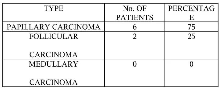
TABLE 11
HISTOLOGICALLY PROVEN MALIGNANCY
TYPE No. OF
PATIENTS PERCENTAGE PAPILLARY CARCINOMA 6 75
FOLLICULAR CARCINOMA
2 25
MEDULLARY CARCINOMA
0 0
The relative incidence of primary malignant tumours in our series is almost in accordance with most of the reported series (DUNHILL36).
In our study, 50% of the cases, all of them males, developed malignancy after 40 years, whereas in females, 75% of cases were younger than 40 years.
COMPARISON OF PRE OPERATIVE FNAC WITH POST OPERATIVE HISTOPATHOLOGICAL EXAMINATION:
Out of 70 cases operated 5 had different histopathology reports as compared to FNAC. If both FNAC & histopathology are benign or malignant they are considered true positive.
[image:59.612.116.501.475.540.2]4 cytologicaly benign lesions were reported as malignant on histopathological examination. So FNAC was false negative in these cases.
TABLE 12
ACCURACY RATE OF FNAC
Result True
Positive case NegativeFalse PositiveFalse Total
No. of Cases 66 4 0 70
Percentage 94 6 0
CANCER RISK IN FOLLICULAR NEOPLA’SM
The cytological appearances of follicular adenoma and follicular carcinoma are very similar. So a cytological diagnosis of follicular neoplasm is only possible, and confirmation of diagnosis of follicular carcinoma depends upon the visualisation of capillary & Vascular invasion in histopathological examination.
Although the Cancer risk is only 20%, CFJ RUSSEL, in common with others advises surgical resection of all solitary thyroid nodules reported as follicular neoplasm cytologically.
In this study, 34 cases were diagnosed as folhicular neoplasm cytologically and of these four were histologically malignant - .12% cancer risk.
SURGICAL PROCEDURES
CARCINOMA
We have done total thyroidectomy for three patients with or without neck node. dissection. All three were papillary carcinoma. Near total thyroidectomy was done for a case, also of papillary carcinoma.
Total Thyroidectomy is considered not only as a measure to reduce the recurrence rate of differentiated carcinoma but also as a means to prevent development of a highly malignant undifferentiated lesion. The percentage of radio iodine pick up can be increased several fold after total thyroidectomy and it also increases the sensitivity of thyroglobin as a post operative marker of residual / recurrent disease.
The risk of permanent hypoparathyroidsm or recurrent laryngeal nerve damage is high.
We have done near total thyroidectomy for one patient, a case of cytological proven papillary carcinoma, which was a small lesion (1.5 cm).
Near total thyroidectomy means leaving a fringe of thyroid tissue to preserve parathyroid.
Indications for near total thyroidectomy includes smaller lesions and better prognostic variants of papillary carcinoma thyroid.
Advantages of near total thyroidectomy are
1. Lesser incidence of permanent or transient hypo parathyroidism 2. Lesser incidence of recurrent laryngeal nerve palsy
Disadvantage of near total thyroidectomy are
1. Follow up is difficult
2. Remaining thyroid tissue has to ablated for control of metastasis.
Out of the four false negative patients (cytologically benign) all were treated with hemi- thyroidectomy. All these patients were taken up for completion thyroidectomy because they fell into the high risk group and they had an uneventful post operative period and follow up.
metastasis, age less than 40 years in males and less than 45 years in females.
So, the above patients were advised completion surgery with suppresive dose of thyroxine and regular follow-up.
The indications for isotope scanning after operation for differentiated cancer are.
1. Unresectable local recurrence 2. Metastatic disease
3. High risk patients and
4. Those with increase in serum Thyroglobulin level.
MALIGNANCY IN A TOXIC NODULE
Majority of cancers arise in hypo or cold thyroid nodules (ALDERSON et al 1976 COX et al 1991). The possibility of malignancy occurring with in a hot nodule is rare.
ADENOMA
Out of the 70 cases operated 30 cases were histolopathologically reported as adenoma. Most of them were macrofollicular adenoma. Hemithyroidectomy was done for all the patients.
Adenoma thyroid commonly occurred in the 3rd decade in this series of study.
NODULAR GOITRE
40% of operated cases were diagnosed as nodular goitre, hemithyroidectomy was done for most of them.
[image:63.612.94.515.428.618.2]4 patients had nodules in the contra lateral lobe also, which were revealed peroperatively, hence sub-total thyroidectomy was done.
TABLE 13
ADENOMA AND NODULAR GOITRE - HISTORICAL COMPARISON
Adenoma Nodular Goitre Usually single Usually multiple nodules
Completely encapsulated Partly encapsulated or unencapsulated Compress the surrounding thyroid
tissue
Doesn’t compress surrounding thyroid tissue.
Compared of fairly uniform follicles smaller than rest of thyroid tissue
Heterogenous appearance often with larger follicles.
Degenerative changes uncommon Hyperplastic follicles with focal haemorrhages, scarring, calcification and micro cyst formation.
OUT COME OF THERAPY
Complications
Two patients had sluggish movement of left vocal cord after total thyroidectomy and improved later.
[image:64.612.112.499.252.529.2]Seven patients had post operative wound infection. We had no mortality during this study.
Table 14
COMPARISION
Study by Cohn FJ Russel FRCS At Royal Victoria Hospital Belfast .U.K. 61 patients operated for solitary thyroid
nodule
Study at Coimbatore Medical College Hospital
Coimbatore 70 patients operated for
solitary thyroid nodule FNAC Pathological Diagnosis Number of Patients .
Percentage Number of Patients
Percentage
Nodular Goitre
30 49 28 40
Follicular
Adenoma 19 31 30 43
Malignancy 8 13 8 11.4
Hashimotos 3 5 2 2.8
Dequervains 1 2 -
CONCLUSIONS
1. FNAC is the gold standard for evaluation of Solitary Thyroid Nodule with an accuracy of 94% in our study.
2. 11.4 percentage of Solitary Thyroid Nodule were Malignant
3. Solitary Thyroid Nodule in males have more malignancy risk than female.
4. Suspect malignancy at extremes of age. Malignancy potential of Solitary Thyroid Nodule after 6th decade is 50%.
5. 50 percentage of thyroid cancer occurred in patients under 40 years of age, of them 75% were papillary carcinoma.
6. A selective surgical policy should be practised to resect Solitary Thyroid Nodule
7. Hemi thyroidectomy is the minimum, surgical procedure for single nodule.
8. Cancer risk in follicular neoplasm is 12%
BIBLIOGRAPHY
1. ALFRED CUSCHERI-Essential surgical practice 4th edition 2. Anatomy regional & applied-RJ LAST 9th edition (430-432)
3. Ashok k mehtha carcinoma thyroid,recent advancr in surgery no.2 ROSHSANLAL GUPTHA
4. ALDERSONS PO Summer HW Siegal BA 1976-the single palpable thyroid nodule,Evaluvation by TC99m pertechnetate
5. Al-SayerHM,Krukowst,,ZH,WILLIAMS VMM et al1985.FNAC in isolated thyroid swelling BMJ 290:1490-1492.
6. Astwood EB CASSIDY CE,A urbach GD 1960. Treatment of Goitre& Thyroid nodules JAMA , 174 :459-469
7. B J S VOL 80 No. 10 oct 1998 concurrent hyperthyroidism&thyroid carcinoma. Terzioghi T
8. CURRENT SURGICAL DIAGNOSIS AND TREATMENT
9. Causo D ,Mazzaferri EL 1991 –FNAC in the management of thyroid nodules
10. Celani MT, Maraini G 1990 - T4 Suppression therapy in the medical management of STN Acta Endocrinol 123 : 603-608
12. COX Mc Spence Raj 1991 – STN- A Prospective evaluvation of nuclear scanning SVSG BJS 78: 90-93
13. ENDOCRINOLOGY , J. DEGROOT Vol 1 835-850
14. General surgical operations R. M. KIRK &R CN Williamson
15. Hamilton Bailey - Demonstation of physical signs in clinical surgery 16. JEMIESON&KAYS- Textbook of Surgical physiology
17. Kendall LW , London RE 1969 – Prediction of malignancy in STN Lancet 1071-73 18. La ROSSA GL, Belfiore A , Giuffrida D et al 1991 –Evaluvation of FNAC in
preoperative selection of cold thyroid nodules.
19. Lowbagen T Granberg P. O. Lundel G et al 1979 –Aspiration cytology in nodule of thyroid suspected to be malignant.
20. Mastery of surgery – Nyhus & Robert JBalwer
21. Manual &Atlas of FNAC –SVANTE R orell Gregory F Stersett eburebil livingstone 1995
22. Nagori LF & MJ Algotor – Solitary Solid thyroid nodule IJ of Surgery 1982 54 (2) : 73-78
23.Pathological Basics of Disease - COTRAN , KUMAR, ROBBINS 6th edition
24. Pathology of tumours - R. A. WILLIS
25.Principles of surgery – SEYMOURI ,SCHWARTZ 8th edition (1670-1671) 26.Principles & Practice of Oncology –VINUNIT. DEVITTA J. R. 7th edition
27. Reverter JL , L ucasa Salimas I etal, 1992 , Suppressive therapy with levothyroxine for STN clin endocrinol 36 :25-28
29. Short Practice of Surgery – Bailey & Love 24 th edition (776-804)
30.Textbook of Surgery – SABISTON 17th edition (961-965)
31.SURGERY – SCIENTIFIC PRINCIPLES AND PRACTICE - Greenfield 3rd edition
(1261-1283)
32. Textbook of Medical Physiology -GUYTON
33. The Otolaryngological clinics of North America vol 29 no 4 aug 1996.
34. Walfish PG , HAZAN E , 1977 combined USG &needle aspiration in the assessment of hypo functioning thyroid nodule Intern, 87: 270-274
35. Psarras A 1972 . The single thyroid nodule BJS 59 : 545-548
36. Frauenhofer, CM etalm 1979. Thyroid. A clinical &pathological study of 125 cases cancer 43 : 2414