Copyright
q
1997, American Society for Microbiology
Inducible Nitric Oxide Synthase in Theiler’s Murine
Encephalomyelitis Virus Infection
EMILIA L. OLESZAK,
1,2,3* CHRISTOS D. KATSETOS,
4JACEK KUZMAK,
1,2AND
ARUN VARADHACHARY
4Fels Institute for Cancer Research and Molecular Biology
1and Departments of Biochemistry,
2Neurology,
3and
Microbiology and Immunology,
4Temple University School of Medicine, Philadelphia, Pennsylvania
Received 19 November 1996/Accepted 10 January 1997
We investigated the role of inducible nitric oxide synthase (iNOS) in Theiler’s murine encephalomyelitis
virus (TMEV) infection of susceptible (SJL) and resistant (C57BL/6 [B6]) strains of mice. TMEV is an
excellent model of virus-induced demyelinating disease, such as multiple sclerosis (MS). Previous studies of
others have suggested that NO may play a role in the pathogenesis of demyelinating disease. The presence and
level of iNOS were determined in the brains and spinal cords of SJL and B6 TMEV-infected mice by the
following methods: (i) PCR amplification of iNOS transcripts, followed by Southern blotting with an
iNOS-specific probe, and (ii) immunohistochemical staining with an anti-iNOS-iNOS-specific affinity-purified rabbit
antibody. iNOS-specific transcripts were determined in the brains and spinal cord of both SJL and B6
TMEV-infected mice on days 0 (control), days 3, 6, and 10 (encephalitic stage of disease), and days 39 to 42,
66, and 180 (demyelinating phase) postinfection (p.i.). iNOS-specific transcripts were found in the brains and
spinal cords of both SJL and B6 TMEV-infected mice at 6, 10, and 39 (SJL) days p.i., but they were absent in
mock-infected mice and in TMEV-infected SJL and B6 mice at 0, 3, 66, and 180 days p.i. Immunohistochemical
staining confirmed the presence of iNOS protein in both TMEV-infected SJL and B6 mice at days 6 and 10 p.i.,
but not at days 0, 3, 66, and 180 days p.i. Weak iNOS staining was also observed in TMEV-infected SJL mice
at 42 days p.i. iNOS-positive staining was found in reactive astrocytes surrounding areas of necrotizing
inflammation, particularly in the midbrain. Weak iNOS staining was also observed in cells of the monocyte/
macrophage lineage in areas of parenchymal inflammation and necrosis (mesencephalon) and in
leptomen-ingeal and white matter perivascular infiltrates of the spinal cord. Rod-shaped microglia-like cells and foamy
macrophages (myelin-laden) were iNOS negative. These results suggest that NO does not play a direct role in
the late phase of demyelinating disease in TMEV-infected mice.
Nitric oxide (NO) is a short-lived, highly reactive molecule
with free radical properties (52, 58). NO production results
from the conversion of
L-arginine to
L-citrulline by NO
syn-thase (NOS) (52, 53, 77). The NO enzyme family consists of
three isoforms: neuronal constitutive NOS (type I NOS),
in-ducible NOS (iNOS or type II NOS), and endothelial
consti-tutive NOS (type III NOS) (30, 52). iNOS expression is
in-duced by lipopolysaccharide or cytokines, such as gamma
interferon (IFN-
g
) or interleukin 1
b
(IL-1
b
), and is calcium
independent (8, 13, 15, 18, 29, 42, 50, 72). The NO generated
by iNOS is the primary form of NO in acute and chronic
inflammation, while rather lower concentrations of NO are
released by constitutive expressed NOS (types I and III), which
act as second messengers in NO signaling pathways in the
neuronal and cardiovascular systems. NO exhibits toxic,
cyto-static, and regulatory functions (3, 6, 9, 12, 23, 25, 33–36, 54, 65,
75, 76). It inhibits T-cell proliferation, and it plays a role in the
antitumor and antimicrobial functions of the immune system
(24, 32, 37, 41, 59, 73, 74). However, type II NOS-directed NO
production is reported to be a potent neurotoxin and at least in
vitro mediates tumor necrosis factor alpha (TNF-
a
) toxicity
toward oligodendrocytes (20, 54, 57). Therefore, NO produced
by iNOS may be both friend and foe. In the central nervous
system (CNS), cytokine-induced iNOS is expressed mainly by
macrophages, microglial cells, and astrocytes, but not by
oli-godendrocytes (11, 26, 29, 43, 55, 71, 78).
iNOS-derived NO has been detected in several
inflamma-tory diseases of the CNS, such as experimental allergic
enceph-alomyelitis (EAE) and viral encephalitis, induced
experimen-tally in rodents by Borna virus, rabies virus, and herpes simplex
virus (17, 21, 31, 38). It is not known whether NO plays a role
in demyelination, which is seen in certain forms of EAE, or
whether NO is associated only with the encephalitic stage of
the disease.
In this report we investigated whether iNOS is expressed in
the CNSs of mice infected with Theiler’s murine
encephalo-myelitis virus (TMEV). This virus induces a biphasic disease in
sensitive strains of mice (such as SJL) (45, 46, 61, 64, 68). The
early phase of the disease is characterized by acute
polioen-cephalomyelitis (early acute disease), in which the virus
repli-cates mainly in neurons and to a lesser extent in astrocytes,
macrophages, and microglia (10, 45, 47, 48). There are
prom-inent neuronophagia and a characteristic intense mononuclear
cell infiltration of neurons in the cerebral cortex and anterior
horn cells of the spinal cord. A few weeks later, mice from
susceptible strains develop the second phase of the disease,
which is characterized by chronic demyelination associated
with heavy mononuclear infiltration (late demyelinating
dis-ease). Pathological changes are limited to the spinal cord.
During chronic demyelinating disease, most of the TMEV
antigens can be detected in macrophages, astrocytes, and
oli-godendrocytes (10, 63, 68). It has been suggested that
demy-elination in TMEV-infected animals is mediated by the
im-mune system (63). In contrast, resistant strains of mice
* Corresponding author. Mailing address: Fels Institute for Cancer
Research and Molecular Biology, Temple University School of
Med-icine, 3420 N. Broad St., Philadelphia, PA 19106. Phone: (215)
707-7657. Fax: (215) 829-1320.
3228
on November 9, 2019 by guest
http://jvi.asm.org/
(C57BL/6 [B6]) develop only the acute phase of the disease,
clear the virus, and do not develop demyelinating disease.
TMEV infection of susceptible and resistant strains of mice
offers a unique opportunity to study the role of NO in
enceph-alitis and demyelination.
MATERIALS AND METHODS
Virus.The wild-type Daniel strain of TMEV was used in all experiments. The origin and propagation of this virus have been described previously (46, 47, 62). Animals.Five- to six-week-old female SJL and B6 mice were purchased from Jackson Laboratory (Bar Harbor, Maine). Mice were housed in microisolators in an individual biohazard unit in a biohazard level II facility. All manipulations and changing of cages were performed in a biohazard hood. Mice were maintained in accordance with the standards of the American Association for Accreditation of Laboratory Animal Care. Sentinel mice were housed together with TMEV-infected mice and examined for the presence of a large number of common mouse pathogens (viruses and mycoplasma). No pathogens were detected. Mice were slightly anesthetized with methoxyflurane (Pitman-Moore, Mundelein, Ill.) and inoculated with 105PFU of TMEV in a 20-ml volume in the right cerebral hemisphere. Control, mock-infected animals were injected with Dulbecco’s mod-ified Eagle medium containing 2% fetal calf serum and standard amounts of penicillin, streptomycin, and glutamine. Medium of identical composition was used to grow TMEV. At days 0, 3, 6, 10, 39, 42, 66 or 67, and 180 (SJL only) p.i., three to five SJL or B6 mice per group were euthanized by the administration of an overdose of methoxyflurane, and their spinal cords and brains were immedi-ately removed and were either snap frozen in liquid nitrogen for RNA isolation or embedded in OCT compound (Tissue-Tech; Sakura, Torrance, Calif.) and stored in2808C. Also, tissue specimens were placed in buffered formalin.
Histopathology and immunohistochemistry.Spinal cords and brains were em-bedded in paraffin, cut into 5-mm-thick sections, and stained with hematoxylin and eosin for light microscopy. iNOS was detected by immunohistochemistry, using an affinity-purified rabbit anti-iNOS antibody (Transduction Laboratories, Lexington, Ky.), according to the manufacturer’s recommendations. Glial fibril-lary acidic protein (GFAP) was detected by a similar approach, as previously described (60), using a rabbit anti-GFAP antibody (DAKO, Carpinteria, Calif.). Briefly, the slides were rehydrated in xylene and alcohols, the sections were washed with phosphate-buffered saline, and endogenous peroxidase was blocked by incubation for 30 min in 1.2% hydrogen peroxide in cold methanol. Alterna-tively, 5-mm-thick frozen sections were fixed for 10 min in cold acetone and stained by the procedures described above. Sections were incubated with goat serum for 45 min to reduce nonspecific binding. iNOS was detected by an immunoperoxidase technique (ABC kit; Vector Laboratories, Burlingame, Cal-if.). Sections were incubated with either anti-iNOS (1.25mg/ml) or anti-GFAP
antibody (diluted 1:1,000) for 1 h. Antigen-antibody complexes were detected with an anti-rabbit–biotinylated avidin–horseradish peroxidase complex accord-ing to the manufacturer’s instructions. The sections were stained with 3,39 -diaminobenzidine as the substrate and then counterstained with Mayer’s hema-toxylin (Sigma Chemical Co., St. Louis, Mo.). Controls included normal rabbit immunoglobulin G (IgG) (1.25mg/ml), which was used instead of a specific rabbit anti-iNOS or anti-GFAP antibody. Staining with control normal rabbit IgG was always negative.
RT-PCR and Southern blot analysis of iNOS mRNA in the brain and spinal cord.Total RNA was isolated from brain and spinal cord tissue of individual mice separately, using the RNA isolation kit of Stratagene (La Jolla, Calif.). The first strand of cDNA was synthesized by reverse transcriptase (RT) (Promega, Madison, Wis.) with 5mg of starting RNA. The reaction was stopped by heating the mixture at 1008C for 10 min. cDNA was amplified by using primers selected from the iNOS cDNA (38) as follows: iNOS antisense 30-mer, 59-GTCGACG AGCCTCGTGCTTTGGGCTCCTC-39; and iNOS sense 30-mer, 59-GTCGAC CTTCCGAAGTTTCTGGCAGCAGCG-39. Amplification was performed as follows: 35 cycles of amplification, with 1 cycle consisting of three steps, namely, denaturation at 948C for 45 s, primer annealing at 508C for 90 s, and primer extension at 728C for 3 min, followed by a final elongation step for 10 min at 728C.b-Actin primers (Clontech, Palo Alto, Calif.) were used as controls to ensure the integrity of RNA. Hybridizations and Southern blotting were per-formed as described, using an iNOS-specific 30-mer hybridization probe, 59-AC GTTCAGGACATCCTGCAAAAGCAGCTGG-39.
RESULTS
[image:2.612.60.555.469.620.2]To elucidate whether iNOS is expressed in the CNS of mice
infected with TMEV, we have investigated the presence of
iNOS transcripts by RT-PCR and the presence of iNOS
pro-tein by immunohistochemical staining. Susceptible (SJL) and
resistant (B6) mice were infected intracerebrally with TMEV
and euthanized at days 0, 3, 6, 10, 39, 42, 66 or 67, and 180 (SJL
only) p.i. Control mice were injected intracerebrally with
me-dium alone, as described in Materials and Methods, and they
were sacrificed 6 and 39 days p.i. The presence of iNOS
tran-scripts and iNOS protein were determined in the brains and
spinal cords of all mice, as described in Materials and Methods.
In addition, two or three animals from the TMEV-infected
group were euthanized at the same time points listed above,
TABLE 1. Histopathology
aand iNOS in TMEV-infected mice
Strain days p.i.No. of
Brain
Leptomeningeal
infiltratese Poliomyelitise
Spinal cord
Encephalitisb iNOS
transcriptsc iNOS stainingd Vacuolar changesor demyelinationf iNOS transcripts iNOS staining
SJL
3
2
Absent
Negative
2
2
2
Absent
Negative
6
11
Present
gStrongly positive
1
11
1
Present
gStrongly positive
10
111
Present
gStrongly positive
11
1
2
Present
gStrongly positive
39–42
1
Present
hWeakly positive
11
2
111
Present
hWeakly positive
66
1
Absent
Negative
11
2
111
Absent
Negative
180
2
Absent
Negative
11
2
111
Absent
Negative
B6
3
1
Absent
Negative
1
2
2
Absent
Negative
6
11
Present
gStrongly positive
1
11
1
Present
gStrongly positive
10
11
Present
gStrongly positive
2
1
2
Present
gStrongly positive
42
2
ND
Negative
2
2
2
ND
Negative
67
2
Absent
Negative
2
2
2
Absent
Negative
aHistopathological changes in the brain (encephalitis) and spinal cord (leptomeningeal infiltrates, poliomyelitis, and vacuolar changes or demyelination) were measured.
bSymbols:2, unremarkable;1, mild leptomeningeal inflammation (no parenchymal inflammation and no necrosis);11, moderate to severe inflammation with or without necrosis;111, severe inflammation with overt necrosis.
cPCR and Southern blotting were carried out as described in Materials and Methods. ND, not determined.
diNOS staining was determined by immunohistochemistry as described in Materials and Methods. iNOS staining was categorized as follows: negative, no staining; strongly positive, robust staining of tissue; weakly positive, weak staining.
eSymbols:2, unremarkable (for leptomeningeal infiltrates) or no infiltrates (for poliomyelitis);1, scant mononuclear infiltrates;11, multifocal, nodular aggregates of inflammatory cells.
fSymbols:2, no vacuolar changes or demyelination;1, focal change involving one white matter funiculus only;111, pronounced change, involving one or more funiculi.
gThe level of iNOS transcripts in the brain was higher than that in the spinal cord. hThe level of iNOS transcripts in the brain was lower than that in the spinal cord.
on November 9, 2019 by guest
http://jvi.asm.org/
and brains and spinal cords were processed individually for
hematoxylin-and-eosin histological evaluation of pathology.
Expression of iNOS in early acute disease.
None of the
TMEV-infected animals showed overt clinical signs of
enceph-alitis, although histological analyses revealed
polioencephalo-myelitis at days 6 and 10 p.i. (Table 1). SJL mice showed more
severe gray matter disease on day 10 p.i. than B6 mice (Table
1). However, B6 mice showed inflammatory infiltrates in the
CNS earlier than SJL mice. We have detected high levels of
iNOS transcripts in the brain and spinal cord in both strains of
mice. Little or no detectable iNOS expression was found in the
brain and spinal cord at day 3 p.i. (data not shown). In contrast,
the level of expression of iNOS transcripts was significant at
days 6 and 10 p.i. in both susceptible and resistant strains of
mice (Fig. 1A, B, and C). At 6 days p.i., a certain fluctuation
was observed in the levels of iNOS transcripts in the CNS of
individual TMEV-infected SJL mice (Fig. 1A). Similar
fluctu-ations in the levels of iNOS transcripts have been reported by
others (38) for virus-induced encephalitis. The induction of
iNOS mRNA on days 6 and 10 p.i. coincided with the severity
of polioencephalomyelitis (Table 1). In general, higher levels
of iNOS expression were detected in the brains of SJL and B6
mice than in the spinal cords during early acute disease (Fig.
1A and C). iNOS transcripts were not detected by RT-PCR,
followed by Southern blotting, in the brains or spinal cords of
mock-infected mice at 6 or 39 day p.i. (Fig. 1B and D), in
agreement with the findings of others (55, 56).
Using antibodies to iNOS protein and immunohistochemical
staining, we have identified the CNS cells that expressed iNOS.
At days 6 and 10 p.i., cells that stained positive with iNOS
antibody were identified in areas of intense inflammation in
both TMEV-infected SJL and B6 mice. In the brains of both
strains of mice, these cells included predominantly reactive
astrocytes (Fig. 2A and B and 3A and B), cells of the
mono-cyte/macrophage lineage, and hypertrophic endothelial cells in
areas of active vascular ingrowth (Fig. 2C and D and 3B). Rare
perivascular monocyte-like cells of the leptomeningeal
infil-trates were also iNOS positive (Fig. 3C), whereas
morpholog-ically overt lymphocytes were as a rule iNOS negative. Normal
interfascicular fibrous and protoplasmic astrocytes,
Berg-mann’s glia, as well as oligodendrocytes, were iNOS negative.
Similarly, no staining was observed in normal endothelial or
smooth muscle cells. To confirm the identity of the cells that
expressed the iNOS protein, we stained adjacent sections
ei-ther with GFAP antibody (Fig. 2A and 3A) or with
anti-iNOS antibody (Fig. 2B and 3B). Cells which were positive for
iNOS were also stained with anti-GFAP antibody. Thus,
reac-tive astrocytes represent the major cell type that expressed
iNOS protein in the brains of TMEV-infected mice. This is in
contrast to the lack of iNOS reactivity in normal astroglia.
iNOS protein was not detected by immunohistochemical
stain-ing in the brains of uninfected, unmanipulated SJL and B6
mice with the rabbit anti-mouse iNOS polyclonal antibody
previously described (data not shown).
The profile of iNOS staining in the spinal cord was similar to
that described for the areas of encephalitis. At 6 and 10 days
p.i., reactive astrocytes near areas of inflammation were iNOS
positive. Occasional or scattered iNOS-positive cells of the
macrophage/monocyte type were detected in foci of spinal
meningitis and in spinal gray matter infiltrates. However,
rod-shaped microglia-like cells were consistently negative (Fig.
3D). There was no difference in the distribution of
iNOS-positive cells in the CNS of TMEV-infected SJL or B6 mice.
Expression of iNOS in late demyelinating disease.
At day
39 p.i., which corresponds to the beginning of demyelinating
disease (Fig. 1D), iNOS transcripts were detected in the spinal
cords of five TMEV-infected SJL mice. Lower levels of iNOS
message were detected in the brains of these mice. However,
iNOS transcripts could not be detected in the brains and spinal
cords of TMEV-infected susceptible SJL mice with extensive
demyelinating disease 66 and 180 days p.i. (Fig. 4). Also, at 67
days p.i., iNOS transcripts could not be detected in the brains
and spinal cords of TMEV-infected resistant B6 mice that were
free of demyelinating disease (Fig. 4).
b
-Actin transcripts were
amplified from each brain and spinal cord cDNA to ensure the
integrity of RNA (Fig. 4). Lack of iNOS expression in the brain
or spinal cord at this time point was confirmed by the lack of
any immunohistochemical staining with anti-iNOS antibody in
both SJL and B6 mice (Fig. 2G and 3E and F and data not
shown). Foamy (myelin-laden) macrophages in areas of
demy-elination in the spinal cords of SJL mice 67 days p.i. were not
stained with anti-iNOS antibody (Fig. 2G). At days 42 and
67 p.i., we did not detect any pathological changes in the brains
and spinal cords of B6 mice (Fig. 3E and F and data not
shown). In contrast, prominent inflammatory infiltrates,
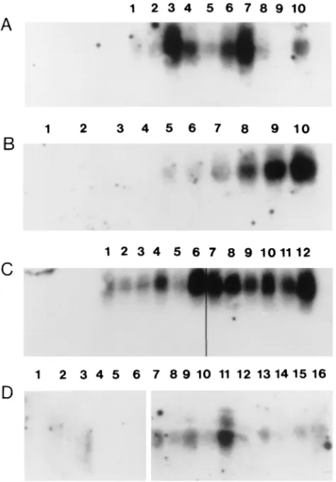
pro-FIG. 1. iNOS transcripts in the CNSs of TMEV-infected and mock-infected mice. Results from individual mice are shown in each lane. (A) iNOS transcripts in the brains (lanes 1 to 5) and spinal cords (lanes 6 to 10) of TMEV-infected SJL mice at 6 day p.i. (B) iNOS transcripts in mock-infected (lanes 1 to 4) or TMEV-infected (lanes 5 to 10) B6 mice at 6 days p.i. iNOS transcripts in spinal cords (lanes 1, 3, 5, 6, and 7) and in brains (lanes 2, 4, 8, 9, and 10) are shown. (C) iNOS transcripts in TMEV-infected SJL (lanes 1 to 6) and TMEV-infected B6 (lanes 7 to 12) mice at 10 days p.i. iNOS transcripts in spinal cords (lanes 1, 3, 5, 7, 9, and 11) and brains (lanes 2, 4, 6, 8, 10, and 12) are shown. (D) iNOS transcripts in mock-infected (lanes 1 to 6) and TMEV-infected (lanes 7 to 16) SJL mice at 39 days p.i. iNOS transcripts in spinal cords (lanes 1, 3, 5, 7 to 9, 10, and 11) and in brains (lanes 2, 4, 6, 12 to 15, and 16) are shown.
on November 9, 2019 by guest
http://jvi.asm.org/
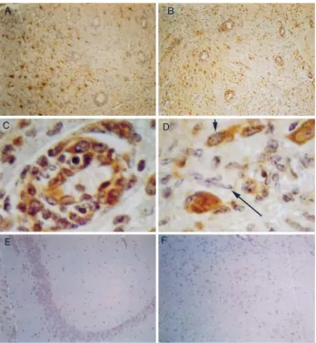
[image:3.612.60.297.70.410.2]FIG. 2. Immunocytochemical detection of GFAP (A) and iNOS protein (B to G) in the CNS of SJL mice at 10 (A to C), 6 (D), 42 (E and F), and 66 (G) days p.i. With the exception of panel E (frozen section), all staining was performed on paraffin-embedded tissue sections. (A) Phase of encephalitis and poliomyelitis of SJL mice 10 days p.i. An area of intense leptomeningeal inflammation surrounding midbrain parenchyma, where a brisk, reactive (principally subpial) astrocytic meshwork shows strong filamentous GFAP staining in glial perinuclear cytoplasm and fibrillary processes, is shown. Compare this with the lack of GFAP staining in mononuclear infiltrates involving the subarachnoid space (top portion of photomicrograph). Original magnification,3400. (B) Same field as in panel A from an adjacent paraffin-embedded section immunostained for iNOS. Diffuse iNOS-like staining is present in reactive subpial astrocytes (arrowheads) and also in occasional monocyte/macrophage cells in the leptomeningeal infiltrate (top portion of photomicrograph). Original magnification,3400. (C) Phase of encephalitis and poliomyelitis of SJL mice 10 days p.i. A high-magnification view of an area of intense (necrotizing) inflammation involving the mesencephalic-diencephalic region is shown. Note iNOS-like localization in hypertrophic endothelial cells representative of active vascular ingrowth (neovascularization). Also, note karyorrhectic debris denoting parenchymal necrosis, adjacent to the vessel. Original magnification,31,000. (D) Phase of encephalitis and poliomyelitis of SJL mice 6 days p.i. A high-magnification view of an area of spinal cord gray matter inflammation involving one of the posterior horns in the thoracic region. There is iNOS-like staining in perivascular inflammatory cells of the monocyte/macrophage lineage and in vascular endothelial cells. Original magnification,31,000. (E) Early phase of spinal cord demyelination of SJL mice 42 days p.i. A lateral column and thoracic cord are shown. Numerous iNOS-immunoreactive fibrillary astrocytes are present between inflammatory cells in a background of incipient vacuolar change (arrowheads) suggestive of early demyelination. Original magnification,3400. (F) Phase of spinal cord demyelination of SJL mice 42 days p.i. Focal, weak, and ill-defined iNOS-like immunoreactivity is detected among leptomeningeal infiltrates (the three arrows) encroaching upon the lateral white matter funiculus of the spinal cord (thoracic level). Note vacuolar change (arrowheads) in the neuropil of the lateral column. Original magnification,3400. (G) Phase of spinal cord demyelination of SJL mice 66 days p.i. An area of overt demyelination involving the posterior white matter funiculus (thoracic level) is shown. The section is immunostained for iNOS. There is no iNOS staining in large foamy (myelin-laden) macrophages that typify these full-blown demyelinating lesions. Original magnification,31,000.
on November 9, 2019 by guest
http://jvi.asm.org/
nounced demyelination, and reactive astrocytes could be
dem-onstrated in the spinal cords of TMEV-infected SJL mice,
while the brains were unremarkable (Table 1). These results
demonstrate that inflammatory infiltrates and gliosis are not
always associated with iNOS expression.
We have also examined the expression of iNOS at the
ter-minal stage of demyelinating disease in TMEV-infected SJL
mice, approximately 6 months p.i. At this time point, mice lost
their righting reflex, their hind limbs were paralyzed, and they
became incontinent. We did not detect any iNOS message in
the brains and spinal cords of SJL mice despite overt
demyeli-nation and inflammatory infiltration of the spinal cord (Fig. 4E
and F). Similarly, iNOS was not detected using anti-iNOS
antibody in reactive astrocytes and in foamy (myelin-laden)
macrophages in areas of demyelination (spinal cord). Cells of
the macrophage/monocyte lineage evident in perivascular cuffs
were also negative (data not shown).
DISCUSSION
The mechanism of the pathogenesis of demyelinating
dis-ease, such as multiple sclerosis (MS), is poorly understood. It
has been suggested that myelin and oligodendrocyte
destruc-tion in MS can be attributed to a direct attack of cytolytic T
cells (1, 63, 69). In addition, CD4
1T cells, macrophages,
microglia, and astrocytes may participate in myelin destruction,
either directly and/or indirectly by secreting cytokines, such as
[image:5.612.135.481.68.444.2]TNF-
a
and lymphotoxin (28, 58). In addition, it has been
FIG. 3. Immunohistochemical detection of GFAP (A) and iNOS protein (B to F) in the CNSa of B6 mice at 6 (A to D) and 42 (E and F) days p.i. (A) Phase of encephalitis and poliomyelitis of B6 mice 6 days p.i. An area of intense parenchymal inflammation (encephalitis) involving the mesencephalic-diencephalic region is shown. Briskly proliferating reactive astrocytes exhibit strong GFAP staining. There is no staining in blood vessels and in mononuclear infiltrates in the Virchow-Robin spaces (perivascular cuffs). Original magnification,3100. (B) Same field as in panel A, from an adjacent paraffin-embedded section immunostained for iNOS. There is widespread iNOS-like immunoreactivity involving reactive astrocytes, blood vessels (particularly endothelial cells), and subpopulations of mononuclear infiltrates most compatible with cells of the monocyte/macrophage lineage. Original magnification,3100. (C) Phase of encephalitis and poliomyelitis of B6 mice 6 days p.i. Transmural and perivascular inflammatory cell aggregates from a focus of necrotizing encephalitis involving the diencephalic-mesencephalic region. Robust iNOS staining is present in monocyte/macrophage cells traversing the vessel wall and in the Virchow-Robin (perivascular) space. Also, note iNOS localization in endothelial cells of the inflamed vessel (vasculitis). Original magnification,31,000. (D) Phase of encephalitis and poliomyelitis of B6 mice 6 days p.i. A focus of poliomyelitis similar to that depicted in panel C is shown. Robust iNOS-like immunoreactivity is present in a reactive astrocyte (short arrow). Compare with lack of iNOS staining in adjoining rod-shaped microglia-like cells (long arrow). Original magnification,31,000. (E) B6 mice 42 days p.i. There is no evidence of iNOS staining in the hippocampus. Original magnification,3100. (F) Cerebral section of a B6 mouse infected with TMEV 42 days p.i. and immunostained for iNOS. Absence of iNOS staining in this field from the diencephalic-mesencephalic region. Original magnification,3100. A similar lack of immunoreactivity was noted in sections of the spinal cord (data not shown).
on November 9, 2019 by guest
http://jvi.asm.org/
proposed that NO may mediate neuron and oligodendrocyte
death and may participate in demyelination (9, 31, 38, 56). NO
is a neurotransmitter in the CNS, which is constitutively
pro-duced by type I NOS in neurons (66). Type II NOS (iNOS) can
be found in activated astrocytes, macrophages, microglia, and
neurons (7, 9, 12, 21, 26, 29, 40, 55–57), while type III NOS is
found in endothelial cells, a subset of neurons of the
hip-pocampus and astrocytes of the CNS (2, 20). Astrocytes and
microglial cells express iNOS in response to endotoxin and to
cytokines such as TNF-
a
, IFN-
g
, and IL-1
b
(8, 13, 15, 42, 50).
In a few in vitro studies, it has been demonstrated that NO may
mediate the destruction of oligodendrocytes (54, 57). NO
tox-icity can be attributed to the nitrosylation of target iron-sulfur
proteins, including key enzymes necessary for DNA replication
and repair, or mitochondrial energy production (44, 65, 66, 75,
76). Direct mitochondrial damage has been detected in
oligo-dendrocytes treated with
S-nitroso-N-acetyl-
DL-penicillamine,
a NO-releasing chemical. In addition, NO may react with the
superoxide anion to form peroxynitrite (ONOO
2) (4, 49, 78),
which induces lipid peroxidation. Membrane peroxidation as
well as swollen oligodendrocyte cell bodies, has been
demon-strated in the brains of patients with MS, which supports the
hypothesis that NO may mediate oligodendrocyte damage.
Furthermore, growing lesions in MS patients are surrounded
by infiltrates of activated macrophages and microglia, which
produce TNF-
a
, a potent inducer of NO. Recently, it was
reported that iNOS mRNA can be detected in microglia
and/or astrocytes of patients with MS (1, 7). Therefore, NO
may be directly involved in the demyelinating process.
In-creased levels of iNOS have also been demonstrated in EAE,
which mimics certain aspects of MS (17, 38), and in astrocytes
of mouse hepatitis virus-infected animals with chronic
demy-elinating disease (67).
[image:6.612.91.263.66.421.2]With the objective of determining whether NO plays a role
in the pathogenesis of inflammatory and/or demyelinating
dis-ease, we have studied iNOS in TMEV infection. TMEV
in-duces a biphasic disease in susceptible strains of mice (14, 68).
The early disease resembles polioencephalomyelitis and is
fol-lowed several weeks later by late chronic demyelination of the
spinal cord (47, 48). In contrast, resistant strains of mice
de-velop only gray matter disease, clear the virus, and do not
develop demyelination. Both acute and chronic demyelinating
disease is characterized by mononuclear cell infiltrates. During
late demyelinating disease, extreme meningeal and
perivascu-lar mononuclear cell infiltrates are found in susceptible SJL
mice with concomitant demyelination in the spinal cord (46).
Using this model of virus-induced demyelinating disease (19,
64), we were able to study separately the induction of iNOS at
both the encephalitic and demyelinating phases of the disease.
We have demonstrated high levels of NO produced in the
brains and spinal cords of both resistant and susceptible strains
of mice during early gray matter disease. The highest level of
iNOS was observed at days 6 and 10 p.i. By
immunohistochem-ical staining with a polyclonal anti-iNOS antibody, we have
identified reactive astroglia and cells of the macrophage or
microglia lineage as the cells that expressed iNOS protein in
the brains and spinal cords of both strains of mice. It remains
to be determined whether cells of the macrophage or microglia
lineage, which expressed iNOS in the CNS, were part of
in-flammatory infiltrates coming from the periphery or were
ac-tivated resident CNS cells. iNOS transcripts and low levels of
iNOS protein were detected in TMEV-infected SJL mice at 39
days p.i., at the beginning of the demyelinating phase of the
disease. However, despite pronounced demyelination and
ful-minant mononuclear infiltration of the spinal cords of
TMEV-infected SJL mice at 66 and 180 days p.i., iNOS transcripts
could not be detected. Similarly, immunohistochemical
stain-ing confirmed the absence of iNOS-expressstain-ing cells in the
spinal cords of these mice. iNOS transcripts were not detected
in the brains or spinal cords of mock-infected mice. This is in
agreement with reports of others (55, 56), who did not find
iNOS transcripts in neurons of mock-infected mice. Therefore,
we demonstrated the presence of iNOS mRNA and iNOS
protein in the brains and spinal cords of both susceptible and
resistant strains of mice infected with TMEV, but only during
early gray matter disease. In contrast, both iNOS transcripts
and iNOS protein were absent at 60 and 180 days p.i. from the
CNSs of B6 and SJL mice, although SJL mice showed severe
progressive demyelination and inflammatory lesions.
FIG. 4. iNOS transcripts could not be detected in the CNS of TMEV-in-fected resistant B6 mice at 67 days p.i. (A and B) or susceptible SJL mice at the late phase of demyelinating disease at 66 (C and D) and 180 (E and F) days p.i. Results from individual mice are shown in each lane. (A) Southern blot with an iNOS-specific probe of transcripts in the brains (lanes 1 to 4) and spinal cords (lanes 5 to 8) of TMEV-infected B6 mice at 67 days p.i. (B) Actin transcripts in the brains (lanes 1 to 4) and spinal cord (lanes 5 to 8) corresponding to the brains and spinal cords of TMEV-infected B6 mice described above for panel A. (C) Southern blot with an iNOS-specific probe of transcripts in the brains (lanes 1 to 3) and spinal cords (lanes 4 to 6) of TMEV-infected SJL mice at 66 days p.i. (D) Actin transcripts in the brains (lanes 1 to 3) and spinal cords (lanes 4 to 6) corresponding to the brains and spinal cords of TMEV-infected SJL mice de-scribed above for panel C. (E) Southern blot with an iNOS-specific probe of transcripts in the brains (lanes 1 and 3) and spinal cords (lanes 2 and 4) of TMEV-infected paralyzed SJL mice at 180 days p.i. (F) Actin transcripts in the brains (lanes 1 and 3) and spinal cords (lanes 2 and 4) corresponding to the brains and spinal cords of TMEV-infected SJL mice described above for panel E. Control actin transcript (Clontech) is shown in lane 5.
on November 9, 2019 by guest
http://jvi.asm.org/
Our results suggest that at least in TMEV-induced
demyeli-nating disease, NO is not involved in the late phase of
demy-elinating disease. The significance of iNOS expression during
acute gray matter disease in both SJL and B6 mice is not well
understood, although it has been reported for other acute viral
infections of the CNS. It has been reported that NO produced
during acute viral infections actually inhibits viral replication of
poxvirus, herpesvirus, coxsackievirus, and rhabdovirus but not
flavivirus (5, 16, 35, 39, 51). Inhibition of replication of DNA
viruses by IFN-
g
-induced iNOS has been attributed to the
inhibition by NO of ribonucleotide reductase, an enzyme that
play a role in viral DNA synthesis (35, 44) or deamination of
viral DNA (75). The mechanism of inhibition of RNA virus
replication (such as vesicular stomatitis virus or coxsackie B3
virus) by NO is less understood. NO may play a
neuroprotec-tive role in virus-induced diseases of the CNS by inhibiting viral
replication. On the other hand, treatment of mice infected with
lymphocytic choriomeningitis virus (11) or Sindbis virus (70)
with inhibitors of iNOS or NOS, respectively, resulted in
en-hancement of severity of clinical symptoms and decreased the
survival of infected animals, without affecting replication of the
virus in the CNS. The protective effect of NO in Sindbis
virus-induced encephalitis has been attributed to the enhancement
of the ability of neurons to survive viral infection by
modifica-tion or inducmodifica-tion of new protein(s), until a specific immune
response can be mounted (70). On the other hand, NO
in-duced by certain neurotropic viruses may contribute to the
pathogenesis of the infection, as has been demonstrated for
Borna disease virus or flavivirus-induced encephalitis (38, 39).
Treatment of tick-borne encephalitis virus-infected mice with
aminoguanidine (an inhibitor of iNOS) increased their
sur-vival. Thus, NO may play an antiviral, neuroprotective, or
neurotoxic role in viral infections of the CNS. Experiments
using specific inhibitors of iNOS, such as aminoguanidine (27),
should help determine whether NO can affect replication of
TMEV.
ACKNOWLEDGMENT
This work was supported in part by a grant to E.L.O. from the
Eleanor Naylor Dana Charitable Trust.
REFERENCES
1.Bagasra, O., F. H. Michaels, Y. M. Zheng, L. E. Bobroski, S. V. Spitsin, Z. F. Fu, R. Tawadros, and H. Koprowski.1995. Activation of the inducible form of nitric oxide synthase in the brains of patients with multiple sclerosis. Proc. Natl. Acad. Sci. USA92:12041–12045.
2.Barna, M., T. Komatsu, and C. S. Reiss.1996. Activation of type III nitric oxide synthase in astrocytes following a neurotropic viral infection. Virology 223:331–343.
3.Beasley, D., J. H. Schwartz, and B. M. Breener.1991. Interleukin 1 induces prolonged L-arginine-dependent cyclic guanosine monophosphate and ni-trite production in rat vascular smooth muscle cells. J. Clin. Invest.87:602– 608.
4.Beckman, J. S., and J. P. Crow.1993. Pathological implications of nitric oxide, superoxide and peroxynitrite formation. Biochem. Soc. Trans.21:330– 334.
5.Bi, Z., and C. S. Reiss.1995. Inhibition of vesicular stomatitis virus infection by nitric oxide. J. Virol.69:2208–2213.
6.Bianco, F. J., R. L. Ochs, H. Schwarz, and M. Lotz.1995. Chondrocyte apoptosis induced by nitric oxide. Am. J. Pathol.146:75–85.
7.Bo¨, L., T. M. Dawson, S. Wesselingh, S. Mo¨rk, S. Choi, P. A. Kong, D. Hanley, and B. Trapp.1994. Induction of nitric oxide synthase in demyeli-nating regions of multiple sclerosis brains. Ann. Neurol.36:778–786. 8.Bogdan, C., Y. Vodovotz, J. Paik, Q. W. Xie, and C. Nathan.1994.
Mecha-nism of suppression of nitric oxide synthase expression by interleukin-4 in primary mouse macrophages. J. Leukocyte Biol.55:227–233.
9.Boje, K. M., and P. K. Arora.1992. Microglia-produced nitric oxide and reactive nitrogen oxides mediate neuronal cell death. Brain Res.587:250– 256.
10. Brahic, M., W. G. Stroop, and J. R. Baringer.1981. Theiler’s virus persists in glial cells during demyelinating disease. Cell26:123–128.
11. Campbell, I. L.1996. Exacerbation of lymphocytic choriomeningitis in mice treated with the inducible nitric oxide synthase inhibitor aminoguanidine. J. Neuroimmunol.71:31–36.
12. Chao, C. C., S. Hu, T. W. Molitor, E. G. Shaskan, and P. K. Peterson.1992. Activated microglia mediate neuronal cell injury via a nitric oxide mecha-nism. J. Immunol.149:2736–2741.
13. Chesrown, S. E., J. Monier, G. Visner, and H. S. Nick.1994. Regulation of inducible nitric oxide synthase mRNA levels by LPS, IFN-g, TGFb, and IL-10 in murine macrophage cell lines and rat peritoneal macrophages. Biochem. Biophys. Res. Commun.200:126–134.
14. Clatch, R. J., S. D. Miller, R. Metzner, M. C. Dal Canto, and H. L. Lipton. 1990. Monocytes/macrophages isolated from the mouse central nervous sys-tem contain infectious Theiler’s murine encephalomyelitis virus (TMEV). Virology176:244–254.
15. Corradin, S. B., N. Fasel, Y. Buchmu¨ller-Rouiller, A. Ransijn, J. Smith, and J. Manuel.1993. Induction of macrophage nitric oxide production by IFN-g
and TNF-ais enhanced by interleukin-10. Eur. J. Immunol.23:2045–2048. 16. Croen, K. D.1993. Evidence for an antiviral effect of nitric oxide. Inhibition
of herpes simplex virus type 1 replication. J. Clin. Invest.91:2446–2452. 17. Cross, A. H., T. P. Misko, R. F. Lin, W. F. Hickey, J. L. Trotter, and R. G.
Tilton.1994. Aminoguanidine, an inhibitor of inducible nitric oxide synthase, ameliorates experimental autoimmune encephalomyelitis in SJL mice. J. Clin. Invest.93:2684–2690.
18. Cunha, F. Q., S. Moncada, and F. Y. Liew.1992. Interleukin-10 (IL-10) inhibits the induction of nitric oxide synthase by interferon-gin murine macrophages. Biochem. Biophys. Res. Commun.182:1155–1159. 19. Dal Canto, M. C., and H. L. Lipton.1977. Multiple sclerosis. Animal model:
Theiler’s virus infection in mice. Am. J. Pathol.88:497–500.
20. Dawson, V. L., T. M. Dawson, E. D. London, D. S. Bredt, and S. H. Snyder. 1991. Nitric oxide mediates glutamate neurotoxicity in primary cultures. Proc. Natl. Acad. Sci. USA88:6368–6371.
21. Dighiero, P., I. Reux, J. J. Hauw, A. M. Fillet, Y. Courtois, and O. Goureau. 1994. Expression of inducible nitric oxide synthase in cytomegalovirus-in-fected glial cells of retinas from AIDS patients. Neurosci. Lett.166:31–34. 22. Dinerman, J. L., T. M. Dowson, M. J. Schell, A. Snowman, and S. H. Snyder.
1994. Endothelial nitric oxide synthase localized to hypocampal pyrimidal cells: implication for synaptic plasticity. Proc. Natl. Acad. Sci. USA91:4214– 4218.
23. Dorheim, M. A., W. R. Tracey, J. S. Pollock, and P. Grammas.1994. Nitric oxide synthase activity is elevated in brain microvessels in Alzheimer’s dis-ease. Biochem. Biophys. Res. Commun.205:659–665.
24. Eisenstein, T. K., D. Huang, J. J. Meissler, and B. Al-Ramadi.1994. Mac-rophage nitric oxide mediates immunosuppression in infectious inflamma-tion. Immunobiology191:493–502.
25. Fu, Y., and E. P. Blankenhorn.1992. Nitric oxide-induced anti-mitogenic effects in high and low responder rat strains. J. Immunol.148:2217–2222. 26. Galea, E., D. J. Reis, and D. L. Feinstein.1994. Cloning and expression of
inducible nitric oxide synthase from rat astrocytes. J. Neurosci. Res.37:406– 414.
27. Griffiths, M. J. D., M. Messent, R. J. MacAllister, and T. W. Evans.1993. Aminoguanidine selectively inhibits inducible nitric oxide synthase. Br. J. Pharmacol.110:963–968.
28. Hauser, S. L., A. K. Bhan, F. Gilles, M. Kemp, C. Kerr, and H. L. Weiner. 1986. Immunohistochemical analysis of the cellular infiltrate in multiple sclerosis lesions. Ann. Neurol.19:578–582.
29. Hewett, S. J., J. A. Corbett, M. L. McDaniel, and D. W. Choi.1993. IFN-g
and IL-1binduce nitric oxide formation from primary mouse astrocytes. Neurosci. Lett.164:229–232.
30. Hibbs, J. B., R. R. Taintor, Z. Vavrin, and E. M. Rachlin.1988. Nitric oxide: a cytotoxic activated macrophage effector molecule. Biochem. Biophys. Res. Commun.157:87–94.
31. Hopper, D. C., S. T. Ohnishi, R. Kean, Y. Numagami, B. Dietzschold, and H. Koprowski.1995. Local nitric oxide production in viral and autoimmune diseases of the central nervous system. Proc. Natl. Acad. Sci. USA92:5312– 5316.
32. Ialenti, A., A. Ianaro, S. Moncada, and M. Di Rosa.1992. Modulation of acute inflammation by endogenous nitric oxide. Eur. J. Pharmacol.211:177– 182.
33. Isomura, K., O. Fukase, and H. Watanabe.1976. Cytotoxicities and muta-genicities of gaseous air pollutants on cultured cells. Taiki Osen Kenkyu 11:59–64.
34. Isomura, K., M. Chikahira, K. Teranshi, and K. Hamada.1984. Induction of mutations and chromosome aberrations in lung cells following in vivo expo-sure of rats to nitrogen oxides. Mutat. Res.136:119–125.
35. Karupiah, G., and N. Harris.1995. Inhibition of viral replication by nitric oxide and its reversal by ferrous sulfate and tricarboxylic acid cycle metab-olites. J. Exp. Med.181:2171–2179.
36. Kitajima, I., K. Kawahara, T. Nakajima, Y. Soejima, T. Matsuyama, and I. Maruyama.1994. Nitric oxide-mediated apoptosis in murine mastocytoma. Biochem. Biophys. Res. Commun.204:244–251.
37. Kolb, H., and V. Kolb-Bachofen.1992. Nitric oxide: a pathogenic factor in autoimmunity. Immunol. Today13:157–160.
on November 9, 2019 by guest
http://jvi.asm.org/
38. Koprowski, H., Y. M. Zhen, E. Heber-Katz, N. Fraser, L. Rorke, Z. F. Fu, C. Hanlon, and B. Dietzschold.1993. In vivo expression of inducible nitric oxide synthase in experimentally induced neurologic diseases. Proc. Natl. Acad. Sci. USA90:3024–3027.
39. Kreil, T. R., and M. M. Eibl.1996. Nitric oxide and viral infection: no antiviral activity against a flavivirus in vitro, and evidence for contribution to pathogenesis in experimental infection in vivo. Virology219:304–306. 40. Kro¨ncke, K. D., K. Fehsel, K. Alsdorff, and V. Kolb-Bachofen.1994.
Expres-sion of inducible NO-synthase in human monocytes. Immunobiology191: 267–268.
41. Kubes, P., M. Suzuk, and D. N. Granger.1991. Nitric oxide: an endogenous modulator of leukocyte adhesion. Proc. Natl. Acad. Sci. USA88:4651–4655. 42. Lamas, S., T. Michel, B. M. Brenner, and P. A. Marsden.1991. Nitric oxide synthesis in endothelial cells: evidence for a pathway inducible by TNF-a. Am. J. Physiol.261:C634–C641.
43. Lee, S. C., D. W. Dickson, W. Liu, and C. F. Brosnan.1993. Induction of nitric oxide synthase activity in human astrocytes by IL-1b and IFN-g. J. Neuroimmunol.46:19–24.
44. Lepoivre, M., F. Fieschi, J. Coves, L. Thelander, and M. Fontecave.1991. Inactivation of ribonucleotide reductase by nitric oxide. Biochem. Biophys. Res. Commun.179:442–448.
45. Levy, M., C. Aubert, and M. Brahic.1992. Theiler’s virus replication in brain macrophages cultured in vitro. J. Virol.66:3188–3193.
46. Lindsley, M. D., and M. Rodriguez.1989. Characterization of the inflam-matory response in the central nervous system of mice susceptible or resis-tant to demyelination by Theiler’s virus. J. Immunol.142:2677–2682. 47. Lipton, H. L.1975. Theiler’s virus infection in mice: an unusual biphasic
disease process leading to demyelination. Infect. Immun.11:1147–1155. 48. Lipton, H. L.1978. Characterization of the TO strains of Theiler’s mouse
encephalomyelitis viruses. Infect. Immun.20:869–872.
49. Lipton, S. A., Y. B. Choi, Z. H. Pan, S. Z. Lei, H. S. V. Chen, N. J. Sucher, J. Loscalzo, D. J. Singel, and J. S. Stamier.1993. A redox-based mechanism for the neuroprotective and neurodestructive effects of nitric oxide and related nitrosocompounds. Nature364:626–631.
50. Lowenstein, C. J., E. W. Alley, P. Raval, A. M. Snowman, S. H. Snyder, S. W. Russell, and W. J. Murphy.1993. Macrophage nitric oxide synthase gene: two upstream regions mediate induction by interferongand lipopolysaccha-ride. Proc. Natl. Acad. Sci. USA90:9730–9734.
51. Lowenstein, C. J., S. L. Hill, A. Lafond-Walker, J. Wu, G. Allen, M. Landa-vere, N. R. Rose, and A. Herskowitz.1996. Nitric oxide inhibits viral repli-cation in murine myocarditis. J. Clin. Invest.97:1837–1843.
52. Marlena, M. A.1993. Nitric oxide synthase structure and mechanism. J. Biol. Chem.268:12231–12234.
53. Marletta, M. A., P. S. Yocr, R. Lyengar, C. D. Leaf, and J. S. Wishnok.1988. Macrophage oxidation of L-arginine to nitrite and nitrate: nitric oxide is an intermediate. Biochemistry27:8706–8711.
54. Merrill, J. E., L. J. Ignarro, M. P. Sherman, J. Melinek, and T. E. Lane. 1993. Microglial cell cytotoxicity of oligodentrocytes is mediated through nitric oxide. J. Immunol.151:2132–2141.
55. Minc-Golomb, D., I. Tsarfaty, and J. P. Schwartz.1994. Expression of inducible nitric oxide synthase by neurones following exposure to endotoxin and cytokine. Br. J. Pharmacol.112:720–722.
56. Minc-Golomb, D., G. Yadid, I. Tsarfaty, J. H. Resau, and J. P. Schwartz. 1996. In vivo expression of inducible nitric oxide synthase in cerebellar neurons. J. Neurochem.66:1504–1509.
57. Mitrovic, B., L. J. Ignarro, S. Montestruque, A. Smoll, and J. E. Merrill. 1994. Nitric oxide as a potential pathological mechanism in demyelination: its differential effects on primary glial cells in vitro. Neuroscience61:575–585. 58. Nathan, C., and Q. W. Xie.1994. Regulation of biosynthesis of nitric oxide.
J. Biol. Chem.269:13725–13728.
59. Nussler, A. K., and T. R. Billiar.1993. Inflammation, immunoregulation, and inducible nitric oxide synthase. J. Leukocyte Biol.54:171–178.
60. Oleszak, E. L., G. Murdoch, L. Manuelidis, and E. E. Manuelidis.1988.
Growth factor production by Creutzfeldt-Jacob disease cell lines. J. Virol. 62:3103–3108.
61. Oleszak, E. L., J. Kuzmak, R. A. Good, and C. D. Platsoucas.1995. Immu-nobiology of TMEV infection. Immunology of Theiler’s murine encephalo-myelitis virus infection. Immunol. Res.14:13–33.
62. Oleszak, E. L., J. L. Leibowitz, and M. Rodriguez. 1988. Isolation and characterization of two plaque size variants of Theiler’s murine encephalo-myelitis virus (DA strain). J. Gen. Virol.69:2413–2418.
63. Rodriguez, M., L. R. Pease, and C. S. David.1986. Immune-mediated injury of virus-infected oligodendrocyte: a model of multiple sclerosis. Immunol. Today7:359–363.
64. Rodriguez, M., E. Oleszak, and J. Leibowitz.1987. Theiler’s murine enceph-alomyelitis: a model of demyelination and persistence of virus. Crit. Rev. Immunol.7:325–365.
65. Stuehr, D. J., and C. F. Nathan.1989. Nitric oxide. A macrophage product responsible for cytostasis and respiratory inhibition in tumor cells. J. Exp. Med.169:1543–1555.
66. Stuehr, D. J., and O. W. Griffith.1992. Mammalian nitric oxide synthases. Adv. Enzymol. Relat. Areas Mol. Biol.65:287–346.
67. Sun, N., D. Grzybicki, R. F. Castro, S. Murphy, and S. Perlman.1995. Activation of astrocytes in the spinal cord of mice chronically infected with a neurotropic coronavirus. Virology213:482–493.
68. Theiler, M.1937. Spontaneous encephalomyelitis of mice: a new virus dis-ease. J. Exp. Med.65:705–719.
69. Traugott, U., E. L. Reinherz, and C. S. Raine.1983. Multiple sclerosis: distribution of T cell subsets within active chronic lesions. Science219:308– 310.
70. Tucker, P. C., D. E. Griffin, S. Choi, N. Bui, and S. Wesselingh.1996. Inhibition of nitric oxide synthesis increases mortality in Sindbis virus en-cephalitis. J. Virol.70:3972–3977.
71. Van Dam, A.-M., J. Bauer, W. K. H. Man-A-Hing, C. Marquette, F. J. H. Tilders, and F. Berkenbosch.1995. Appearance of inducible nitric oxide synthase in the rat central nervous system after rabies virus infection and during experimental allergic encephalomyelitis but not after peripheral ad-ministration of endotoxin. J. Neurosci. Res.40:251–260.
72. Vodovotz, Y., C. Bogdan, J. Paik, Q. W. Xie, and C. Nathan.1993. Mecha-nisms of suppression of macrophage nitric oxide release by transforming growth factor-beta. J. Exp. Med.178:605–613.
73.Wei, X., I. G. Charles, A. Smith, J. Ure, G. Feng, F. Huang, D. Xu, W. Muller, S. Moncada, and F. Y. Liew.1996. Altered immune responses in mice lacking inducible nitric oxide synthase. Nature375:408–411.
74.Weinberg, J. B., D. Granger, D. S. Pisetsky, M. F. Seldin, M. A. Misukonis, S. N. Mason, A. M. Pippen, P. Ruiz, E. R. Wood, and G. S. Gilkeson.1994. The role of nitric oxide in the pathogenesis of spontaneous murine autoim-mune disease: increased nitric oxide production and nitric oxide synthase expression in MRL-Ipr/lpr mice and reduction of spontaneous glomerulo-nephritis and arthritis by orally administered NG-monomethyl-L-arginine. J. Exp. Med.179:651–660.
75. Wink, D. A., K. S. Kasprzak, C. M. Maragos, R. K. Elespuru, M. Misra, T. M. Dunams, T. A. Cebula, W. H. Koch, A. W. Andrews, J. S. Allen, and L. K. Keefer.1991. DNA deaminating ability and genotoxicity of nitric oxide and its progenitors. Science254:1001–1003.
76. Wink, D. A., and J. Laval.1994. The Fpg protein, a DNA repair enzyme, is inhibited by the biomediator nitric oxidein vitroandin vivo. Carcinogenesis 15:2125–2129.
77. Xie, Q. W., H. J. Cho, J. Calaycay, R. A. Mumford, K. M. Swiderek, T. D. Lee, A. Ding, T. Troso, and C. Nathan.1992. Cloning and characterization of inducible nitric oxide synthase from mouse macrophages. Science256:225– 228.
78. Zielasek, J., M. Tausch, K. V. Toyka, and H. P. Hartung.1992. Production of nitrite by neonatal rat microglial cells/brain macrophages. Cell. Immunol. 141:111–120.