“SURGICAL CLOSURE OF ATRIAL SEPTAL DEFECT (ASD) – A COMPARATIVE STUDY OF DIRECT SUTURE CLOSURE AND PERICARDIAL PATCH CLOSURE
TECHNIQUE”
Submitted in partial fulfillment of requirements of
M.Ch DEGREE EXAMINATION BRANCH I
CARDIO VASCULAR AND THORACIC SURGERY
August 2013
MADRAS MEDICAL COLLEGE AND GOVERNMENT GENERAL HOSPITAL CHENNAI – 600 003.
CERTIFICATE
This is to certify that the dissertation entitled “SURGICAL CLOSURE OF ATRIAL SEPTAL DEFECT (ASD) – A COMPARATIVE STUDY OF DIRECT SUTURE CLOSURE AND PERICARDIAL PATCH CLOSURE TECHNIQUE” presented here is the original work done by Dr. SHEGU G, in the department of Cardio Thoracic Surgery, Rajiv Gandhi Government General Hospital, Madras Medical college, Chennai 600003, in partial fulfillment of the University rules and regulations for the award of Branch I M.Ch Cardio Vascular and Thoracic Surgery degree under our guidance and supervision during the academic period from 2011 - 2013.
Prof.V. KANAGASABAI,M.D., THE DEAN,
Madras Medical College ,
Rajiv Gandhi Govt.General Hospital, Chennai – 600 003.
Prof .S.MANOHARAN.,MS.,MCh.,
PROFESSOR and HOD, Department of CVTS, MMC/RGGGH, Chennai – 600 003.
DECLARATION
I, Dr.SHEGU.G, hereby solemnly declare that this dissertation titled
“SURGICAL CLOSURE OF ATRIAL SEPTAL DEFECT (ASD) – A
COMPARATIVE STUDY OF DIRECT SUTURE CLOSURE AND
PERICARDIAL PATCH CLOSURE TECHNIQUE” was done by me in the
Department of Cardio Thoracic Surgery, Madras Medical College & Rajiv
Gandhi Govt. General Hospital, Chennai-3 during the period from Jan 2011 to
Dec 2012 under the guidance and supervision of
Prof.Dr.T.S.MANOHARAN, MS., M.Ch., This dissertation is submitted to
the Tamil Nadu Dr.M.G.R.Medical University towards the partial fulfillment of
requirement for the award of M.Ch Degree in Cardio Thoracic Surgery.
Signature of the Candidate
ACKNOWLEDGEMENT
Foremost, I would like to thank Prof. V. Kanagasabai MD, the DEAN, Madras Medical College, for allowing me to conduct my thesis study in department of Cardiothoracic Surgery.
I would like to express my sincere gratitude to My Chief and Head of Department,
Prof.T.S.Manoharan.MS, MCh, a teacher and a surgeon par excellence, for his encouragement, guidance, and for his patience, motivation, enthusiasm, and support. His guidance helped me at all the time of doing research and writing of this thesis.
I thank my professors Dr.P.Moorthy MCh, Dr.K.Sundaram MCh, Dr.K.RajaVenkatesh MCh, Dr.A.Varadharajulu MCh, Dr.R.K.Sasankh MCh, Dr.N.Nagarajan MCh, Dr.T.M.Ponnusamy MCh, Dr.B.Kasinathan MCh, Dr. Ganesan Mch,and Dr. Mariyappan Mch,Dr. Jaikaran, Mch, and our registrar Dr. Sivaraman, Mch for their invaluable suggestions towards my carrier and my thesis. Thank you for your encouragement and support.
It is a privilege to be a part of this institution and I also thank the ethical committee members.
I thank all my post graduate colleagues and all cardiothoracic department staffs for extending their support in carrying out my thesis work.
INDEX
S.NO CONTENTS PAGE. NO
1. INTRODUCTION 01
2. AIMS AND OBJECTIVES 05
3. REVIEW OF LITERATURE 07
4. MATERIALS AND METHODS 40
5. RESULTS 46
6. DISCUSSION 55
7. SUMMARY 59
8. CONCLUSION 60
10. BIBLIOGRAPHY 61
ABBREVIATION
ASD : Atrial Septal Defect
PPC : Pericardial Patch Closure
DC : Direct Closure
MR : Mitral Regurgitation
TR : Tricuspid Regurgitation
PAP : Pulmonary Artery Pressure
AR : Aortic Regurgitation
PHT : Pulmonary Hypertension
LVD : Left Ventricular Dimension
RVD : Right Ventricular Dimension
INTRODUCTION
Atrial septal defect (ASD) accounts for one third of congenital
heart disease in adults. 90% of the ASDs are fossa ovalis defect.
Natural History is death by fourth or fifth decade when not repaired
and when repaired at an earlier stage the survival approaches the rate
for that of the normal population. Surgical closure of the ASD has a
low morbidity and mortality. Surgical closure is usually accomplished
by two methods, direct suture closure and pericardial patch closure.
Cardiac surgeons preference to choose either of these two techniques
to close the ASDs are very much variable. Small to moderate sized
ASD were closed using direct suture technique and large ASDs were
closed using pericardial patch technique.
In the present study, we tried to identify those characteristics of the
ASD and the surgical outcomes when either of these two techniques
were involved. It is a retrospective study of two groups of patients, the
direct closure group and pericardial patch closure group.
A comparison is made between these two groups in terms of –
1) The mortality and morbidity associated with each group
2) Associated anomalies in both the groups
4) To compare the effectiveness of symptom relief between each
group
5) To compare the effect of these repairs on associated conditions
like mitral regurgitation (MR), mitral valve prolapse (MVP) and
tricuspid regurgitation (TR)
6) To compare the effects of these techniques on right ventricle,
left ventricle and mitral valve geometry
7) To compare the cardiopulmonary bypass time, aortic cross
clamp time and duration of surgery
8) To compare the differences in the duration of mechanical
ventilation time and duration of hospital stay
9) To evaluate the postoperative complications like residual shunt
and thromboembolism between the two groups
Atrial septal defect ostium secundum (ASD OS) accounts for
5- 10% of all congenital heart disease. The gold standard for ASD is
surgical closure of the ASD. The surgeon has the advantage of closing
the ASD invariable of its location, size and proximity to vital
structures. The criterion standard in the treatment of atrial septal
defect (ASD) is direct closure of the defect by using an open approach
with extracorporeal support. 9John Gibbon performed the first
techniques and equipment have since improved to the point that the
mortality rate from this repair approaches zero.
In the usual procedure, a median sternotomy incision is
made, and the sternum is split in the midline. Direct arterial and
double venous (superior vena cava and inferior vena cava)
cannulation are performed. By applying cardiopulmonary
bypass, the aorta is clamped, and the heart is arrested with a
cardioplegia solution. The caval snares are tightened, and the
right atrium is opened. Most secundum defects can be closed by
using a direct continuous suture of 3-0 or 4-0 polypropylene
(Prolene).
Caution must be taken when large defects are directly
closed because this closure can distort the atrium. Large defects
that rise superiorly can distort the aortic annulus if closed
directly. These ASDs are best closed by using autologous
pericardium or synthetic patches made of polyester polymer
(Dacron) or polytetrafluoroethylene (PTFE). Care must be taken
ventricle before cardiopulmonary bypass is discontinued.
Temporary pacing wires are left in place on the right ventricle
before the chest is closed over the drains.
In this present study, comparison between the outcomes
of direct suture and pericardial patch closure of ASD were
analysed. The Comparison of outcomes of both the surgical
techniques- duration of the surgery, cardio pulmonary by pass
time, changes in chamber geometry, aortic cross clamp time,
need for blood products, thromboembolism, infections, other
complications, duration of hospital stay and relief from
AIM AND OBJECTIVES
AIM
To compare the outcomes following direct suture and
pericardial patch closure in Atrial septal defect
OBJECTIVE OF THE STUDY
To compare the mortality and morbidity
To identify associated anomalies in both groups
To compare the effect of these techniques in those patients
with Mitral valve prolapse (MVP) and Mitral regurgitation
(MR) and Tricuspid regurgitation (TR)
To compare the effects of these techniques in right ventricle
and left ventricle chamber geometry
To compare the complications (Residual shunt and
Thromboembolism)
To compare the effectiveness of the symptom relief
To compare the duration of cardio pulmonary bypass time
and surgery between the two groups
To compare the differences in duration of ventilator time
To compare the amount of blood transfusion needed in the
REVIEW OF LITERATURE
An ASD is a hole of variable size in the atrial septum.
Recognition of ASD has been possible only in the last 60 years. By
1941, Bedford and colleagues had started to diagnose ASDs clinically.
With the introduction of catheterization studies in 1950s a correct
diagnosis was possible. With the invention of echocardiogram, one
could easily visualize an ASD by the bedside without causing much
difficulty for the patient.
In the initial period many ingenious methods were discovered
for the closure of ASD. They were atrioseptopexy, external suturing,
Sondergrads external suture closure and inflow stasis method.
Another interesting method discovered by Grass was the atrial
well technique. With the invention of cardiopulmonary bypass
machine by John gibbon in 1953, a golden era for cardiac surgery had
started. Open heart surgery had become possible. He first used this
technique and closed the ASD in a young woman using a pump
Embroyology:
Formation of the atrial septum starts in the 4th week of
gestation. Two septae develop parallel to each other. The septum
primum starts to develop as a crescentric septum at the postero
superior aspect of the left of the atrial heart field. It grows towards the
endocardial cushions which are separating the ventricles. The ostium
primum is the gap between the endocardial cushion and the septum
primum. The ostium secondum (OS) is formed when there is
resorption of the septum primum‟s superior aspect. During this time
the septum secondum starts to develop immediately rightward of the
septum primum, it closes the ostium primum and circumscribes the
fossa ovalis. The septum secondum‟s free edge acts as a flap valve for
the fossa ovalis allowing only the right to left shunt.
Conditions that affect the formation of the valve or impairment
of the valve results in persistent inter atrial communication. The
secondum defect lies within the perimeter of the fossa ovalis and its
morphology varies from a slit like patent foramen ovale (PFO) to a
confluent defect involving a part or all of the fossae. ASD-OS can also
form due to maldevelopment of the septum secondum or septum
Natural History:
PFO is present in 60% of newborns. At one year of life it is
present in 92-99% of babies. Secondum ASD in children accounts for
10% of congenital heart disease and 20-40% of adult ASDs (Sabiston
et al).
Spontaneous closure of ASD occurs only when the size of ASD
is less than 4millimeters at infancy10. Spontaneous closure does not
occur after three to four years of age. When the size is more than 10
millimeters at diagnosis then the ASD does not close spontaneously.
Adults with ASD are symptomatic mostly and they would have
already developed pulmonary hypertension. Even when symptoms are
absent there is reduced exercise capacity. When adult ASDs are not
repaired the life expectancy is reduced to 40 to 50 years. The
incidence of pulmonary hypertension increases after 30 years of age in
those patients who have an ASD.
In most adult patients with RV dysfunction, right ventricular
volume overload is the best predictor of exercise capacity8. The shunt
through the defect increases with age. Hemodynamically small ASDs
need not be closed. Subacute endocarditis may occur in ASD patients
occurred in patients with ASDs and also by septal involvement of
endocarditis by endocarditis involving the other structures of the heart.
Associated defects:
ASDs can occur isolated. But, it is found many times to be
associated with defects like ventricular septal defect (VSD), patent
ductus arteriosis (PDA) and mitral valve prolapse (MVP). VSD was
present in 18% of ASD patients. Left sided obstruction like mitral
stenosis (MS) and aortic stenosis (AS) are present in 29% of patients
with ASD. Right sided obstruction like pulmonary stenosis was
present in 31% of ASD patients. ASD can be associated with mitral
stenosis and dilated pulmonary artery, a condition termed
Lutembacher‟s syndrome. This was more common in the era when
rheumatic heart disease was common. Non rheumatic mitral stenosis
can also be associated with ASD. Some patients with ostium primum
ASD present with cleft anterior mitral leaflet. MR is associated with
ASD and it is present 2.5- 10% of patients with large ASD. MVP is
present in 20% of patients. MR in ASD patients occurs due to MVP.
MVP is due to volume over load of right ventricle due to septal
distortion which affects the mitral valve geometry. As an evidence of
Tricuspid regurgitation (TR) is present in most of the patients
with large ASD. This is due to annular dilatation. This condition
reverses after repair of the ASD. Other associated anomalies with
ASD are VSD and patent ductus arteriosis (PDA) (1%), valvular
pulmonary stenosis (VPS) (4%), left superior venacava (LSVC) (5%),
peripheral pulmonary artery stenosis (1%) and azygoz extension of
IVC (1%)1.
„P‟ wave abnormalities usually accompany patients with ASD.
P wave duration is increased. The P wave duration reduces after the
repair of ASD in young patients implying that problem is due to
chronic stretching of the atrial wall. Although in elderly patients and
those who have prior paroxysmal atrial fibrillation this reversal does
not occur. ASD is associated with syndromes like Noonan‟s syndrome
where there is associated VPS also, Holt Oram syndrome and in
trisomy 21 as the part of the endocardial cushion defect.
Pathophysiology
During the early neonatal period pulmonary vascular resistance is
very high and the RV and LV chamber pressures are equal, hence only
slight shunting of blood between the two chambers. With growth of
shunting, hence there is shunting of blood from left to right. Increase
in the shunt results in volume overload and hypertrophy of the right
ventrticle and the flow through the pulmonary valve also increases.
Due to volume overload of the RV the interventricular septum (IVS)
bulges into the LV. This can impede LV filling and also causes
systolic anterior motion (SAM) of the mitral valve. This results in LV
dysfunction. RV hypertrophy causes reduced coronary reserve. LV
dysfunction becomes more prominent with exercise and it is more
often a diastolic dysfunction. This dysfunction reverts to normal 6
months after surgery.
Pulmonary vascular disease:
Pulmonary hypertension (PHT) develops in 35 – 40% of
patients with ASD by the age of forty years. Development of
pulmonary hypertension disease is not uniformly related to the age and
size of the shunt. PHT can develop earlier in patients with trisomy 21
and premature infants. Histological examination of some ASD patients
revealed intra-acinar and pre-acinar pulmonary vascular disease which
suggests that this could have been a primary one or that the ASD
could have been an incidental finding in patients with PHT disease.
In patients with ASD, due to increased pulmonary flow the
vessels significantly dilate to accommodate the increased flow in the
pulmonary vascular system. This causes reduced flow velocity and
apparently stagnation leading to pulmonary artery thrombosis. These
thrombi can embolise to different parts of the lung and cause multiple
infarctions. This leads to pulmonary vascular occlusive disease.
Clinical presentation:
Most ASD patients are asymptomatic. Symptoms are more
common in the later stages of life. Symptoms of ASD are
breathlessness, recurrent respiratory tract infections, syncope and
palpitations. Breathlessness and palpitations are more common in the
elderly, while in children, breathlessness is present only on severe
exertion. Rarely chylothorax was the presenting feature of the ASD.
Some ASD patients are cyanosed as in the case of Eisenmengar‟s
syndrome where there reversal of the shunt. Some patients without
pulmonary vascular disease also presents with cyanosis. Cyanosis may
also be due to preferential flow of IVC blood into the large ASD,
Physical examination:
Signs of RV volume overload and left to right shunt are present
in patients with ASD. RV parasternal left, precordial bulge and shift of
the apex to the left are also seen. Auscultatory findings are an ejection
systolic flow murmur in the pulmonary area, an apical mid-diastolic
murmur on inspiration due to increased flow across the tricuspid
valve. A loud P2 component is present and a split S2 fixed throughout
the respiratory cycle is prominent. The chest X ray shows an increased
cardiothoracic ratio with prominent pulmonary vascular markings.
Electrocardiography (ECG) shows RV hypertrophy, prolonged PR
interval, RSR in V1 and incomplete right bundle branch block
(RBBB). RV enlargement in ECG is seen in children. RBBB with
right axis deviation is seen in ASD OS and RBBB with left axis
deviation is seen in ostium primum ASD. Catheterisation studies are
routinely performed for ASD patients. This study shows an oxygen
step up at atrial level due to left to right shunt. A gradient of less than
25 mm Hg across the pulmonary valve is noted due to the increased
flow.
Transthoracic echocardiogram (TTE) is sensitive, specific and
widely available. The morphology and characteristics of the ASD can
interatrial septum. Colour Doppler can be used study the direction of
the shunt. Bubble contrast echocardiography is standardly used to
diagnose smaller intra-cardiac shunts. High resolution computed
tomography (HRCT) and magnetic resonance imaging (MRI) are also
used to diagnose ASDs when it is difficult to diagnose ASD by
ECHO. MRI helps in visualizing PAPVC lying adjacent to the lung
and airways.
Clinical presentation of patients with ASD is dependent upon
the type of defect, the magnitude of intracardiac shunt, and presence
or absence of associated anomalies. The volume overload from
interatrial shunting is generally well tolerated for long periods of time.
Asymptomatic patients with small intracardiac shunt (QP:QS < 1.5:1),
no cardiomegaly on chest X-ray, and only minimal enlargement of the
right ventricle have little to no risk of developing symptoms or
pulmonary vascular complications and repair is unnecessary. For
patients with larger shunts (QP:QS > 1.5:1), onset of symptoms may be
expected beyond the second decade of life. The magnitude of
intracardiac shunting may increase with age as left ventricular
compliance decreases. The most common presenting symptom is
reduced exercise tolerance with dyspnea and fatigue resulting from
age, there is increasing risk of palpitations and arrhythmia resulting
from atrial dilation. The onset of arrhythmia, either atrial flutter or
fibrillation, usually causes a rapid deterioration in functional status
because of loss of coordinated atrial contraction in the setting of right
ventricular dysfunction. Patients who develop SVT will probably have
persistent arrhythmia after successful ASD closure, especially if
beyond 40 years of age. These patients should be considered for
arrhythmia ablation during ASD repair.
Older patients with an unrepaired hemodynamically important
ASD may develop pulmonary vascular occlusive disease, although
this is far less common and delayed in comparison to patients with
VSD. Pulmonary vascular disease leads to a reversal of intracardiac
shunting and systemic desaturation. Overall, patients with unrepaired
ASD with QP:QS > 1.5:1 have a decreased life expectancy with an
average life expectancy of 45 to 50 years.
Associated Lesions
Frequently older patients with an ASD develop tricuspid
insufficiency and subsequent atrial arrhythmia. Other long-term
complications of ASD such as mitral valve incompetence, pulmonary
observed. Because of the potential for bidirectional shunting at the
atrial level, patients with ASD are at increased risk of paradoxic
embolization. Emboli within the systemic venous circulation that are
normally cleared by the lungs may cross the ASD and enter the
systemic arterial circulation. Cryptogenic stroke (i.e., a stroke with no
source other than a paradoxic embolus) may occur in patients with
patent foramen ovale (PFO).
SURGICAL INTERVENTION
Indications
The patients benefiting most from ASD closure are those for
whom pulmonary hypertension will develop, but once pulmonary
hypertension is present, surgical risk increases. This principle is the
basis for the recommendation to close all significant ASDs. Elective
closure of ASD is generally recommended when the Qp:Qs is 1.5:1 or
greater, ideally performed at age 2 to 5 years, before exercise capacity
changes, while chest wall compliance is optimal, and before school
age2. An echo diagnosis of a significant defect with right ventricular
volume overload is common and sufficient indication to close an
ASD. Long-term follow-up data after surgical ASD closure show
in life, with age-related diminution in survival. Twenty-seven-year
survival for those operated on after 40 years of age is only 40%.
Irreversible pulmonary hypertension is the only contraindication
to ASD closure. It is important to consider that, in a high flow state,
with a large Qp:Qs, high pulmonary artery pressure may not represent
fixed pulmonary hypertension. Generally, irreversible pulmonary
hypertension is characterized by a pulmonary vascular resistance
(PVR) 8–12 wood units/m2, with Qp:Qs <1.2:1, despite a vasodilator
challenge.
Moderate pulmonary hypertension with a reactive component is
not a contraindication to ASD closure, though pulmonary
hypertension may progress in these patients regardless of closure.
Guidelines for inoperability are largely based on VSD data. Generally,
the PVR must fall below 7 U/m2 with vasodilator therapy at cardiac
catheterization for ASD closure risk to be less than prohibitive.
Vasodilators used at cardiac catheterization to determine the reversible
component of pulmonary hypertension include hyperoxia, inhaled
Device versus Surgical Closure
A majority of ASDs today are closed by a variety of
catheter-based devices, although the Amplatzer ASD occluder is the only Food
and Drug Administration (FDA)-approved device at this time. The
success rate and morbidity are nearly equal with the two approaches.
Current published studies comparing device closure versus surgical
closure with anatomically similar defects show a device success rate
of 80% to 95.7%, compared with 95% to 100% success of surgical
closure, though the success of device closures continues to evolve.
Complications requiring treatment occur in 0% to 8% of device
closures and 23% to 24% of surgical closures, and mean length of
hospital stay is 1 day in the device group versus 3.4 days in the
surgical group. Continual advances in the hardware and experience
with device closures are improving the success rate of these
catheter-based approaches.
Conflicting cost data currently fail to definitively favor device
or surgical closure as the more cost-effective approach. The major cost
at present for the surgically closed ASD is intensive care unit cost,
whereas the major cost of the device closure is the device itself. Cost
strategies of early extubation and accelerated postoperative
management protocols.
Anatomical determinants that prohibit device closure remain the
major indications for surgical ASD closure in the current era. Defects
unsuitable for device closure include those that have failed attempted
device closure, common atria or those without sufficient septal rim to
engage the device, and sinus venosus defects for which device closure
would threaten obstruction of pulmonary veins, IVC, or SVC.
Anterior-inferior septal deficiency can be prohibitive of device
closure, as the device can interfere with the tricuspid valve, mitral
valve, or coronary sinus. Individual deficient septal rims, while
originally constituting contraindication to device closure, no longer
are absolute contraindications but may reduce success rates. The
largest Amplatzer septal occlusion device presently available in the
United States is 38 mm, and defects exceeding this size would require
surgical closure. Multiple defects can be closed with multiple devices,
though the cost of multiple device closures may exceed the cost of
surgery3. Determinants of the limitations to device closure are under
evolution as devices and their delivery systems continue to undergo
OPERATIVE TECHNIQUE:
Secundum ASD
The standard surgical incision for the repair of ASD is the
median sternotomy. A portion of the anterior pericardium is preserved
for use as a patch. Although other materials can be used, we prefer an
autologous pericardial patch, treated with glutaraldehyde. Bicaval
venous cannulation, mild hypothermia, and antegrade cardioplegia are
employed to provide a still, blood-free field through which to expose
the interatrial septum via right atriotomy made in parallel to the
atrioventricular groove. A careful examination of the interatrial
septum is carried out to ensure the correct identification of the margins
of the defect. The SVC and IVC are identified, with special attention
to any structures that might represent partial anomalous pulmonary
venous return to the right atrium or vena cavae. The Eustachian valve
is carefully identified to avoid the error of baffling the IVC to the left
atrium. The coronary sinus is identified and protected from inclusion
in the suture line, as impaired venous effluent from the coronary
circulation can result in precipitous cardiac edema and heart failure
after separation from cardiopulmonary bypass. A determination is
made to close the defect primarily where there is sufficient septum
firmly into surrounding tissue but without interfering with the adjacent
non-coronary sinus of the aorta superiorly, the tricuspid or mitral
valves anteriorly, the coronary sinus and atrioventricular AV node
inferoanteriorly, the IVC and right lower pulmonary vein orifice
inferiorly and posteriorly, or the right upper pulmonary vein and SVC
superoposteriorly
Minimally Invasive Approaches
A variety of alternatives to the median sternotomy have been
described for the repair of numerous cardiac defects, most notably the
ASD. An inframammary incision with right anterolateral thoracotomy,
bilateral anterior thoracotomy, or median sternotomy provides
exposure of the right atrium with a scar that is more easily concealed
than the full median sternotomy, though these approaches may risk
phrenic nerve palsy, lung herniation, scoliosis, and breast or chest
muscle deformity4. The subxiphoid “ministernotomy” or partial lower
sternotomy can be performed safely through incisions as small as 3.5
cm, with cannulation through the incision. Smaller incisions still,
without any sternotomy, have been described, with video-assistance
and femoral cannulation. Video-assist technology also permits ASD
closure through a small right thoracotomy. Though these alternative
incision and median sternotomy, it has been difficult to demonstrate
objective advantages in chest wall stability, pulmonary physiology,
pain, or length of hospital stay. Robotic-assisted closure of ASD is not
widespread, but advances in the field of robotics promise to introduce
newer approaches to ASD repair as well as other cardiac procedures
through diminishing invasive incisions.
Complications of Surgery
Complications following the surgical closure of ASD include
early- or late-patch dehiscence, thromboembolism, and arrhythmias
such as heart block, sinus node dysfunction, and atrial fibrillation or
flutter. In the rare context of ASD closure with pulmonary
hypertension, systemic venous hypertension, right ventricular failure,
and low cardiac output can result acutely, necessitating a return to
cardiopulmonary bypass to fenestrate the closure.
Though early sinus node dysfunction occurs in 9% of patients
undergoing repair of superior sinus venosus defects by either Warden
procedure or baffle and SVC patch, 8-year follow-up data show no
persistent late sinus node dysfunction following these procedures.
At examination of outcome 27 to 32 years after surgical repair
complications, including late cardiac failure, stroke, and atrial
fibrillation, all of which are more frequent when the age at repair is
older than 25 years. Independent risk factors for the development of
atrial fibrillation with ASD, repaired or not, include age over 25 years,
left atrial enlargement, and mitral or tricuspid regurgitation. Thirty to
forty percent of patients over 40 who exhibit atrial fibrillation after
ASD repair may have an embolic event within 10 years of ASD repair,
and systemic anticoagulation is recommended in this group.
Catheter-Based Treatment
King and Mills reported the first catheter-delivered ASD
closure in 1976, using a double umbrella device and a 23-Fr delivery
catheter. The large-delivery catheter size precluded its use in children.
The clamshell occlusion device, reported in 1990, could be delivered
through an 11-Fr sheath, bringing device closures to the pediatric
population. Device arm fracture resulted in its redesign, and a variety
of other devices appeared and remain in use5. Current devices include
the CardioSEAL (Nitenol Medical, Boston, MA), the Amplatzer
(AGA Medical Corp., Golden Valley, MN), the Sideris buttoned
Wings device (Microventa Corp., White Bear Lake, MN),[23] the
ASDOS device (Osypka Corp., Rheinfeldon, Germany), the Helix
septal occluder (W.L. Gore & associates, Inc., Flagstaff, AZ), and a
transcatheter polyurethane foam patch.
Complications of Device Closure
The reported overall complication rate following
catheter-deployed ASD closure devices is about 8%. Included among cardiac
complications are device malposition or dislocation, early or late
embolization, arrhythmia, pericardial effusion, left or right atrial
thrombus, atrial or ventricular perforation, mitral or tricuspid
regurgitation, aortoatrial fistula, eustachian valve entrapment, and
sudden death6. Mitral or tricuspid regurgitation can result from device
entrapment within chordae, chordal rupture, or leaflet perforation by
the device or the delivery system. Reported noncardiac complications
include iliac vein dissection, retroperitoneal or groin hematoma, and
OUTCOME
Physiology of ASD Closure
Subtle changes in exercise performance in ASD patients can be
measured even in childhood7. An abnormal ventilatory threshold
during submaximal exercise returns to normal by 6 months after repair
of ASD in patients under 5 years of age, but remains subnormal for
patients repaired older than 5 years. Most patients older than 5 years
old at the time of repair have at least some residual RV dilation and
abnormal septal wall motion after ASD closure, not predicted by
preoperative shunt or ASD size. The clinical significance of this
finding is unclear. These data may further support a strategy of ASD
closure before school age.
Adult
Though the adult with ASD clearly benefits by improvement in
exercise physiology and reduction of RV dilation after closure of
ASD, the improvements are less pronounced with advancing age.
There is a clear survival advantage and a reduction in the incidence of
medical management. Some controversy remains as to the best
treatment strategy in the older adult population11.
Regardless of the presence of symptoms, RV and RA
enlargement in adults decreases after surgical or device closure of
ASD, though right atrial enlargement persists proportional to the age
at repair. Though younger adults demonstrate an improved VO2 max
within months of ASD closure, patients older than 40 years who
undergo repair may take several years to show exercise improvement.
The incidence of atrial fibrillation is similar for the patient older than
40 years, whether surgically or medically treated12. Though its onset is
sooner after surgical closure of ASD, the arrhythmia has a higher
relation to long-term mortality in the medically treated group. The
significant incidence of sustained postoperative atrial fibrillation
suggests that a Maze procedure concurrent with ASD closure may be
advisable for the patient older than 40.
Prior to ASD closure, >60% of patients older than 40 are
NYHA class III to IV, whereas after ASD closure, >80% are NYHA
class I to II. Patients older than 60 show functional class improvement,
immediate and late reduction in pulmonary artery pressure, and
improved 5- and 10-year survival after ASD closure, by comparison to
regardless of age for the symptomatic patient, though some
controversy remains over the closure of the asymptomatic ASD after
age 40, as functional class deterioration and arrhythmia has been
observed in this group after ASD closure.
In a study done by Kambet et al on cross sectional ECHO on
mitral valve prolapse associated with ostium secondum ASD, a
preoperative and postoperative comparative study on 71 patients, the
incidence of MVP was 53.2%. Anterior MVP was found in 36%
patients. Posterior MVP was found in 3 patients and 2 patients had
both. Anterior MVP decreased in incidence from 28 patients to 17
following ASD closure, whereas posterior MVP remained unchanged
even after surgery.
Speechly et al. has done a study on long term surgical outcome
and the problem of late MR in secondum ASD on 52 patients with a
mean age of 33 years with ASD. 52 patients underwent direct suture
closure and 3 patients underwent pericardial patch closure. In this
series surgical mortality was zero. Late postoperative morbidity
happened in 2 patients, one of them required mitral and tricuspid wall
replacement and the other patient developed cardiac failure. Both
these patients were above 60 years of age. 2 patients developed sinus
(AF) persisted even after surgery in 6 patients. New AF developed in
two elderly patients. MVP was present pre operatively in 9 (16%)
elderly patients (p < 0.001). These patients had high pulmonary artery
pressure (p<0.001). MVP persisted in these 9 patients postoperatively.
MVP and MR developed newly in 5 patients postoperatively of which
one patient needed MVR and TVR.
Takahashi H et al. in a study on MVP in patients with
postoperative ASD involving 90 patients reported that the preoperative
incidence of MVP in their study was 78% and postoperative incidence
was 56%. Improvement of MVP was noted only in 24% of patients.
No case of MVP newly developed after the surgery. In those patients
with persistent MVP after surgery was associated with a large QP/QS,
high pulmonary artery pressure and right ventricular dilatation. In
those patients with MVP after surgery the resultant MR was not
significant.
In a study done by Gatoulis M A et al. on atrial arrhythmias
after ASD closure in adults on 213 patients of which 82 were men and
131 were women. The preoperative incidence of AF was 19% (40
patients). AF was not present in the age group between 37±13 years
(p< 0.01) and the pulmonary arterial pressure (PAP) was 19.7+8.2 mm
this group of patients was 29±9.7 mmHg. There was no perioperative
deaths reported in this study. After a mean follow up of 3.8±2.5 years,
24 out of 40 patients (60%) continued to have AF. Mean age of these
patients was greater than that of 16 patients whose rhythm converted
to sinus rhythm (p<0.02)19. Late events (one month after surgery)
included stroke in 6 patients and death from noncardiac cause in 2
patients. The risk of AF in adults with ASD was related age at the time
of surgery and the pulmonary arterial pressure.
Horer J et al. did a study on surgical closure of ASD in
patients older than 30 years and evaluated the risk factors for late
death from arrhythmias and heart failure. The long term follow up of
281 adult patients who underwent ASD closure was reviewed. The
mean age at surgery was 43.8 ±10 years (30-76 years). There were 2
early deaths. Death from arrhythmia or heart failure was present in 9
patients at a mean time of 8.5± 6.6 years after operation. Patients more
than 43 years exhibited a high PAP. Preoperative pulmonary artery
systolic pressure (PASP) more than 36 mmHg and mean PAP more
than 21 mm Hg were predictive of late death due to arrhythmia or
heart failure.
Jose M et al in their study on surgical closure of ASD before or
unoperated patients. In this study the age group of patients were
between 31 ± 19 years. 70% of the patients were of female sex, age at
surgery was 31± 20. The diastolic RV size was 27± 7 mm and PAP
was 34 ± 14 mm Hg. Degree of mitral insufficiency was 0.7± 0.8.
Degree of tricuspid insufficiency was 1.2 ± 0.8. The LV diameter was
42 ± 11 mm. The incidence of AF was 17% and 9% (25 of 280
patients) had tricuspid regurgitation (TR). 2 % of the patients who had
surgery before the age of 25 years presented with serious TR, whereas
18% of patients who were operated after the age of 25 years had TR.
The left atrial size was more than 40 mm in 54% of patients and more
than 50 mm in 20% of patients.
Attie F et al. did a study on 521 patients on surgical treatment
of secundum ASD in patients more than 40 years old, a randomized
control trial study. It was a prospective study measuring the long term
clinical outcome. Of these 521 patients 232 underwent surgery and
241 underwent medical management. The primary and secondary end
points were 1) Major cardiovascular events (functional class
detoriation or heart failure, pulmonary embolism, major arrhythmic
event, cerebro vascular event, recurrent pulmonary infection and
death) 2) Overall mortality. Risk of mortality was high in the medical
attempted as a first treatment of ASD in adults older than 40 years of
age with PASP less than 70mm Hg and QP:QS ≥ 1.7. The operation
must be done earlier when symptoms of hemodynamic effects seem to
be minimal.
Kodo T et al. in their study titled surgery of ASD in patients
aged over 40 years: comparative study of direct suture closure and
pericardial patch closure on 39 patients, evaluated the postoperative
complications. Patients who underwent patch closure (13 patients) had
a larger defect. In the group of patients who underwent direct closure
(27 patients), there was one patient with pulmonary stenosis. In the
other group 2 patients had mitral valve disease and 6 patients had
pulmonary hypertension (PHT). In the direct closure group, incidence
of heart failure was 29% and arrhythmia was 47% whereas in the other
group incidence of heart failure was 61% and arrhythmia was 69%. In
33% of patients aged over 40 years with ASD heart failure developed
after ASD closure.
In a study that evaluated left ventricular adaptation after ASD
closure by increased concentration of N-terminal pro-brain natriuretic
peptide (BNP) and cardiac MRI in adult patients showed that NT-pro
BNP increased after interventional closure of the ASD. This increase
MRI. Later after surgery NT-pro BNP returned to baseline20. This
suggested that the presence of transient hemodynamic stress on left
ventricle leading to acute heart failure that explains the late
improvement that occurs in exercise capacity after ASD closure.
In a study done by Beyer J at al. on 716 patients evaluating
acute left heart failure following repair of ASD and its treatment by
ASD. 15 of these patients developed acute left heart failure requiring
partial reopening. Some patients had severe PHT which led to right
ventricular hypertrophy (RVH). There was marked difference in
stroke work between the right and left ventricle. High incidence of left
heart failure as a cause of death after repair of an ASD indicates the
importance of the complications.
In a study of ASD over 60 years of age and older evaluated the
operative results and long term postoperative follow up. 56 patients
were divided into 3 groups according to the PASP. In these groups
there was no significant difference in QP/QS, RA, LA pressures, right
and left ventricular end diastolic pressure but pulmonary vascular
resistance index was more significant when the PASP was more than
60 mm Hg. Operative mortality was 6%. All of these patients
underwent appropriate additional procedures. The operative mortality
vascular resistance (PVR). The patients improved symptomatically
regardless of the functional class, PAP or PVR. There was significant
longevity at 5 and 10 years after surgery when compared to the
medical treatment.
Felix et al. studied the incidence of atrial fibrillation and atrial
flutter in adults with ASD before and after surgery. The study was
done on 211 adult patients with ASD. Holter monitoring was done
preoperatively and early and late postoperatively. These patients were
divided into 3 groups according to their age groups. Atrial flutter was
significant in the age group of 54±12 years whereas atrial fibrillation
was significant in the age group of 59± 8 years. Atrial flutter
converted to sinus rhythm in 10 out of 18 patients postoperatively and
atrial fibrillation converted into sinus rhythm in 21 out of 28 patients.
The surgical repair of ASD to treat supraventricular tacchyarrhythmias
(SVT) is warranted in adults but when atrial fibrillation is present,
sometimes it may have to be combined with Maze operation.
Shah et al13. published a prospective study on the natural
history of OS-ASD in adults after medical and surgical treatment. In
the nonsurgical group 13 patients (43%) had TR, mild TR was present
in 7 patients and moderate TR in 6 patients. Average TR velocity was
patients (17%) had mild TR with TR velocity was 2.7 m/sec and the
peak gradient was 29.5mm Hg. In the medically managed group atrial
fibrillation was present in 45% of patients above the age of forty
years, which was comparable to 51% of patients as reported by
Murpy et al. in the same age group.
Sutton et al18. reported an incidence of atrial fibrillation to be
58% in his series. The percentage of atrial fibrillation in surgical group
was 40% in the age group of 40 years and 58% for patients in the age
group of 60 years. All systemic embolic episodes occurred in patients
who had atrial fibrillation and their incidence was equal in both
groups. Warfarin was the treatment of choice to prevent embolism in
patients with atrial fibrillation. 45% of the medically managed group
and 50% of the surgically treated patients was receiving diuretics to
treat congestive cardiac failure. All these patients had atrial
fibrillation.
In the above mentioned study the authors reported that the LV
dysfunction could not be detected by ECHO. RV was dilated in both
the operated and unoperated group of patients but their function was
good in both the groups. RV thickness was essentially the same in
both groups. Cardiac failure was due to TR and atrial fibrillation. The
treatment though TR was detected more significantly by Doppler
ECHO in the medical group.
M Vogel et al14. studied the incidence of secondary PHT in
adults with ASD and sinus venosus defect. This study involved 31
patients with SVC type ASD and 138 patients with ASD-OS. In the
SVC defect group there was significantly higher systolic, diastolic and
mean PAP and PVRI than the ASD-OS group. PHT was present when
mean PAP was more than 30 mmHg. Incidence of PHT was 26 % in
SVC ASD and 9% in ASD-OS group. Raised PVRI (Rp/Rs > 0.3) was
present in 60% of SVC group and 4% with ASD-OS group. Raised
PAP and PVRI was more apparent in the age group of more than 40
years than the 18-40 years group and there was no progression of
pulmonary vascular disease (PVD) in patients more than 60 years21.
Significant correlation was present between age and New York heart
association (NYHA) class in both the groups however the correlation
was more significant for the SVC group. 2 patients in each group were
listed for heart lung transplant. 1 patient with SVC ASD died. He was
58 years old. In this patient the PAP was 68 mm and Rp/Rs was 0.4.
He had a left to right shunt with a QP/QS of 1.8: 1. The left to right
patient developed PHT crisis six hours after surgery and the patient
died
C A Boucher et al15. did a retrospective analysis of 235 adults
with ASD-OS and evaluated the incidence of MR, its management and
morphologic basis. In this study 4% (10) of patients had significant
MR. 3 of them required MVR along with the ASD surgery. 4 patients
had ASD closure alone, in which one of them required MVR after 5
years. 3 patients did not undergo surgery due to coexisting comorbid
illness. 6 patients had their mitral valve studied pathologically and it
revealed that 3 had myxomatous disease and the other 3 where of
rheumatic origin. Their data shows that the association between MR
and ASD was not significant. The morphology of the mitral valve
were mainly chordal and leaflet thickening, fibrosis and deformity and
not thinning and ballooning as in MVP.
All the patients in the above mentioned study presented with
symptoms and were on digoxin and diuretics.one patient had
congestive cardiac failure (CCF). 7 had atrial fibrillation and left atrial
enlargement in ECG. 3 patients had LVH on ECG. All of them had
generalised cardiomegaly in their chest X-ray right ventricle and
pulmonary artery was prominent. 8 patients had radiological evidence
patients are in NYHA class I after 2 and 6 years. Third patient died of
severe right and left heart failure. In the 4 patients with isolated ASD
closure postoperative LA pressure was elevated and all of them
continued to demonstrate significant clinical MR and one of them
subsequently required MVR after 5 years but he died one year after
the surgery. The fifth patient developed grade II symptoms and left
atrial enlargement over 5 years. Sixth and seventh patients were in
grade I after 5 years. Incidence of MR was uncommon in young
patients with ASD and more common in elderly patients. The residual
MR is significant after ASD closure. Pulmonary venous and left atrial
pressure increased and caused symptoms in 2 patients.
MVR at the time of ASD repair should be waited against the
long term risk of anticoagulation. The decision to replace the valve
should be done after inspecting the valve elements, partially occluding
the ascending aorta to increase the afterload and by volume
overloading. These maneuvers unmasks significant MR. When MR is
severe MVR is done in the same setting. When MR is less significant
MVR is delayed but it may be required subsequently. Hence surgery
in patients with ASD and MR were delayed until significant symptoms
MJ Davis et al16. in the study of 16 adult hearts with ASD
studied the morphology of the mitral valve. 15 of these hearts showed
thickening of medial half of the anterior cusps and fusion of chordae,
fibrosis and neovascularization and myxomatous degeneration. The
function abnormality in mitral valve was due the changes caused by
increased flow and altered LV geometry.
TL Schreiber et al17. in their study on effect of atrial septa
defect repair on LV geometry and degree of MVP in 14 patients
reported that cross sectional ECHO showed MVP in 7 patients (50%)
preoperatively. The degree of prolapse was studied by measuring the
net algebraic area subtended by the area of apposed surface of the
leaflet in systole with respect to mitral ring. In patients with MVP the
area was 0.3 ± 3.1 Units and in patient with no MVP the area was 12.5
± 3.1 Units. Post-operative prolapse was reduced or abolished in 6 of 7
patients (86%) and the mitral valve apposing surface area was 14.7 ±
4.4 Units after ASD closure. RV internal dimension index (RVidi)
reduced from a preoperative value of 2.69 ± 0.16 cm /m2 to 2.19 ±
0.17 cm /m2 post operatively. Mean LV internal dimension (LVidi)
reduced from 3.16 ± 0.21 cm /m2 preoperatively to 3.64 ± 0.19 cm /
m2 but it was not significant. In 1 patient RVidi remained in the upper
right shunting. No significant changes noted in the LA dimension.
Septa motion normalized in 86% of the patients.
MG St John Sutton et al18. did a study in 53 patients with ASD
assessing the left ventricular function by complete analysis of the
M-mode ECHO. LV function was assessed by computer assessed
analysis of LV Echo and cardiac catheterization. There were two
groups of patients one above 60 years and other was below 60 years.
Cavity size and septal motion were abnormal in 86% but cardiac index
left ventricular end diastolic pressure, fractional shortening, rapid
filling and LV filling were grossly normal. They concluded that in
patients with ASD the LV function is normal irrespective of increased
age RV failure and PHT. The abnormal septal motion is compensated
by enhanced septal and posterior wall percentage thickening.
MATERIALS AND METHODS
Study design
Retrospective study
Methodology
Subject Selection: 100 patients
Inclusion Criteria:
All patients with Atrial septal defect ofostium secundum type between 12 to 60 years of age with a
significant left to right shunt with Qp /Qs of 1.5: 1 or greater
and patients with ASD OS with symptoms
Exclusion Criteria:
ASD Ostium Secundum (OS) typeassociated with complex congenital malformations, ASD OS
with severe Mitral or Tricuspid regurgitation, angiographically
confirmed acquired coronary artery disease, ASD associated
with Partially Anomalous Pulmonary Venous Connections
(PAPVC)
Analysis Plan:
Comparison of outcomes of both the surgicaltechniques- duration of the surgery, cardio pulmonary by pass
time , changes in chamber geometry, aortic cross clamp time,
complications, duration of hospital stay and relief from
symptoms
Screening Procedures / Visits:
PostoperativeTransthoracic echocardiography
Follow up Procedures / Visits:
Transthoracicechocardiography
Assessments of Parameters:
ECG- Reduction in QRS duration
ECHO- Left ventricle internal diastolic dimension (LVID),
Right ventrical internal diastolic dimension (RVID), Mitral
regurgitation (MR), shunt fraction, residual shunt. In this study
transthoracic ECHO was done in all patients preoperatively,
postoperatively and on follow up. It was a comprehensive study
that included M mode, two dimensional ECHO, continuous
wave, pulsed wave and colour Doppler. In the apical four
chamber view, the transverse diameter measured was taken as
the right ventricle size. The tricuspid regurgitant jet velocity
was used to calculate pulmonary artery pressure. The shunt ratio
was measured by taking into account the velocity time integral
(VTI) and the cross sectional area at the corresponding sites in
was diagnosed when there was a sudden mid to late systolic
posterior displacement of a part of the mitral valve to atleast
2mm below the line that joins the point of closure of the mitral
valve in systole to the point of opening of the mitral valve in
diastole, with leaflet displacement superiorly, confirmed by
cross sectional ECHO. MVP was also diagnosed when there
was pan systolic prolapse with displacement 3mm below the
line joining the closure and opening of the mitral valve, with the
peak in the mid systole confirmed by bulging of the mitral
ASD OSTIUM SECUNDUM TYPE PERICARDIAL
PATCH CLOSURE
RESULTS
Clinical data
In the study of 100 patients, 54 patients presented with
symptoms while the remaining had no symptoms. Patients presented
with symptoms like breathlessness on exertion, palpitations, fainting
attacks, tiredness, recurrent respiratory tract infections and atypical
chest pain. The mean age of patients in the pericardial patch closure
group was 38 ± 6 years and in that of the direct closure group mean
age was 41 ± 7 years (Table 1).In the first group 62% were females
and in the second group 50% were females. 27 % of patients in the
first group and 30% in the second group were in NYHA class II-III
respectively. Atrial fibrillation was present in 24 of the 100 patients at
the time of diagnosis. 65% of the patients were females with the
median age of 26 years and 35% were males with the median age of
Comparison between the two study groups
Table 1Variables Pericardial patch closure group
Direct suture closure group
Age (years) 38±6 41±7
Female 31 28
Male 19 22
NYHA class II & III 27% 30%
ASD average size 26±8 mm 24±7 mm
Total cross clamp time 16±3 mins 8±3 mins
Cardiopulmonary bypass time
35±2 mins 25±3 mins
Residual shunt None None
Duration of hospital stay 6±2 days 6±3 days
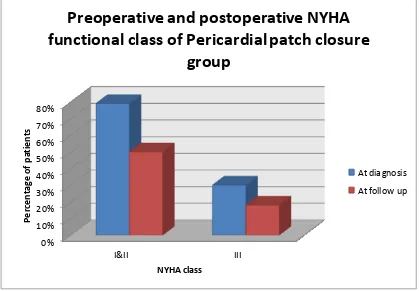
Graph 1 0% 10% 20% 30% 40% 50% 60% 70% 80% I&II III P e rc e nt ag e of pa ti e nt s NYHA class
Preoperative and postoperative NYHA
functional class of Pericardial patch closure
group
Graph 2 0% 10% 20% 30% 40% 50% 60% 70%
I& II III
P e rc e nt ag e of pa ti e nt s NYHA class
Preoperative and postoperative NYHA
functional class of direct suture closure
group
Echocardiographic (ECHO) data:
Average size of the ASD was 26±8mm, 24±7mm in the
pericardial patch closure and direct suture closure group respectively.
Mitral valve prolapse (MVP) was noted in 16 patients (30%) in the
pericardial patch closure group (Table 2) and 12 patients (24%) in the
direct suture closure group (Table 3). In the patients with MVP the
pulmonary artery mean pressures were higher and these patients were
older, most of them being above the age of forty years. Mitral
regurgitation (MR) was present in 9 patients in the pericardial patch
closure group, 7 of them had mild MR and 2 had moderate MR. In the
direct suture closure group 11 patients had mild MR. None of the
patients in both groups had a severe MR. 34 patients in the pericardial
patch closure group and 38% in the direct suture closure had no MVP
and their mean age was 25 years. We did not note any significant
correlation between the prolapse and the shunt size of the ASD. All
patients underwent prospective follow up ECHO at a mean period of
four months from the time of surgery. 4 patients in the pericardial
patch closure group and 3 patients in the direct suture closure group
developed MR in the follow up period. All these patients were elderly
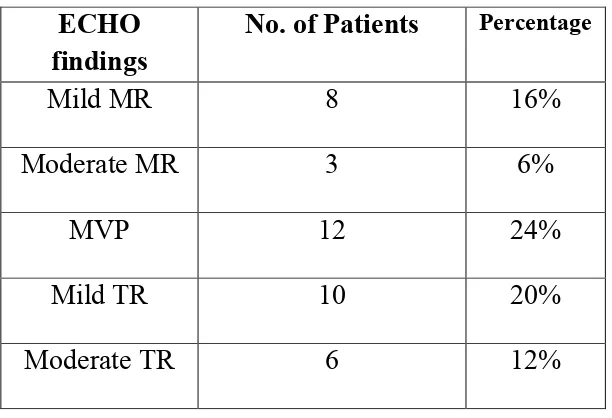
Pericardial patch closure- Preoperative ECHO findings
Table 2
Direct closure group- Preoperative ECHO findings
Table 3
MR-Mitral regurgitation, MVP-Mitral valve prolapse, TR-Tricuspid regurgitation, PHT-Pulmonary hypertension
ECHO findings No. of Patients Percentage
Mild MR 7 14%
Moderate MR 2 4%
MVP 16 30%
Mild TR 11 22%
Moderate TR 12 24%
ECHO findings
No. of Patients Percentage
Mild MR 8 16%
Moderate MR 3 6%
MVP 12 24%
Mild TR 10 20%
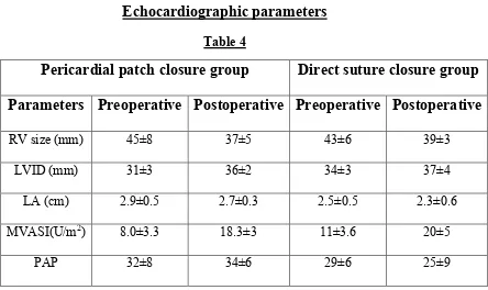
[image:57.595.155.461.439.648.2]Echocardiographic parameters
Table 4
Pericardial patch closure group Direct suture closure group
Parameters Preoperative Postoperative Preoperative Postoperative
RV size (mm) 45±8 37±5 43±6 39±3
LVID (mm) 31±3 36±2 34±3 37±4
LA (cm) 2.9±0.5 2.7±0.3 2.5±0.5 2.3±0.6
MVASI(U/m2) 8.0±3.3 18.3±3 11±3.6 20±5
PAP 32±8 34±6 29±6 25±9
RV- Right ventricle at end diastole, LVID-Left ventricular internal dimension at end diastole, LA-Left atrium, MVASI- Area subtended by the opposed mitral valve in systole in the long axis, cross sectional ECHO with respect to the mitral ring
Surgical data:
In this study 50 patients underwent pericardial patch closure and
another 50 patients underwent direct suture closure for ASD. We
observed that in those patients who underwent pericardial patch
closure the defect size was not significantly larger than in those who
had direct repair of their defects. There was no mortality in both the
groups. Surgical morbidity was present in 2 patients in the pericardial
patch closure group in the form of bradyarrythmia but they
up period after 2 months the drug was slowly withdrawn and
bradyarrhythmia did not recur in these patients. None of them needed
a permanent pace maker implantation. Atrial fibrillation was present
postoperatively in all the patients in both groups who had developed it
preoperatively (Table 5). 2 patients in the pericardial patch closure
group and 1 patient in the direct suture closure group developed new
atrial fibrillation the follow up. 3 patients in the pericardial patch
closure group and 4 patients in the direct suture closure group had
transient tachyarrhythmia their mean ages were 53±5. In the
pericardial patch closure group 1 patient aged 60 years developed
cardiac failure and required treatment with diuretics. 2 patients in the
direct suture closure group and 1 patient in the pericardial patch
closure group developed acute pericarditis in the postoperative period
and needed treatment with aspirin and steroids. 46% in the pericardial
patch closure group and 32% in the direct closure group had tricuspid
regurgitation. The mean pulmonary artery pressure in the first group
was 32±8 mm Hg preoperatively and in the postoperative follow up
period it was 34±6 mm Hg .In the direct closure group the mean
pulmonary artery pressure was 29±6 mm Hg preoperatively and
reduced to 25±9 mm Hg in the postoperative follow up period (Table
4). Two patients in the PPC group and one patient in the DC group
five patients in the DC group with preoperative atrial fibrillation
reverted to sinus rhythm. The right ventricular size in the PPC group
reduced from 45±8mm in the preop period to 37±5mm in the postop
follow up period. In the DC group it reduced from 43±6mm before
surgery to 39±3mm in the follow up period after surgery. Mitral valve
apposition area in systole (MVAS) improved from 8±3.3 to 18.3±3 in
the post op follow up in the PPC group. In the DC group the MVAS
improved from 11± 3.6 to 20±5 in the post op follow up.
Preoperative atrial flutter of fibrillation
Table 5
Variables Pericardial patch closure group
Direct suture closure group
Number of patients 14 10
Qp:Qs 2.3±4 1.9±7
PAP(mm Hg) 31±8 30±6
Electrocardiogram and chest X-ray findings
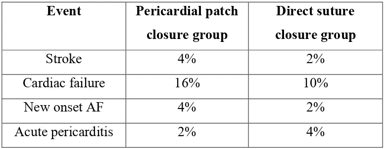
Table 6Cardiac events and clinical outcome after surgery
Table 7Event Pericardial patch closure group
Direct suture closure group
Stroke 4% 2%
Cardiac failure 16% 10%
New onset AF 4% 2%
Acute pericarditis 2% 4%
Pericardial patch closure group
Direct suture closure group
Variables Pre operative Post operative Pre operative Post operative
Axis 68±30 54±20 70±20 57±30
PR interval (m sec.)
184±6 152±5 179±8 140±3
QRS duration (m sec.)
123±2 107±2 128±3 111±5
R wave in lead V1
2.4±2 2.2±3 2.6±5 2.3±3
CT ratio in chest
X-ray (%)
[image:61.595.124.514.506.657.2]DISCUSSION
Kambet et al in their study on incidence of MVP associated
with atrial septal defect mentioned that the incidence of MVP was
53.2% whereas in our study the incidence of MVP was 54 %. 30 % of
patients in the PPC group had MVP and 24% in the DC group had
MVP. Postoperatively in the follow up ECHO the MVP reduced to
24% in the PPC group and to 13% in the DC group.
Comparing our results with the study done by Speechly et al,
surgical mortality in both study were zero. Late postoperative
morbidity was present in 7 patients in the PPC group and 6 patients in
the DC group in the form of atrial fibrillation, failure and acute
pericarditis whereas in the Speechly‟s study group one patient
required mitral valve replacement and tricuspid valve replacement and
the other one had developed cardiac failure, two patients had
developed sinus node dysfunction, new AF developed in two elderly
patients. In our study, 54% of patients who had MVP also had high
pulmonary artery pressures. MVP improved after surgery in 10 of
patients in the PPC group and 12% of patients in the DC group. While
in Speechly‟s study nine preoperative patients had high PAP and MVP
and MVP developed in five patients. In the PPC group four patients
and three patients in the DC group had developed new mitral
regurgitation. These results were also comparable to the study done by