Copyright © 1998, American Society for Microbiology
Resistance of Human Cytomegalovirus to Benzimidazole
Ribonucleosides Maps to Two Open Reading
Frames: UL89 and UL56
PAULA M. KROSKY,
1,2MARK R. UNDERWOOD,
3STEVEN R. TURK,
1† KATHRYNE W.-H. FENG,
1RAJEEV K. JAIN,
1ROGER G. PTAK,
1ALLISON C. WESTERMAN,
1KAREN K. BIRON,
3LEROY B. TOWNSEND,
2ANDJOHN C. DRACH
1,2*
Department of Biologic and Materials Sciences, School of Dentistry,
1and Interdepartmental Graduate Program in
Medicinal Chemistry, College of Pharmacy,
2University of Michigan, Ann Arbor, Michigan 48109, and
Department of Virology, Glaxo Wellcome Inc., Research Triangle Park, North Carolina 27709
3Received 26 November 1997/Accepted 4 March 1998
2,5,6-Trichloro-1-
b
-
D-ribofuranosyl benzimidazole (TCRB) is a potent and selective inhibitor of human
cytomegalovirus (HCMV) replication. TCRB acts via a novel mechanism involving inhibition of viral DNA
processing and packaging. Resistance to the 2-bromo analog (BDCRB) has been mapped to the UL89 open
reading frame (ORF), and this gene product was proposed as the viral target of the benzimidazole nucleosides.
In this study, we report the independent isolation of virus that is 20- to 30-fold resistant to TCRB (isolate C4)
and the characterization of the virus. The six ORFs known to be essential for viral DNA cleavage and packaging
(UL51, UL52, UL56, UL77, UL89, and UL104) were sequenced from wild-type HCMV, strain Towne, and from
isolate C4. Mutations were identified in UL89 (D344E) and in UL56 (Q204R). The mutation in UL89 was
identical to that previously reported for virus resistant to BDCRB, but the mutation in UL56 is novel. Marker
transfer analysis demonstrated that each of these mutations individually caused
;
10-fold resistance to the
benzimidazoles and that the combination of both mutations caused
;
30-fold resistance. The rate and extent
of replication of the mutants was the same as for wild-type virus, but the viruses were less sensitive to inhibition
of DNA cleavage by TCRB. Mapping of resistance to UL56 supports and extends recent work showing that
UL56 codes for a packaging motif binding protein which also has specific nuclease activity (E. Bogner et al.,
J. Virol. 72:2259–2264, 1998). Resistance which maps to two different genes suggests that their putative
proteins interact and/or that either or both have a benzimidazole ribonucleoside binding site. The results also
suggest that the gene products of UL89 and UL56 may be antiviral drug targets.
Human cytomegalovirus (HCMV) can cause significant
morbidity and mortality in immunocompromised populations
(3). It is a common opportunistic disease in patients with AIDS
and is often a factor in their death (38). HCMV infection has
been implicated in increased risk of organ rejection following
heart (28) and kidney transplants (8) and in restenosis of
diseased arteries following angioplasty (41, 63). It is also a
leading cause of birth defects (16).
Current therapies for HCMV infection include ganciclovir
(GCV) (22), cidofovir (30), and foscarnet (20). Each of these
drugs has several limitations to its use: none are orally
bio-available, all have dose-limiting toxicity, and resistance has
developed to each (26). Because all three of these drugs inhibit
viral replication through an interaction with the virally
en-coded DNA polymerase (25, 31, 37), the possibility of
cross-resistance exists. Thus, additional drugs with unique
mecha-nisms of action are needed for the treatment of HCMV
infections.
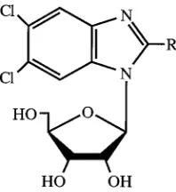
In 1995, we reported that 2-bromo-5,6-dichloro-1-(
b
-
D-ribo-furanosyl)benzimidazole (BDCRB; Fig. 1) and the 2-chloro
ana-log [2,5,6-trichloro-1-(
b
-
D-ribofuranosyl)benzimidazole TCRB]
are potent and selective inhibitors of HCMV replication (55).
These compounds have a novel mechanism of action, which
unlike the current therapies for HCMV infection, does not
involve inhibition of DNA synthesis. The benzimidazole
ribo-nucleosides prevent the cleavage of high-molecular-weight
vi-ral DNA concatemers to monomeric genomic lengths (57).
Resistance to BDCRB has been mapped to the HCMV UL89
open reading frame (ORF), which, by analogy to gene gp17
from bacteriophage T4, may be a terminase (23, 57).
Conse-quently, we have proposed that the benzimidazole
ribonucleo-sides inhibit the product of this gene and that the UL89 gene
product is involved in the viral DNA concatemer cleavage
process (57).
HCMV replication proceeds in a manner which is conserved
among herpesviruses. The virally encoded DNA polymerase
produces large, complex head-to-tail concatemers (10, 29, 33)
which must be cleaved into genomic-length pieces before
in-sertion into preformed capsids (59). With herpes simplex virus
type 1 (HSV-1), temperature-sensitive mutants which are
un-able to cleave and package the concatemeric DNA have been
derived (1, 2, 4, 45, 49, 50, 61). By this process, six HSV-1 genes
have been found to be involved in concatemer cleavage and
packaging. They are UL6, UL15, UL25, UL28, UL32, and
UL33. In addition, recent studies in Homa’s laboratory have
established that the product of UL25 is required for viral DNA
encapsidation but not cleavage (39). Homologs of these genes
exist in HCMV and are UL104, UL89, UL77, UL56, UL52,
and UL51, respectively (18).
In our continuing investigation of the mode of action of
benzimidazole nucleosides, we report herein the independent
* Corresponding author. Mailing address: School of Dentistry,
Uni-versity of Michigan, Ann Arbor, MI 48109-1078. Phone: (734)
763-5579. Fax: (734) 764-7406. E-mail: [email protected].
† Present address: Division of AIDS, National Institute of Allergy
and Infectious Diseases, National Institutes of Health, Bethesda, MD
20892.
4721
on November 9, 2019 by guest
http://jvi.asm.org/
isolation of HCMV strains resistant to TCRB, characterization
of these strains, and identification of the mutations responsible
for the development of resistance. The results demonstrate
that the mechanism of action of the benzimidazole
ribonucleo-sides is more complex than previously proposed and that a
second gene product implicated in DNA cleavage and
packag-ing is involved.
MATERIALS AND METHODS
Chemicals.BDCRB and TCRB (Fig. 1) were synthesized in the laboratory of L. B. Townsend as previously described (55). GCV was provided through the courtesy of Syntex Laboratories, Inc., Palo Alto, Calif.
Cell and viral culture.Monolayer cultures of human foreskin fibroblasts (HFF cells) and MRC-5 cells were grown in minimal essential medium with Earle’s salts [MEM(E)] plus 10% fetal bovine serum at 37°C in a humidified atmosphere of 3% CO2–97% air. They were regularly passaged at 1:2 dilutions, using
con-ventional techniques, with 0.05% trypsin plus 0.02% EDTA in HEPES-buffered saline (51). HCMV strain Towne was kindly provided by Mark Stinski, University of Iowa. Stocks of HCMV were prepared by infecting HFF cells at a multiplicity of infection (MOI) of,0.01 PFU per cell, and stock viral titers were determined in monolayer cultures of HFF cells as described earlier (46, 56). Viral assays, but not the preparation of viral stocks, were done in the presence of penicillin-streptomycin (100 U of penicillin G per ml, 100mg of streptomycin per ml).
HCMV antiviral assays.Both plaque and yield reduction assays were used to compare the sensitivities of isolates to BDCRB and TCRB. For plaque reduction assays, HFF cells in 24-well cluster dishes were infected with approximately 100 PFU of HCMV per well as described earlier (56). Following virus adsorption, compounds dissolved in growth medium were added to duplicate wells at four to eight selected concentrations. After incubation at 37°C for 10 days, cell mono-layers were stained with crystal violet, and plaques were enumerated at 40-fold magnification. Drug effects were calculated as a percentage of reduction in number of plaques observed in the presence of each drug concentration com-pared to the number observed in the absence of drug. For yield reduction assays, the procedure devised by us (46) was used. Briefly, HFF cells were planted in 96-well cluster dishes, incubated overnight, and infected with HCMV at an MOI of 0.5 to 1. After virus adsorption, inoculum was replaced with 0.2 ml of fresh medium containing test compounds in quadruplicate on a single plate in a manner which provided drug concentrations from 100 to 0.14mM, using one-half log10dilutions. Plates were incubated at 37°C for 4 days and subjected to one
cycle of freezing and thawing. Aliquots from each of the wells were transferred to a fresh 96-well monolayer culture of HFF cells and serial diluted across the plate. Cultures were incubated, cells were stained, plaques were enumerated at 40-fold magnification, and titers were calculated as described.
For both plaque and yield assays, dose-response relationships were con-structed by plotting the percent inhibition of plaque number or the log10of the
percent inhibition of virus titer against log10drug concentrations. The 50 or 95%
inhibitory concentration (IC50or IC95) and corresponding 95% confidence
in-tervals were calculated by using the variable-slope dose-response algorithm of GraphPad Prism (GraphPad, San Diego, Calif.). Samples containing GCV as a positive control were used in all assays.
Selection of TCRB-resistant virus.HFF cells were infected with HCMV strain Towne, and the virus was grown in 10mM TCRB for 45 days. The progeny were collected and grown for an additional 7 days in 10mM TCRB. The resulting virus was passaged in 30mM TCRB for 13 days. The duration of each passage was a function of the viral input and the rate of viral growth. The resulting virus stock was plaque purified three times by limiting dilution, and purified isolates were designated D10 and B11. Isolate D10 was further passaged three times in 50mM TCRB, and the highly resistant viral stock was plaque purified three times by limiting dilution; this isolate was designated C4. Virus isolates D10 and C4 were tested for drug resistance by plaque reduction assay and yield reduction assay
and were further characterized by pulsed-field electrophoresis as described pre-viously (57).
Growth characteristics.Viral growth rates were measured both over a single cycle of viral replication and over multiple cycles of replication. For observation of a single cycle, HFF cells were plated at 10,000 cells per well in 96-well cell culture plates and infected at an MOI of 2 PFU/cell. At time points spaced 8 to 12 h apart, the cultures were frozen at280°C. Samples were collected for 4 days, and after all were collected, they were titered across microplates as described previously (46). For assays involving multiple cycles of replication, growth was determined by infecting 96-well cell culture plates with 10,000 HFF cells per well at an MOI of 0.01 PFU/cell and freezing samples every 12 h over a course of 10 days.
Analysis of DNA processing.Samples were prepared for contour-clamped homogeneous electric field electrophoresis with a ChefMapper (Bio-Rad, Her-cules, Calif.) as described previously (57). Briefly, HFF cells were infected at an MOI of 3 PFU/cell and treated with selected drug concentrations for 3 days. Cells were removed from monolayers by treatment with a solution of 0.5 mM EDTA and 0.05% trypsin, suspended in 0.8% agarose, and digested with 100 mg of proteinase K (PK) per ml in 1% sarkosine for 48 h. Aliquots were loaded into the wells of a 1% agarose gel, and electrophoresis was performed as detailed previously (52). Upon completion of the electrophoresis procedure, gels were stained with ethidium bromide and photographed. Figure 4 was obtained by scanning the photographs with a Microtek Scanmaker III using Adobe Photo-shop. The colors were inverted and selected portions of the gels were transferred to Abobe Pagemaker, aligned, and labeled.
DNA sequencing.ORFs UL51, UL52, UL56, UL77, UL89, and UL104 were sequenced from wild-type HCMV, strain Towne, and isolate C4. Additionally, the UL56 and UL89 ORFs were sequenced from isolate D10 and, later, from recombinant isolates r56 and rC4. Viral stocks (;106PFU/ml) were lysed in an
equal volume of 13PCR buffer (Perkin-Elmer, Foster City, Calif.)–0.05% Non-idet P-40–0.05% Tween 20. These were digested with PK (100mg/ml) at 55°C for 1 h, and the enzyme was inactivated at 95°C for 15 min. Specific viral genes were amplified from this lysate by PCR. Primers for PCR and sequencing were de-signed by using the PRIME program in the Genetics Computer Group package and are based on the published sequence of the AD169 strain of HCMV (18, 27). A primer list is available upon request. The PCR mixtures included 20% viral lysate, 10% dimethyl sulfoxide, deoxynucleoside triphosphates at 250 nM (Gibco BRL, Grand Island, N.Y.), primers at;1.5mM (Midland Certified Reagents, Midland, Tex.), and 2.5 U of AmpliTaq (Perkin-Elmer). The reagents were prepared in an ice bath, and the tubes were placed in a Perkin-Elmer GeneAmp 2400 PCR system preheated to 95°C. The reactions were cycled 35 times as follows: 95°C for 45 s, 58°C for 1 min, and 72°C for 3 min. PCR products were purified by using a QiaQuick PCR purification kit (Qiagen, Chatsworth, Calif.) or a Centricon 100 column (Amicon, Beverly, Mass.). DNA was quantified by comparison with DNA mass markers (Gibco BRL) and sequenced using a Dye Terminator Cycle Sequencing Ready Reaction kit (Perkin-Elmer). The sequenc-ing reactions were purified with a CentriSep spin column (Princeton Separations, Aldephia, N.J.) and were separated and detected with an Applied Biosystems Inc. 377 DNA sequencer. The data were aligned and edited by using Sequencher software (GeneCodes, Ann Arbor, Mich.).
Marker transfer studies. Genomic DNA was prepared using two different methods. For wild-type (wt) virus, strain Towne, a modification of the package insert from a RecoverEase DNA isolation kit (Stratagene, La Jolla, Calif.) was used. Supernatant virus from HCMV-infected cells was pelleted at;10,0003g for 2.5 h. Pellets were suspended in 1 volume of digestion buffer plus 1 volume of PK solution and were incubated at 55°C for 2 h with gentle rocking every 15 min. The resulting suspension was dialyzed against TE (10 mM Tris-HCl [pH 7.5], and 1 mM EDTA) overnight. For wt virus, strain AD169, and isolate D10, a second method was used (52). Briefly, virus pellets were suspended in TNE (10 mM Tris-HCl [pH 7.5], 100 mM NaCl, 1 mM EDTA) with 0.05% sodium dodecyl sulfate and digested overnight with PK (100mg/ml) at 37°C. The resulting DNA-containing solutions were gently extracted twice with 1 volume of phenol and once with 1 volume of 24:1 chloroform-isoamyl alcohol. The DNA was precipitated overnight at 220°C with 2.5 volumes of ice-cold 95% ethanol. Precipitates were collected by centrifugation at 12,0003g for 30 min and pellets were dissolved in TE by standing undisturbed at room temperature for several days.
[image:2.612.120.219.69.177.2]Calcium phosphate transfections were performed as described by Sambrook et al. (48). Infectious DNA (0.3 to 2mg) from either wt Towne or D10 virus isolate was coprecipitated with 12- to 25-fold molar excess of a PCR product encoding the portion of interest of UL56 from either wt Towne or C4 isolate (Fig. 2). Low (10 to 15)-passage HFF cells were exposed to the precipitate for 4 to 5 h, and then the precipitate was removed. The cells were treated with 15% glycerol in buffered saline for 90 s, washed briefly with MEM(E) containing 5% fetal bovine serum and penicillin-streptomycin, and incubated overnight in this medium. The medium was replaced with fresh medium the following morning. A PCR product from exon II of UL89 (Fig. 2) from B11 was similarly transfected with infectious wt AD169 DNA into MRC-5 cells. Transfected cultures were incubated in the absence of drug until maximum cytopathic effect was observed (usually around 21 days), and the supernatant progeny virus was clarified by centrifugation (2,0003g for 5 min) and stored in liquid nitrogen. Titers of viral stocks were determined as described previously (46).
FIG. 1. Structure of benzimidazole ribonucleosides. TCRB, R 5 Cl; BDCRB, R5Br.
on November 9, 2019 by guest
http://jvi.asm.org/
Viral plating efficiencies were determined by infecting five flasks (75 cm2) each
with 20,000 to 70,000 infectious progeny virions under a methylcellulose overlay containing BDCRB. The concentrations of BDCRB used to screen the various recombinants are given in Table 3. A parallel experiment was performed in the absence of BDCRB to determine the actual number of infectious virions plated in a given experiment. Five additional flasks each were infected with 1/10 the virus used in the first experiment. Recombination efficiencies were calculated by subtracting the number of plaques produced by wt progeny virus in the presence of BDCRB from the number of plaques produced by recombinant progeny virus populations in the presence of BDCRB. The resulting number of plaques ob-served under drug was divided by 10 times the number of plaques obob-served in the absence of drug, and the result was converted to a percentage.
The recombinant, mutant viruses were isolated and plaque purified by the method of Klein (34). The first round was performed in the presence of 2, 5, or 15mM BDCRB, depending on the resistance of the virus, and two subsequent rounds were performed in the absence of drug.
Nucleotide sequence accession numbers. Nucleotide and amino acid se-quences were obtained from GenEMBL. Accession numbers are as follows: HCMV strain AD169, X17403; HSV-1, X14112; human herpesvirus 6, X83413; human herpesvirus 7, U43400; and varicella-zoster virus, X04370. The sequences from HCMV strain Towne of UL51, UL52, UL56, UL77, UL89 exon 1, UL89 exon 2, and UL104 as reported herein are AF039234, AF047521, AF047523, AF047522, AF047525, AF047526, and AF047524, respectively.
RESULTS
Initial isolation and characterization of resistant virus.
Prior studies in our laboratories have shown that BDCRB
inhibits viral DNA packaging through an interaction with the
gene product of UL89 (57). The resistant isolates (1038rA, -B,
and -C) were obtained by growth of HCMV strain AD169 in
the presence of BDCRB. Independently, another clone of
re-sistant virus was isolated by passaging HCMV strain Towne in
the presence of increasing concentrations of TCRB up to 30
m
M. Isolates resulting from plaque purification of the
hetero-geneous mixture have been designated D10 and B11. Isolates
D10 and B11 were approximately 10-fold resistant to the
benz-imidazole ribonucleosides in plaque reduction assays (Table
1). This was nearly identical to resistance observed with strains
1038rA and 1038rC (57). Moreover, all four isolates were
sen-sitive to GCV and grew in the absence of drugs at rates nearly
identical to those for wt HCMV (data not presented), further
illustrating that a unique mechanism of action was involved.
Sequence analysis of B11 and D10.
Based on the similarity
of phenotypic characteristics among B11, D10, and 1038rA and
1038rC plus the known mutation of aspartate to glutamate at
position 344 (D344E) in UL89, which caused the resistance of
the AD169 strains (57), UL89 of B11 and D10 was sequenced.
A change of C to G in nucleotide 1032 of UL89 resulting in an
inferred D344E amino acid mutation was found in B11 and
D10 (Table 2). This change was identical to the C-to-G
nucle-otide change found in AD169 strains 1038rA and 1038rC but
different from the C-to-A change found in strain 1038rB, which
conferred the identical D344E change (57). In addition to the
C-to-G change, a difference between AD169 and Towne
strains was found in the next nucleotide (1033, T to G),
result-ing in a codresult-ing change of serine to alanine in position 345.
[image:3.612.53.288.71.204.2]To determine if the D344E change was necessary and
suffi-cient for the Towne strain to be resistant to the benzimidazole
ribonucleosides, drug resistance was transferred by
transfect-ing exon 2 of the UL89 ORF from isolate B11 into MRC-5
cells with infectious, genomic-length AD169 DNA. The plating
efficiency difference of 0.43% between populations transfected
with wt and B11 UL89 DNA (Table 3) represents the
percent-age of drug-resistant virions in the heterogeneous progeny and
demonstrated that the mutation in UL89 generated resistance
to the benzimidazoles. This recombinant (termed rT89E2-4)
was isolated, and its resistance to the benzimidazole
ribo-nucleosides was similar to that of isolates B11 and D10 (Table
1), demonstrating that the mutation in UL89 was sufficient to
cause the level of resistance observed. The recombinant was
[image:3.612.308.548.89.171.2]FIG. 2. Schematic of the genome of HCMV showing the locations and ori-entations of the UL56 and UL89 ORFs. A PCR product encoding exonII of UL89 was used in the marker transfer experiments, and a 39-truncated portion of UL56 was similarly used. The numbers shown are from the published sequence of HCMV strain AD169 and are only relative for the Towne-derived viruses reported in this paper.
TABLE 1. Resistance of HCMV strains to benzimidazole
ribonucleosides and GCV
HCMV strain
mean IC50(mM)a6SD
TCRB BDCRB GCV
Towne
3.4
6
1.4
0.92
6
0.61
4.9
6
2.8
D10
29
6
9.9
7.4
6
5.7
5.8
6
2.8
B11
28
6
15
14.3
6
7.4
3.5
6
0.4
AD169
1.8
6
0.1
0.3
6
0.1
4.6
6
3.3
rT89E2-4
ND
b6.3 (4.9–8.1)
cND
aResults from plaque reduction assays performed in duplicate, using the
indicated strains of HCMV as described in the text. IC50s were calculated from
2 to 20 experiments using at least four drug concentrations each.
bND, not determined.
cResults presented with 95% confidence interval (in parentheses) because
data are from a single experiment performed in duplicate which used eight drug concentrations.
TABLE 2. Sequence of the UL89 ORF from drug-resistant isolates
HCMVstrain Position no.a Sequenceb
AD169
c1021
ACT ACC AGT GAC TCC ACG TGT
341
Thr Thr Ser Asp Ser Thr Cys
Towne
1021
ACT ACC AGT GAC GCC ACG TGT
341
Thr Thr Ser Asp Ala Thr Cys
D10
1021
ACT ACC AGT GAG GCC ACG TGT
341
Thr Thr Ser Glu Ala Thr Cys
B11
1021
ACT ACC AGT GAG GCC ACG TGT
341
Thr Thr Ser Glu Ala Thr Cys
rT89E2-4
1021
ACT ACC AGT GAG TCC ACG TGT
341
Thr Thr Ser Glu Ser Thr Cys
C4
1021
ACT ACC AGT GAG GCC ACG TGT
341
Thr Thr Ser Glu Ala Thr Cys
rC4
1021
ACT ACC AGT GAG GCC ACG TGT
341
Thr Thr Ser Glu Ala Thr Cys
r56
1021
ACT ACC AGT GAC GCC ACG TGT
341
Thr Thr Ser Asp Ala Thr Cys
aFor the first nucleotide or amino acid listed.
bThe nucleotide sequence of the entire UL89 ORF was determined as
de-scribed in the text. Only the portion of exon 2 which contained the mutation is presented. Nucleotide and inferred amino acid changes are shown in boldface.
cNucleotide sequence from GenBank.
on November 9, 2019 by guest
http://jvi.asm.org/
[image:3.612.307.548.510.685.2]sequenced and had the expected C-to-G change in nucleotide
1032 in exon 2 of UL89 resulting in the D344E mutation
(Table 2). Surprisingly, T was found in position 1033, inferring
serine in position 345 which is characteristic of the parental
AD169 DNA used in the transfection, not an alanine which
would be expected based on Towne strain as the source of the
drug-resistant DNA. We speculate that because there were two
adjacent mismatches due to homologous recombination
be-tween genomic DNA from wt AD169 and PCR product of
mutant UL89 from Towne, the second mismatched nucleotide
(position 1033) was repaired in the recombinant DNA.
Isolation and characterization of highly resistant virus.
In
our prior study, isolate 1038rB was more resistant to the
benz-imidazole ribonucleosides than 1038rA and 1038rC due to an
additional mutation from Ala to Thr at position 355 (57). To
explore the possibility that the region of UL89 around amino
acids 344 to 355 was critically involved in the binding of
benz-imidazole ribonucleosides, we attempted to isolate additional
drug-resistant mutants by passaging strain D10 (with the single
D344E mutation) in a higher concentration of TCRB. Upon
further passage of isolate D10 in the presence of 50
m
M
TCRB, progeny virus eventually grew; another mutant was
selected and plaque purified, and the resulting isolate was
designated C4. It was
;
30-fold resistant to the benzimidazole
ribonucleosides (Table 4). Based on the prior results and the
increased resistance to the benzimidazoles, virus isolate C4 was
expected to encode additional mutations in UL89, but none
were detected (Table 2). C4 was further characterized
geno-typically by sequencing the other five genes known to be
in-volved in viral DNA processing and packaging (ORFs UL51,
UL52, UL56, UL77, and UL104). The only ORF of these six
that had any mutation was UL56. A mutation from A to G in
position 611 resulted in a coding change from glutamine to
arginine at position 204 (Q204R) (Table 5).
Marker transfer analysis of highly resistant virus.
Marker
transfer techniques were used to explore the significance of the
mutation identified in the UL56 ORF. This mutation was
in-troduced into D10 virus through homologous recombination.
The plating efficiency of the progeny (Table 3) strongly
sug-gested that the Q204R mutation was responsible for the
in-crease in drug resistance between isolates D10 and C4. Since
none of the progeny from the recombination between D10
DNA and wt UL56 grew under the selection conditions (20
m
M BDCRB), the increased drug resistance did not appear to
develop spontaneously during the course of the experiment
and the resistant virus observed was a result of the mutation in
the UL56 ORF.
The drug-resistant recombinant was plaque purified and
designated rC4. Its susceptibility to the benzimidazole
ribo-nucleosides and to GCV was similar to that of C4 (Table 4).
The similarity in the resistance patterns of virus isolates C4 and
rC4 confirmed that the mutation in UL56 was responsible for
the increase in drug resistance between isolates D10 and C4
and substantiated that it was the only additional mutation
involved in the phenotype of a high level of drug resistance
(20-to 30-fold) in isolate C4. UL89 and UL56 from virus isolate
rC4 were sequenced, and each encoded only the expected
mutations (Tables 2 and 5).
Homologous recombination also was used to selectively
in-troduce the mutation in UL56 from isolate C4 into wt Towne
virus. The progeny from this experiment had a plating
effi-ciency of 5.4% in 5
m
M BDCRB, which demonstrated that the
single nucleotide change in the UL56 ORF could cause drug
resistance in the absence of any changes in the UL89 ORF
(Table 3). This recombinant virus was plaque purified and
designated r56. It was 5- to 10-fold resistant to the
benzimid-azole nucleosides (Table 4), demonstrating that this mutation
also was necessary and sufficient for drug resistance. Virus r56
TABLE 3. Efficiencies of marker transfer experiments
Virus strain for:
Gene for PCR product BDCRB concn (mM) Plating efficiencya(%) Recombinant strain Parental DNA PCR product
AD169
wt (Towne)
UL89
2
0.070
AD169
B11 (Towne)
UL89
2
0.50
rT89E2-4
D10
wt (Towne)
UL56
20
,
0.003
D10
C4 (Towne)
UL56
20
0.29
rC4
Towne
wt (Towne)
UL56
5
,
0.005
Towne
C4 (Towne)
UL56
5
5.4
r56
[image:4.612.51.548.82.170.2]aHFF cells were cotransfected with the indicated sources of parental and mutated DNA (PCR product). Following transfections, stocks of progeny virus were grown and titers were determined. Five flasks (20,000 to 70,000 PFU per flask) were infected with this virus and incubated in the presence of the indicated concentrations of BDCRB. Five flasks also were infected in the absence of drug, using 1/10 the amount of virus. Plating efficiency is the percentage of plated recombination progeny which formed plaques under the indicated concentration of BDCRB as described in the text.
TABLE 4. Susceptibilities of HCMV strains and recombinants to benzimidazole ribonucleosides and GCV
aHCMV strain Plaque reduction assay (IC50[mM]) Yield reduction assay (IC95[mM])
TCRB BDCRB GCV TCRB BDCRB GCV
Towne
2.4 (2.2–2.7)
2.3 (1.5–3.5)
4.9 (3.7–6.4)
1.1 (0.9–1.2)
0.18 (0.15–0.22)
3.1 (2.1–4.7)
D10
18 (14–22)
13 (11–16)
5.4 (3.9–7.5)
7.9 (5.5–11)
2.0 (1.4–2.9)
3.4 (2.6–4.4)
C4
57 (43–76)
33 (29–38)
6.1 (4.9–7.7)
25 (18–34)
6.8 (4.5–10)
1.8 (0.85–3.7)
rC4
68 (28–163)
32 (24–42)
2.5 (2.1–2.9)
39 (32–48)
4.8 (2.8–8.3)
3.7 (2.7–5.0)
r56
12 (10–15)
7.6 (6.0–9.6)
7.0 (5.0–9.8)
5.2 (4.0–7.0)
3.0 (2.4–3.8)
2.9 (1.9–4.4)
aPlaque and yield reduction assays were performed with the indicated strains of HCMV as described in the text; 95% confidence intervals (in parentheses) were calculated by using the variable-slope dose-response algorithm of GraphPad Prism. Plaque assay data are averages of three experiments, each done in duplicate except for r56, in which case two experiments were done. Yield reduction assays were done once, in quadruplicate.
on November 9, 2019 by guest
http://jvi.asm.org/
[image:4.612.48.547.619.700.2]was sequenced through ORFs UL89 and UL56 to verify that it
had only one mutation in the UL56 ORF (Q204R) and did not
have any mutations in UL89 (Tables 2 and 5).
Growth studies.
The replication characteristics of wt Towne,
D10, C4, rC4, and r56 were measured to determine if any of
the isolates were growth defective. The rate of replication of
each virus isolate initially was measured following infection at
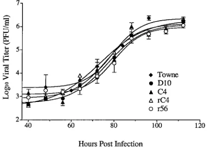
a high MOI (2 PFU/cell). Figure 3 shows that there were no
differences in the rates of replication of the various isolates
over the course of a single cycle of replication. Statistical
com-parisons showed that the slopes of all curves were similar
(slopes
6
95% confidence intervals
5
7.1
6
2.5, 5.7
6
1.5,
6.0
6
1.4, 7.1
6
1.7, and 5.4
6
1.4 h
21, respectively for Towne,
D10, C4, rC4, and r56). Additionally, there were no statistically
significant differences in the total amounts of infectious virus
produced during the course of the infection (yield
6
95%
confidence intervals
5
6.4
6
0.3, 6.0
6
0.2, 6.1
6
0.2, 6.1
6
0.2,
and 5.9
6
0.1 log10
PFU/ml at 112 h postinfection, respectively,
for Towne, D10, C4, rC4, and r56) (Fig. 3). Thus, all of the
mutants were fully capable of replicating at normal levels.
Each isolate was also observed over multiple cycles of
replica-tion by infecting cells at a low MOI (0.01 PFU/cell). Samples
were frozen twice daily for 10 days, and the viral titer of each
sample was determined. No statistically significant differences
in the slopes of the growth curves or in the total yield of
HCMV were detected among the mutants (slopes
6
95%
confidence intervals
5
25
6
2.6, 35
6
8.4, and 31
6
7.3 h
21and
yield at plateaus
5
7.4
6
0.2, 8.2
6
0.7, 7.0
6
0.3, and 8.4
6
0.9
log10
PFU/ml at 204 h postinfection, respectively, for D10, C4,
rC4, and r56).
Analysis of DNA processing by the recombinant viruses.
Benzimidazole ribonucleosides inhibit the cleavage of
concate-meric, viral DNA to monoconcate-meric, genomic-length units at
con-centrations at and above the IC50
for both wt virus and HCMV
isolates resistant to BDCRB due to the mutation in UL89 (57).
To explore if this also were true for resistant isolates with
mutations in UL56, HFF cells were infected with the mutant
HCMV isolates and grown for a single cycle of viral
replica-tion. Intracellular DNA was separated by pulsed-field
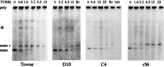
electro-phoresis and stained with ethidium bromide. Figure 4 shows
gels obtained from wt Towne and three mutant viruses. The
concentration of TCRB required to inhibit monomer
forma-tion by each of the mutants was proporforma-tional to the IC50
of the
isolate. The phenotype did not differ between D10, with a
single mutation in UL89, and r56, with a single mutation in
UL56, nor was there any difference with C4, which contains
both mutations. These data further confirm that the
benzimid-azole ribonucleosides target viral DNA processing.
We also observed a band migrating slightly slower than the
monomer band (labeled “mono
1
” in Fig. 4), similar to
pre-vious studies with BDCRB-resistant viruses (57). The amount
of this apparently higher-molecular-weight DNA species first
appeared at drug concentrations around the IC50
and
in-creased with increasing drug concentrations. At particularly
high concentrations of TCRB, this band disappeared with wt
virus and isolate C4. With isolates D10 and r56, it persisted at
the highest drug level tested, but we have no data to explain
why this persistence was observed only with the
single-muta-tion viruses. The identity and structure of this DNA species is
not known but is under investigation (57).
DISCUSSION
There is substantial evidence from our laboratories
indicat-ing that the benzimidazole ribonucleosides inhibit viral
repli-cation during the DNA processing and packaging stage (57).
Most significant is that polygenomic concatemeric viral DNA is
not cleaved to monomeric lengths and is not encapsidated (57).
This is a phenotype consistent with that of
temperature-sensi-tive and null mutant HSV-1 isolates with aberrations in one of
six ORFs: UL6, UL15, UL25, UL28, UL31, or UL32 (1, 2, 4,
5, 45, 49, 50, 54, 61, 62). Each of these genes is essential for
viral replication and is presumed to be involved in viral DNA
maturation. The similarity in phenotype between these
mu-tants and wt HCMV treated with the benzimidazole
ribo-nucleosides is consistent with the hypothesis that
benzimid-azole ribonucleosides inhibit viral DNA processing.
HSV-1 ORFs UL15 and UL28 are homologs of HCMV
ORFs UL89 and UL56, respectively. Previous studies have
shown that null mutants of HSV-1 UL15 and UL28 have the
same phenotype of inability to process viral DNA (5, 54, 62).
Our isolates, D10 and r56, respectively, have similar resistance
to the benzimidazole ribonucleosides in antiviral assays. Thus,
our mutants with a single mutation in either HCMV UL89 or
UL56 also have identical phenotypes. These studies suggest
that the proteins encoded by UL56 and UL89 either have very
similar functions or participate in an intertwined, concerted
series of activities.
[image:5.612.50.290.81.211.2]Antiviral drug resistance that maps to two different genes is
[image:5.612.64.275.531.682.2]FIG. 3. Single-cycle growth curves comparing mutant viruses. HFF cells were infected at an MOI of 2 PFU/cell and were harvested at time points spaced 8 to 12 h apart over the course of 4 days. After all samples were collected, they were titered across 96-well plates. Curves were fitted and data were analyzed by using the exponential growth algorithm of Prism.
TABLE 5. Sequences of UL56 from drug-resistant virus isolates
HCMV strain Position no.a Sequenceb
AD169
c601
ATC CCG AAT CAG GGC CGC TCG
201
Ile Pro Asn Gln Gly Arg Ser
Towne
601
ATC CCG AAT CAG GGC CGC TCG
201
Ile Pro Asn Gln Gly Arg Ser
D10
601
ATC CCG AAT CAG GGC CGC TCG
201
Ile Pro Asn Gln Gly Arg Ser
C4
601
ATC CCG AAT CGG GGC CGC TCG
201
Ile Pro Asn Arg Gly Arg Ser
rC4
601
ATC CCG AAT CGG GGC CGC TCG
201
Ile Pro Asn Arg Gly Arg Ser
r56
601
ATC CCG AAT CGG GGC CGC TCG
201
Ile Pro Asn Arg Gly Arg Ser
aFor the first nucleotide or amino acid listed.
bThe nucleotide sequence of the entire UL56 ORF was determined as
de-scribed in the text. Only the portion which contains the mutation is presented. Nucleotide and inferred amino acid changes are shown in boldface.
cNucleotide sequence from GenBank.
on November 9, 2019 by guest
http://jvi.asm.org/
not unprecedented. With GCV, resistance maps both to the
kinase responsible for activation of the drug (53) and to the
DNA polymerase which GCV triphosphate inhibits (52).
How-ever, this is very different from our current results in that these
two steps in GCV action are not directly related in viral
rep-lication. Furthermore, BDCRB does not require
phosphoryla-tion for activity, and there is no evidence for any other form of
activation (36). Consequently, this does not explain the pattern
of resistance to the benzimidazoles.
Mutations in proteins known to interact with the target
pro-tein of a drug have been shown to cause altered drug sensitivity
(21). For example, selected mutations in the gene for viral
DNA polymerase that result in resistance to phosphonoacetic
acid concurrently result in hypersensitivity to aphidicolin (9,
21). In addition, temperature sensitive mutants with lesions in
the single-stranded DNA binding protein (UL29) have
in-creased sensitivity to phosphonoacetic acid and aphidicolin
(19). That mutations in one protein (UL29) could affect the
activity of another (DNA polymerase) led to the proposal of an
interaction between the viral DNA polymerase and the
single-stranded DNA binding protein (reviewed in reference 42).
Likewise, the fact that drug resistance maps to both UL89
and UL56 strongly suggests an interaction between their two
gene products. This is consistent with the proposed role of
each in viral DNA maturation and with observations of the
pseudorabies virus (PRV) homolog of UL56 (UL28). This
protein is found primarily in the nuclei of infected cells (44),
but it is limited to the cytoplasm in the absence of other viral
proteins. When it is coexpressed with HSV-1 UL15, it regains
nuclear localization (35). These findings support an interaction
between the two proteins which is required for proper
intra-cellular localization of the PRV UL28 gene product. It is
possible that in HCMV such an interaction between UL56 and
UL89 proteins results in a complex which functions during
viral DNA maturation.
There also are similarities to bacteriophage. Bacteriophage
replication has long been proposed as a model for that of the
herpesviruses because of the similarity in genome size and the
large number of gene products required for DNA replication
and packaging (60). Both synthesize high-molecular-weight
DNA concatemers, cleave these concatemers into
genomic-length units, and package the pieces into preformed capsids. A
two-subunit terminase complex is common among
bacterio-phages (12). In bacteriophage T4, gp17 is part of a two-subunit
terminase complex with gp16 (12). The gp17 subunit appears
to have constitutive endonucleolytic activity which seems to be
regulated by an association with gp16 (47). gp17 also interacts
directly with another protein—p20, the bacteriophage capsid
vertex protein (32). A mutant with a lesion in p20 displays the
same phenotype as the HSV-1 packaging mutants; viral DNA
is synthesized but not cleaved or encapsidated.
HCMV UL89 and homologs are highly conserved among
herpesviruses and are invariably expressed as the spliced
prod-uct of two or more exons. The HSV-1 homolog, UL15, has
been studied extensively (5–7, 24, 45, 57, 62) but the function
of this protein has not been observed directly. In 1992, Davison
reported some homology between UL89 and the
endonucleo-lytic portion of the terminase complex of bacteriophage T4,
protein gp17 (23), thereby suggesting that UL89 could be the
HCMV terminase. Both proteins encode a potential
nucleo-tide binding motif, HCMV amino acids GTK219 to DE310. In
a linear sense, this motif is distal to the mutations in UL89 that
confer resistance. It is possible that protein folding juxtaposes
the two areas and allows the mutations to alter the nucleotide
binding site and thereby affect endonuclease activity (57).
However, we know of no direct evidence for nuclease activity
of the UL89 protein.
In contrast, the recent studies of Bogner et al. (13) provide
direct evidence for specific nuclease activity of the UL56 gene
product. Until now, little has been known about the function of
this protein. It is a minor structural protein and can be
de-tected with human convalescent serum (14, 15). There were
early suggestions that the HSV-1 homolog could be involved in
both DNA processing (2) and glycoprotein expression (43).
However, later studies negated its involvement in glycoprotein
trafficking, and a null mutant of the HSV-1 UL28 ORF was
fully capable of glycoprotein expression (17, 54). This finding
suggests that UL28 may have multiple, independently
func-tioning domains. Bogner et al. also provide direct evidence for
such a suggestion by establishing that the gene product of
UL56 has not only nuclease activity but also specific DNA
binding affinity (13).
[image:6.612.134.466.75.212.2]A potentially corroborating observation for specific DNA
binding emerges from a comparison of the predicted amino
acid sequences of some of the homologs of the UL56 ORF
(Table 6). First, the amino acid change which resulted in drug
resistance (Q204R) is in a position that is completely
con-served but the change did not affect the viability of the virus.
Second, three cysteines and one histidine also are completely
conserved in all 12 herpesviruses that have been compared.
FIG. 4. Pulsed-field electrophoresis study of monomer formation by the mutant viruses in the presence of TCRB. HFF cells were infected at an MOI of 3 PFU/cell and treated with selected concentrations (micromolar) of TCRB for 3 days. Cells were embedded in agarose and digested with PK. DNA was separated with a ChefMapper, stained with ethidium bromide, and photographed. Bands: poly, polygenomic, concatemeric HCMV DNA; di, digenomic HCMV DNA; mono1, 270-kb HCMV DNA; mono, 230-kb genomic-length HCMV DNA.on November 9, 2019 by guest
http://jvi.asm.org/
These amino acids could represent a metal binding motif (11);
in accord with observations of Bogner et al. (13), this suggests
DNA sequence-specific binding capability for the UL56
pro-tein.
Such specific binding may account for one of the major
differences in DNA packaging between bacteriophage T4 and
the herpesviruses. Bacteriophage T4 DNA is packaged in a
head-full manner (12), whereas herpesvirus DNA is cleaved
and packaged in a manner that is both sequence specific and
measured (40, 58, 59). The protein encoded by UL56
appar-ently binds the pac sequences (cleavage recognition sites) and
endonucleolytically cleaves viral DNA. The role of the gene
product of UL89 is unknown but it may be an accessory protein
to the UL56 protein. This may account for the putative
inter-action between these two proteins that is implied by the results
reported herein.
For TCRB or BDCRB to inhibit viral DNA processing, it is
possible that both UL89 and UL56 proteins interact directly
with the benzimidazole ribonucleoside. Consequently
resis-tance to the benzimidazoles would be a function of altered
binding of the drug to each protein or to a common binding
pocket formed by a complex of two (or more) proteins. On the
other hand, the situation may be similar to that of the DNA
polymerase/single-stranded DNA binding protein where a
mu-tation in one protein affects the activity of the other protein
(21). In this case, we hypothesize that the UL89 protein is the
direct target of the benzimidazole ribonucleosides because
drug resistance has mapped to this gene in most of the mutants
found to date. When the UL89 protein binds to BDCRB,
binding may alter the interaction between this protein and the
UL56 protein, thereby decreasing nuclease activity and/or
DNA binding. The mutation in UL56 may compensate for the
interference by altering the stringency of UL56 protein binding
to pac sites thereby facilitating a more productive interaction.
In summary, resistance to the benzimidazole ribonucleosides
has been mapped to both HCMV ORFs UL56 and UL89.
Mapping of resistance to UL56 complements and extends the
recent work by Bogner et al. (13), who showed that UL56
encodes a pac motif binding protein which has specific
nucle-ase activity. Our results also suggest that the proteins encoded
by UL56 and UL89 interact and that both are potential
anti-viral drug targets.
ACKNOWLEDGMENTS
We thank Faris Albayya for expert technical assistance, Jonathon
Winger for help with DNA sequencing, Jennie Jacobson for assistance
with aligning sequences and isolating infectious DNA, and David
Min-dell for use of an ABI model 377 sequencer.
These studies were supported by research grant UO1-AI31718 from
the National Institute of Allergy and Infectious Diseases and Research
Agreement DRDA-942921 with Glaxo Wellcome Inc. Use of the GCG
programs was supported by grant M01RR00042 to the University of
Michigan General Clinical Research Center. P.M.K. was supported in
part by NIH training grant GM07767.
REFERENCES
1. Addison, C., F. J. Rixon, J. W. Palfreyman, M. O’Hara, and V. G. Preston. 1984. Characterization of a herpes simplex virus type 1 mutant which has a temperature sensitive defect in penetration of cells and assembly of capsids. Virology 138:246–259.
2. Addison, C., F. J. Rixon, and V. G. Preston. 1990. Herpes simplex virus type 1 UL28 gene product is important for the formation of mature capsids. J. Gen. Virol. 71:2377–2384.
3. Alford, C. A., and W. J. Britt. 1993. Cytomegalovirus, p. 227–255. In B. Roizman, R. J. Whitley, and C. Lopez (ed.), The human herpesviruses. Raven Press, New York, N.Y.
4. al-Kobaisi, M. F., F. J. Rixon, I. McDougall, and V. G. Preston. 1991. The herpes simplex virus UL33 gene product is required for the assembly of full capsids. Virology 180:380–388.
5. Baines, J. D., C. Cunningham, D. Nalwanga, and A. Davidson. 1997. The UL15 gene of herpes simplex virus type 1 contains within its second exon a novel open reading frame that is translated in frame with the UL15 gene product. J. Virol. 71:2666–2673.
6. Baines, J. D., A. P. W. Poon, J. Rovnak, and B. Roizman. 1994. The herpes simplex virus 1 UL15 gene encodes two proteins and is required for cleavage of genomic viral DNA. J. Virol. 68:8118–8124.
7. Baines, J. D., and B. Roizman. 1992. The cDNA of UL15, a highly conserved herpes simplex virus type 1 gene, effectively replaces the two exons of the wild-type virus. J. Virol. 66:5621–5626.
8. Balfour, H. H., B. A. Chace, J. T. Stapleton, R. L. Simmons, and D. S. Fryd. 1989. A randomized, placebo-controlled trial of oral acyclovir for the pre-vention of cytomegalovirus disease in recipients of renal allographs. N. Engl. J. Med. 320:1381–1387.
9. Bastow, K. F., D. D. Derse, and Y.-C. Cheng. 1983. Susceptibility of phos-phonoformic acid-resistant herpes simplex virus variants to arabinosyl nucleosides and aphidicolin. Antimicrob. Agents Chemother. 23:914–917. 10. Ben-Porat, T., and F. J. Rixon. 1979. Replication of herpesvirus DNA. IV.
Analysis of concatemers. Virology 94:61–70.
11. Berg, J. M. 1986. Potential metal-binding domains in nucleic acid binding proteins. Science 232:485–487.
12. Black, L. W. 1989. DNA packaging in dsDNA bacteriophages. Annu. Rev. Microbiol. 42:267–292.
13. Bogner, E., M. Radsak, and M. Stinski. 1998. The gene product of human cytomegalovirus open reading frame UL56 binds the pac motif and has specific nuclease activity. J. Virol. 72:2259–2264.
14. Bogner, E., M. Reschke, B. Reis, T. Mockenhaupt, and K. Radsak. 1993. Identification of the gene product encoded by ORF UL56 of the human cytomegalovirus genome. Virology 196:290–293.
15. Bradshaw, P. A., M. R. Duran-Guarino, S. Perkins, J. I. Rowe, J. Fernandez, K. E. Fry, G. R. Reyes, L. Young, and S. K. H. Foung.1994. Localization of antigenic sites on human cytomegalovirus virion structural proteins encoded by UL48 and UL56. Virology 205:321–328.
16. Britt, W. J., R. F. Pass, S. Stagno, and C. A. Alford. 1991. Pediatric cyto-megalovirus infection. Transplant. Proc. 23:115–117.
17. Cavalcoli, J. D., A. Baghian, F. L. Homa, and K. G. Kousoulas. 1993. Resolution of genotypic and phenotypic properties of herpes simplex virus type 1 temperature-sensitive mutant (KOS) tsZ47: evidence for allelic complementation in the UL28 gene. Virology 197:23–34.
18. Chee, M. S., A. T. Bankeir, S. Beck, R. Bohni, C. M. Brown, R. Cerny, T. Horsnel, C. A. I. Hutchinson, T. Kouzarides, J. A. Martignetti, E. Preddie, S. P. Satchwell, P. Tomlinson, K. M. Weston, and B. G. Barrel.1990. Analysis of the protein-coding content of the sequence of human cytomeg-alovirus strain AD169. Curr. Top. Microbiol. Immunol. 154:125–170. 19. Chiou, H. C., S. K. Weller, and D. M. Coen. 1985. Mutations in the herpes
simplex virus DNA-binding protein gene leading to altered sensitivity to DNA polymerase inhibitors. Virology 145:213–226.
20. Chrisp, P., and S. P. Clissold. 1991. Foscarnet: a review of its antiviral
TABLE 6. Amino acid sequence comparison of homologs of UL56
Virus Amino acid sequencea
HCMV...
EVYVEGTT-CAQCYEELMAVPNQGRSLNKRLQGLLCNHIAVHRPSS
HSV-1...
ELS-DPSHPCAVCFEELCVTANQGATIARRLADRICNHVTQQAQVR
Varicella-zoster virus ...
VELFDPAHPCAICFEELCITANQGETLHRRLLGCICDHVTKQVRVN
Human herpesvirus 6...
ESYLETKT-CMKCYEELTLTPNQGKSLRRRLHGKFCNHLTEQKAFF
Human herpesvirus 7...
EVFSETTT-CLKCYEELSLVPNQGKSIRKRLAGKFCNHLTETHMVS
aEach amino acid in boldface represents the location of the mutation in virus isolate C4. Amino acids that are conserved among listed herpesviruses plus PRV, equine herpesvirus type 1, bovine herpesviruses 1 and 2, murine cytomegalovirus, rat cytomegalovirus, and herpesvirus saimiri are underlined. The cysteines and histidine suggest a potential metal binding site. From the Swiss Protein database.on November 9, 2019 by guest
http://jvi.asm.org/
activity, pharmacokinetic properties, and therapeutic use in immunocompro-mised patients with cytomegalovirus retinitis. Drugs 41:104–129. 21. Coen, D. M., P. A. Furman, D. P. Aschman, and P. A. Schaffer. 1983.
Mutations in the herpes simplex virus DNA polymerase gene conferring hypersensitivity to aphidicolin. Nucleic Acids Res. 11:5287–5297. 22. Crumpacker, C. S. 1996. Ganciclovir. Drug Ther. 335:721–729.
23. Davidson, A. J. 1992. Channel catfish virus: a new type of herpesvirus. Virology 186:9–14.
24. Dolan, A., M. Arbuckle, and D. J. McGeoch. 1991. Sequence analysis of the splice junction in the transcript of herpes simplex virus type 1 gene UL15. Virus Res. 20:97–104.
25. Erriksson, B., B. O¨ berg, and B. Wahren. 1982. Pyrophosphate analogs as inhibitors of DNA polymerases of cytomegalovirus, herpes simplex virus and cellular origin. Biochim. Biophys. Acta 696:115–123.
26. Field, A. K., and K. K. Biron. 1991. “The end of innocence” revisited: resistance of herpesviruses to antiviral drugs. Clin. Microbiol. Rev. 7:1–13. 27. Genetics Computer Group. 1991. Program manual for the GCG package, 7th
ed. Genetics Computer Group, Madison, Wis.
28. Grattan, M. T., C. E. Moreno-Cabral, V. A. Starnes, E. B. Stinson, and N. E. Shumway.1989. Cytomegalovirus infection is associated with cardiac allo-graph rejection and atherosclerosis. JAMA 261:3561–3566.
29. Hirsch, I., G. Cabral, M. Patterson, and N. Biswal. 1977. Studies on intra-cellular replicating DNA of herpes simplex virus type 1. Virology 81:48–61. 30. Hitchcock, M. J. M., H. S. Jaffe, J. C. Martin, and R. J. Stagg. 1996. Cidofovir, a new agent with potent antiherpesvirus activity. Antimicrob. Agents Chemother. 7:115–127.
31. Ho, H.-T., K. L. Woods, J. J. Bronson, H. DeBoeck, J. C. Martin, and M. J. M. Hitchcock.1992. Intracellular metabolism of the antiherpes agent (S)-1-[3-hydroxy-2-(phosphonylmethoxy)propyl]cytosine. Mol. Pharmacol. 41:197–202.
32. Hsiao, C. L., and L. W. Black. 1977. DNA packaging and the pathway of bacteriophage T4 head assembly. Proc. Natl. Acad. Sci. USA 74:3652–3656. 33. Jacob, R. J., L. S. Morse, and B. Roizman. 1979. Anatomy of herpes simplex virus DNA. XII. Accumulation of head-to-tail concatemers in the nuclei of infected cells and their role in the generation of four isomeric arrangements of viral DNA. J. Virol. 29:448–457.
34. Klein, R. J. 1975. Isolation of herpes simplex virus clones and drug resistant mutants in microculture. Arch. Virol. 49:73–80.
35. Koslowski, K. M., P. R. Shaver, X.-Y. Wang, D. J. Tenney, and N. E. Pederson.1997. The pseudorabies virus UL28 protein enters the nucleus after coexpression with the herpes simplex virus UL15 protein. J. Virol. 71:9118–9123.
36. Krosky, P. M., K. Z. Borysko, M. R. Nassiri, R. G. Ptak, S. S. Good, M. R. Underwood, K. K. Biron, L. B. Townsend, and J. C. Drach.1997. Unpub-lished data.
37. Mar, E., J. Chiou, Y. Cheng, and E. Huang. 1985. Inhibition of cellular DNA polymeraseaand human cytomegalovirus-induced DNA polymerase by the triphosphates of 9-(2-hydroxyethoxymethyl)guanine and 9-(1,3-dihydroxy-2-propoxymethyl)guanine. J. Virol. 53:776–780.
38. McKenzie, R., M. W. D. Travis, S. A. Dolan, S. Pittaluga, I. M. Feuerstein, J. Shelhamer, R. Yarchoan, and H. Masur.1991. The cause of death in patients with human immunodeficiency virus infection: a clinical and patho-logical study with emphasis on the role of pulmonary disease. Medicine 70:326–343.
39. McNab, A. R., P. Desai, S. Person, L. L. Roof, D. R. Thomsen, W. W. Newcomb, J. C. Brown, and F. L. Homa.1998. The product of the herpes simplex virus type 1 UL25 gene is required for encapsidation but not for cleavage of replicated viral DNA. J. Virol. 72:1060–1070.
40. Mocarski, E. S., and B. Roizman. 1982. Structure and role of the herpes simplex virus DNA termini in inversion, circularization, and generation of virion DNA. Cell 31:89–97.
41. Nieto, F. J., E. Adam, P. Sorlie, H. Farzadegan, J. L. Melnick, G. W. Comstock, and M. Szklo.1996. Cohort study of cytomegalovirus infection as a risk factor for carotid intimal-medial thickening, a measure of subclinical atherosclerosis. Circulation 94:922–927.
42. Olivo, P. D., and M. D. Challberg. 1990. Functional analysis of the herpes simplex virus gene products involved in DNA replication, p. 137–150. In E. Wagner (ed.), Herpesvirus transcription and its replication. CRC Press, Boca Raton, Fla.
43. Pancake, B. A., D. P. Aschman, and P. A. Schaffer. 1983. Genetic and phenotypic analysis of herpes simplex virus type 1 mutants conditionally
resistant to immune cytolysis. J. Virol. 47:568–585.
44. Pederson, N. E., and L. W. Enquist. 1991. Overexpression in bacteria and identification in infected cells of the pseudorabies virus protein homologous to herpes simplex virus type 1 ICP18.5. J. Virol. 65:3746–3758.
45. Poon, A. W., and B. Roizman. 1993. Characterization of a temperature-sensitive mutant of the UL15 open reading frame of herpes simplex virus 1. J. Virol. 67:4497–4503.
46. Pritchard, M. N., S. R. Turk, L. A. Coleman, S. L. Englehardt, C. Shipman, Jr., and J. C. Drach.1990. A microtiter virus yield reduction assay for the evaluation of antiviral compounds against human cytomegalovirus and her-pes simplex virus. J. Virol. Methods 28:101–106.
47. Rao, V. B., and L. W. Black. 1988. Cloning, overexpression, and purification of the terminase proteins gp16 and gp17 of bacteriophage T4: construction of a defined in vitro DNA packaging system using purified proteins. J. Mol. Biol. 200:475–488.
48. Sambrook, J., E. F. Fritsch, and T. Maniatis. 1989. Molecular cloning: a laboratory manual, 2nd ed., vol. 3. Cold Spring Harbor Laboratory Press, Plainview, N.Y.
49. Sherman, G., and S. L. Bachenheimer. 1988. Characterization of intranu-clear capsids made by ts morphogenic mutants of HSV-1. Virology 163:471– 480.
50. Sherman, G., and S. L. Bachenheimer. 1987. DNA processing in tempera-ture-sensitive morphogenic mutants of HSV-1. Virology 158:427–430. 51. Shipman, C., Jr. 1969. Evaluation of
4-(2-hydroxyethyl)-1-piperazinee¨thane-sulfonic acid (HEPES) as a tissue culture buffer. Proc. Soc. Exp. Biol. Med. 130:305–310.
52. Sullivan, V., K. K. Biron, C. Talarico, S. Stanat, M. Davis, L. M. Pozzi, and D. M. Coen.1993. A point mutation in the human cytomegalovirus DNA polymerase gene confers resistance to ganciclovir and phosphonylmethoxy-alkyl derivatives. Antimicrob. Agents Chemother. 37:19–25.
53. Sullivan, V., C. L. Talarico, S. C. Stanat, M. Davis, D. M. Coen, and K. K. Biron.1992. A protein kinase homologue controls phosphorylation of gan-ciclovir in human cytomegalovirus-infected cells. Nature 358:162–164. 54. Tengelsen, L. A., N. E. Pederson, P. R. Shaver, M. W. Wathen, and F. L.
Homa.1993. Herpes simplex virus type 1 DNA cleavage and encapsidation requires the product of the UL28 gene: isolation and characterization of two UL28 deletion mutants. J. Virol. 67:3470–3480.
55. Townsend, L. B., R. V. Devivar, S. R. Turk, M. R. Nassiri, and J. C. Drach. 1995. Design, synthesis, and antiviral activity of certain 2,5,6-trihalo-1-(b-D -ribofuranosyl)benzimidazoles. J. Med. Chem. 38:4098–4105.
56. Turk, S. R., C. Shipman, Jr., M. R. Nassiri, G. Genzlinger, S. H. Krawczyk, L. B. Townsend, and J. C. Drach.1987. Pyrrolo[2,3-d]pyrimidine nucleosides as inhibitors of human cytomegalovirus. Antimicrob. Agents Chemother. 31:544–550.
57. Underwood, M. R., R. J. Harvey, S. C. Stanat, M. L. Hemphill, T. Miller, J. C. Drach, L. B. Townsend, and K. K. Biron.1998. Inhibition of human cytomegalovirus DNA maturation by a benzimidazole ribonucleoside is me-diated through the UL89 gene product. J. Virol. 72:717–725.
58. Vlazny, D. A., and N. Frenkel. 1981. Replication of herpes simplex virus DNA: localization of replication recognition signals within defective virus genomes. Proc. Natl. Acad. Sci. USA 78:742–746.
59. Vlazny, D. A., A. Kwong, and N. Frenkel. 1982. Site specific cleavage and packaging of herpes simplex virus DNA and the selective maturation of nucleocapsids containing full length viral DNA. Proc. Natl. Acad. Sci. USA 79:1423–1427.
60. Weller, S. K. 1995. Herpes simplex virus DNA replication and genome maturation, p. 189–213. In G. M. Cooper, R. G. Temin, and B. Sugden (ed.), The DNA provirus: Howard Temin’s scientific legacy. American Society for Microbiology, Washington D.C.
61. Weller, S. K., E. P. Carmichael, D. P. Aschman, D. J. Goldstein, and P. A. Schaffer.1987. Genetic and phenotypic characterization of mutants in four essential genes that map to the left half of HSV-1 UL DNA. Virology 161:198–210.
62. Yu, D., A. K. Sheaffer, D. J. Tenney, and S. K. Weller. 1997. Characterization of ICP6::lacZ insertion mutants of the UL15 gene of herpes simplex virus type 1 reveals the translation of two proteins. J. Virol. 71:2656–2665. 63. Zhou, Y. F., M. B. Leon, M. A. Waclawiw, J. J. Popma, Z. X. Yu, T. Finkel,
and S. E. Epstein.1994. Association between prior cytomegalovirus infection and the risk of restenosis after coronary atherectomy. N. Engl. J. Med. 335:624–630.
on November 9, 2019 by guest
http://jvi.asm.org/