INVITRO ANTIMICROBIAL ACTIVITY OF PLANT EXTRACTS
BY TURBIDITY METHOD
Thesis submitted to
The Tamilnadu Dr. M.G.R Medical University, Chennai
In partial fulfillment of the requirements
For the award of the degree of
MASTER OF PHARMACY
IN
PHARMACOLOGY
Submitted by
MR. HARIT KUMAR RAWAL
Reg.No: 26074743
Under the guidance of
INSTITUTIONAL GUIDE
ADVISOR
Mr.R. Suresh, M.Pharm
Dr.B.N. Shringi
Professor, Pharmacology
Asst.Professor
Department,
College Of Veterinary& Animal
RVS College of Pharmaceutical Rajasthan Agricultural
Science, Sulur, Coimbatore, University, Bikaner
Tamilnadu. Rajasthan.
DEPARTMENT OF PHARMACOLOGY
RVS COLLEGE OF PHARMACEUTICAL SCIENCE,
SULUR, COIMBATORE
ACKNOWLEDGEMENT
I offer flowers of gratitude to the almighty God & to my Parents who have been the source of strength
throughout my life.
I express my warmest gratitude to our beloved Principal
Dr. R.
Venkatnarayanan
, There constructive criticism, perpetual encouragement,
timely advice and meticulous attention were the real driving forces as well as
his keen interest in my project encouraged me a lot.
I wish my Keep sense of gratitude and sincere thanks to Guide
Mr.R.Suresh,
M.Pharm, Dept. of Pharmacology, RVS College of Pharmaceutical Sciences
Sulur, Coimbatore (TN), under whose active guidance, innovative ideas,
constant inspiration, encouragement valuable suggestions, the work was
entitled as
“IN VITRO ANTI-MICROBIAL ACTIVITY OF
PLANT EXTRACTS BY TURBIDITY METHOD”
My special thanks to my coordinator
Dr. B.N Shringi ,
College of Veterinary &
Animal Science, Rajasthan Agricultural University, Bikaner for his valuable
guidance, inspiration and also for providing the excellent facilities for my
work. It is hard to put in words, in its real sense, the care and affection given
by him for my work.
I express my sincere and heartily thanks to
Dr.Benito Johnson,
Pharm
Ph.D, and Head of dept. of Pharmacology, RVS College of Pharmaceutical
Science Sulur, Coimbatore (TN) for his scholastic guidance, moral support,
and persistent motivation entire period of course.
I am deeply indebted to
A.K. Gahlot
, Dean. Rajasthan Agricultural
University, Bikaner who established this reputed organization and provided
opportunity to the students like me to work with such a prestigious
Department.
It gives me immense pleasure in expressing my sincere regards and gratitude
to
Dr. S.K. Kashyap,
Head Department of Veterinary Microbiology &
I extend my thanks to
Miss. C.Maheswari ,
lecturer Pharmacology
for their
valuable guidance for my dissertation work.
I sincerely acknowledge
Mr. Sakthivel
, Lab Technician for their constant
valuable suggestions during my work.
I am hearty thankful to all my batch mates especially, Mr.Irfan, Mr.Manoj, Mr.Narendra, Mr. Samba,
Mr.NVSSKishor, Mr.Chandrashekhar, Mr.Kishor, Mr.Vivek
for giving me moral support and help during my dissertation work.
I am hearty thankful to my batch mate
Mr. Dinesh Patidar
for his timely
help in Cooperation Whenever I needed.
It will be my Pleasant duty to express my infinite thanks to
Mrs. Varnika
H.Rawal
for her timely help and cooperation.
Last, but not least, I express my gratitude and apologize to everybody whose
contributions, I could not mention in this page.
With This I Remain
List of Content
S.No Title Page
No
1 Introduction
1
2 Review
of
Literature
5
3 Material
and
Method
29
4 Result
48
5 Discussion
62
6 Conclusion
70
7 Summary
72
8 Bibliography
74
List of Table
S.No Title
Page.
No
1
Biochemical and Metabolic reaction of
staphylococcus aureas.
49
2
Biochemical and metabolic reaction of
Escherichia coli.
50
3
Antibiotic sensitivity and resistance of
different test organism in sensidics
diffusion test.
51
4
Antibacterial and Antifungal Activity of
5
Antibacterial and Antifungal Activity of
Withania Somnifera Leaf Extract.
54
6
Antibacterial and Antifungal Activity of
Citrullus Colosynthis fruits Extract
55
7
Antibacterial and Antifungal Activity of
Salvadora Oleoids Leaf Extract.
56
8
Antibacterial and Antifungal Activity of
Swerita Chirata Leaf Extract.
57
9 Summary
of
Antibacterial Activity of
different Plant Extracts
58
10 Summary
of
Antifungal Activity of Plant
Extract against Aspergillus Species.
59
11
MIC of Aqueous Plant Extract by
Turbidity Method.
60
12
Comparison of MIC and MLC of Aqueous
INTRODUCTION
Introduction
Microbiology is emerging as the key biological science. Microorganisms provide the models used in molecular biology for research. This research at the molecular level has provided, and continues to provide, the answers to numerous fundamental questions in genetics, metabolism, and cell forms and functions. Microorganisms also provide model systems for studying the relationships between species in mixed populations.
The course of an infection is determined by three interacting factors: the microorganism, host resistance and treatment. The most important of these is the interaction between the host and the pathogenic microorganisms, i.e. the balance between the virulence of the pathogens and the resistance of the host to the pathogens. The role of antimicrobial agents, although often decisive, is mainly to shift the balance in favor of the host, giving the host time to metabolize its resistance mechanisms. Some bacterial species are naturally resistant to certain classes of antibiotics, either because they lack the necessary receptor or because their cell wall is impenetrable to the drug. There are several ways in which bacteria may acquire resistance. The most common mechanism of resistance is that the microorganisms acquire an enzyme that destroys the antibiotics. An important factor in the spread of resistance is the transfer of genetic material from one microorganism to another, even from a non-pathogen to a pathogen. Many pathogenic bacteria have developed resistance to the commonly used antibiotics.
Antibiotics resistant in the bacteria spread at three levels: I. By transfer of bacteria between people.
II. By transfer of resistant genes between bacteria (usually on plasmids).
III. By transfer of resistant genes between genetic elements within the bacteria, on transposons. Tranposons: Some stretched of DNA can be fairly readily transferred (transposed) from one plasmid to another and also from plasmid to chromosomes or vice versa. This is because integration of these segments of DNA, which are called transposons.
Antimicrobial drugs have greatest contribution to therapeutics. They are one of the few curative drugs. Antibiotics are the substances produced by microorganisms, which suppress the growth of or kill other microorganisms at very low concentrations. An antibiotic is said to have a narrow spectrum of activity, if it is effective against either Gram-positive or Gram-negative bacteria. Antimicrobial drugs can be classified in many ways according to their chemical structure, mechanism of action, types of organisms, spectrum of activity, type of action, source of origin. Antibiotics are bactericidal or bactriostatic. Bacteriostatic antibiotics inhibit the growth and multiplication of bacterial cell, without killing them, but allow host factors to eliminate the pathogens. Bactericidal antibiotics kill and sometimes lyse the cells. According to their nature and dose concentration they are given. Bactericidal drugs are Penicillin, Cephalosporin, Cephamycin, Aminoglycocides, Glycopeptides, Polymixin, Bacitracin, Monobactams, Carbapenems and bacteriostatic drugs are Tetracycline, Chlormpenicol, Clindamycin, Sulphonamides, Trimethoprim, and Macrolides.
Sensitivity testing for antibiotics is based mainly on quantitative criteria, such as the minimum inhibitory concentration (MIC). The MIC is the minimum concentration that prevents visible growth of standard inoculum of bacteria after 18-24 hours incubation.
Major part of the Rajasthan state covers unique ecosystem i.e. Thar Desert that is rich in several unique plants and shrubs advocated to this climate. Traditional healers are known to employ many applications in the treatment of infectious and none infectious diseases which are derived from locally available medicinal plants. The description about such plants and medicines are available in local literature it thus become important to explore the therapeutic potential variety of the plants of the area in a systematic manner.
It thus becomes important to survey and study systematically the availability of antimicrobial compounds in the plants which are used in traditional clinical practice or that are available in the local deserts area rich in ecophytodiversity.
The present work was under taken in the view of following Objectives: 1. To evaluate the antimicrobial activity of selected plant extracts.
2. To measure the minimum inhibitory and minimum lethal concentration of the plant extracts. 3. To determine antifungal activity of some plant extracts.
Review of literature
Review of literature
parts of the world. Purified active principles of many indigenous plants are still practiced in modern medicine.
The Thar desert encompassing Bikaner and adjoining areas is rich in many unique flora which are important component of desert ecosystem Some of these plants are rich sources of nutrients and thus they are used by animals for grazing. They are found in the greater India which are selected, and also used by the physician from a long time for treatment of many disease. They have a lot of pharmacological activity from which selected only antimicrobial activity and antifungal activity. In the present study the potential antimicrobial activity of some of these plants was explored and the available literature on these plants is reviewed as under.
PLANTS DESCRIPTION:
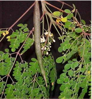
1. MORINGA OLEIFERA:
Or MORINGA PTERYGOSPERMA (Family: Moringaceae)
English: Horse radish tree, Drumstick tree Hindi: Sahijan, Sahnjana, Sanjan, Mungana
It is an unarmed middle sized tree, with grayish brown trunk, and easily breakable branches; leaves usually tripinnate, rachises slender thickened and articulated at the base, leaflets elliptic or obviate, rounded at the apex, nerves obscure; 4-6 pairs and an odd one. Flowers are creamy white or yellow in colour. Capsule is acutely 3-quetrous and slightly constricted between the seeds. Seeds are 3-winged. This plant commonly grows in wasteland and cultivated in gardens and house. Fast growth can be achieved by cuttings (Shetty and Singh, 1991).
and antibacterial activity against both the organism in the from of zone of inhibition in the culture media.
Cacereers et al. (1991) found preliminary screening of antimicrobial activity of Moringa oleifera. Leaves, root and seeds were tested against bacteria, yeast dermatophytes and helminthes by using disc diffusion methods. The result showed that fresh leaf juice and aquous extracts of seed inhibit the growth of Pseudomonas aeruginosa and Staphylococcus aureus
When extraction temperature was above 56 oC, this activity was inhibited. No activity was observed against other pathogenic bacteria and Candida albicans. A method was standardized for studying the effect of aqueous extract on Ascaris-lumbricides eggs, but no activity was exhibited by any part of tree in contrast to Chenopodium ambrosioides leaf extracts. Emeruwa (1991) observed antimicrobial activity of aqueous extracts from seed of Moringa oleifera against fungi including Candida and Penicillum and bacteria including Proteus, Streptococcus and Mycobacterium spp. Remarkable results were obtained against all the fungi and bacteria tested.
Singh et Al. (2003), observed antimicrobial activity of leaves, root, bark and seeds of Moringa oleifera against bacteria, yeast, dermatophytes and helminthes. The fresh leaf juice and water extracts tested against green algae, E. Coli, Pseudomonas aeruginosa, and Staphylococcus auerus, Bacillus sterothermophilus and Herpes simplex virus type I and Polio virus type I. The antibacterial effect of aqueous methonolic extract and water extract showed a fluctuation in its effects. Pseudomonas aeruginosa was more sensitive to all Moringa oleifera extracts; bacillus sterothermophilus was more sensitive than other organism to all extracts.
Dried leaves ground with garlic, salt, black pepper and turmeric are used as a treatment for dog bites or infections. Fresh leaf juice, mixed with honey, is used as an ointment for sore eyes. Decoction of dried leaves is taken orally for abortion and externally for rheumatism and wound healing.
Leaves are taken orally as an aphrodiasic and to treat wounds, the leaves are powered with turmeric and buttermilk and then applied (R. N. Chopra, 1932). As it contains sulphur, it is recommended for rheumatism, ascites and venomous bites; as a poultice for neuralgia of the face. Ethanol extracts (95 %) of dried flowers, dried fruits, dried leaves and dried root, undiluted on agar plate, was active on Escherichia coli and Staphylococcus aureus.
of 100 micro liters on agar plate was active on Pseudomonas aeruginosa and inactive on Escherichia coli, Staphylococcus aureus and Streptococcus pyogenes.
Water and hexane extracts of dried seeds, applied externally to mice at a dose of 10.0 %, were active on Staphylococcus aureus. Powdered dried seeds at a concentration of 100 micro liters, were active on Staphylococcus aureus and inactive on Escherichia coli, Pseudomonas aeruginosa and Streptococcus pyogenes.
Water extracts of dried seeds, at a concentration of 1: 10 on agar plate, was active on Bacillus cereus, Bacillus megaterium, Bacillus subtilis, Sarcina lutea and Staphylococcus aureus. The extract was equivocal on Escherichia coli, Salmonella edinburgi and Serratia marcesens; inactive on Klebsiella aerogenes and produced weak activity on Proteus mirabilis and Streptococcus faecalis.
Powdered dried bark, powdered dried root, powdered dried seeds fresh leaves and powdered dried leaves at a concentration of 1 ml on agar plate, were inactive on Epidermophyton flaccosum, Microsporum canis, Microsporum gypseum, Tricophyton mentagrophytes and Tricophyton rubrum.
Water extracts of dried seeds at a concentration of 1: 10 on agar plate, was active on Botrytis allii, Coniophora cerebella, Penicillum expansum, Phytopthera cactorum, and Polyporus vesicular. The extract was equivocal on Fusarium oxysporum and inactive on Aspergillus oryza (Antifungal activity).
An extract of the entire plant, on agar plate was active on Mycobacterium tuberculosis. Water extract of dried seeds at a concentration of 1: 10 on agar plate, was active on Mycobacterium phlei (Schramm, 1956).
PHARMACOLOGICAL ACTIVITIES: Antibacterial activity.
Anti-inflammatory activity. Antimalarial activity. Antimycobacterial activity.
Antispasmodic activity (unspecified type). Antitumor activity.
Antiyeast activity.
Barbiturate sleeping time decrease. Carcinogenesis inhibition.
CNS depressant activity. Diuretic activity. Embryotoxic effect. Hyperglycemic activity. Hypoglycemic activity. Hypocholesterolemic effect. Hypoproteinemia activity. Hypotensive activity.
Myocardial depressant activity.
Polygalacturonase inhibition. Protopectinase inhibition.
Skeletal muscle relaxant activity. Thyroid hormone effect.
Figure : I MORINGA OLEIFERA
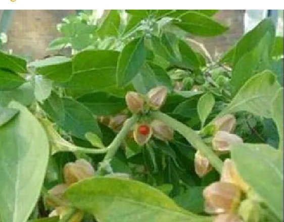
2. WITHANIA SOMNIFERA : DUNAL or
English : Winter cherry.
Hindi : Asgandh; Punir; Ashvagandha
The name Ashvagandha, means ‘‘the thing that has the smell of a horse ‘‘- a reference to the horse's strength and vitality rather than its odour. Ashvagandha is a wonder herb of India; regarded as 1st class adaptogenic tonic. It is a small woody herb belonging to family Solanaceae; an erect branching undershurb reaching about 150 cm in height, usually clothed with minutely stellate tomentum; leaves ovate up to 10 cm long; flowers greenish or lurid yellow in axillary fascicles; fruits globose berries which are orange coloured when mature, enclosed in a persistent calyx. The fleshy roots when dry are cylindrical, gradually tapering down with a brownish white surface and pure white inside when broken.
Constituents:
Withanolide (steroid). Withasomnine.
Sitoindosides (glycowithanolides). Withaferin A.
Sominiferin. Withanine. Anahygrine. Pseudotropine.
Reducing sugar Phytosterol and Ipuranol. Mixture of saturated and unsaturated acids.
Ray and Majumdar (1976) studied the antimicrobial activity of different plant parts of 105 Indian species. Only 30 species showed antibacterial activity (of which 20 also had antifungal activity). These include roots of Withania somnifera.
Jaffer et al. (1988) studied the antimicrobial activity of Withania somnifera extract against different gram positive, gram negative and candida species and no antimicrobial activity against gram negative bacteria was observed. However leaf chloroformic, leaf methanolic and stem chloroformic extract displayed most significant antibacterial activity against gram positive bacteria.
Kazmi et al. (1991) analysed the antimicrobial activity of Withania somnifera. The crude extract of Withania somnifera inhibited the growth of Tricophyton mentagrophyte, Microsporium cannis and Aspergillus boydii at an MIC of 450 - 500 microgram / ml whereas pure compound inhibited the growth at MIC of 300 - 350 microgram / ml species of Corynebacterium, Bacillus, and Streptococcus spp. and Staphylococcus aureus were found to be highly susceptible to both crude and pure compound.
Ramadan et al. (1994) reported studies on alchoholic and aqueous extracts from 20 wild medicinal plants from the Qassim region. The sensitivity of 18 microbes (5 -gram positive and 6 - -gram negative bacteria 5 - fungi 2 - yeasts) to the prepared extracts at concentrations of 10, 25, 50, 100 and 200 mg / ml was investigated. The MIC values for different active extracts were also investigated against the bacteria. Alcoholic and aqueous extracts of the studied parts exhibited strong antibacterial activity against Staphylococcus aureus, Staphylococcus aureus (methicillin resistant), and Streptococcus type B and D, Salmonella type C, Escherichia coli, Haemophilus influenzae, Proteus mirabilis and pseudomonas aeruginosa. The MIC values for Centaurea bruguierana, Rhazya stricta, Peganum harmala, Cynomorium coccineum and Withania somnifera extracts against H. influenzae were 8.36, 8.42, 8.47, 23.77 and 23.87 mg / ml respectively. Fungi and yeast were less sensitive, with the exception of Tricophyton mantagrophytes, which was slightly sensitive to some plant extracts.
Dhuley (1998) studied the therapeutic efficacy of Ashwagandha against exprimental aspergillosis in mice and found to have antifungal and immunomobulatory activites.
The stem, flower and fruits extracts showed highest inhibition of Staphylococcus aureus, Streptococcus mutans and Escherichia coli. The aqueous extracts of stem and flowers showed better inhibition of Staphylococcus aureus, Pseudomonas aeruginosa, while stem alcoholic extracts showed better inhibition of Escherichia coli. Flower and fruits extracts showed higher inhibition of Pseudomonas aeruginosa then other plant parts. Salmonella typhi was not inhibited by any of extracts.
Arora et al. (2004) studied the antibacterial activity of Withania somnifera by agar plate disc diffusion methods against Salmonella - typhinurium and E. coli using methanol and ether extract from both leaves and roots of Withania somnifera. The maximum inhibitory concentration assessed was 0.1 mg / ml for Salmonella typhinurium and E coli. From the extracts tested. Only methanol and hexane extracts of both leaves and roots were found to have potent antibacterial activity.
Mothana and lindequist (2005) selected 25 plants belonging to 19 families from different localities of the island Soqutra and extracted with solvents choloroform, methanol and hot water. The extracts were tested for their antimicrobial activity against one yeast species using agar diffusion method. Antimicrobial activity against several gram positive and several gram negative bacteria and one yeast species using agar diffusion method. Antimicrobial activity was demonstrated especially against gram positive bacteria including multiresistant Staphylococcus strains. The greatest activity was exhibited by the methanolic extracts of Withania adunensis and Withania riebeckii.
Owais et al. (2005) evaluated the antibacterial activity of Aswagandha (root and leaves). Both aqueous and alcoholic extraction of plants (root and leaves) were found to possess strong antibacterial activity against a range of bacteria by invitro agar well -diffusion methods. The methanolic extraction was further subfractionated using various solvents the butanolic sub fraction was found to possess maximum minimum inhibitory concentration against a spectrum of bacteria including Salmonella typhy-nurium. In constrat to synthetic antibiotics (Chloramphenicol), these extracts did not lead to lyses of human RBCs on incubation, advocating their safety to the living cell. Finally efficacy of the extracts isolated from plants (leaves and root) was determined against experimental Salmonella in bulb / c mice as revealed by increased survival rate.
Antianxiety effect. Antiarthritic effect. Antibacterial activity. Anticonvulsant activity.
Antipyretic activity, analgesic, antiinflammatory activities. Antispasmmodic effect.
Antistress agent. Aphrodisiac activity. Antisterility effects. CNS depressant activity.
Haematics and growth promoters in growing children. Hypotensive eefect.
Immunomodulatory activity. Immunosuppressive effect. In the management of amla -pitta.
Antiinflammatory and liver protective activities.
Tumouricidal activity and gastric cytoprotective effects. Sedative and sleeping inducing effects.
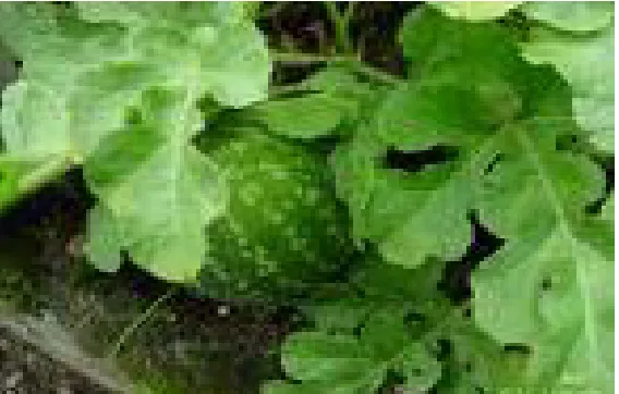
3. CITRULLUS COLOCYNTHIS: (Family: Cucurbitaceae)
English : Colocynth, Bitter apple.
Hindi : Badi Indrayan, Mekkal , Visala, Mahendravaruni, Tumba.
Its Hindi name is tumba and is a perennial, and an extensively annual herbs with bifid tendrils, angular branching stems woolly tender shoots; leaves deeply divided and crisped, lobes narrow, thick, glabrous or some what hairy flowers monoecious, yellow, both male and females solitary, corolla pale yellow, fruit s globosely or oblong fleshy indehiscent berry and variegated with green and white; seed pale brown. It is found in desertic zone and traditionally used as a drastic purgative (Shetty and Singh, 1991).
Constituents:
Colocynthin. A glucoside.
Colocynthein ( Resin). Colocynthitin.
Pectin. Albuminoids.
Adam et al. (2000) studied the effect of oral administration of tumba fruits alone and combined along with Rhazya stricta use in Najdi sheep. The result were indicating that the oral administration of 0.25 g/kg/day of tumba fruit or 0.25 g/kg/day of Rhazya stricta leaves for 42 days did not prove fatal but that mixture of both plants (0.25 g + 0.25 g/kg/day) proved fatal with profuse diarrhea, ataxia prior to death.
Memon et al. (2003) studied the antibacterial properties of Citrullus colocynthis against gram positive and gram negative bacilli using ethanolic extract of fruits, leaves, stem and their roots. Ethanolic extracts of fruits, leaves, stems and roots were found to be against gram positive bacilli, viz. Bacillus pumilus and Staphylococcus aureus while fruits and roots extracts in double strength gave positive results against bacillus subtilis. No activity was found against E coli. and Pseudomonas aeruginosa.
PHARMACOLOGICAL ACTIVITIES: ROOTS :
Treatment of Uteralgia. Treatment of Mammillitis. Treatment of Rheumatalgia. Treatment of Visceromegaly. Treatment of Ophthalmia. Treatment of Ascites. Treatment of Jaundice. Treatment of Uropathy.
FRUITS : Purgative. Antipyretic. Anthelmintic.
Treatment of Bronchitis. Treatment of Urethrorrhea. Treatment of Jaundice. Treatment of Dyspepsia. Treatment of Constipation. Treatment of Elephantiasis.
Treatment of Tubercular glands of the neck. Treatment of Splenomegaly.
Treatment of Migraine.
Figure III : CITRULLUS COLOSYNTHIS
4. SALVADORA OLEOIDS : (Family : Salvadoraceae)
English : Tooth Brush Tree.
Hindi : Pilu, Kankhina, Jhal, Kharkanella, Khara jhal.
somewhat fleshy when mature, linear lanceolate, acute or sub obtuse, often mucronate, glabrous; main nerves indistinct. Flowers are greenish white, sessile, in errect axillary panicled spikes, often clustered. The fruits have a sharp, pungent, acrid and sweet, sour taste with a flavour.
Constituents:
Alkaloids Trimethylamine. Aromatic oil and fixed oil.
Akpata and Akinrimisi (1977) found the antibacterial activity of some extract from some African Chewing stick including Salvadora Species which inhibited the growth of periodontal pathogen Porphyromons gingivalis and Bacteriodes melaninogenus invitro.
Albaghieh et al (1994) observed the antimycotic effect of the aqueous extracts of roots of salvadora oleoids several concentration of aqueous extracts of miswak prepared with Sabourauds medium were inoculated and incubated at 37 oC and turbidity was determined at 600 nm wavelength measured at specific interval over a period of 48 hours. At a concentration of 15 % the extract had fungi static effect for up to 48 hours.
Allafi and Ababneh ( 1995 ) described the effect of extract of miswak (Chewing sticks) used in Jordan and middle east Arabia on oral bacteria. Three methods of determining antibacterial activity were carried out as streaked plate method, disc plate method, tube dilutation method for MIC. It was found that extract of these sticks had a drastic effect on growth of Staphylococcus aureus with MIC of 69 mg/ml/100CC.
Ahmad (2001) studied the antimicrobial effect of Salvadora oleoids from India. He used the disc plate method, to test the antimicrobial and fungicidal activity of different plant extracts. The inhibition zone up to 1.8 mm was found in the extract of Salvadora plant.
Alali et al (2004) determined the antimicrobial activity of volatile oil and aqueous and alcoholic extract of the Salvadora oleoids. Among all test fractions the volatile oil exhibited potent activity against Pseudomonas aeruginosa and Staphylococcus aureus.
Krishanan (1998) studied the antimicrobial activity of Salvadora oleoids. He used above methods along with it buffering capacity and fluoride contents from the aqueous and alcoholic extacts of plant parts.
PHARMACOLOGICAL ACTIVITIES:
Treatment of painful Rheumatic affections (Stimulating effect). Stomachic.
Vesicant.
Purgative. (Given to horses). Aphrodisiac activity.
Treatment of Enlarged Spleen.
Treatment of Rheumatism and low fever. Treatment of Snake bite.
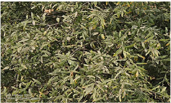
5. SWERTIA CHIRATA (Family - Gnetianaceae)
English : Swerita chirata
Hindi : Chirayata, Mamajaka, Meetha kirayata,
Swerita chirata is an erect annual herb found throughout the greater part of India. The stem, which attains about 6 mm in thickness, is of a yellowish brown colour, glabourous, slightly winged. The lower part of the stem is rounded, and the upper part of the stem produces in the axils of the opposite leaves numerous slender, elongated bearing fruits and occasional flowers. The drug has no marked odor, but all parts have an extremely bitter taste.
Constituents:
Ophelic acid Chiratin
The leaves are fed to cattle to increase appetite. Plant extracts were reported for the biological activities such as antidiabetic, anti-inflammatory, stimulant, astringent and diuretic and anthelmintic propertite.
It also acts as ethno medicine for snake bite. The plant is used to cure leucorrhoea. The root extracts showed antimalerial activity both invitro and in vivo. Methanolic extract showed antidiabtic effect in alloxon induced diabetic rats. It inhibited carrageen-induced edema and its anti-inflammatory activity is comparable to that of hydrocatisone. The plants are extremely bitter due to bitter principle Ophelic acid and amrogentian.
PHARMACOLOGICAL ACTIVITIES: Antispasmodic effect.
Figure V : SWERITA CHIRATA
MATERIALS
AND
MATERIALS AND METHODS:
1. MATERIALS
A). Ingredients of bacteriological media:
I. Agar -agar type I (Hi - media lab Pvt. Ltd.) II. Beef extract (Glaxo - Lab Chemical Division) III. D - Mannitol (Hi - media Laboratories) IV. Dextrose sugar (Hi - media Lab Pvt. Ltd) V. D - lactose (Sarabhai M. Chemicals)
VI. Eosin Water Soluble Yellowish (George T. Gurr Ltd ) VII. Mc - conkey Agar Base (Hi - Media Lab. Pvt. Ltd.
VIII. Methylene Blue M. S. (S. D. Fine Chem. Pvt Ltd.) IX. Peptone- Bacteriological (Glaxo Lab Chemical Divison ) X Sodium Chloride (Glaxo Lab Chemical Divison)
B). Chemicals and Reagents: I. Alpha napthol
II. Barium Chloride Powder
III. Buffer tablets pH 7.0 ( Glaxo Lab and Fine Chemical) IV. Crystal violet (Glaxo Lab and Fine Chemical)
V. Ethyl alcohol.
VI. Heparin sodium injection, 25000 I U in 5 ml (Biological Ltd ). VII. Kovac's Reagentes.
VIII. Methyl Red Indicator. IX. Neutral red.
X. Phenol- red -pH indicator. XI. Potassium hydroxide. XII. Potassium iodide. XIII. Sulphuric acid.
C). Other materials: I. Distilled water
II. Hi - Media antibiotics disc.
III. Mastitis cattle milk samples and calf diarrhea sample. IV. Normal saline solution.
V. Rabbit plasma.
VI. Selected indigenous plant parts. VII. Sheep blood.
2. Preparations of stains:
A large number of coloured compounds (dye) are available for staining microorganisms. These compounds are generally rather complex in terms of molecular structure. Fixed staining preparation are most frequently used for the observation of the morphological characteristics of bacteria. The advantages of this procedure are that;
1). the cells are made more clearly visible after they are coloured.
2). Differences between cells of different species and with in the same species can be determined by use of appropriate staining solutions (differential or selective staining).
I. Stain for Gram's staning:
Gram staining is one of the most important and widely used differential techniques. This technique was introduced by Christian Gram in 1884. In this process the fixed bacterial smear is subject to the following staining reagents in the order listed.
(a). Crystal violet ( Primary stain )
1 % crystal violet aqueous solution.W / V
(b). Neutral red
1 gm of neutral red dissolved in 2ml of 1 % acetic acid solution made to 1000 ml in distilled water.
(c). Gram’s iodine solution
20 gm of potassium iodide and 10 gm of iodine crystals dissolved in 1 L of distilled water.
II. Lacto phenol cotton blue stain:
Phenol crystal 20.0 g, Glycerin 20.0 g, Lactic acid 20.0 g and Water 20.0 ml were mixed with gentle heating. Cotton blue 0.05 g added and dissolved.
3. Preparation of bacteriological media:
I. Nutrient agar
10 g of peptone was mixed with 5 g of beef extracts and 5 g of sodium chloride. These ingredients were mixed to 1000 ml of distilled water and pH was adjusted to 7.2. Agar - agar type I was added at the rate of 2 %. The media was autoclaved at 121 oC and 15 lb pressure, dispersed in Petri dish and stored in refrigerator at 4 oC till use.
II. Blood agar
Nutrient agar basal media was prepared and autoclaved. The temperature of the medium was brought to 50 oC and sheep blood was added at rate of 5 % and then dispersed in Petri dishes.
III. MacConkey agar base media
Readymade MacConkey agar base media (Hi Media) was used. Lactose was sterilized at 10 lb pressure in autoclave and added to basal media.
IV. Mannitol salt agar
10 g of mannitol was sterilized in water bath at 90 oC for 30 minutes and added to basal media.
V. Hugh and Leifson’s medium
2 g of Peptone, 5 g of Sodium chloride, 0.3 g of di - basic Potassium phosphate, and 1 % Bromothymol blue (3ml) were mixed in 1000 ml of distilled water. Agar - agar at rate of 0.5 % was added and then pH was adjusted to 7.1. Glucose was added to the final concentration of 10 %. This medium was autoclaved and distributed in tubes.
VI. Eosin methylene blue agar
Peptone 10 g, lactose 10 g, Di potassium hydrogen phosphate 2 g, Eosin yellow 0.4 g, and Methylene blue 0.065 g were dissolved in 1000 ml of distilled water. Agar - agar was added at rate of 2 % and final pH was adjusted to 6.8. The media was autoclaved at 121 oC for 15 minutes at 15 lb pressure.
VII. Sabouraud's dextrose agar
Dextrose sugar 10 g, Peptone 10 g, and Agar 20 g mixed in 1000 ml distilled water, and then pH was adjusted to 5.0 - 6.0 and the media was autoclaved at 121 oC for 10 minutes at 10 lb pressure.
4. METHODS
The proceeding of the methodology was as follow:
A. Collection of plants
Antimicrobial activity of plants was carried out by testing them against gram positive bacteria and fungi. The following plants were selected for determination of antimicrobial activity.
1. Moringa oleifera
3. Citrullus colocynthis
4. Salvadora oleiodes
5. Swerita chirata
All the above mentioned plants were collected from College of Veterinary and Animal Science campus, Bikaner, Rajasthan Agricultural University campus, Beechwal, Bikaner and herbal medicinal plant library, Dungar college, Bikaner and herbal medicinal garden, Jhalrapatan and identified by dept. of botany, Dungar college, Bikaner. The following parts from the plant were collected for determimnation of antimicrobial activity.
1. Moringa oleifera : Leaves
2. Withania somnifera : Leaves
3. Citrullus colocynthis : Fruits
4. Salvadora oleiodes : Leaves
5. Swerita chirata : Leaves
Collected plant parts were washed and cleaned by muslin cloth and kept for drying for 7 days at 40 oC. Then plant parts were ground in to a powder form.
B). Extraction of plant phytochemical
Extraction may be defined as the process of removal of removal of desirable soluble constituent from a substance, leaving out those which are not wanted, with the aid of solvent and standardized processes.
Extraction is a process in which generally a part is treating with solvent for separating out the active constituents completely or partially.
desired therapeutic effect. In recents years active principles from both plants have been isolated or obtained as purified products of precisely known potency and stability.
The solvent used for extraction is known as “Menstruum” and the undissolved residue left behind after the process is called “Marc”. The process of drug extraction can be summarized in to these steps.
1. Penetration of the solvent in to the drug. 2. Dissolution of the constituent.
3. Outward diffusion of the solutions from the cells. 4. Separation of dissolved portion.
I. Preparation of Aqueous extract
Aqueous extraction was carried out by decoction process. This was carried out by boiling in hot water. In this process I part of dried powder of plant and 5 part of sterilized distilled water were taken in a boiling water flask and boiled for 15 minutes. After boiling the extract was filtered through a What man filter paper no. I, autoclaved at 121 oC for 15 minutes and kept in clean and sterilized test tube and stored at 4 oC till further use.
II. Preparation of alcoholic extract
Alcoholic extract of indigenous plants were prepared according to the methods described by Davis. The Alcoholic extract was prepared by continuous hot percolation process which is known as “Soxhlet Extraction”. In this process the dried powder form of plant material was extracted by using a little volume of a hot menstrum repeatedly. The hot menstrum was ethyl alcohol and it extracted out the active components of plant when it repeatedly passed through a packed column of plant material. The apparatus used for continuous hot percolation process is known as Soxhlet apparatus and process is known as Soxhlation.
side tube and condensed there. The condensed hot Ethyl alcohol fell on the packed column of the plant material and extracted out the ingredients of plants which moved downward through the packed column and collected in an extractor.
As more and more menstrum passed through the packed column of plant material, the level of liquid in extractor as well as in the siphon went on increasing. When the level in the siphon reached at the highest position, it carried down the extract from extractor to flask. On further heating, the vapour of Ethyl alcohol left the flask while the soluble active constituents remained in it. The process of filling and emptying of the extractor was repeated for 14 - 15 times and it required 4 - 6 hours, for the complete extraction of active constituent from the plant material. After completion of the process the concentrated active constituents from plant material were kept in sterilized test tubes stored in refrigerator till further use. The traces of ethanol were removed by keeping the tubes at 50 oC for 1 hour.
5. Isolation and identification of test bacteria
Milk samples from 4 mastitic cattle and one fecal sample from calf diarrhea were screened for the presence of bacteria by cultivation, isolation and identification using standard procedure of Cown and Steel (1975). The mastitis cattle milk sample and calf diarrheal sample were withdrawn with an inoculating loop aseptically and streaked on blood agar, nutrient agar and MacConkey agar culture media plates in primary, secondary and tertiary fashion in order to obtain isolated colonies of bacteria. These Petri plates were inoculated for 24 hours at 37 oC and if colonies did not appear or were found to be small, the plates were incubated for further 24 hours.
Following incubation, the plates were observed for colonies characteristics and haemolytic zone on blood agar plates; the different colonies were selected out and subculture separately for obtaining the pure culture of the bacterial isolates.
MacConkey agar culture media plates were observed for the appearence of pink coloured colonies. Idenificaion of the pathogen was done by carrying out of the following procedure :
Primary the smear were prepared from the bacterial pure colonies, fixed by gentle heating and stained by Gram's methods. The stained smear was examined under oil immersion objective for determining Gram's reaction, morphological characteristic so as to ascertain homogenicity of the organism.
Primary identification up to generic level
I. Morphology
Colonies of bacteria on nutrient agar plate were purified and bacteria were observed for thier size, shape arrangement, sporulation, capsulation and presence of any other distinctive feature.
II. Motility
Motility was studied in hanging drop preparation of broth culture of bacteria.
III. Growth in air
Growth in air was studied to confirm whether the bacterial isolates were able to grow under aerobic or anaerobic condition.
IV. Acid fastness
Acid-fast staining property was determined by Ziehl Neelson method as per the technique of John (1977). Acid - fast reaction of bacterial cells was recorded as ' + ' and non acid fastness was recorded ' - '.
V. Gram's Reaction
Smear of young culture of bacterial isolates were stained by modified Gram’s Method of staining described by Hucker and Cohn (1923). The results were noted as Gram positive (+) for organisms staining blue and Gram negative (-) for those isolates taking pink colour of counter stain.
Spore - formation was observed in smear prepared from colonies.
VII. Catalase Activity
Catalase activity was tested for the confirmation of bacteria producing catalase and the technique of Thomas (1963) was adopted. One ml of three percent solution of hydrogen peroxide was placed on a clean glass slide. Pure culture was picked up from nutrient agar slant with an inoculating straight wire in front of flame and placed on drop of reagent on glass slide. Culture was properly emulsified and cover slip was placed. The production of gas bubble confirmed a positive reaction.
VIII. Oxidase Activity
It confirms the production of cytochrom oxidase by certain bacteria. The culture from nutrient agar slant was picked up with an inoculating loop and rubbed on filter paper. Simultaneously a drop of oxidase reagent (N, N, N, N - p, Phenylene - diamine dihydrochloride) was added. Colonies producing oxidase gave coloured reaction, the colour of filter paper turning to deep blue in a few seconds.
IX. Oxidation and fermentation test
This test was used to differentiate oxidative bacteria from fermenters following the technique of Hugh and Leifsons. Hugh and Leifsons medium was used containing glucose and bromothymol blue as indicator. Semisolid medium was inoculated in pairs by culture of bacteria to be tested. One tube of pairs was kept open, while the others tube was covered with 1 - 2 mm l ayer of sterilized paraffine to provide anaerobic condition.
The tubes were inoculated for 24 hours. Those bacteria that oxidised the sugar showed acid production and yellow discolouration of the medium in open tube. Bacteria that ferment the sugar showed acid production and yellow discoloration in both the paired tubes.
Secondary identification test and use of selective media ( up to species level)
generic identification reactions recommended by Cown and Steel (1975) for gram's positive and gram's negative bacterial genera.
I. Coagulase test
Haemolytic bacterial colonies from blood agar were isolated and preserved on nutrient agar slant. These were tested for the confirmation of Staphylococcus. Coagulase production by Staphylococcus is an important criterion of its pathogenecity which was evaluated by the coagulase test.
(a) Collection of plasma
Rabbit blood was taken aseptically in tube containing 0.5 ml of heparin sodium by intra cardiac puncture. The tube containing blood was centrifuged at 2500 rpm for 15 minutes to separate the plasma. Clear plasma supernatant was taken in to another sterilized test tube.
(b) Tube coagulase test
Plasma was diluted 1: 5 in saline solution and 0.5 ml of plasma was taken in a sugar tube and culture of organism from slant was fished out and inoculated in to plasma and mixed thoroughly. The tubes were inoculated in water bath at 37 oC and were observed at 1, 4, and 8 hours intervals. A partial clotting of plasma was considered positive test.
II. Identification of selective media
(a) Mannitol salt agar
Mannitol salt agar plates were streaked with the test culture from slants and incubated for 24 to 28 hours. The mannitol fermenting pathogenic organism caused yellow discoloration of the media due to production of acid by mannitol fermentation which decolorized phenol red indicator to yellow, whereas the colonies of non pathogenic cocci were small and red.
(b) Eosin Methylene Blue agar (EMB)
6. Biochemical Test
I. Indole Test
Indole test was conducted to access the ability of bacteria to decompose amino acid trytophan to indole. Peptone water was inoculated with isolated test culture and incubated for 48 hours at 37 oC. After incubation, 0.5 ml of Kovac's reagent was added and shaken gentle. Appearance of red colour indicated positive reaction.
II. Methyl Red (MR), Voges Proskaure (VP) Test
MR - VP broth was prepared and inoculated with test culture and incubated for 4 days. To 5 drops of methyl red indicator were added. A positive reaction was shown by red colouration, indication of production of acid bringing pH down to 4.0.
III. Voges Proskaur (VP) Test
VP test was performed to detect the formation of acetyl - methyl - carbinol which is an intermediate product of carbohydrate metabolism. This test was performed after the conduction of methyl red (MR) test. First, 5 ml of 10 % KOH solution was added to neutralize the acidity, produced in MR test. Later 1 ml of 5 % L -naphthol reagent was added to broth culture. The tubes were shaken well and kept undisturbed for 5 - 10 minutes.
IV. Citrate Utilization test
This test was carried out to test the ability of an organism to utilize citrate at the sole source of carbon. Simmon's citrate agar was used. The pure colony cultures from nutrient agar slant were fished out and streaked over citrate agar plates and incubated for 24 hours.
Appearance of blue colour was considered positive while original green colour of media showed that citrate was not utilized.
V. Nitrate Reduction
It is a test to determine the presence of the enzymes nitrate reductase which cause reduction of nitrate and tested by appropriate colorimetric reagent. Nitrate broth was inoculated with the test culture and incubated for 2 - 3 days. To 5 ml of broth culture was added 0.1 ml of test reagent. A red colour developing within a few seconds indicated the presence of nitrate and hence the ability of organism to reduce nitrate.
VI. Antibiogram determination
The following antibiotics discs (Hi-Media) were used for determination of the isolates.
Ampicillin (A) 10 mcg Amoxycillin (Am) 10 mcg Chloramphenicol (C) 30 mcg Doxycycline (D) 30 mcg Kanamycin (K) 30 mcg Bacitracin (B) 10 unit Gentamycin (G) 10 mcg Penicillin (P) 10 unit Vancomycin (Va) 30 mcg Sulfadiazine (Sz) 300 mcg Neomycin (N) 30 mcg Ciprofloxacin (Cf) 5 mcg
from slants. After 6 - 7 hours, when the bacteria were in exponential phase of growth, the broth culture was swabbed on the Muller - Hinton agar plates by sterile cotton swab. When broth culture was dried, eight antibiotics discs were placed with the aid of automatic disc dispensor in front of flame.
The petriplates were incubated for 15 - 20 hours and observed for the zone of inhibition. The diameter of zone of inhibition was determined with the help of measuring scale and compared with the standard scale of inhibition for each antibiotics disc as per the instruction provided by manufacture (Hi-Media).
7. Determination of concentration of test organisms
The concentration (total count) of test bacteria (S.aureus and E.coli.) Was determined by nephelometry using McFarland scale (McFarland, 1977). The standard tubes were prepared by mixing varying amount of 1 % barium chloride and 1 % sulphuric acid in last stopper tubes as follows.
Table: McFarland scale
Scale 1% Bacl2 (ml) 1% H2SO4 (ml) No. of bacteria
value listed x 106
approximately
1. 0.1 9.9 300
2. 0.2 9.8 600
3. 0.3 9.7 900
4. 0.4 9.6 1200
5. 0.5 9.5 1500
6. 0.6 9.4 1800
7. 0.7 9.3 2100
8. 0.8 9.2 2400
9. 0.9 9.1 2700
10. 1.0 9.0 3000
The turbidity of over night broth culture of test organism was compared with that of McFarland scale tubes against white back ground and concentration was approximated according to the table.
8. Preparation of dilution of plant extracts
Two fold serial dilution of aqueous and alcoholic plant extracts were prepared in sterilized test tubes with sterile normal saline solution beginning from 1:1 undiluted and 1:2, 1:4, 1:8, 1:16, 1:32, 1:64, 1:128, 1:256, 1:512. The total amount of each dilution was kept 2 ml.
I. Inoculation of test organisms
Each dilution of plant extracts was added with equal volume of double strength (2x) of nutrient broth so as to make normal concentration of nutrients after the addition of the medium. Along with the desired plant extracts two set of control tubes were simultaneously taken one of which lacked plant extracts the other one was kept for un-inoculated control.
All the dilutions of plants extracts and control tubes (except UN - inoculated control tubes) were inoculated with 0.1 ml of broth culture of test organism having turbidity comparative to McFarland tube no. 1 with approximated number of organism as 300 x 106 per ml. Following inoculation all the tubes along with the control tubes were incubated at 37 oC. Turbidity of all the tubes was measured after 2, 4, 6, 8 hours. The highest dilution of plant extracts that showed inhibited growth of test organism as compared with the control was considered as MIC and determined using the methods adopted by Tsuchiya et al (1996).
II. Determination of MLC
extracts that was found to kill the test organism was considered as per the methods of Sato et al (1997).
9. Isolation and identification of test fungi
Sheep nasal swab was taken and cultured on Sabouraud's dextrose agar and the plates were incubated at room temperature for 2 days. Grayish brown mycelia were seen which turned later to black colour. Smear was prepared and stained with lacto phenol cotton blue stain and observed under high power microscope.
I. Preparation of spores
10 ml of sterile normal saline solution was added to the Petri plates containing fungal growth i.e. Showing fungal mycelium and was shaken gently. The fluid containing fungal spores was collected. With the help of sterile pipette, and centrifuged at 1500 rpm for 10 minutes. The supernatant was discarded and the sediment was washed twice with normal saline solution. Finally the spore sediment was suspended in normal saline at a concentration of about 100 - 200 spores per high power field.
II. Germination of spores
To a glass cavity slide (normally used for hanging drop preparation), 30 ml of Sabouraud's dextrose agar (0.5 % agar) was placed in a cavity portion while in molten (at temperature 50 oC ) state. It was then added with 10 ml of test fungi spore preparation under possibly aseptically conditions. The cavity portion of the slide was covered with a sterile and clean cover slip and its margins were sealed with sterile paraffin wax. It was then incubated for two days in moist chamber at room temperature and observed under low power microscope for germination of spores.
III. Inhibition of spore germination activity
and 40 micrometer of each dilution were added along with inoculation of spores as described above in spore germination techniques. The micro culture slides were incubated at room temperature in moist condition and observed for inhibition of spore germination at 24 hours and 48 hours of inoculation following the method described by Rana et al, (1997).
RESULTS
The present investigation involves aqueous and alcoholic extraction of some of the plant commonly found in this area. The aqueous and alcoholic extracts were used to determine their antibacterial and antifungal potential using pathogenic Staphylococcus aureus and Escherichia coli as test bacterial and Aspergillus spp. as test fungi. The results obtained are presented as follows:
I. Isolation and identification of test bacteria
(A). Isolation and identification of Staphylococcus aureus
Staphylococcus aureus could be isolated and identified from mastitic milk sample of cattle. The biochemical and metabolic property of Staphylococcus aureus isolated are presented in Table.
Table 1: Biochemical and metabolic reaction of Staphylococcus aureus
identification
1. Gram reaction + Growth in MSA + 2. Morphology Cocci Coagulase + 3. Motility - Growth in EMB -
4. Spore - Indole test -
5. Growth on McConkey agar
- MR test +
6. Catalase + VP test +
7. Oxidase - Citrate utilization - 8. O-F test F Nitrate reduction test +
(B). Isolation and identification of Escherchia coli
[image:50.612.77.497.65.272.2]Escherichia coli could be isolated from faecal sample of calf diarrhoea. The biochemical and metabolic property of Escherichia coli isolate are presented in Table
Table 2: Biochemical and metabolic reaction of Escherichia coli
S.No. Primary Identification Secondary identification
1. Gram reaction + Growth in MSA + 2. Morphology Rods Coagulase + 3. Motility - Growth in EMB Metalli
c sheen
4. Spore - Indole test -
5. Growth on McConkey agar
- MR test +
7. Oxidase - Citrate utilization - 8. O-F test F Nitrate reduction test +
II. Isolation and identification of test Fungi
The test fungi i.e Aspergillus fumigatus could be isolated from nasal swab of sheep. The identification of Aspergillus was based on its cultural and morphological characteristics. Their appearance were white puffy colony when it first appeared, rapidly become velvety, granular, green blackish in colour and hyphae showed parallel wall and dichotomous branching often showing ballooning.
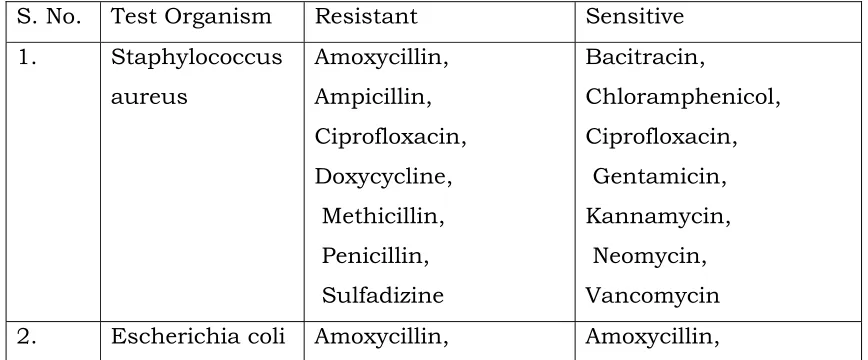
III. Antibiotic sensitivity pattern of test bacteria
[image:51.612.77.511.546.726.2]The test organism that is Staphylococcus aureus and Escherichia coli isolates were subjected to their sensitivity and resistance pattern to commonly used antibiotics. The results of antibiotic sensitivity pattern of test bacterial isolates are presented in Table.
Table 3 Antibiotic sensitivity and resistance of different test organism in sensidisc diffusion test
S. No. Test Organism Resistant Sensitive 1. Staphylococcus
aureus
Amoxycillin, Ampicillin, Ciprofloxacin, Doxycycline, Methicillin, Penicillin, Sulfadizine
Bacitracin,
Bacitratin, Ciprofloxacin, Penicillin, Sulfadizine, Vanocomycin, Ciprofloxacin
Chloramphenicol, Ciprofloxacin, Gentamicin, Kannamycin, Methicillin, Neomycin
IV. Extraction of plant phytochemicals
Extract could be prepared from five selected plant using hot water and Ethanol as a solvent. From 15 g of each plant part 4 - 5 ml aqueous extract could be prepared. In alcoholic extracts after removal of traces of ethanol 2-3 ml extract was left from each plant which was dark coloured and pasty in consistency and thus it was found difficult to measure the turbidity of dilutions of the extract before and after inoculation.
V. Antibacterial and antifungal activity of plant extracts
The alcoholic and aqueous extracts of selected plant were tested for antibacterial and antifungal activity. The results are presented in table as follows:
1. Moringa oleifera : Table No. IV
2. Withania somnifera : Table No. V
3. Citrullus colocynthis : Table No. VI
4. Salvadora oleoides : Table No. VII
The antibacterial activity of these plant extracts has been summarized in T