DISSERTATION ON
STUDY OF BACTERIOLOGIC PROFILE IN CRITICAL
CARE SETTINGS AND EFFECTS OF PREVENTIVE
MEASURES
Submitted to
THE TAMIL NADU DR. M.G.R. MEDICAL UNIVERSITY
In partial fulfilment of the regulations for the award of the degree of
M.D. BRANCH - I
GENERAL MEDICINE
MADRAS
MADRAS MEDICAL COLLEGE AND GOVERNMENT GENERAL HOSPITAL, CHENNAI – 3
THE TAMIL NADU DR. M.G.R. MEDICAL UNIVERSITY CHENNAI, INDIA
2
DECLARATION
I solemnly declare that this dissertation entitled “STUDY OF BACTERIOLOGIC PROFILE IN CRITICAL CARE SETTINGS AND
EFFECTS OF PREVENTIVE MEASURES” was done by me at Madras
Medical College and Rajiv Gandhi Government General Hospital during 2009-2012 under the guidance and direct supervision of Prof. C.RAJENDIRAN, M.D., Director and Professor of Medicine, Institute of Internal Medicine, Madras Medical College and Rajiv Gandhi Government General Hospital, Chennai-3. This dissertation is submitted to the Tamil Nadu Dr. M.G.R. Medical University towards the partial fulfillment of requirements for the award of M.D. Degree in General Medicine (Branch-I).
Place: Chennai (DR. S. ANNE PRINCY)
Date:
3
ACKNOWLEDGEMENT
At the outset, I thank Prof. V.KANAGASABAI M.D., Dean, Madras Medical College and Rajiv Gandhi Government General Hospital, Chennai-3 for having permitted me to use the hospital data for the study.
I am very much thankful to Prof. V.PALANI M.S., Medical Superintendent, Rajiv Gandhi Government General Hospital, Chennai-3 for permitting me to carry out my study.
I am indebted to Prof. C.RAJENDIRAN M.D., Director and Professor of Medicine, Institute of Internal Medicine, Madras Medical College and Rajiv Gandhi Government General Hospital, Chennai-3 for his support and his painstaking efforts and guidance in scrutinizing the study.
I thank Prof. S.RAGUNANTHANAN M.D., Professor of Medicine, Institute of Internal Medicine, Madras Medical College and Rajiv Gandhi Government General Hospital, Chennai-3 for his guidance throughout the study.
I thank Dr. S.BASKER M.D., Dr. M.ANUSUYA M.D., Dr. SUNDAR
M.D., Dr. V.RAJENDRAN M.D., Dr. D.RAMESH M.D., Dr.
D.THANGAM M.D., Assistant Professors of Medicine, Institute of Internal Medicine, Madras Medical College and Rajiv Gandhi Government General Hospital, Chennai-3 for their contributions to the study.
4
Sciences (IBMS), Taramani and their Post Graduate students who rendered active support and guidance in testing the samples of this study.
I thank all my professional colleagues for their support and valuable contributions and criticisms.
5
CERTIFICATE
This is to certify that the dissertation entitled “STUDY OF BACTERIOLOGIC PROFILE IN CRITICAL CARE SETTINGS AND
EFFECTS OF PREVENTIVE MEASURES” is a bonafide work done by
Dr. S.ANNE PRINCY, post graduate student, Institute of Internal Medicine, Madras Medical College, Chennai-3 in partial fulfillment of the University Rules and Regulations for the award of MD Branch – I General Medicine, under my guidance and supervision, during the Academic period from April 2009 to April 2012.
Prof. C. RAJENDIRAN M.D.,
Director, Professor & Unit Chief Guide & Supervisor
Institute of Internal Medicine Madras Medical College &
Rajiv Gandhi Govt. General Hospital Chennai – 3.
Prof. V.KANAGASABAI M.D.,
The Dean
Madras Medical College & Rajiv Gandhi Govt. General Hospital,
6
CONTENTS
SL.NO TITLE PAGE NO
1. INTRODUCTION 7
2. AIMS & OBJECTIVES 9
3. REVIEW OF LITERATURE 10
4. MATERIALS AND METHODS 52
5. OBSERVATIONS AND RESULTS 57
6. DISCUSSION 84
7. CONCLUSION 88
8. LIMITATION 90
ANNEXURES
ABBREVIATIONS
PROFORMA
MASTER CHARTS
BIBLIOGRAPHY
PHOTOGRAPHS
PATIENT CONSENT FORM
INSTITUTIONALETHICALCOMMITTEE
APPROVAL ORDER
91 93
99
116
131
142
7
INTRODUCTION
Health Care Associated Infection (HCAI), also referred to as “nosocomial” or “hospital” infection, is defined as:
“An infection occurring in a patient during the process of care, in a health care facility, which was not present or incubating at the time of admission. An infection manifested >48 hours after admission is defined as hospital acquired. This includes infections acquired in the hospital but appearing after discharge and also occupational infections among the staffs.”
HCAI is acknowledged as the most frequent adverse event in health care, but the global burden remains unknown because of the difficulty of gathering reliable data. This is mainly due to the complexity and lack of uniformity of diagnostic criteria and to the fact that surveillance systems for HCAI are virtually non-existent in most countries.
8
Health Care Associated Infections (HCAI) are preventable errors. The improvement of the quality of the health care is a major concern for intensive care professionals because, the patients of the ICU are thought to be particularly at risk of errors due to complexity of the patients, interdependence of the practitioners, and dependence on team functioning, ensuring patients’ safety during their hospital stay which requires mechanisms to determine the incidence of adverse events. In the ICU, the accumulation of a number of immuno-compromised patients and their nursing and invasive procedures provide a favorable environment to the growth and transmission of nosocomial infections. The use of a ventilator or a central venous catheter, and ICU acquired drug-resistant infections were associated with a high risk of hospital mortality in ICU patients.
9
AIMS & OBJECTIVES
To identify the prevalence and pattern of infections in critical care area.
To identify the predominant infecting organisms.
To determine the bacteriologic profile.
10
REVIEW OF LITERATURE
The word NOSOCOMIAL infection is derived from Latin word
nosocomium 1 hospital, Greek meaning nosokomeion, nosokomos one who tends the sick, from nosos disease + -komos2 ; akin to Greek kamnein to suffer, toil, Sanskrit śāmyati he tires3.
HISTORICAL MILESTONES
One of the earliest records of hospital infections are perhaps those found in an Egyptian papyrus4 written around 3000 B.C. Needless to say, mere absence of documentation of bacterial infection does not exclude its prevalence prior to this time.
Nearer home, in the Indian context, a similar account of hospital infection is available in the ancient Ayurvedic literature (Ca. 600 B.C.). Again the famous Hindu physician Charaka and surgeon Sushuruta5 (Ca. 400 B.C.) have also emphasized the need for prevention of infection in clinical practice. Elsewhere in the world too, there is ample evidence that hospital infections were prevalent and documented in ancient times viz: the records of Herodatus6 on the conditions that prevailed in Greek and Roman hospitals in the period 1000 to 600 B.C., and the Hippocrates treatise (Ca 400 BC) testifying the existence of infections7.
11
other types of air currents. It soon became recognized that certain medicaments were capable of either preventing or checking the progress of infection. Place in 1721 used the term Antiseptics9 to describe these substances and, nearly 30 years later, Pringle in 175010 conducted extensive trials with antiseptics while working with the British army in Flanders.
In 1856 Louis Pasteur conclusively demonstrated that bacteria were responsible for fermentation of wine, which could be prevented by gentle heating whereby the micro-organisms were destroyed11. The existence of such micro-organisms in the atmosphere was proved by him in 1864. In his celebrated lecture to Acadimie de Medicine on April 30th, 187312, Louis Pasteur is quoted13 as having said:
“If I had the honour of being a surgeon, not only would I use
absolutely clean instruments, but after cleaning my hands with
the greatest care would only use sponges previously raised to a
heat of 1300-1500 Fahrenheit. I would still have to fear germs
suspended in the air, and surrounding the bed of the patient”.
12
the disease. A drastic reduction in infection rates was achieved by the introduction of hand washing practices with chlorinated lime16.
In 1969, Lister introduced his antiseptic theory17, following the extensive use of carbolic acid18 to pack wounds, especially of compound fractures, sterilize instruments and sutures, and to decontaminate his hands. He observed that these practices could greatly reduce the incidence of suppuration and gangrene, which quite commonly occurred otherwise.
In 1883, Gustao Neubar introduced the use of masks and gowns in surgery19, and Halsted in 1890, introduced the use of rubber gloves20 in surgery. Steam sterilization21 was discovered by Von Bergman in 1896 and all these measures further increased the safety of surgery and contributed greatly in bringing down rates of infection by use of aseptic and antiseptic techniques. During the period, when many fundamental discoveries in bacteriology were being made, other principles of hospital infection control were also simultaneously established.
BACKGROUND
13
Patients in Intensive Care Units (ICUs) have a higher risk of acquiring hospital associated infections than those in non-critical care areas. ICUs are sites of considerable broad spectrum antibiotic use, and antibiotic resistant pathogens are frequent. Bloodstream infections (BSIs), pneumonias, and Urinary Tract Infections (UTIs) are the most common hospital acquired infections and are most often associated with the use of invasive devices.24
FREQUENCY OF INFECTION
Every year, thousands of patients die of hospital acquired infections (HAI) in India. Death due to HAI is responsible for more mortality than any other forms of accidental death in the country. The irony is, about one–third of all such cases are preventable.
14
mortality for pneumonia occurring in the ICU population alone is between 5 – 14%.
Several studies 26,27,28,29 have shown that the utilisation of invasive devices such as venous and urinary catheter, ETT, intracranial pressure monitoring devices is a major risk factor for the development of nosocomial infections in ICU. Thus the incidences of such infections are expressed as number of infection/1000 device utilisation days. Early removal of such invasive devices will eliminate the risk of such device associated infections. However critical conditions of many ICU patients often require continued use of these catheters, tubes, and drains.
Similarly, contamination during care of the devices also causes infection. Most common HAI is ventilator associated pneumonia (VAP). The incidence of VAP is 11 per 1,000 device days followed by catheter associated blood stream infection (BSI) which is 8 per 1000 device days and then by urinary tract infections.30
Data reveals that HAI increases the length of stay from 2 to 5 days and thereby increasing cost to patients. As per an estimate in Argentina, the increase in cost due to HAI is around $5000 and in India, it could be about Rs. 25,000 to 100,000 depending on severity and hospital31.
FACTORS INFLUENCING THE DEVELOPMENT OF NOSOCOMIAL
15
THE MICROBIAL AGENT33
16
infection). Most infections acquired in hospital today are caused by micro-organisms which are common in the general population, in whom they cause no or milder disease than among hospital patients (Staphylococcus aureus,
coagulase-negative staphylococci, Enterococci, Enterobacteriaceae).
PATIENT SUSCEPTIBILITY34
Important patient factors influencing acquisition of infection include age, immune status, underlying disease, and diagnostic and therapeutic interventions. The extremes of life — infancy and old age — are associated with a decreased resistance to infection. Patients with chronic diseases such as malignant tumors, leukemia, diabetes mellitus, renal failure or the acquired immunodeficiency syndrome (AIDS) have an increased susceptibility to infections with opportunistic pathogens. Immunosuppressive drugs or irradiation may lower resistance to infection. Injuries to skin or mucous membranes bypass natural defense mechanisms. Malnutrition is also a risk. Many modern diagnostic and therapeutic procedures, such as biopsies, endoscopic examinations, catheterization, intubation/ventilation and suction and surgical procedures increase the risk of infection.
ENVIRONMENTAL FACTORS35
17
hospital are a further source of infection. Crowded conditions within the hospital, frequent transfers of patients from one unit to another, and concentration of patients highly susceptible to infection in one area (e.g. newborn infants, burn patients, and intensive care) all contribute to the development of nosocomial infections. Microbial flora may contaminate objects, devices, and materials which subsequently contact susceptible body sites of patients
NOSOCOMIAL INFECTION SITES
URINARY INFECTIONS
18
The bacteria responsible arise from the gut flora, either normal (Escherichia coli) or acquired in hospital (multi-resistant Klebsiella).
NOSOCOMIAL PNEUMONIA
Nosocomial pneumonia39 occurs in several different patient groups. The most important are patients on ventilators40 in intensive care units, where the rate of pneumonia is 3% per day. There is a high case fatality rate41 associated with ventilator associated pneumonia, although the attributable risk is difficult to determine because patient’s co morbidity is so high.
The definition of pneumonia may be based on clinical and radiological criteria which are readily available but non-specific: recent and progressive radiological opacities of the pulmonary parenchyma, purulent sputum, and recent onset of fever. Diagnosis is more specific when quantitative microbiological samples are obtained using specialized protected bronchoscopy methods. Known risk factors42 for infection include the type and duration of ventilation, the quality of respiratory care, severity of the patient’s condition (organ failure), and previous use of antibiotics.
Apart from ventilator associated pneumonia, patients with seizures or decreased level of consciousness are at risk for nosocomial infection, even if not intubated. Viral bronchiolitis (respiratory syncytial virus, RSV) is common in children’s units, and influenza and secondary bacterial pneumonia may occur in institutions for the elderly. With highly immune-compromised patients,
19
NOSOCOMIAL BACTERAEMIA
These infections represent a small proportion of nosocomial infections (approximately 5%) but case fatality rates43 are high — more than 50% for some micro-organisms. The incidence is increasing; particularly for certain organisms such as multi-resistant coagulase negative Staphylococcus and
Candida spp. Infection may occur at the skin entry site of the intravascular device44, or in the subcutaneous path of the catheter (tunnel infection). Organisms colonizing the catheter within the vessel may produce bacteraemia without visible external infection. The resident or transient cutaneous flora is the source of infection. The main risk factors are the length of catheterization, level of asepsis at insertion, and continuing catheter care.
OTHER NOSOCOMIAL INFECTIONS
20
Skin and soft tissue infections: open sores (ulcers, burns and bedsores) encourage bacterial colonization and may lead to systemic infection.
Gastroenteritis is the most common nosocomial infection in children, where rotavirus is a chief pathogen: Clostridium difficile is the major cause of nosocomial gastroenteritis in adults in developed countries.
Sinusitis and infections of the eye and conjunctiva.
Endometritis and other infections of the reproductive organs following childbirth.
MICRO-ORGANISMS
Many different pathogens may cause nosocomial infections. The infecting organisms vary among different patient populations, different health care settings, different facilities, and different countries.
BACTERIA
These are the most common nosocomial pathogens. A distinction may be made between:
Commensal bacteria45 found in normal flora of healthy humans. These have a significant protective role by preventing colonization by pathogenic micro-organisms. Some commensal bacteria may cause infection if the natural host is compromised.
Pathogenic bacteria46 have greater virulence, and cause infections (sporadic or epidemic) regardless of host status. For example:
21
Gram-positive cocci - Staphylococcus aureus (cutaneous bacteria that colonize the skin and nose of both hospital staff and patients) cause a wide variety of lung, bone, heart and bloodstream infections and are frequently resistant to antibiotics; beta hemolytic streptococci are also important.
Gram-negative bacteria- Enterobacteriacae (e.g. Escherichia coli,
Proteus, Klebsiella, Enterobacter, Serratia marcescens), may colonize sites when the host defenses are compromised (catheter insertion, bladder catheter, cannula insertion) and cause serious infections (surgical site, lung, bacteraemia, peritoneum infection). They may also be highly resistant. Gram-negative organisms such as Pseudomonas spp. are often isolated in water and damp areas. They may colonize the digestive tract ofhospitalized patients.
Selected other bacteria are a unique risk in hospitals. For instance,
Legionella species may cause pneumonia (sporadic or endemic) through inhalation of aerosols containing contaminated water (air conditioning, showers, and therapeutic aerosols).
VIRUSES
22
viruses such as cytomegalovirus, HIV, Ebola, influenza viruses, herpes simplex virus, and varicella-zoster virus, may also be transmitted.
PARASITES AND FUNGI
Some parasites (e.g. Giardia lamblia) are transmitted easily among adults or children. Many fungi and other parasites are opportunistic organisms and cause infections during extended antibiotic treatment and severe immune-suppression (Candida albicans, Aspergillus spp., Cryptococcus neoformans,
Cryptosporidium). These are a major cause of systemic infections among immune-compromised patients. Environmental contamination by airborne organisms such as Aspergillus spp. which originate in dust and soil is also a concern, especially during hospital construction. Sarcoptes scabies (scabies) is an ectoparasite which has repeatedly caused outbreaks in health care facilities
RESERVOIRS AND TRANSMISSION
Bacteria that cause nosocomial infections can be acquired in several ways:
23
2. Flora from another patient or member of staff (exogenous cross-infection48)
Bacteria are transmitted between patients:
a) Through direct contact between patients (hands, saliva droplets or other body fluids)
b) In the air (droplets or dust contaminated by a patient’s bacteria) c) Via staff contaminated through patient care (hands, clothes, nose
and throat) who become transient or permanent carriers, subsequently transmitting bacteria to other patients by direct contact during care
d) Via objects contaminated by the patient (including equipment), the staff’s hands, visitors or other environmental sources (e.g. water, other fluids, food)
3. Flora from the health care environment49(endemic or epidemic exogenous environmental infections)
Several types of micro-organisms survive well in the hospital environment: • In water, damp areas, and occasionally in sterile products or
disinfectants (Pseudomonas, Acinetobacter, Mycobacterium)
24
• As most micro-organisms require humid or hot conditions and nutrients to survive
• In food
• In fine dust and droplet nuclei generated by coughing or speaking (bacteria smaller than 10 µm in diameter remain in the air for several hours and can be inhaled in the same way as fine dust)
SOURCES OF CROSS INFECTION (50, 51, 52) IN THE ICU
o Hands of staff and attendants (via two bowl hand washing and
communal towels or no hand washing)
o Assisted ventilation equipment o Suction and drainage bottles o I.V. lines – central and peripheral o Urinary catheters
o Wounds and wound dressings o Disinfectant containers
o Dressing trolleys (on which disinfectants jars/bottles are stored) SURVEILLANCE
25
growing hospital and public health concern. Both the prevalence of antibiotic resistant organisms and of a vulnerable, immuno-compromised population is increasing in hospitals and long term care homes. There is conclusive evidence to show that the establishment of a surveillance system for HAIs is associated with reductions in infection rates. Surveillance is also useful in monitoring the effectiveness of preventive and infection control programs.
There are several established components to an active & effective surveillance system:
1. PLANNING
Because it is not feasible to monitor all types of infections at all times, choosing which infections will be surveyed is based upon an initial assessment that will establish the priorities for the surveillance system54. An initial assessment will include:
• The types of patients/residents that are served by the health care setting • The key medical interventions and procedures that are provided in the
health care setting
• The frequency of particular types of infections within a particular health care setting
• The impact of the infection (including per cent case fatality and excess costs associated with the infection)
26
Surveillance for some types of infections and syndromes, such as Febrile Respiratory Illness (FRI) and Gastrointestinal Illness (GI) are currently part of routine practice in all health care settings.
2. DATA COLLECTION
Collection of infection data for surveillance purposes55 must be done using validated, published definitions for HAIs. In order to generate valid HAI rates, information must be collected on those who develop a HAI and those who do not develop infection. Electronic screening56 of patient records is an emerging tool for identification of potential HAIs. These computerized systems of case finding will reduce the time spent by infection control professionals in case finding.
3. DATA ANALYSIS
It is recommended that incidence density rates be calculated57 i.e., the measurement of new cases of infection (incidence) based on the time at risk in the patient/resident population, e.g., length of stay in a hospital. It may be useful in hospitals to stratify rates of surgical site infections by standardized risk scores58 in order to compare the rates to other hospitals. An electronic spreadsheet/database and/or statistical analysis program should be used in hospitals and long-term care homes to store data and calculate HAI rates, to maximize infection prevention and control resources and reduce the potential for errors associated with manual calculations.
27
Surveillance data requires interpretation to identify areas where improvements to infection prevention and control practices can be implemented to lower the risk of HAI. This investigation is particularly essential where major deviations from the baseline HAI rate may indicate the presence of an outbreak59. Analysis and interpretation of infection data may be done with the facility’s Infection Prevention and Control Committee or other advisory body to the Infection Control Team. HAI rates may be compared to both the facility’s own previous HAI rates and benchmarks, or to external standards or benchmarks set by other health care settings60.
5. COMMUNICATION OF RESULTS
Communication of surveillance data should take place on an ongoing, systematic basis and be targeted61 to those with the ability to change infection prevention and control practice. Communication may be targeted to:
• A health care setting’s Infection Prevention and Control Committee, which provides an aggregate picture of all infections of interest in the hospital
• A particular patient/resident care area or specialty care area, focused on the risk of specific types of infections that are of importance to these groups
28
6. EVALUATION
29
DESIRED CHARACTERISTICS OF A NOSOCOMIAL INFECTION
SURVEILLANCE SYSTEM64, 65, 66
*
1. CHARACTERISTICS OF THE SYSTEM
• Timeliness, simplicity and flexibility • Acceptability and reasonable cost • Representativeness (or exhaustiveness)
2. QUALITY OF THE DATA PROVIDED
• Sensitivity and specificity
• Predictive value (positive and negative)
• Usefulness, in relation to the goals of the surveillance (quality indicators) * Adapted from Thacker SB, 1988 (4).
KEY POINTS IN THE PROCESS OF SURVEILLANCE FOR
NOSOCOMIAL INFECTION RATES
• Active surveillance (prevalence and incidence studies) • Targeted surveillance (site, unit, priority-oriented) • Appropriately trained investigators
• Standardized methodology
• Risk adjusted rates for comparisons
30
collaboration with patient care units, to improve practice, and to define and monitor new prevention policies. The final aim of surveillance is to decrease nosocomial infections and reduce costs.
Surveillance is a continuous process which needs to evaluate the impact of interventions to validate the prevention strategy, and determine if initial objectives are attained.
PREVENTIVE MEASURES
ENVIRONMENTAL MANAGEMENT PRACTICES
A clean environment plays an important role in the prevention of hospital associated infections (HAI) 68. Many factors, including the design of patient care areas, operating rooms, air quality, water supply and the laundry can significantly influence the transmission of HAI.
CLEANING OF THE HOSPITAL ENVIRONMENT
31
Any areas visibly contaminated with blood or body fluids should be cleaned immediately with detergent and water. Isolation rooms and other areas that have patients with known transmissible infectious diseases should be cleaned with a detergent disinfectant solution at least daily. All horizontal surfaces and all toilet areas should be cleaned daily.
ENVIRONMENTAL CLEANING71
DAILY
• Cleaning must be done daily with the hospital approved cleaner. All surfaces must be wiped with a damp cloth to remove dust and dirt
• Cleaner/disinfectants should be identified by the Intensive care team and used as indicated. High level disinfectants (HLD) are not used for environmental cleaning.
• Cleaner/disinfectants should be kept closed when not in use.
TERMINAL
• When patients are discharged from the unit, a thorough cleaning of the bed and bedside equipment must be completed before admitting new patients.
SCHEDULED
• A total cleaning of all areas, including the store clean and soiled storage areas should be done at least every 1-2 weeks.
32
• Cleaning equipment should be wiped & properly stored when not in use.
UNIT DESIGN72
Unit design should consider the following to enhance infection control strategies.
SPACE
BEDS
The beds should be 2.5 - 3 meters (7-9 feet) apart, to allow free movement of staff and equipment, reducing risk of cross contamination. Ideally, a sharps container should be within easy access of each bed.
PARTITIONS
Privacy partitions should be of material that is easily cleaned and should be cleaned weekly and any time that it becomes soiled or contaminated. If curtains are used, they should be changed weekly and between patients.
MEDICATION PREPARATION
Medication preparation areas should be separate from patient care areas and should be maintained as a clean area.
CLEAN STORAGE
An area should be identified and maintained for clean storage and should be separate from care and waste disposal areas.
SOILED AND WASTE STORAGE
33
Ideally, this area should have a clinical sink for the disposal of blood and body fluid waste. The area should include storage of filled sharps containers until these containers can be removed.
TOILETS
May be located outside the ICU.
SINKS AND WATERLESS HAND RUB DISPENSERS
Sinks should be placed near the ICU entrance and at key points, within the unit in order to provide ease of access to the care givers. If this is not feasible, waterless hand rub dispensers73 should be available at the ICU entrance and at each bedside.
VENTILATION
TYPE
The source of clean air should be determined including central or through the wall air conditioning units. System should be evaluated for proper functioning and preventive maintenance.
34
WINDOWS
Windows should remain closed in order to control all airborne risks.
VISITORS
Design of the unit should permit staff to assess visitors for communicable disease (e.g. rash, respiratory infection) before permitted to enter unit. They should be instructed in washing their hands if assisting the patient.
WATER
Drinking water should be safe75 for oral ingestion. National norms and international recommendations define appropriate criteria for clean drinking water. Even water that conforms to accepted criteria may carry potentially pathogenic micro-organisms. Organisms present in tap water have frequently been implicated in nosocomial infections. These micro-organisms have caused infection of wounds (burns, surgical wounds), respiratory tract, and other sites (semi critical equipment such as endoscopes rinsed with tap water after they have been disinfected). Legionella spp. lives in hot water networks where the temperature promotes their development within protozoan phagosomes; tap aerators facilitate proliferation of these and other micro-organisms, such as
Stenotrophomonas maltophilia.
FOOD76
Quality and quantity of food are key factors for patient convalescence. Ensuring safe food is an important service delivery in health care.
35
Maintain scrupulous personal hygiene among food handlers, especially hand washing, as hands are the main route of contamination.
Staff should change work clothes at least once a day, and keep hair covered.
Avoid handling food in the presence of an infectious disease (cold, influenza, diarrhoea, vomiting, throat and skin infections), and report all infections.
Use appropriate cooking techniques and follow recommendations to prevent growth of micro-organisms in food.
Food handlers should receive continuing instruction in safe practices.
Separate raw and cooked food to avoid cross contamination.
The catering system environment must be washed often and regularly with tap water and appropriate detergents (and/or disinfectants).
LAUNDRY
General instructions
LINEN77
The basic principles of linen management are as follows:
Place used linen in appropriate bags at the point of generation.
Contain linen soiled with body substances or other fluids within suitable impermeable bags and close the bags securely for transportation to avoid any spills or drips of blood, body fluids, secretions or excretions.
36
Separate clean from soiled linen and transport/store separately.
Wash used linen (sheets, cotton blankets) in hot water (70°C to 80°C) and detergent, rinse and dry preferably in a dryer or in the sun. (Heavy duty washers/dryers are recommended for the hospital laundry.)
BEDDING
Mattresses and pillows with plastic covers should be wiped over with a neutral detergent.
Mattresses without plastic covers should be steam cleaned if they have been contaminated with body fluids. If this is not possible, contaminations should be removed by manual washing, ensuring adequate personnel and environmental protection.
Wash pillows either by using the standard laundering procedure or dry clean if contaminated with body fluids.
WASTE MANAGEMENT
Hospital waste is a potential reservoir of pathogenic micro-organisms and requires appropriate, safe and reliable handling. The main risk associated with infection is sharps contaminated with blood78. There should be a person or persons responsible for the organization and management of waste collection, handling, storage and disposal. Waste management should be conducted in coordination with the infection control team. Steps79 in the management of hospital waste include:
37
Segregation/separation.
Collection.
Transportation.
Storage.
Treatment.
Final disposal.
METHODS OF DISPOSAL
SHARPS
Autoclave, shred and land fill or microwave, shred and land fill or treat by plasma pyrolysis of puncture proof containers storing discarded sharps.
Deep burial in a secure area. Burial should be 2 to 3 meters deep and at least 1.5 meters above the groundwater table.
Waste requiring incineration:
Anatomical parts and animal carcasses.
Cytotoxic drugs (residues or outdated).
Toxic laboratory chemicals other than mercury. Waste that may be incinerated:
Patient contaminated non-plastics and non-chlorinated plastics. Waste that should not be incinerated:
Chlorinated plastics.
38
Plastics, non-plastics contaminated with blood, body fluids, secretions. and excretions and infectious laboratory wastes.
Radioactive waste (should be dealt with according to national laws).
PERSONAL HYGIENE
All staff must maintain good personal hygiene. Nails must be clean and kept short. Hair must be worn short or pinned up. Beard and moustaches must be kept trimmed short and clean.
HAND WASHING
Appropriate hand washing can minimize micro-organisms acquired on the hands by contact with body fluids and contaminated surfaces. Hand washing breaks the chain of infection transmission and reduces person-to-person transmission80, 81.
Hand washing is the simplest and most cost effective way82 of preventing the transmission of infection and thus reducing the incidence of health care associated infections.
All health care personnel and family care givers of patients must practise effective hand washing. Patients and primary care givers need to be instructed83 in proper techniques and situations for hand washing.
TYPES OF HAND WASHING
HAND WASHING
39
HAND ANTISEPSIS/DECONTAMINATION87, 88
Hand antisepsis removes or destroys transient micro-organisms and confers a prolonged effect. It may be carried out in one of the following two ways:
Wash hands and forearms with antimicrobial soap and water, for 15-30 seconds (following manufacturer’s instructions).
Decontaminate hands with a waterless, alcohol based hand gel or hand rub for 15-30 seconds. This is appropriate for hands that are not soiled with protein matter or fat.
Immersion of hands in bowls of antiseptics is not recommended.
SURGICAL HAND ANTISEPSIS
Surgical hand antisepsis removes or destroys transient micro-organisms and confers a prolonged effect.
The hands and forearms are washed thoroughly with an antiseptic soap for a minimum of 2-3 minutes.
The hands are dried using a sterile towel.
Surgical hand antisepsis is required before performing invasive procedures
MATERIALS USED FOR HAND WASHING/HAND ANTISEPSIS84
1. Soap: Plain or antimicrobial soap depending on the procedure.
2. Plain soap: Used for routine hand washing, available in bar, powder or liquid form.
40
• 2%-4% chlorhexidine • 5%-7.5% povidone iodine • 1% triclosan
• 70% alcoholic hand rubs
Waterless, alcohol based hand rubs85, 90, 91 with antiseptic and emollient gel and alcohol swabs, which can be applied to clean hands.
FACILITIES FOR DRYING HANDS
Disposable towels, reusable single use towels or roller towels, which are suitably maintained, should be available. If there is no clean dry towel, it is best to air dry hands86.
CLOTHING
WORKING CLOTHES74
Staff can normally wear a personal uniform or street clothes covered by a white coat. In special areas such as intensive care units, uniform trousers and a short sleeved gown are required for men and women. The working outfit must be made of a material easy to wash and decontaminate. If possible, a clean outfit should be worn each day.
SHOES74
In aseptic units and in operating rooms, staff must wear dedicated shoes, which must be easy to clean.
41
In aseptic units, operating rooms, or performing selected invasive procedures, staff must wear caps or hoods which completely cover the hair.
MASKS 74
Masks of cotton wool, gauze, or paper are ineffective. Paper masks with synthetic material for filtration are an effective barrier against micro-organisms. Masks are used in various situations; mask requirements differ for different purposes.
GLOVES74
Hands must be washed when gloves are removed or changed.
Disposable gloves should not be reused.
Latex or polyvinyl chloride is the materials mostly used for gloves.
Gloves should be selected according to need (e.g., sterile for procedures using aseptic technique such as insertion of central venous catheter and non-sterile for procedures such as emptying urinary drainage bags, insertion of peripheral IV catheters, contact with contaminated surfaces or equipment).
Change gloves and decontaminate hands, as above: • Between contacts with different patients.
• After handling respiratory secretions or objects contaminated with secretions from one patient.
42
• Between contacts with a contaminated body site and the respiratory tract of, or respiratory device on, the same patient.
CARE OF HEALTH CARE WORKERS
Health care workers (HCW) are at risk of acquiring infection through occupational exposure74, 92. Hospital employees can also transmit infections to patients and other employees. Employees’ health should be reviewed at recruitment, including immunization history and previous exposures to communicable diseases (e.g. tuberculosis) and immune status. Some previous infections such as varicella-zoster virus may be assessed by serological tests. Immunization recommended for staff includes: hepatitis A and B, influenza, measles, mumps, rubella, tetanus, and diphtheria. Immunization against varicella, rabies may be considered in specific cases. The Mantoux skin test will document a previous tuberculosis (TB) exposure.
EXPOSURE TO HUMAN IMMUNODEFICIENCY VIRUS (HIV)
43
guidelines must include screening of patients, disposal of sharps and wastes, protective clothing, managing inoculation accidents, sterilization and disinfection. Post exposure prophylaxis97 should be started as per local or national guidelines.
EXPOSURE TO HEPATITIS B VIRUS98
Following standard precautions is important, but immunization99 is the best way of preventing transmission to health care staff. All HCWs at risk must be vaccinated. Staff infected with blood borne pathogens may transmit these infections to patients and require careful evaluation with respect to their duties. This status should not be used as cause for discrimination.
EXPOSURE TO HEPATITIS C VIRUS
The route of infection is mainly parenteral. Sexual transmission does occur but is far less frequent. No post exposure therapy is available100 for hepatitis C, but seroconversion (if any) must be documented. As for hepatitis B viral infection, the source person must be tested for HCV infection. For any occupational exposure to blood borne pathogens, counselling and appropriate clinical and serological follow-up must be provided.
TUBERCULOSIS
44
been exposed to TB, they should report to the Infection Control Practitioner or the Staff Health Nurse depending on the hospital protocol for health care worker exposures.
SHARP INJURIES
Needle stick injuries are the most common of sharps injuries101, although other contaminated sharp instruments may also cause injuries. All health care workers with potential exposure should be vaccinated. For other personnel, the risk of hepatitis B, hepatitis C and HIV infection should be assessed and appropriate immunization or chemoprophylactic steps taken. Immediate treatment of such injuries should encourage washing thoroughly with running water and an antiseptic solution. An incident reporting system should be in place102. It should not be seen as punitive; active support by managers should encourage prompt and accurate reporting.
SAFE INJECTION PRACTICES103
To prevent transmission of infections between patients with injections:
Eliminate unnecessary injections
Use sterile needle and syringe
Use disposable needle and syringes, if possible
Prevent contamination of medications
Follow safe sharps disposal practices
PATIENT CARE EQUIPMENT
45
EQUIPMENT & PATIENT
CARE ARTICLES
REPROCESSING METHOD
1. Ventilatory circuits • Disposable tubing does not routinely need to be changed for
a single patient unless it becomes visibly contaminated,
malfunctions or within 3-4 days.
• Multiple use tubing must be heat disinfected for at least 76°C
for 30 minutes or sterilized (see manufacturer’s guidelines).
• The use of non-disinfected tubing between patients increases
the risk of chest infection due to gram-negative bacilli, e.g.
Pseudomonas aeruginosa.
• If properly maintained, a ventilated patient may use the same
circuit for 3-4 days before reprocessing becomes necessary.
• When cost-effective and unless medically contraindicated,
use a heat-moisture exchanger (HME) to prevent pneumonia in
a patient receiving mechanically assisted ventilation. Change
the HME when it malfunctions mechanically or becomes
visibly soiled.
• Do not routinely change a HME more frequently than every
48 hours. Install filters, e.g. heat-moisture exchangers with
filters (HMEF) on the expiratory and inspiratory ends of the
ventilator to prevent contamination.
2. Endotracheal suction
catheters
• Closed suction catheters that incorporate a protective sleeve
do not need to be changed every 24 hours. Studies have
46
EQUIPMENT & PATIENT
CARE ARTICLES
REPROCESSING METHOD
until the device is contaminated or malfunctions.
• More often, disposable suction catheters are used for
respiratory tract suctioning. This device should be discarded
after each use or may be used maximum for up to 6 hours on
the same patient.
• The water used for flushing the catheter after each suction
must be sterile and changed every time.
3. Endotracheal tubes • These may be recycled after thorough cleaning and
autoclaving.
• Disposable endotracheal tubes are better.
4. Ambu-bags • Ambu-bags are extremely difficult to disinfect and become
contaminated very quickly.
• Heat is the most reliable method of disinfection; 2%
glutaraldehyde is a less acceptable method.
• The bags must be rinsed thoroughly in sterile water after
immersion in glutaraldehyde. This will reduce the risk of
chemical irritation, which can itself precipitate respiratory
infection.
5. Oxygen delivery masks These can be disposable or reusable;
• Wash thoroughly.
• Soak in alcohol for 10 minutes or soak in chlorine (500 ppm),
rinse, dry and store.
47
EQUIPMENT & PATIENT
CARE ARTICLES
REPROCESSING METHOD
container held in a clear plastic outer container.
Non-disposable bottles:
• Before buying a system, ensure that the outer container can
be heat disinfected or autoclaved.
• Must be changed every 24 hours (or sooner if full).
• The contents may be emptied down in the toilet.
• Must be rinsed and autoclaved.
• If sterilizing facilities are not available, wash thoroughly, dry
and perform high level disinfection.
• Recyclable connector tubing should be cleaned thoroughly
and sterilized. The system must be closed and risk to staff from
body fluids should be minimal.
7. Resuscitaires
• Disconnect all connections.
• Wash thoroughly with a soft brush and autoclave.\
PROCEDURES REQUIRING ASEPTIC TECHNIQUE
INTRAVASCULAR DEVICE104,105,106
INSERTION
48
b) Use aseptic technique including a cap, mask, sterile gown, sterile gloves, and a large sterile sheet for the insertion of central venous catheters (including PICCs) or guide wire exchange.
c) After insertion, the area surrounding the catheter (introducer) will be cleansed with povidone iodine or chlorhexidine.
d) Apply a sterile transparent adhesive dressing to cover the insertion site. e) Note down the date and time of insertion of the catheter.
CARE AND MAINTENANCE
All insertion sites will be evaluated daily (SOS) for the continued need for the device, signs/symptoms of infection, and the response to evidence of infection. Such evaluation will be documented in the patient chart. Dressings will be changed regularly.
DURATION OF THE IV DEVICES
1. CENTRAL CANNULA:
a) All central cannulas shall be removed when no longer medically indicated or if they are strongly suspected of causing sepsis.
b) On the 7th consecutive day (and intervals thereafter) of an individual catheter via the same access, the physician shall evaluate the insertion site and catheter and assess the continued need for the same, and document in the notes of the patient’s chart that the catheter is still medically indicated and the absence or presence of signs of sepsis.
49
2. PERIPHERAL CANNULA:
a) Peripheral IV access devices should be replaced every 72 hours.
REMOVAL OF IV DEVICES
a) Guide wire assisted catheter exchange may be used to replace a malfunctioning catheter or to convert an existing catheter if there is no evidence of infection at the catheter site. Attire and antiseptic technique should parallel that of insertion.
b) If catheter related sepsis is suspected, but there is no evidence of local catheter related infection (e.g., purulent drainage, erythema, tenderness) the catheter may be changed over a guide wire.
c) Do not use guide wire assisted catheter exchange whenever catheter related infection is documented. If the patient requires continued vascular access, remove the implicated catheter and replace it with another catheter at different insertion site.
URINARY CATHETER: 107,108,109
Avoiding urethral catheterization unless there is a compelling indication limiting the duration of drainage, if catheterization is necessary.
50
Hygienic hand wash or rub prior to insertion and following catheter or drainage bag manipulation.
Sterile gloves for insertion.
Perineal cleaning with an antiseptic solution prior to insertion.
Non-traumatic urethral insertion using an appropriate lubricant.
Maintaining a closed drainage system.
Other practices which are recommended, but not proven to decrease infection include:
Maintaining good patient hydration.
Appropriate perineal hygiene for patients with catheters.
Appropriate staff training in catheter insertion and care.
Maintaining unobstructed drainage of the bladder to the collection bag, with the bag below the level of the bladder.
Generally, the smallest diameter catheter should be used. Catheter material (latex, silicone) does not influence infection rates.
For patients with a neurogenic bladder:
Avoid an indwelling catheter if possible.
If assisted bladder drainage is necessary, clean intermittent urinary catheterization should be used.
RESPIRATORY CARE
51
mechanically assisted ventilation and/or who has an enteral tube in place, at an angle of 30-45 degrees.
Periodically drain and discard any condensate that collects in the tubing of a mechanical ventilator, taking precautions not to allow condensate to drain toward the patient.
If available, use an endotracheal tube with a dorsal lumen above the endotracheal cuff to allow drainage (by continuous suctioning) of tracheal secretions that accumulate in the patient's subglottic area.
52
MATERIALS AND METHODS
STUDY DESIGN
Retrospective and Prospective Prevalence study
STUDY CENTRE
Intensive Medical Care Unit
Government General Hospital, Chennai
SELECTION OF PATIENTS
1. INCLUSION CRITERIA
Patients with negative baseline cultures at the time of admission.
Health care professional who are not incubating any infection at the time of study.
Inanimate objects present in the intensive care units.
2. EXCLUSION CRITERIA
All infected and septicemic patients at the time of admission are excluded from the study
3. SAMPLE SIZE
Cultures from the environment, health care providers are taken weekly from critical care area.
53
MATERIALS AND METHODS
Phase I:
Analysis of case sheets
Phase II:
Uricol bottle
Tracheal aspirate sample containers
Blood culture bottle
Ames swabs
Culture media
Phase III:
Uricol bottle
Tracheal aspirate sample containers
Blood culture bottle
Ames swabs
Culture media
Aprons, caps, masks, slippers
Running water and soap
Antiseptic lotion
METHODOLOGY
54
of organisms prevalent before the study .The study was then further spread over two phases of three months each, Phase II and Phase III. In the Phase II of our study i.e., during the first three months, the regular ward routine were not disturbed and no new specialized preventive measure was introduced. The nursing staff continued to attend to their patients as per routine. Ward disinfection routine continued as before and waste disposal was carried out as per the standard policy of the hospital. Bacterial flora of ICU was monitored by weekly cultures from wall, floor, and other patient contact items like bed, oxygen giving masks, nasal catheters and antiseptic in use lotions (table below).
ICU ENVIRONMENT PATIENT STAFF
Wall Urine if catheterized Swabs from palms
Floor Blood samples Nail bed
Air settle plates Tracheal swabs
Beds If necessary:
Ventilator tubing Suction catheter tip with aspirate Oxygen masks CVP cath tip
Nasal catheters Intra cath tip Antiseptic inuse lotion Wound swabs
Linen/dress Tracheal swabs
55
tracheal aspirates, blood and CVP catheter tip and intracath tips were cultured whenever changed. All the swabs were taken before any antiseptic procedure was carried out.
All the ICU staffs were educated about the aim of the study and details of the protocol to ensure their participation from time to time. The entry of the visitors was restricted to only one at a time for maximum five minutes during the visiting hours. Hand washing facilities were improved by providing running warm water and liquid soft soap near the central nursing station. The staff were explained the importance of repeated hand washing immediately before and after every episode of patient contact or nursing activity in prevention of cross infection. They were instructed to wash hands immediately before and after handling each patient in specific manner as advocated to prevent contact of washed areas with unwashed areas of hands. In the Phase III study, the bacterial monitoring of the ICU environment, surveillance of the staff and the patients was continued as in Phase II.
56
infection and also the efficacy of various existing infection control methods being followed.
The results obtained in both phases for each category were finally compared as percentage and further statistically analysed for any significant change, by using t test and P values as test of significance. Attempt was also made to link the difference in observations in both phases to the financial implications in provision of disposables for infection control programme.
DATA COLLECTION AND ANALYSIS
Collection of data: As per Performa attached.
Analysis of data: Using statistical package SPSS software Conflict of interest: NIL
57
OBSERVATIONS AND RESULTS
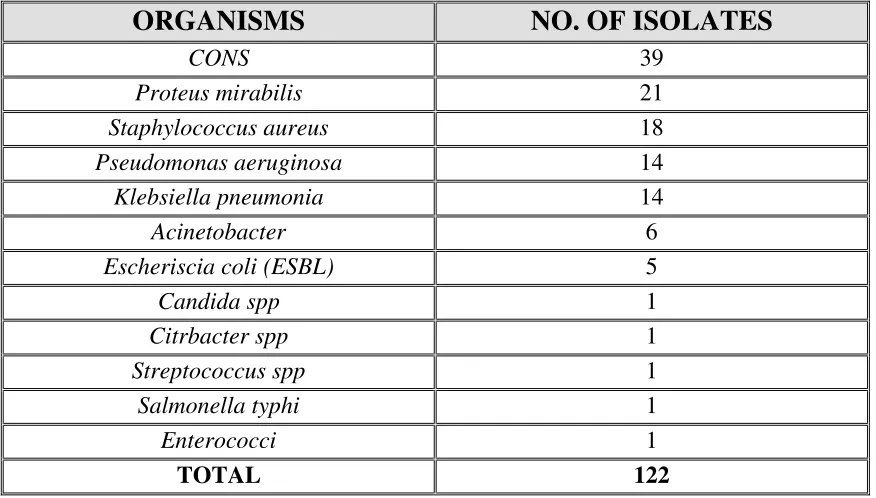
In Phase I trial, which was carried out between the months of August to October 2010, 177 case sheets were analysed (retrospectively the bateriologic profile for these months were analysed) in order to know about the pattern of organisms prevalent before the study.
[image:57.595.80.518.366.615.2]Overall estimates showed that CONS was the most common organism isolated followed by Proteus. Out of the 122 isolates gram +ve cocci were 58, gram -ve bacilli were 63, others 1.
TABLE 1. BACTERIOLOGIC PROFILE IN PHASE I TRAIL
ORGANISMS NO. OF ISOLATES
CONS 39
Proteus mirabilis 21
Staphylococcus aureus 18
Pseudomonas aeruginosa 14
Klebsiella pneumonia 14
Acinetobacter 6
Escheriscia coli (ESBL) 5
Candida spp 1
Citrbacter spp 1
Streptococcus spp 1
Salmonella typhi 1
Enterococci 1
58
[image:58.595.86.514.68.316.2]In the Phase II, trial samples were taken from different IMCU environments and from the patients, and were carried out between the months of December 2010 to February 2011.
TABLE 2. TOTAL NO.OF PATIENT SAMPLES PHASE II TRIAL
SAMPLES NO. OF SAMPLES
Urine 47
Tracheal aspirate 29
Blood 17
CVP catheter tip 02
TOTAL 95
Out of the 95 samples analysed, 93 organisms were isolated from patients’ samples. The most prevalent organism being Candida spp, followed by
Psuedomonas aureginosa.
[image:58.595.181.418.470.608.2]59
ORGANISM NO. OF ISOLATES
Candida species 22
Pseudomonas aeruginosa 15
E.coli 11
Non fermentative gram negative bacilli 9
Enterococcus species 8
Enterobacter species 8
Klebsiella species 7
Staphylococcus aureus 4
Citrobacter species 4
Coagulase negative Staphylococci 3
Proteus species 2
TOTAL 93
60
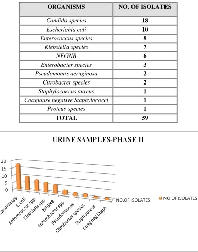
TABLE 4. URINE SAMPLES - PHASE II TRIAL
ORGANISMS NO. OF ISOLATES
Candida species 18
Escherichia coli 10
Enterococcus species 8
Klebsiella species 7
NFGNB 6
Enterobacter species 3
Pseudomonas aeruginosa 2
Citrobacter species 2
Staphylococcus aureus 1
Coagulase negative Staphylococci 1
Proteus species 1
TOTAL 59
61
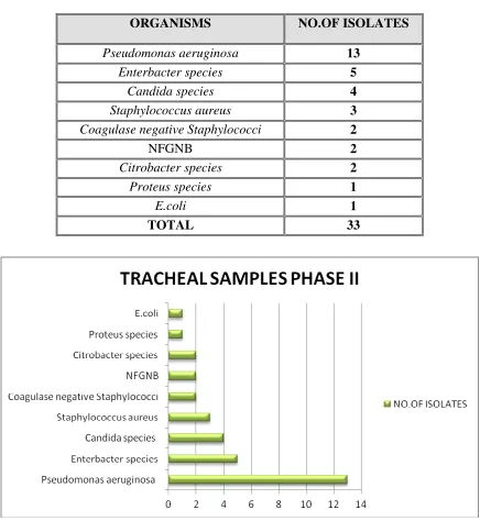
TABLE 5. TRACHEAL ASPIRATE SAMPLES - PHASE II TRIAL
ORGANISMS NO.OF ISOLATES
Pseudomonas aeruginosa 13
Enterbacter species 5
Candida species 4
Staphylococcus aureus 3
Coagulase negative Staphylococci 2
NFGNB 2
Citrobacter species 2
Proteus species 1
E.coli 1
TOTAL 33
62
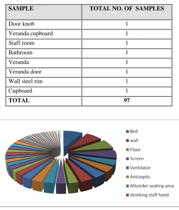
TABLE 6. TOTAL NO.OF ENVIRONMENTAL SAMPLES PHASE II TRIAL
SAMPLE TOTAL NO. OF SAMPLES
Bed 8
Wall 7
Floor 7
Screen 6
Ventilator 4
Antiseptic 4
Attender seating area 4
Working staff hand 4
Ventilator Stand 3
Window 3
IV stand 3
Multi Monitor 3
Cubicle Glass 3
Bed spread 2
Oxygen Mask 2
Ambu bag 2
Drug Tray 2
X-ray lobby 2
Emergency drug tray 2
Computer 2
Phone 2
Doctor’s table 2
Working table 2
Pendent 2
Cubicle Tray 1
Ounce glass 1
Mortar and pestle 1
Laryngoscope 1
Circuit box 1
Feeding syringe 1
Drug table 1
63
SAMPLE TOTAL NO. OF SAMPLES
Door knob 1
Veranda cupboard 1
Staff room 1
Bathroom 1
Veranda 1
Veranda door 1
Wall steel rim 1
Cupboard 1
TOTAL 97
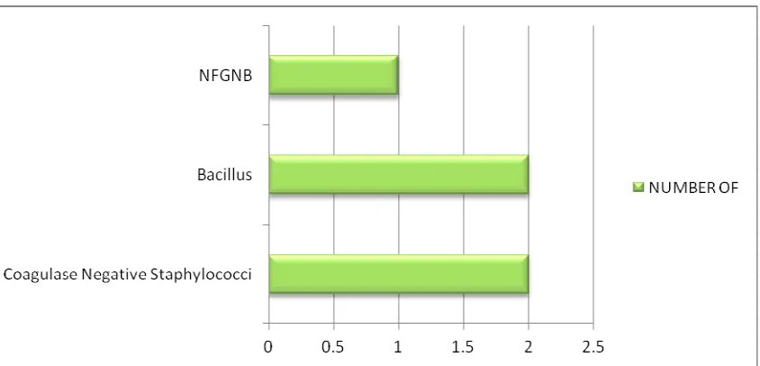
[image:63.595.121.477.64.482.2]Coagulase negative Staphylococci was the most common organism isolated from the environment followed by Bacillus spp and Pseudomonas aureginosa.
TABLE 7. BACTERIOLOGIC PROFILE ENVIRONMENTAL SAMPLES PHASE II TRIAL
ORGANISM NUMBER OF ISOLATES
CONS 66
Bacillus 29
Pseudomonas 26
NFGNB 23
Micrococci 11
64
ORGANISM NUMBER OF ISOLATES
Enterobacter 8
Candida 6
Staphylococcus aureus 5
Klebsiella 4
Citrobacter 1
E.coli 1
TOTAL 190
WALL
Total no of sample: 7 Total no of isolates: 11
0 50 100 C O N S B a c il lu s P se u d o m o n a s N F G N B M ic ro c o c c i E n te ro c o c c i E n te ro b a c te r C a n d id a S ta p h y lo c o c c u s … K le b si e ll a C it ro b a c te r
ENV SAMPLE-PHASE II
65
FLOOR
Total no of sample: 7 Total no of isolates: 18
BED
66
VENTILATOR SAMPLE
Total no of samples: 4 Total no of isolates: 6
VENTILATOR STAND
67
SCREEN SAMPLE
Total no of samples: 6 Total no of isolates: 14
BED SPREAD
Total no of samples: 2 Total no of isolates: 8
ATTENDER SEATING AREA
68
CUBICLE SEPARATING GLASS
Total no of samples: 3 Total no of isolates: 8
WINDOWS
69
[image:69.595.77.499.69.272.2]In the Phase III, trial samples were taken from different IMCU environments and from the patients, and were carried out between the months of July 2011 to Sep 2011.
TABLE 8. TOTAL NO.OF PATIENT SAMPLES - PHASE III TRIAL
SAMPLES NO. OF SAMPLES
Urine 51
Tracheal aspirate 31
Blood 12
CVP cather tip 2
TOTAL 96
Out of the 96 samples analysed, 86 organisms were isolated from patients’ samples. The most prevalent organism being Candida spp, followed by
[image:69.595.171.414.386.511.2]Staphylococcus aureus.
TABLE 9.BACTERIOLOGIC PROFILE PATIENT SAMPLES - PHASE III TRIAL
ORGANISM NO. OF ISOLATES
Candida species 19
Staphylococcus aureus 14
Acinetobacter species 9
Corynebacterium species 8
70
ORGANISM NO. OF ISOLATES
Enterococcus species 7
Klebsiella pneumoniae 6
Klebsiella oxytoca 5
Coagulase negative Staphylococci 5
E.coli 4
Non fermentative gram negative bacilli 2
TOTAL 86
PATIENT SAMPLE PHASE III TRIAL
Candida species
Staphylococcus aureus
Acinetobacter species
Corynebacterium species
Pseudomonas aeruginosa
Enterococcus species
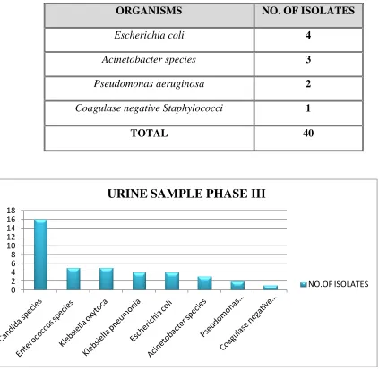
Among these, out of the 51 urine samples 40 organisms were isolated with Candida being the most commonly isolated organism. No growth was found in 11 samples.
TABLE 10. URINE SAMPLES - PHASE III TRIAL
ORGANISMS NO. OF ISOLATES
Candida species 16
Enterococcus species 5
Klebsiella oxytoca 5
71
ORGANISMS NO. OF ISOLATES
Escherichia coli 4
Acinetobacter species 3
Pseudomonas aeruginosa 2
Coagulase negative Staphylococci 1
TOTAL 40
0 2 4 6 8 10 12 14 16 18
URINE SAMPLE PHASE III
NO.OF ISOLATES
[image:71.595.81.504.69.483.2]Out of the 31 tracheal aspirate samples, 46 organisms were isolated and was polymicrobial. Staphylococcus aureus being the most common among them. No growth was noted in 7 samples.
TABLE 11. TRACHEAL ASPIRATE SAMPLES - PHASE III TRIAL
ORGANISMS NO.OF ISOLATES
Staphylococcus aureus 14
Corynebacterium species 8
Acinetobacter species 6
72
ORGANISMS NO.OF ISOLATES
Coagulase negative Staphylococci 4
Candida species 3
Enterococci species 2
Klebsiella species 2
Non fermentative gram negative bacilli 2
TOTAL 46
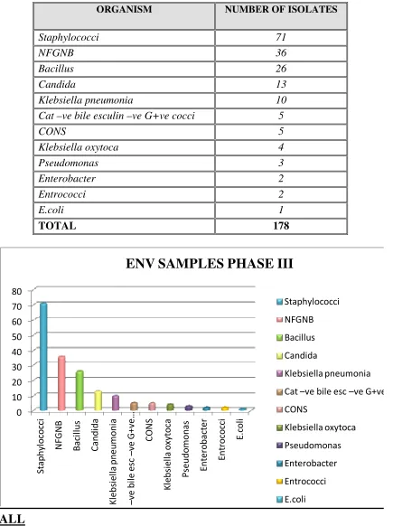
There was no organism isolated from the blood culture samples & CVP catheter tip. 98 environmental samples were analysed and 178 organisms were isolated.
TABLE 12. TOTAL NO.OF ENVIRONMENTAL SAMPLES - PHASE III TRIAL
SAMPLE NO OF SAMPLE
AMBOBAG 2
BED 10
BED SPREAD 4
BIOLOGICAL BINS 2
COMPUTER 2
73
SAMPLE NO OF SAMPLE
CURTAIN 4
DRUG TABLE-2 2
DRUG TRAY 4
FLOOR 10
FRIDGE INSIDE 2
GENERAL TABLE 4
IV STAND 3
MILK GLASS 3
MORTAR AND PESTLE 2
MULTI MONITOR 4
PENDENT 3
SCREEN 8
VENTILATOR OUTSIDE 2
VENTILATOR INSIDE 2
WALL 10
WINDOW 4
X RAY LOBBY 2
ANTISEPTIC 2
WORKING STAFF HAND 4
TOTAL 98
AMBOBAG
BED
BED SPREAD
BIOLOGICAL BINS
COMPUTER
CUBICLE SEPERATING GLASS
CURTAIN