AGE ESTIMATION FROM THE PHYSIOLOGICAL
CHANGES OF TEETH USING MODIFIED GUSTAFSON’S
METHOD
Dissertation Submitted to
THE TAMIL NADU DR.M.G.R.MEDICAL UNIVERSITY
Towards the partial fulfillment for the degree of
MASTER OF DENTAL SURGERY
BRANCH – IV
ORAL PATHOLOGY & MICROBIOLOGY
ACKNOWLEDGEMENTS
I begin with expressing my profound thanks to the Almighty for everything.
I sincerely thank our Principal Dr. K.S.G.A
.Nasser, MDS,for providing me an
opportunity to study in this esteemed institution and constant encouragement.
I am deeply indebted to our beloved former Professor Dr.Shaheen Ahmed,
MDS, department of oral pathology & Microbiology, for her support, constant
encouragement, and guidance.
I would like to extend my deepest gratitude to my teacher Dr. M.R.C. Rajeswari,
MDS, department of oral pathology & Microbiology for her encouragement, motivation,
help and support .
I also extend my thanks to Reader Dr. I. Ponniah, MDS, Assistant professor
Dr.R.Bharathi, MDS, department of oral pathology & Microbiology, for their guidance,
constant support and kind words of encouragement.
Forensic Medicine, Dr.N.Hemalatha, PG Student in forensic medicine, Madras
Medical College, Chennai for their timely co-operation and kind assistance in collecting
the samples.
My Sincere thanks to Dr.Ravanan, Reader Department of Statistics, Presidency
College, Chennai for his help during statistical analysis of this study.
My special thanks to my colleagues Dr .S. Gnanadeepam and Dr. N.Lavanya for
their kind help and support.
I would like to thank my Junior Colleagues, specially Dr. N.V.Vani, Dr. Bhawna
Gupta and Dr. V. Ilayaraja for their cooperation.
CONTENTS
SL. NO
TITLE PAGE.NO
1.
Introduction
1
2.
Aims and objectives
4
3.
Review of literature
5
4.
Materials and methods
25
5.
Tables and Charts
30
6.
Results
37
7.
Discussion
39
8.
Summary and conclusion
49
INTRODUCTION
AGE ESTIMATION FROM THE PHYSIOLOGICAL CHANGES OF TEETH USING MODIFIED GUSTAFSON’S METHOD
Odontological examinations have been a critical determinant in the search
for identifying the humans where positive identification is not practical due to
decomposition or destruction of the soft tissues.
“Oh! Look the dead teach the living”
Winternitz.
Forensic identification is based on finding differences, polymorphism’s
between different individuals. These differences can take many forms, such as
differences in facial appearances, hair color, height, ear lobe confrontation, retinal
arterial structure etc. Some variations are unique and some are not. Indeed individual
variation is a tenet of biology.
The ability of inert mineralized structures of teeth to resist postmortem
degradation and to survive deliberate accidental or natural change has lead to analysis to
Teeth can be used in forensic investigations in identification of dismembered
remains of mass disasters, fires and in high impact crashes.
Though human dentition is considered as unique and hard tissue analog to the
fingerprints, it may change during the lifetime of an individual due to various
physiological and pathological process.
Different methods have been used for age estimation in different ranges of
age. The most common method in adults is using dental parameters used by Gustafson
in 1947. He presented his models based on microscopic and macroscopic features of
teeth in 1950.
Gustafson 14,30 first formulated observations of macrostructural changes in
teeth into a workable system for adult age estimation. His method was based upon six
age related changes, assigning points upon an ascending scale of 0 to 3 according to the
severity of the change.
These changes are;
1.Attrition- The gradual wear of the enamel on the occlusal surface, used as a method
of aging adult populations.
2.Secondary dentine apposition - Age related build up of dentine on the walls of the
pulpal chamber.
3.Periodontitis – The irregularity in the form of the cementum and root dentine caused
4.Cementum build up , related to periodontitis, where the continuous repositioning of
the tooth in the alveolar bone necessitates extra layers of cementum.
5.Root resorption – The gradual resorption of the root apex ( a process little understood
in terms of oral biology).
6.Root transparency – The tendency of root dentin in thin sections to appear to be
transparent in transmitted light from the apex upwards
( termed sclerotic dentin).
Using this six parameters, age is calculated. Each factor will get some points according
to severity and total score is calculated adding points obtained by all factors. A formula
is obtained using regression analysis between the total score obtained and known age.
AIMS AND OBJECTIVES
AIMS AND OBJECTIVES
1. To evaluate the physiological changes occurring in the teeth with the advancing age
using undecalcified ground sections of teeth obtained from cadavers with known age.
2. To derive a formula for age estimation using multiple regression analysis in our
population by modified Gustafson’ s method
3. To estimate age of an individual using the formula obtained by modified Gustafson’s
method.
REVIEW OFLITERATURE
REVIEW OF LITERATURE
Identification of humans using the unique features of the teeth and jaws has
been used since Roman times. 57
Forensic odontology may have been born at the battle of Nancy in 1477, when
the body of Charles the Bold was identified by the absence of a lower tooth.
Throughout history, various stories have been recorded in which a person’s unusual
smile, crowded or fractured teeth, or a single darkened tooth have been used to identify a
corpse to the exclusion of all other people.
In 1835, when a gold denture helped to identify the burned body of the
Edwin Saunders in 1837, who claimed the teeth provided the most reliable guide to age
estimation from height which was a standard method used during that time.
Lascassagne 19,39 in 1889 was the first to characterize changes in fully formed
teeth with aging.
In 1925, Bodecker 7 established that the apposition of secondary dentin was
correlated to age.
In 1941, Schour and Massler chart ( development of human dentition) was
published which is periodically updated by ADA. The drawings show development of
dentition during various age period, which are of life size and can be used to compare
with radiographs or individual tooth. This chart does not differentiate between males and
females.
Moores et al have used a chart for the age estimation using fourteen stages of
mineralisation. This can be done by using a panaromic or lateral oblique projections.
The results are expressed as the mean age of attainment for each fourteen stages for the
developing tooth studied. Using this chart, age from six months to twelve years in males
and females can be estimated.
six dental factors known to change with advancing age. They are attrition, gingival
attachment and shape of the pulp chamber, which may be altered due to secondary
dentin deposition, transparency of the root, thickness of the cementum and apical root
resorption.
Root dentin sclerosis spreads crownward is consistent with the findings of
Nalbadian et al 40,55 (1960) and it seems to increase linearly with age (Azaz et al 1977).55
Stack 41 (1960) evolved a method to know the age of infants and children from
the weight and height of the erupting teeth of a child. This method can be used on both
deciduous and permanent teeth during their erupting phase.
In 1962 Dalitz 10 disregarded cementum apposition and root resorption. He
presented his model by classifying the factor into five categories.
The first person to propose a seriation based on attritions was Miles (1963)
who worked with the Anglosaxon skeletons from breedon on the hill. Miles 37 (1963)
remarked that, of the changes used by Gustafson in his point formula, root transparency
Shafer et al 55 (1963) pointed that attrition is a result of occlusal function
which starts at the time of occlusal contact between teeth therefore, attrition increases
with increasing age.
Boyde 41 (1963) found out a method of studying the cross striation which
develop in the enamel of teeth till the enamel goes on depositing on the teeth. It is thus
useful to estimate the age of a dead infant when death occurs before the end of complete
formation of enamel on the teeth. However, as the cross striation lines represent daily
incremental lines of the enamel, by this method age of the infant can be estimated in
terms of days, but the process of counting the number of cross striations is very tedious.
Harcourt 55 (1964), Nalbadian, et al 55 (1960) said that sclerosis, as an aging
phenomenon occurs not only in the Coronal dentin but also in the root and root apex.
According to Philippas & Applebaum55 (1966), reparative dentin can also form
under normal physiologic functions of teeth, without severe attrition, caries or erosion,
sclerosis, can result from the aging process of the tooth. He believed that the increased
amount of reparative dentin was not related to the intensity of attrition but rather to the
Bang and Ramm 6 concentrated on measurements of root dentin transparency as
the sole age indicator. In 1970, Bang and Ramm have shown mean error of estimation to
be + 4.7 years in 58% of cases, to be + 10years in 79% of the subjects.
In 1971, Johanson19 found that the Gustafson’s method to give less accurate
results and modified the Gustafson’s Method by multiple regression analysis and
proposed a more accurate formula for age estimation with standard error of five to
sixteen years. Johanson suggested the use of a 0.25mm thick ground section, mounted in
a photographic enlarger for the production of an enlarged accurate tracing or an enlarged
photograph from which the different changes are easily and reproducibly evaluated.
According to Johanson, the correlation of the transparency of dentin with age is the
highest, while that of apical resorption is the lowest.
Tronstad 55 (in 1972) pointed out, however, that the optical and radiographic
variations in the incisal dentin are not caused by age or external irritation, but rather are
a normal feature of anterior teeth.
Demirjian’s et al 63 (1973) used a Technique which is based on
children based on dental age.
Pillai et al 43 (1974) showed in India that Gustafson’s method is under
influence of external factors such as race and culture. According to him, congenital and
environmental patterns, including eating habits, which seems to be determinant of dental
factors.
According to Bhaskar 55 (1976) , the root dentin of elderly people can become
so sclerotic that it assumes a transparent glass like appearance.
In 1978, Maples 33 used factors like secondary dentin and translucency of root
of the second molar teeth for age estimation. His method was suggested for use as a
complementary method along with other methods.
In 1979, Helm et al 37 used the severity of attrition of molar teeth to estimate
age and showed that attrition factor had a medium accuracy for age estimation.
In 1980, Wegener et al 61 studied the correlation coefficient between root dentin
transparency and age. It was 0.67 and the best range of age was 30 and above years
Metzger et al 35 (1980) prefer the use of thick ground (1mm) sections instead
of thin ground (0.25mm) section for evaluating dentin transparency value. It is useful to
minimize the variability and inaccuracy in the evaluation of dentin transparency value
and secondary dentin value which have the highest correlation with age.
Brothwell (1981) used skeletal material from Neolithic and Medieval
Britian to compile an ageing method that looked at the rate of attrition in molar.
Brothwell used ten year increments to categorise the amount of dentin exposed to
attrition ranging from 17-25, 25-35, 35-45, >45. This technique allows for a less rigid
age group and would benefit from multifactorial analysis due to the different rates of
wear that a population may exhibit.
Stanley et al 55 (1983) demonstrated a close correlation between the
dentin changes observed in un-decalcified ground sections and microradiographs with
the staining characteristics of the decalcified sections. They found that the pollak
trichrome stain and pollak trichrome
variation No.6 Stain, were most effective in revealing dentin sclerosis. Sclerosis appears
as a red-orange zone with the former staining technique and orange with the latter.
According to him, dentin sclerosis and reparative dentin can be detected by ground
sections, microradiographs and staining techniques, although undecalcified ground
section was the most reliable. He demonstrated that the root and furcation dentinal
unrelated to particular lesions but did relate to increasing age. Root dentinal sclerosis
extended from apical to cervical area with increasing age.
In 1983 , Altinini 2,39 said that age related changes occur in teeth between
approximately 10weeks in utero to old age.
Ketterl 21 (1983) demonstrated age induced changes in the teeth. Enamel of old
people undergoes attrition and dentin is characterized by continuous narrowing of the
lumen of the dentinal tubule, increasing calcification, reduction in the amount of
peritubular fluid and reduced sensitivity. With age, cementum undergoes continuous
deposition and volume of the pulp declines owing to the deposition of secondary dentin.
Lovejoy et al 29 (1985) showed that upon using a high sample size, a correlation
coefficient of 0.93 could be found between the attrition factor and the age for a group of
American Indians.
Mean error of Gustafson’s method was shown to be + 4.6 years by Haertig’s et
al 37 study in France (1985). Sabaghian 37(1988) and Savabi 37 (1989) had also used
Gustafson’s linear regression without new modeling with a lower sample size in a group
Hillson (1986) pointed out that the rate of attrition may fluctuate within a
population due to different wear patterns of different people within the same group at
different times in their lives.
Mc Kee and Molnar (1988) state that “Rate and patterns of wear are governed
by tooth developmental sequences, tooth morphology, tooth size, internal crown
structure, tooth angulations, non dietary tooth use, the biomechanics of chewing and
diet. ”
Tooth selection was based on Solheim’s 37 (1989) study and included right 2nd
premolar, left 2nd premolar, right 1st premolar, left 1st premolar, right canine, left canine,
right lateral, left lateral, right incisor and left incisor in descending order.
In 1989, Solheim et al 52 showed that correlation coefficients between
translucency factor and age were 0.68 to 0.86 in different methods of measurement and
0.57 to 0.83 in different teeth. The increase in the translucent zone with advancing age
was found to be linear and was not affected by periodontal destruction.
Lorentsen M et al 28 (1989) examined the relationship between age and the
area of translucent dentin (ATD) at root apex. For statistical analysis, an XT
microcomputer and SPSS/PC regression were used. The correlation between age and
Solheim T 52 (1989) said that the cervical pulp width of mixed human teeth,
was found to reduce by 2mm over a mean patient age range of between 28 and 74 years,
giving an approximate rate of secondary deposition of 43µm per year or 0.119µm per
day.
In 1990, W.R. Maples 32 found that the six factors used in Gustafson’s
method, root transparency was the most reliable one followed by deposition of
secondary dentin formation, attrition, migration of periodontal ligament, cemental
apposition and root resorption.
Woods et al 34 (1990) concluded that the timing of secondary dentin formation
is more closely fit by a curved than a straight line.
In 1990, However, Santini et al 37 showed that the attrition factor of molar
teeth based on Miles method was not useful for age estimation.
In 1990, Solheim 37 showed that the highest correlation coefficient between age
and cementum thickness in the lower third of root. It ranged from 0.40 to 0.67 by
According to Stein TJ et al 56 (1990), there is a positive correlation, which
could not occurred by chance, that as age increases, the deviation and the width of the
foramen opening both increases. This increase appears to be a result of apical cemental
thickening that occurs as the patient ages.
Drusini A et al 12 (1990) applied Bang and Ramm equation using the
percentage ratio hx100/H (after Lamendin & Cambray 1981). Where h is the extension
of the root transparency zone (in mm) and H is the total root length (in mm), some
regression functions have been elaborated.
The error of the age estimation obtained following Bang & Ramm was quite high in
percentage, being comprised between +/-5 years only in 21.13% of the cases.
In 1991, Kambe et al 20 have found a correlation coefficient of 0.93 between
attrition and age using computer assisted image analyzer.
Morse DR 38 (1991) showed that the dentinal thickness has been calculated
as increasing at a rate of approximately 0.5 micrometer per day.
Lamendin et al 24 (1992) proposed a technique to estimate age as a function
with a mean error of + 10years on their working sample and + 8.4 years on a forensic
control sample.
In 1993, Tomaru et al 58 showed that the correlation coefficient between
incisors of lower jaw and age was 0.607 based on their findings.
Morse(1993) studied aging changes of the dental pulp and dentin in normal
teeth by radiographic method and found that root canal shrinkage increased with
advancing age.
Solhiem T 53(1993) used scoring system for surface roughness (surface
roughness scores - SRS). However the SRS could not be assessed with sufficient
reproducibility and the estimates were therefore too subjective to be used as the sole
criterion for age estimation.
Lopez et al 27(1993) studied age determination on the basis of image
analysis of scanning electron microscopicy using root transparency and dentinal tubule
diameter as parameters. The results showed limited age estimation due to individual
In 1993, Drusini 13 published a study that confirmed the negative
correlation between the coronal index after the actual age of individuals using soft x-ray
photos of intact adult teeth. The author was able to show that the correlation coefficients
range from – 0.73 to 0.89.
Huda et al 18(1995) determined age in dental microstructure using
incremental markers which are thought to be formed in circadian and circaseptan
rhythms in Juveniles.
Lic et al 25 (1995) estimated age from the permanent molar by the
method of average stage of attrition (ASA). The ASA method gave an estimated age at
death from only one molar either first molar or second molar on either maxilla or
mandible. The maximum error of these equations was 4.53 years. The results show that
the ASA method can or does reflect the attrition condition of the whole occlusal surface
more objectively than some methods using dental wear because the wear degree is
estimated by averaging the wear stages of all the cusps rather than of only one or partial
cusps.
different rations of the two dimensional pulp size, which depends on the amount of
secondary dentin and chronological age.
Lucy et al 30 (1995) pointed another statistical analysis of Gustafson’s data
and find that the errors calculated by Maples and Rice were also in error, being about a
year too small.
Whittaker et al 62 (1996) pointed out that the effect of racial origin should
be considered when using sclerosis as a means of age determination in forensic cases.
Hopp R et al 17 used length of translucency zone so that the mean error of
estimation was 5 years with 90% reliability.
Sengupta et al 47 (1999) showed difficulties in estimating age using root
dentin translucency in human teeth of varying antiquity. The percentage length of RDT
in sectioned teeth was found to correlate well with chronological age in the modern
sample but not in the archaeological sample.
Amariti et al 3 (2000) studied a new technique where a photomicrographic
image of a cross section of sclerotic dentin was converted to a grey scale of 256 tones
and then reduced to black and white and read by computer using specially developed
limit of 11 years was obtained.
Kim et al 22(2000) scored the degree of occlusal wear for all premolar and
molar teeth using dental stone cast. The degree of tooth wear showed a significant
positive correlation with age in each and every examined tooth of both males and
females. Tooth wear score of males were higher than those of females. Kim’s new
system for scoring tooth wear is a reliable and accurate method of age estimation.
Williem SG et al 63(2001) used Demirjian’s Technique and this study
confirmed significant overestimation of the dental age and is basically due to different
rates of dental development in different populations.
Ajmal et al 1 (2001) studied three methods namely, Johanson method,
methods of Kashyap and Koteswar Rao and the average stage of attrition method (ASA).
In all the three methods overestimates of age were common in mandibular teeth and in
teeth taken from female individuals and ASA method was found to be the best method.
In 2002 Ball J 5 , highlighted the weaknesses and limitations of age
Prince et al 44 (2002) applied Lamendin’s method (using only two factors) to
estimate age. Results are with a mean error of 8.2 years standard deviation 6.9 years and
standard error of the mean 0.34 years. When ancestry and sex are accounted, the mean
errors are reduced for each group.
In 2002, Murray et al 39 demonstrated that the degree of age related
changes in teeth appeared to be asymmetrical, with decreases in the root being greater
than the crown. With increasing patient age, in both crown and root aspects of teeth,
dentinal thickness increased.
Valenzuela et al 59 (2002) recommended different regression models to
calculate age depending on the postmortem interval.
In 2003, Soomer et al 54 studied the reliability and validity of eight
different dental age estimation methods for adults. The method for sectioned teeth gave
more reliable results when compared to methods for intact teeth.
In 2003, Babak et al 37 showed that among the different Mandibular teeth,
best correlation coefficient with age. Mean error upon estimation of age by type of tooth
appeared to be 6.4, 7.0, 6.7,5.2 and 6.2 years for regression lines of central, lateral,
canine, first and second premolar tooth respectively.
Olze et al 42 (2004) studied age estimation of unidentified corpses by
measurement of root translucency and said that to avoid seriously inaccurate estimates in
individual cases, the result should always be verified critically against an assessment of
the overall stomatognathic system and other Post-Mortem findings of relevance to
age.
Paewinsky et al 34 (2005) verified the applicability of Kvaal et al method and
found a significant negative correlation between the width ratios of the pulp cavity and
chronological age.
In 2006, Vicek 60 used the modified Gustafson technic for the determination
of age by teeth from paleoantheropological material. The modification makes it
possible for anthropologists and forensic experts to use the histological method of
Gustafson’s section in estimating the age both in prehistorical and in recent bone
material.
transparency, second is using root and root canal analysis from the x-ray and third is
using six parameters on each teeth. The coefficient of correlation of third method was
0.85 and they are in the significant strong correlation with the known age. He also
suggested that the teeth of the maxilla are more convenient for the age determination
than the teeth of mandible.
Yun JII et al 64 (2007) studied modified Kim’s scoring system and showed
that it is a reliable and accurate method for age estimation. Tooth wear scores of all
teeth except the two lower central incisors were higher in males than in females.
SOME OTHER METHODS OF AGE DETERMINATION
Cementum is continuously deposited at the root end and seen as
incremental lines and there is referred to as cemental annulations. Many researches have
used cemental annulations to determine the age of the adults. At present there is
controversy using this method, because difference studies shows vast discrepancies in
the results.
An interesting method using intensity of fluorescence from dentin and
between age, depending of the colour of the tooth and increase in the intensity of the
fluorescence. I t has been proposed that colour changes in the dentin and the cementum
are caused by infusion of decomposition products from red blood cells.
Katsuichi Yamamoto studied racemisation of aminoacids, in the field of
archaeology as a means of determining the era of geochemical materials such as animal
or plant fossil and accumlation strata. The same method was applied for the first time by
Helfman et al 16 (1975) to estimate the age of teeth . They determine separately the
amount of Land D type of aspartic acid in their enamels and found that the d/L ratio of a
MATERIALSAND METHODS
MATERIALS AND METHODS
The medico legal cases received for the autopsy by the Department of
forensic medicine, Government General Hospital , Chennai, were taken for the study.
Total number of 50 cases were studied. Age of cases ranged from 21 to85.
The apparatus used in the study are:
1.Tooth extraction forceps
2. Probe
3. Lathe
4. Carborundum stone
5. Alcohol and xylene
6.Formalin
7.Microscope and slides, etc
The details of the deceased(age of patient) were noted from the relatives
accompanying. After collecting the details, teeth to be studied were selected. Our
priority in tooth selection was based on solheim’s 52 study and included right second
incisors in descending order.
Degree of attrition and extent of periodontal disease were recorded before the
extraction of the tooth. After the tooth selection, the distance between sulcus of gingiva
and cervix of tooth in medial aspect of buccal surface was measured with a probe in
millemeters. This is measured to calculate the periodontitis factor.
Upon presence of trauma or laceration of gingiva, the distance between
junctional epithelium on root and cementoenamel junction was measured after extraction
of the tooth.
Tooth extraction was based on rotational technique using lower jaw forceps.
Upon fracture of a tooth due to severe curvature of the root, the tooth was disregarded
and the next tooth was selected based on the above mentioned priority.
After extraction, the tooth were cleaned and put in tubes containing alcohol
and xylene. Alcohol and xylene show a better presentation by dehydration of translucent
area of root.
and then with rough carborundum stone until a section of 1mm was obtained and at this
thickness, the root translucency was noted.
Grinding was further done using fine stone until the section of 0.25mm
thickness is left, finally cleaned , and dried section was mounted on slide and viewed
under microscope for secondary dentin formation, cementum apposition and root
resorption.
The factors seen in the tooth before and after sectioning were recorded using 4
points allotment system 37 as follows:
1.Periodontitis factor
P 0- No periodontitis
P1 - Beginning of periodontitis
P2 - Periodontitis more than one third of root coronally
P3 - Periodontitis more than two third of the root coronally
2.Attrition factor
A0 - No attrition
A1 - Attrition up to enamel level
A2 - Attrition up to dentin level
3.Secondary dentin apposition factor
S0 - No secondary dentin
S1 - Secondary dentin up to upper part of pulp cavity
S2 - Secondary dentin up to half of pulp
S3 - Diffuse calcification of the entire pulp
4. Root resorption factor
R0 - No resorption
R1 - Spotted like resorption
R2 - Root resorption at the level of cementum
R3 - Extensive resorption of cementum and dentin
5. Cementum apposition factor
C 0 -Normal thickness
C1 - Thickness more than normal { detectable}
C2 - Generation of thick cementum
C3 - Hypercementosis
6. Translucency of root factor
T 0 - No translucency
T1 - Beginning of translucency of root
T2 - Translucency more than one third of apical root
T3 - Translucency more than two thirds of apical root
After collecting the data and calculating the total score, multiple regression
analysis done using total score and known age. Multiple regression analysis yielded a
new formula for the values obtained by all factors and known age.
Formula obtained;
Y = 12.29+ 4.42 X
Y denotes estimated age
X denotes total score
Using this formula, age is calculated and tabulated. Difference between known age and
TABLES AND CHARTS
TABLE:2
TABLE:3
TEETH WISE DISTRIBUTION
TEETH No of Cases
Maxillary Incisors 12
Mandibular Incisors 6
Maxillary Canine 6
Mandibular Canine 7
Maxillary Premolar 10
Mandibular Premolar 9
Total 50
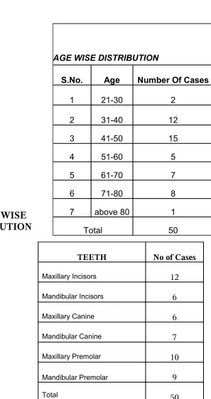
AGE WISE DISTRIBUTION
S.No. Age Number Of Cases
1 21-30 2
2 31-40 12
3 41-50 15
4 51-60 5
5 61-70 7
6 71-80 8
7 above 80 1
TABLE:4
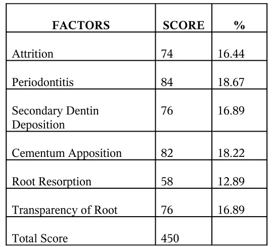
PERCENTAGE DISTRIBUTION OF FACTORS
FACTORS SCORE %
Attrition 74 16.44
Periodontitis 84 18.67
Secondary Dentin Deposition
76 16.89
Cementum Apposition 82 18.22
Root Resorption 58 12.89
Transparency of Root 76 16.89
Total Score 450
TABLE:5
CORRELATION CO-EFFICIENT BETWEEN FACTORS AND AGE
S.No. Factor Correlation Co-efficient
1. Attrition 0.6222**
2. Periodontitis 0.5888**
3. Secondary dentin deposition 0.6090**
4. Cementum Apposition 0.4937**
5. Root Resorption 0.5222**
6. Transparent of Root 0.8721**
[image:35.612.88.507.475.725.2]** denotes significance at 1% level
TABLE:6
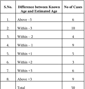
DISTRIBUTION OF DIFFERENCES
S.No. Difference between Known Age and Estimated Age
No of Cases
1. Above –3 6
2. Within –3 10
3. Within – 2 4
4. Within – 1 9
5. Within +1 5
6. Within +2 3
7. Within +3 6
8. Above +3 9
Table:1
It Shows type of tooth selected, scores obtained by six factors like attrition,
periodontitis, secondary dentin deposition, cementum apposition root resorption and
translucency of root. Total score is calculated and age is estimated. Total score,
estimated age, known age as well as difference in age are also shown in the tables.
Table:2
It Shows Age wise distribution of cases. Maximum number of cases were selected in
the age range of 41-50 years. Only one case was selected above 80.
Table:3
It Shows Teeth Wise distribution of cases. Maxillary teeth were selected than mandibular teeth. Maximum number of cases were selected in the maxillary arch than mandibular arch. Incisors were mostly selected in the maxillary arch than Mandible.
It Shows percentage of distribution of each factors, Total score of 450 was obtained
when calculating the scores of each factor in 50 cases. Periodontitis factor has got
highest value and Root resorption has got lowest value.
Table:5
It Shows correlation coefficient between each factor and age. The correlation
coefficient between total score and age is 0.977. The correlation coefficient between
translucency of root and age is 0.872 and this factor is highly correlated with age among
six factors.
Table:6
It Shows distribution of differences between known age and estimated age.
RESULTS
RESULTS
The total number of 50 cases were selected, Age wise distribution of cases are
shown in the table2. The maximum number of cases were selected in the age range of
41-50 years. Percentage of distribution of the six factors, including attrition,
periodontitis, root resorption, secondary dentin apposition, cementum apposition, and
translucency of the root, are shown in Table 3. periodontitis factor has got highest score
among the six factors. Root resorption factor has got lowest score and contributes lesser
percentage to the determination of the age. Attrition, secondary dentin deposition, and
translucency of root scores are more or less in the same range. Over estimation of age
range from 0.13 to 12.19 years. Under estimation of age range from 0.18 to 6.66
years.Mean error of estimation is + 2.33 years.
Over estimation of age occurs in 21 cases in a total of 50 cases under estimation
of age occurs in 29 cases in a total of 50 cases.In six cases only difference between
Difference between known and estimated age is within –3 in 23 cases
(underestimated). The difference between known and estimated age is within +3 (over
estimated) in 14 cases. In nine cases only, the difference between known and estimated
age is above +3 value (over estimated).
The correlation coefficient between total score and age is 0.977** The
correlation coefficient between translucency of root and age is 0.872** and this factor,
translucency of root is highly correlated with age among six factors. The correlation
coefficient between root resorption factor and age is 0.522** and this factor is least
correlated with age among six factors. In our study, the correlation coefficients of age
with each of single factors are less than the coefficient of age with the sum of factors so
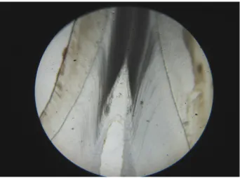
PHOTO
MICROGRAPHS
FIGURE .1 ARMAMENTARIUM
FIGURE .2
ATTRTION UPTO ENAMEL LEVEL, DENTIN LEVEL AND PULP LEVEL
FIGURE .3
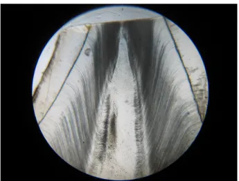
BEGINNING OF TRANSLUCENCY OF ROOT
FIGURE .4
TRANSLUCENCY MORE THAN ONE THIRD OF APICAL ROOT
[image:43.612.67.419.341.599.2]
FIGURE.5
PHOTOMICROGRAPH
[image:44.612.130.472.440.694.2]
FIGURE .1
SECONDARY DENTIN UP TO UPPER PART OF PULP CAVITY
FIGURE .2
FIGURE .3
[image:45.612.125.470.410.676.2]DIFFUSE CALCIFICATION OF THE ENTIRE PULP
FIGURE .4
FIGURE .5
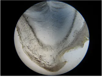
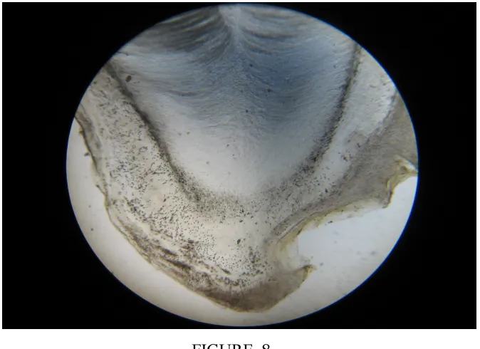
GENERATION OF THICK CEMENTUM
[image:46.612.143.481.405.659.2]
FIGURE .7
SPOTTED LIKE RESORPTION
FIGURE .8
[image:47.612.144.481.453.698.2]
FIGURE .9
DISCUSSION
DISCUSSION
There have been two major series of methods for age estimation based on
dental parameters, which are single and multiple factor methods.
In 1979, Helm et al 37 used the severity of attrition of molar teeth to
estimate age of Medieval Danes. These findings showed that attrition factor had a
medium accuracy for age estimation.Lovejoy et al 29showed that upon using a high
sample size, a correlation coefficient of 0.93 could be found between the attrition factor
and the age of a group of American Indians. Hillson (1986) pointed out that the rate of
attrition may fluctuate within a population due to different wear patterns of different
people within the same group at different times in their lives.
In 1991, Kambe et al 20 have found a correlation coefficient of 0.93 between
attrition and age using computer assisted image analyzer.In 1993, Tomaru et al 58
showed that the correlation coefficient between incisors of lower jaw and age was 0.607
based on their findings.
However, Santini et al 37 showed that the attrition factor of molar teeth based
on miles method was not useful for age estimation. Lic et al 25 (1995) estimated age
from the permanent molar by the method of average stage of attrition. The ASA method
gave an estimated age at death from only one molar either first molar or second molar on
either maxilla or mandible. The maximum error of these equations was 4.53 years.
Kim et al 22 (2000) scored the degree of occlusal wear for all premolar and
molar teeth using dental stone cast. The degree of tooth wear showed a significant
positive correlation with age in each and every examined tooth of both males and
females. Tooth wear score of males were higher than those of females. In 2002 Ball J 5,
highlighted the weaknesses and limitations of age estimation by examination of dental
attrition as the sole indicator of age. Yun JII et al 64 (2007) studied modified Kim’s
scoring system and showed that it is a reliable and accurate method for age estimation.
Tooth wear scores of all teeth except the two lower central incisors were higher in males
In our study, attrition factor has correlation coefficient of 0.6222** which is
concurrent with Tomaru et al study.
Translucency of dentine can also be used for age estimation as another possible
single factor method. Bang and Ramm 6 concentrated on measurements of root dentine
transparency as the sole age indicator.In 1970, Bang and Ramm have shown mean error
of estimation to be + 4.7 years in 58% of cases to be + 10years in 79% of the cases.
In 1980, Wegener and Albrecht’s 61 study correlation coefficient between root
dentin transparency and age was 0.67 and the best range of age was 30 and above years
using the translucency factor. On the otherhand, Hopp R et al 17 used length of
translucency zone so that the mean error of estimation was ± 5 with 90% reliability.In
1989, Solheim et al 37 showed that correlation coefficients between translucency factor
and age were 0.68 to 0.86 in different methods of measurement and 0.57 to 0.83 in
different teeth. The increase in the translucent zone with advancing age was found to be
linear and was not affected by periodontal destruction. Drusini A et al12applied Bang and
Ramm equation and the error of age estimation obtained following Bang & Ramm was
quite high in percentage, being comprised between +/-5 years only in 21.13% of the
Sengupta et al 47 (1999) showed difficulties in estimating age using root dentin
transluency in human teeth of varying antiquity.The percentage length of root dentin
translucency in sectioned teeth was found to correlate well with chronological age in the
modern sample but not in the archaeological sample.
In 2004, Olze et al 42 studied age estimation of unidentified corpses by
measurement of root translucency and said that to avoid seriously inaccurate estimates in
individual cases, the result should always be verified critically against an assessment of
the overall stomatognathic system and other Post-Mortem findings of relevance to age.
In our study, translucency of root has got 0.8721** correlation value This factor has
got highest correlation coefficient than all other factors of aging suggested, which is
concurrent with Johanson method which is once again proved in our study also.
In 1992, Solheim T 52 said that the cervical pulp width of mixed human teeth
was found to reduce by 2mm over a mean patient age range of between 28 and 74 years,
giving an approximate rate of secondary deposition of 43 micrometer per year or 0.119
micrometer per day.
Kvaal et al 34 (1995) demonstrated negative correlation of a composition of different
rations of the two dimensional pulp size, which depends on the amount of secondary
method and found a significant negative correlation between the width ratios of the pulp
cavity and chronological age. In our study , secondary dentine deposition factor scored a
value of 0.6090**( correlation coefficient).
In 1990, solheim showed that the highest correlation coefficient between
age and cementum thickness in the lower third of root. It ranged from 0.40 to0.67
by different methods of measurements. cementum deposition is least correlated in our
study which has got value of 0.4937.
The multiple factor method was first used by Gustafson 14 in 1950. He
developed a system of dental age determination using six dental factors known to
change with advancing age. They are attrition, gingival attachment and shape of the pulp
chamber , which may be altered due to secondary dentin deposition, transparency of the
root , thickness of the cementum and apical root resorption.
In 1962 Dalitz 10 disregarded cementum apposition and root resorption. He
presented his model by classifying the factor into five categories. In 1971, Johanson19
found the Gustafson’s method to give less accurate results and Modified the Gustafson’s
Method by multiple regression analysis and proposed a more accurate formula for age
According to Johanson, the correlation of the transparency of dentin with age is
the highest, while that of apical resorption is the lowest.In 1978, Maples 33 has used
secondary dentin and translucency of root of the second molar teeth. His method was
suggested for use as a
complementary method along with other methods. Mean error of Gustafson’s method
was shown to be + 4.6 years by Haertig’s et al 37study in france (1985). Sabaghian37
(1988) and Savabi 37 (1989) had also used Gustafson’s linear regression without new
modeling with a lower sample size in a group of Iranians.
Lamendin et al (1992) proposed a technique to estimate age as a function of two
factors, translucency of the tooth root and periodontitis.
He estimated age at death with a mean error of + 10years on their working
sample and + 8.4 years on a forensic control sample. Lopez et al 27 studied age
determination on the basis of image analysis of scanning electron microscopic image
using root transparency and dentinal tubule diameter as parameters. The results showed
limited age estimation due to individual variations caused by genetic factors and
chewing habits.
Ajmal et al 1 studied three methods namely, Johanson method,
(ASA). In all the three methods overestimates of age were common in mandibular teeth
and in teeth taken from female individuals and ASA method was found to be the best
method. Prince applied Lamendin’s method to estimate age. Results are with a mean
error of 8.2 years, standard deviation 6.9 years and standard error of the mean 0.34
years. When ancestry and sex are accounted, the mean errors are reduced for each group.
In 2003, Babak et al 37 showed that among the different Mandibular teeth,
the sum of ranks of the first premolar factors had the best correlation coefficient with
age.
Mean error upon estimation of age by type of tooth appeared to be 6.4, 7.0,6.7,5.2
and 6.2 years for regression lines of central, lateral, canine, first and second premolar
tooth respectively. In 2003, Soomer et al 54 studied the reliability and validity of eight
dental age estimation methods for adults. The method for sectioned teeth gave more
reliable results when compared to methods for intact teeth.
In 2006 Brkic et al 8 determined age by three ways, one is using root
dentin transparency, second is using root and root canal analysis from the x-ray and third
is using six parameters on each teeth. The coefficient of correlation of third method was
0.85 and they are in the significant strong correlation with the known age. The teeth of
the maxilla are more convenient for the age determination than the teeth of mandible.
These studies show different results with different accuracies based on
dental factors that may be due to different methodologies, race, and environmental
factors.
In our study , mean error of estimation is +2. 33 years, regardless of tooth
type and it is less compared to Gustafson method. The mean error of estimation is +
3.63 in gustafson’s method. The difference between known age and estimated age is
within +3 years in 70% of cases ( 35cases)
In our study, the correlation coefficients of age with each of single factors are less
than the coefficient of age with the sum of factors so that the best estimation is achieved
by combination of all six dental factors.
The correlation coefficient between total score and age is 0.9773**.The
correlation coefficient between each factor and age is less than 0.9773**. This results
show that the best correlation is achieved by combination of factors. In 2003 babak et al
37 also showed best estimation of age done by combination of factors.
According to Johanson 19, the correlation of the transparency of dentin with age is
In ourstudy also,highest correlation value obtained by transparency of
dentin and lowest correlation value obtained by apical resorption. The correlation
between age and transparency of root is 0.8721** The correlation between age and
root resorption is 0.5222**.
The formula for age estimation obtained by Gustafson’s method in various
groups showed various constant value. This is because, the aging factors are also
influenced by the environmental factors like food, eating habits, morphology of tooth,
race and general health status. so there is a need to derive a formula for
different groups of peoples. Hence we have arrived a formula for age estimation which
is
Y = 12.29 +4.42X
Using the formula we can estimate age of an individual in our population.
This method can be used either before or in conjunction with other accurate
methods , such as aminoacid analysis of D\ L ratio of aspartic acid crystals in enamel
SUMMARY
AND CONCLUSION
SUMMARY AND CONCLUSION
In the field of forensic medicine, odontology has got an important role in the determination of age as well as sex.
This study is based on modified Gustafson’s method to evaluate the
physiological changes occurring in teeth during aging process and to estimate the age
using multiple regression analysis.
The results of this study show that the correlation coefficients of age with each
of six single factors are less than the coefficient of age with the sum of factors ,so that
best estimation of age is achieved by combination of all six factors.
The correlation coefficient between total score and age is 0.9773** .The
translucency of root has got highest correlation value. Mean error of age estimation is +
2.33 years.
practical method and should be used in the first step before more sophisticated methods
of age estimation in unknown cadavers.
Eventhough the sample size is very small, we derived a new formula for our
population. Using this derived formula, we can estimate age of unknown cadavers in
our population.
Larger sample will give more precise or better results according to the
BIBLIOGRAPHY
BIBLIOGRAPHY
1. Ajmal M, Mody B, Kumar G, “Age estimation using three established methods - a study on Indian population.” Forensic sci int 2001; (2-3): 150-154.
2. Altinini M, Age determination from teeth - a review. JDent Assoc South Am 1983;38:275-279
3. Amariti ML, Restori M, Deferrari F, PaganelliC, Faglia R, Legnani G. “A histological procedure to determine dental age.” J forensic odontostomatol 2000; 18(1):1-5.
4. Aykroyd RG, Lucy D, Pollard AM, Solheim T. “Technical Note: regression Analysis in Adult age estimation.” Am J.Phys. Anthropol 1997;104(2):259-265
5. Ball J ,“A critique of age estimation using attrition as the sole indicator” J forensic odontostomatol. 2002;20(2):38-42.
6. Bang G and Ramm E, “Determination of age in humans from root dentine transparency.” Acta odontol scand. 1970:28:3-35
8. Brkic H, Milicevic M, PetroveckiM, “Age estimation methods using Antaropological parameters on human teeth.” Forensic sci int. 2006;162 (1-3): 13-16
9. Chomette G, Auriol M, Koulibaly M, BellefqihS, GuilbertF, Vaillant JM, “Approach to age determination based on dental morphological criteria obtained in Microradiography, steromicroscopy and scanning electron microscopy.Rev stomatol chir maxillofac 1986; 87(1):33. 10. Dalitz GD. “ Age determination of adult human remains by teeth examination” J. Forensic sci. Soc. 1962;3:11-21.
11.Dayal P and Srinivasan K, “Text book of forensic and odontology, paras publication Ist edition, 1998.
12.Drusini A, Volpe A, Dovigos. “Age determination in human adults by dental histology” dz Morphol Anthropol. 1990;78(2):169-74.
13. Drusini AG, “ Age estimation from teeth using soft x-ray findings” anthrop anz 1993; 51:41 –46
14. Gustafson G, “Age determinations on teeth”, J Am dent Assoc. 1950;41:45-54.
15. Haertig A, Crainic K, Durigon M “Medico legal identification by the dental system.” Presse Med 1985; 14(9):543-545.
16. HelfmanPM and BadaJL, “Aspartic acid racemization tooth enamel from living humans”, Proc.Nat Acad Sci-USA 1975;72: 2891-2894.
17. HoppR, Blick U. “Age determination by teeth”. Dtsch Zahnavztl Z 1980;35(2):244-245.
18. Huda TF, Bowman JE, “Age determination from dental Microstructure in Juveniles”. Am J phys Anthropol 1996;101(2):305-306.
19.Johanson, G, “Age Determination from human teeth” Odontologisk Revy, 1971;22(Supp):126.
number of sites of dental attrition and its use for age estimation.” Forensic sci int 1991;50(1):97-109.
21.Ketterl W, “Age induced changes in the teeth and their attachment apparatus”. Int.Dent J. 1983;33(3):262-271.
22. Kim YK, KhoHS, Lee KH, “Age estimation by occlusaltooth wear”. J forensic science 2000;45(2):303-309.
23. Kvalls, Solheim T, “A Non-Destructive dental Method for age estimation”. J Forensic Odontostomatol. 1994;12(1):6-11.
24. Lamendin H, Baccino E, Humbert JF, Tavernier JC Nossintchovk Zerilli A. “A simple technique for age estimation in adult corpses: the criteria dental method J forensic sci.
1992;37(5):1373-1379.
25. Lic, JiG, “Age estimation from the parmanent molar innortheast china by the method of average stage of attrition”. Forensic sci int
1995;75(2-3):189-196.
26. Lopez – Nicolas M, MoralesA, Luna A “Application of dimerphism in teeth to age calculation”. J Forensicodontostomatol
1996;14(1):9-12.
27.Lopez Nicolas M, Morales A, Luna A “Morphometric study of teeth in age calculation”. Journal of Forensic odontostomatol
1993;11(1):1-8.
28.Lorentsen M, Solheim T. “Age assessment based on translucent dentine”. J Forensic odontostamatol 1989;7(2):3-9.
29.Lovejoy CO, Meindl RS, Mensforth RP, Barton TJ, “Multifactorial determination of skeletal age at death- amethods and blind tests of its accuracy”. Am J phys. Anthropol 1985;68:1-14.
30.Lucy D and pollard AM, “Further comments on the estimtion of error associated with the Gustafson Dental Age estimation
method,” J forensic sci 1995;40:222-227.
Luna JD. “Differences in morphological age related Dental changes depending on postmortem interval”. J Forensic sci
2001;46(4):889-892.
32.Maples WR, An improved technique using dental histology for estimation of adult age. J Forensic sci, 1978;23(4):764-770.
33.Maples WR, Rice PM, “Some difficulties in the Gustafson dental age estimations.” J Forensic sci. 1979;24:168-172.
34.Meinl A, Tangl S, Pernicka E, fenes C and watzekG.“On the applicability of secondary Dentin formation to radiological age estimation in young adults.” J Forensic sci, 2007; 52(2):438-441.
35.Metzger Z, Buchner A, and Gorsky M, “ Gustafson’s Method for age determination from teeth – a modification for the use of dentists in identification teams” J Forensic sci 1980;25 (4): 742-749.
36.Miles A, “ Dentition in the estimation of age” J Dent. Res 1963;42:255-263.
37.MonzaviFB, Ghodoosi M, Ghodoosi A, Savabi O and Hasanzadeh A, “ Model Age estimation based on dental factors of unknown cadavers among factors of unknown cadavers among Iranians.” Journal of Forensic Science 2003;48:379-381.
38.MorseDR, “ age – related changes of the dental pulp complex and their relationship to systemic ageing”. Oral surg oral med oral pathol 1991;72:721- 745
39.Murray PE, Stanley HR, Maththews JB, Sloan AJ, Smith AJ. “Age related odontometric Changes of Human teeth”. Oral Surg, Oral med, Oral Path, Oral radio and endo 2002;93(4):474-482.
40.Nalbandian J., Gonzales, F and Sognnaes R., “ Sclerotic age changes in root dentin of human teeth as observed by optical Electron and X-ray Microscopy”. J dent Res, 1960;39:598-607.
Reprinted 2003 Page 65.
42.Olze A, Geserick G, Schmeling A. Age estimation of unidentified corpses by measurement of root translucency. J Forensic
odontostomatol. 2004;22(2):28-33.
43.Pillai PS, Bhaskar GR, “Age estimation from teeth using Gustafson’s method- a study in india.” J Forensic sci
1974;3:135-141.
44.Prince DA, Ubelaker DH. Application of Lamendin’s adult dental aging Technique to a diverse skeletal sample. J Forensic sci
2002; 47 (1): 107-116.
45.Reppien K, Sejrsen B, Lyurerup N. “Evaluation of post-mortem estimated dental age versus real age. a retrospective 21 year survey. Forensic sci int 2006;159( supp): 84-88.
46.Santini A, Land M, Raab GM. “ The accuracy of simpleordinal scoring of tooth attrition in age assessment.” Forensic sci. int 1990;48(2):175-184.
47.Sengupta A, Whittaker DK, Shellis RP, “Difficulties in estimating age using root dentine translucency in humanteeth of
varying antiquity”. Arch oral Biol. 1999;44(11):889-899. 48.Singh A, Gorea RK and Singla U, “Age estimation from
the physicological changes of teeth. JIAFM, 2004;26(3):94-96. 49.Solheim T, “Amount of secondary dentin as an indicator of age”. scand J Dent Res. 1992;100(4):193-199.
50.Solheim T, “A new method for dental age estimation in adults”. Forensic sci int. 1993;59:137-147.
51.Solheim T , “Dental color as an indicator of age” Gerodontics. 1988;4:114-118.
52.Solheim T, “Dental root translucency as an indicator of age”. Scand J Dent Res 1989;97(3):189-197.
age”. J Forensic odontostomatol1993;11(1):9-21.
54.Soomer H, Ranta H, Lincoln MJ, Penttila A, Leibur E, “Reliability and validity of eight dental age estimation methods for
adults”. J Forensic sci 2003;48 (1):149-152.
55.Stanly HR, Pereira JC, Spiegel E, Broom C, Schultz M,“The detection and prevalence of reactive and physiological sclerotic dentin, reparative dentine and dead tracts beneath various types of
dental legions according to tooth surface and age”. J oral path 1983;12(4):257-289.
56.Stein JJ, Corcoran JF, “Anatomy of the root apex and its histologic changes with age”. oral surg, oral med, oral path 1990;69(2):238- 242.
57. Sweet D, Why a dentist for identification? Dent clin NorthAm 2001;45 (2):237-251.
58. Tomaru Y, uchiyamaY, Kobayashi K, et al. “ Age estimation from tooth attrition of lower incisors – discussion on the “Amano’s
method” Nippon Hoigaku Zasshi 1993; 47(1):13-7.
59. Valenzuela A, Martin –De Las Heras S , Mandojana JM, Dedios Luna J, Valenzuela M, Villanueva E. “Multiple regression models for age estimation by assessment of morphologic dental changes according to teeth source. Forensic med. Pathol.
2002;23(4):386-389.
60. Vice KE use of the modified Gustafson Technic for the determination of age by teeth from paleoanthropological material of
Czech ruling princes at the turn of the 9th and 10th centuries. (Forensic
science international 2006). Cesk, patol, 1997;13(4):49-55.
transparency”. Z Rechtsmed 1980; 86(1):29-34.
62. Whittaker DK, Bakri MM, “Racial variations in the extent of tooth root translucency in aging individuals”. Arch oral biol
1996;41(1):15-19.
63. Williems G, Vanolmen A, Spiessens B, Carels C. “Dental Age estimation in Belgian children. Demirjian’s technique revisited.” J Forensic sci. 2001;46(4):893-895.
64. Yun JII, Lee JY, Chung JW, KhoHS and Kim YK. “Age
estimation of Korean adults by occlusal tooth wear”. J Forensic sci