A PROSPECTIVE STUDY OF VISUAL
OUTCOME IN LENS INDUCED GLAUCOMAS
FOLLOWING SURGERY
DISSERTATION SUBMITTED FOR
MASTER OF SURGERY DEGREE
BRANCH – III - OPHTHALMOLOGY
MARCH 2008
THE TAMILNADU
DR.M.G.R. MEDICAL UNIVERSITY
Dept. of Ophthalmology, Govt. Rajaji Hospital, Madurai.
CERTIFICATE
This is to certify that this dissertation entitled “A PROSPECTIVE STUDY OF VISUAL OUTCOME IN LENS INDUCED GLAUCOMAS FOLLOWING SURGERY” has been done by DR. M. SMITHA under my guidance in Department of OPHTHALMOLOGY, Madurai Medical College, Madurai.
I certify regarding the authenticity of the work done to prepare this dissertation.
DR. R.UNNAMALAI. M.S., D.O.,
PROFESSOR & H.O.D.
DEPARTMENT OF OPHTHALMOLOGY
GOVT. RAJAJI HOSPITAL & MADURAI MEDICAL COLLEGE
DECLARATION
I, Dr. M. SMITHA solemnly declare that the dissertation titled
“A PROSPECTIVE STUDY OF VISUAL OUTCOME IN LENS
INDUCED GLAUCOMAS FOLLOWING SURGERY” has been prepared by me.
This is submitted to The Tamil Nadu Dr. M.G.R. Medical University, Chennai, in partial fulfillment of the requirement for the award of M.S.,(Ophthalmology) Branch - III degree Examination to be held in MARCH 2008.
Place : Madurai
ACKNOWLEDGEMENT
I am deeply indebted to Dr.R.UNNAMALAI, MS., D.O, Professor and Head of the department of Ophthalmology, Madurai Medical college, Madurai for the able guidance, inspiration and encouragement she rendered at every stage of this study.
I express my heartful gratitude to Dr. R. GEETHA RAMANI, M.S., DO, Formerly Professor and Head of the department of Ophthalmology, Madurai Medical College, Madurai for her valuable advice and help in carrying out this study.
I acknowledge with gratitude the dynamic guidance and persistent
encouragement given to me by my guide Dr. G.S. SRINIVASAN, M.S., DO, and Dr. A.R. ANBARASI, M.S., DO, Assistant Professors in Ophthalmology, Department of Ophthalmology, Madurai Medical College, Madurai for having taken keen interest in sharing their ideas throughout the study period. Their valuable suggestions and patronage has
My Sincere thanks to Dr.V. RAJI. M.D., Dean, Madurai Medical College, Madurai for permitting me to utilize the clinical materials of the hospital.
Last, but not the least, my profound gratitude to all the ‘patients’, to
whom I owe everything because, this venture would not have been possible
CONTENTS
S.NO. TOPIC PAGE NO.
PART – 1
1. AIM OF THE STUDY
2. INTRODUCTION 1
3. CLASSIFICATION OF GLAUCOMA 3
4. PATHOGENESIS OF LENS INDUCED GLAUCOMA 5
5. CLINICAL PRESENTATION 8
6. MANAGEMENT OF EACH TYPE 11
7. REVIEW OF LITERATURE 31
PART - II
1. PATIENTS & METHODS 35
2. PROFORMA 38
3. RESULTS 41
4. DISCUSSION 52
5. CONCLUSION 61
AIM OF THE STUDY
INTRODUCTION
Cataract and glaucoma are the leading causes of blindness through out the world.
Of the total 45 million blind persons (Visual acuity < 3/60) in the world, 7 million are in India. Glaucoma accounts for 5.8% of all types of blindness in India with a prevalence of 4% in population aged 30 years and above.
Glaucoma is not a single disease process. It is a group of disorders characterized by widely different clinical and histopathological manifestation.
These disorders share characteristic changes in visual field, optic nerve head and usually have increase in Intra ocular pressure. Intraocular pressure is only a risk factor.
CLASSIFICATION OF GLAUCOMA
Glaucoma may be classified into
Primary:
Here the pathology is confined to AC angle or conventional aqueous outflow pathway with no apparent contribution from other ocular or systemic disorder.
This may be divided into 3 main categories. 1. Open angle
2. Closed angle 3. Congenital
Secondary :
Glaucoma in which there is a predisposing ocular / systemic event. Though this form a small proportion of all types of glaucoma they frequently produce the most difficult problems in diagnosis and management.
1) Outflow obstruction :
a. Pre trabecular block from iris – trabecular contact or adhesion.
i. With pupil block ii. Without pupil block
b) Trabecular block – due to damage to mesh work Eg secondary open angle glaucoma
c) Post trabecular block – from episcleral venous pressure
2) Hypersecretion glaucoma - Rare cause eg. Epidemic dropsy Secondary glaucoma may occur due to many reasons, one of which is a sub group called Lens induced glaucoma, where lens is the prime factor responsible for glaucoma.
Few varieties of lens induced glaucoma are as follows.
a) Phacomorphic glaucoma : Swollen lens causes increased pupillary block and secondary angle closure.
b) Dislocated lens : Dislocated lens causes, increased pupillary block and angle closure.
d) Lens particle glaucoma : Lens matter liberated by means of surgery or trauma blocks outflow.
e) Phacoanaphylaxis : Granulomatous inflammation following sensitization to lens proteins causes secondary glaucoma.
PATHOGENESIS, CLINICAL FEATURES, DIAGNOSIS, MANAGEMENT OF EACH TYPE OF LENS
INDUCED GLAUCOMAS.
PHACOLYTIC GLAUCOMA (Lens Protein Glaucoma) :
Hypermature or mature rarely immature cataracts may leak lens proteins into aqueous humor and obstruct the aqueous outflow channels. This form of lens induced open angle glaucoma is called phacolytic glaucoma although some studies suggest lens protein glaucoma may be an appropriate term.
Flocks et al proposed the term phacolytic glaucoma for the open angle glaucoma associated with a leaking hypermature cataract. They believed that glaucoma was caused by obstruction of the inter trabecular spaces by macrophages. Epstein et al provided evidence that high molecular weight lens protein may be primarily responsible for the obstruction to aqueous outflow in this disorder and the term lens protein glaucoma has been suggested.
PATHOGENESIS :
The lens protein ordinarily are sequestrated within capsule and isolated from the remainder of the eye. During phacolysis microscopic defects occur in the anterior and posterior lens capsule through which soluble lens proteins enter the aqueous humor. These microscopic leaks probably reflect an end stage in the process of cataract formation.
Morphologically, eosnophilic protein like material and macrophages have been observed in the trabecular meshwork of eyes with phacolytic glaucoma. The presence of macrophages is a natural response to the presence of lens material in the AC and studies have demonstrated that lens protein especially, gamma crystalline lens proteins can induce the migration of peripheral blood monocytes. The macrophages have been shown by electron microscopic studies to have phagocytozed degenerated lens material.
aspiration of a cataract, but did not appear to obstruct aqueous outflow.
HMW protein is rare in childhood lenses which may explain why phacolytic glaucoma rarely occurs in children.
CLINICAL FEATURES :
The term phacolytic glaucoma should be reserved for the sudden onset of open angle glaucoma caused by a leaking mature or hypermature cataract.
The typical patient presents with an acute onset of monocular pain and redness. There is usually a history of gradual reduction in visual acuity over the preceding months or years. Vision at the time of presentation may be reduced to light perception. The examination reveals a high IOP, and conjunctival hyperemia.
Aggregates of white material can be seen circulating in Aqueous humor represents small particles of lens material, cellular aggregate or insoluble lens protein. Calcium oxalate crystals or cholesterol crystal may also be present in aqueous humor. The latter appear as iridescent or hyper refringent particle in both lens and aqueous are a helpful diagnostic sign. The cataract is usually mature or hypermature. Soft white patches representing macrophages phagocytozing lens proteins at leaks in the capsule, typically are present on the lens capsule.
On gonioscopy the anterior chamber angle is open, usually without obvious abnormalities. Retinal perivasculitis has been reported with phacolytic glaucoma. The perivasculitis rapidly resolved when lens protein leakage ceased.
Special diagnostic technique :
Diagnostic paracentesis reveal typical engorged macrophages as microscopic examination. Phase contract microscopy and the Millipore filter technique may identify the macrophages.
Differential Diagnosis :
The acute intraocular pressure elevation associated with phacolytic glaucoma must be diffrentiated from that of pupillary block glaucoma, phacomorphic glaucoma, neovascular glaucoma, glaucoma associated with uveitis.
On S/L examination AC cellular reaction in phacolytic glaucoma consists of larger, translucent macrophages and the iridescent particles. This cellular reaction contrasts with the small leucocytes present in most inflammatory glaucoma.
MANAGEMENT Medical Management :
Appropriate management of phacolytic glaucoma includes initial control of IOP and subsequent cataract extraction. The IOP may be lowered by hyperosmotic agents, carbonic anhydrase inhibitiors, topical Beta adrenergic antagonists. Topical corticosteroid is useful to decrease the inflammation response preoperatively. A trial of diagnostic steroid therapy may differentiate phacolytic glaucoma from uveitic glaucoma.
Surgical therapy :
cataract surgery the glaucoma usually clear and there is often a return of good vision, despite a significant preoperative reduction.
Trabeculectomy was added to standard cataract surgery if symptoms endured for more than 7 days or if preoperative control of IOP with maximum medical therapy was inadequate. It prevents post operative rise in IOP and decrease the need for systemic hypotensive medications.
PHACOMORPHIC GLAUCOMA :
Aqueous humor dynamics - Aqueous is secreted by ciliary body into the posterior chamber bounded posteriorly by lens anteriorly by iris.
Aqueous then passes through the pupil into AC bounded by iris posteriorly and cornea anteriorly. Thus pupil forms a window between anterior and posterior chamber. It then exits the interior of the eye through the angle between iris and cornea across the trabecular mesh work and through the draining channels.
It can be seen that lens guarding the posterior aspect of aqueous pathway can cause havoc if it swells. This is what happens in phacomorphic glaucoma when the lens swells.
EVOLUTION OF CATARACT
An opacifying lens may go through stages of lamellar separation, cleft formation, incomplete (immature) and mature (complete) opacification. It may even progress to total cortex liquefaction with cortical escape and capsule wrinkling (hypermature) stage.
Pathologically clefts are watery or eosnophilic material probably representing altered protein fragments and broken up cortical cells that round up at these edges. These form the Morgagnian globules. The cytoplasm is liquefied. More such corpuscles are formed and replace normal cortex.
This together with the denatured protein increases osmolarity of the substance within the lens capsule and absorbs fluid. This increase in antero posterior diameter causes intumescence.
morgagnian) cataract. The nucleus then sinks inferiorly due to gravity.
In cases where the fluid particles are of sufficiently small size, they may escape through the intact capsule and result in a smaller than normal lens with wrinkled capsule forming hypermature cataract.
Disproportionate increase in lens size very rarely can happen in densely sclerosed lens too.
Other causes of lens swelling can be trauma and inflammation leading to rapid lens swelling resulting in pupillary block or forward displacement of lens iris diaphragm.
The enlarging lens then pushes the iris forwards and narrows the irido-corneal angle. Increased apposition between iris and lens obstructs the aqueous flow from posterior to anterior chamber through the pupil and leads to accumulation of aqueous behind iris, pushing it anteriorly and causing iris bombe configuration leading to closure of drainage angle.
pushing against the cornea and angle structures, increased IOP all can initiate intra ocular inflammation.
This over time can lead to all complications of inflammation including adhesion between the angle structures and permanent angle closure.
Thus the factors leading to raised IOP are
a) Pupillary block
b) Appositional angle closure c) Associated inflammation d) synechial angle closure
CLINICAL PRESENTATION :
Typical history would be gradual impairment of vision with sudden onset of pain, redness and there may be vomiting also, with worsening of vision like acute angle closure attack.
Examination might show some lid edema, superficial and circumcorneal ciliary congestion, corneal edema may be variable, proportional to increase in IOP.
sluggish or no reaction or even RAPD depending on the optic nerve damage inflicted.
Lens will be usually swollen and soft. IOP is elevated. Gonioscopy if possible (with medical treatment and corneal deturgescence with topical glycerin) reveals initially appositionally closed and later synechialy closed angles.
It is extremely important to evaluate other eye for correct diagnosis of phacomorphic glaucoma. A gonioscopic examination of the other eye has to be done in all cases. If the angle is found to be open, then diagnosis in the affected eye is established to be strictly a secondary angle closure glaucoma.
Conversely, if the fellow eye has occludable angles, then one is dealing with an accelerated form of primary angle closure glaucoma in an eye already predisposal to papillary block thus questioning the diagnosis and being more in favor of primary angle closure.
MANAGEMENT:
vision is lens extraction. The immediate medical treatment is identical to primary acute angle closure glaucoma.
Regarding the use of miotics different people have different opinion. According to few pilocarpine plays a vital role as it prevents the formation of peripheral anterior synechiae by constricting the pupil. Some people says it aggravates pupillary block by increasing the lens thickness and causes anterior lens movement and thus further shallow the anterior chamber.
The goal of treatment is to reduce IOP to safe levels before proceeding for surgery.
Topical Beta adrenergic blocks, topical alpha agonists topical and systemic carbonic anhydrase inhibitors, and hyperosmotic agents can be used alone or in combination as necessary with topical steroids.
With these IOP and inflammation will come down and patient improves symptomatically and corneal edema decreases. Analgesics, antiemetics may also be used as required.
identical to those with primary angle closure attack namely by iridotomy after the intraocular pressure is lowered medically and pupil is constricted as much as possible with a miotic agent.
It is important to re evaluate the anterior chamber angle after iridotomy. Iridotomy may not be curative in all cases especially those in which direct pressure from lens is playing a greater role than the pupillary block.
If the patient has medically unresponsive attack of angle closure, Argon laser iridoplasty as the initial laser procedure may be also successful in opening the angle.
This approach allows sometime for the intraocular inflammation to resolve before performing laser iridotomy, there by eliminating the need for intraocular surgery on an inflammed eye with a markedly raised intraocular pressure.
Regarding the surgical management of phacomorphic glaucoma different surgeons have different opinion.
extraction is adequate while others believe that trabeculectomy should be combined with this.
According to Lyme B kaplinsky and B. Thomas Hutchison in cases in which synechial angle closure is evident a chamber depending procedure with cataract extraction and IOL placement may be warranted while combined cataract extraction and trabeculectomy should be performed if angle remains closed after therapeutic chamber deepening.
Technique :
A paracentesis is made and aqueous is drained. Anterior chamber is injected with high molecular weight viscoelastic substance. Intra operative gonioscopy is performed to visualize extent of synechial closure and to decide if filtering surgery is needed. Early synechiae can sometimes be lysed.
In practice, management comes to dwell on surgeon’s preference some preferring plain cataract extraction and others combining it with filtering surgery (especially if presentation is late)
The prognosis for good post operative visual recovery and intraocular pressure control is guarded and depends on duration of presentation after the onset of acute symptoms.
LENS PARTICLE GLAUCOMA :
This type of glaucoma is caused by liberation of lens particles and debris after the disruption of lens capsule and the term lens particle glaucoma has been proposed.
Clinical Features :
accompany this condition with heavy flare and cellular reaction in anterior chamber. The circulating white cells may include small leucocytes and larger more translucent macrophages. If a larger amount of free lens materials is present a hypopyon is observed. On gonio angle is open, cortical lens material is often present in angle. PAS may result from inflammation.
Some times a similar form of glaucoma occurs many years after ECCE surgery when lens material is freed into anterior chamber. Circulating chunks of whitish lens material then are seen in the anterior chamber. The cellular reaction is more macrophagic than in the immediate post operative condition.
PATHOGENESIS :
DIAGNOSIS :
In the typical post operative or post penetrating trauma patient lens particle glaucoma usually is diagnosed when clinical findings indicate significant quantities of free cortical lens material in the aqueous humor and increase in IOP. If only small amounts of lens material can be seen or if the clinical presentation is atypical AC tap or microscopic examination of fluid may aid in the diagnosis by identifying macrophages and fragments of suspected lens material. MANAGEMENT :
MEDICAL THERAPY :
SURGICAL THERAPY :
If glaucoma from retained lens cortex does not respond quickly and adequately to the previously mentioned medical therapy lens material should be surgically removed without delay. In early stages of the disorder, lens material is loose and is early aspirated from the anterior chamber resulting in dramatic cure of glaucoma. If surgery is delayed lens material may be trapped with in the capsule or inflammatory membrane. In this situation microvitrectomy instruments are valuable is removing trapped lens material through a closed incision. Surgical removal of lens material is usually sufficient to control IOP elevation and additional glaucoma surgery is rarely required.
PHACOANAPHYLAXIS GLAUCOMA :
CLINICAL FEATURES :
Phacoanaphylaxis is a rare granulomatous inflammation that develops uncommonly after lens injury. It infrequently causes glaucoma and infact is usually associated with hypotony.
Phacoanaphylaxis occurs in the following clinical situations that allow sensitization to previously isolated lens proteins.
1. After extracapsular extraction or phacoemulsification with out vitreous loss.
2. After extracapsular extraction or phaco in one eye, with subsequent ECCE or phacoemulsification or a leaking mature or hypermature cataract in the other involved eye.
fragments may be present in AC. KP frequently present on cornea. Peripheral anterior and posterior synechiae commonly form as a part of inflammation. The uveitis can involve vitreous and inflammatory membrane can form. Typically the uveitis does not respond to topical, subconjunctival or systemic steroids. Removing an intraocular lens has no effect on uveitis. The process is nearly always uniocular, although there have been reports of a sympathizing reaction in the fellow eye.
PATHOGENESIS :
Phacoanaphylaxis was long believed to be due to an immune rejection of a sequestrated foreign material that is released as a result of lens injury. However immunologic studies have shown that lens protein is normally recognized by the immune system and lens protein are found in the aqueous in eye with normal lenses and are found in some normal individuals and are increased in cataracts.
foreign tissue. Histopathologically phacoanaphylaxis is characterized by a typical granulomatous, inflammatory reaction. The damaged lens is surrounded by an extreme polymorpho nuclear leucocyte infiltrate. The infiltrate is surrounded by a zone of granulomatous inflammation containing multi nucleated giant cells and is in turn bordered by a non-specific mononuclear cell infiltrate. Eosnophils, plasmacells and histiocytes containing phagocyte lens material are frequently present.
Glaucoma in phacoanaphylaxis is due to multiple pathologic mechanism. Residual lens particles, proteins or both can obstruct the trabecular meshwork causing lens particle glaucoma. The inflammatory reaction may involve the trabecular meshwork causing or aggravating glaucoma. Topical corticosteroid used to treat the inflammation may cause steroid induced glaucoma. Peripheral anterior synechiae may form and cause angle closure glaucoma. Posterior synechiae may result in pupillary block glaucoma.
DIAGNOSIS :
paracentesis reveal foamy macrophages. Diagnostic vitrectomy also can be done.
DIFFERENTIAL DIAGNOSIS :
Toxic reaction due to foreign material, infective endophthalmitis caused by bacteria of low virulence or fungus, phacolytic glaucoma, sympathetic ophthalmia includes the differential diagnosis .
MANAGEMENT :
Surgical removal of residual lens material is required to cure the condition and a pars plana vitrectomy is the best option. All residual lens material must be removed. The removed lens material should be submitted for pathologic examination to verify the diagnosis.
should be managed by laser or incisional surgical iridectomy followed by gonioscopy so that significant residual synechial closure can be appropriately managed.
ECTOPIA LENTIS
The term ectopia lentis is applied to cases of lens dislocation or subluxation.
Conditions associated with ectopia lentis Trauma is the most common cause Simple ectopia lentis
Ectopia lentis at pupillae Marfan’s syndrome Homocystinuria
Weill-Marchesani syndrome High myopia
Uveitis
Buphthalmos
Ehlers –Danlos syndrome Hyperlysemia
Causes of glaucoma in the presence of ectopia lentis
I - Lens related
a) Pupillary block by lens
b) Pupillary block by lens and vitreous c) Lens in anterior chamber
d) Phacolytic glaucoma
e) Peripheral anterior synechiae caused by chronic angle closure
II Lens Unrelated:
a) Angle recession
b) Chamber angle anomaly
c) Coincident chronic open angle glaucoma
d) Other terms of glaucoma related to an underlying disease process (eg. Ghost cell glaucoma, neovascular glaucoma) MANAGEMENT:
miotic therapy should be avoided when pupillary block is caused by loose zonules as in microspherophakia.
The definitive treatment is laser iridectomy. The iridotomy should be placed peripherally to avoid subsequent obstruction by lens.
REVIEW OF LITERATURE
1. A prospective study of 413 cases of lens – induced glaucoma in Nepal
(Pradhan Damodar, Henning Albreebt, Kumar Jitendra, Foster Alloen. Sangarmatha Choundhaey Eye Hospital, Lahan, Nepal, London school of Hygiene and Tropical Medicine London, United Kingdom. 1JO 2001 volume 49 issue 2 page 103-107
Purpose :
To determine the frequency and type of lens induced glaucoma, reasons for late presentation and outcome of current management
Methods :
413 (1.5%) OF 27,073 senile cataracts seen in the outpatient department of Sagarematha Chaundhaey Eye Hospital, Lahan, Nepal presented with Lens induced Glaucoma. There were 298 (72%) phacomorphic cases and 115 (28%) phacolytic glaucoma. Pain for more than 10 days was reported by 293 (71%) patients. The majority 258 (62.4%) traveled a distance of more than 100 kms to the hospital. The major reasons for late presentation were ‘no escort’ in 143 (34.6%) and lack of money in 128 (31.0%) cases. At presentation IOP was more than 30 mmHgin 327 (79%) eyes. Following cataract surgery, 251 (80.7%) had IOP 21mm Hg or less at discharge. The visual acuity was hand movement or less before surgery in all eyes ; at discharge 120 of 311 operated eyes (38.6%) achieved 6/60 or better, 97 (37.2%) less than 6/60, and 94 (30.2%) less than 3/60. The main causes for poor vision in 94 cases were optic atrophy in 32 (34%) uveitis in 25 (26.6%) eyes and corneal edema in 24 (25.5%) eyes.
2. Lens induced Glaucomas – visual results and risk factors for final visual acuity. (Prajana N Venkatesh, Ramakrishnan R, Krishnadas R, Manoharan N, Aravind Eye Hospital and Post Graduate Institute of Ophthalmology, Madurai IJO 1996, volume-44 issue 3 page 149-155.
Lens induced glaucomas are a common occurrence in India. An attempt was made to understand the clinical modes of presentation and post operative visual results in 93 patients with lens induced glaucoma, 49 phacomorphic and 44 phacolytic, attending Aravind institute during 1994. All these patients were subjected to a planned extra capsular extraction. Forty four percent had a posterior chamber Intra ocular lens implantation following surgery.
whom glaucoma was present for more than 5 days had a significantly higher risk of poor visual outcome post operatively.
3. VISUAL OUTCOME IN LENS INDUCED GLAUCOMAS
(Dr. V. Velayutham, Dr. T. Nirmal Fredrik, Dr.V. Nagarathinam, Dr.P.R. Sankari, TNOA, March 1996)
A total number of 240 eyes who attended the glaucoma clinic in RIO GOH and were analysed and among them 100 cases were phacomorphic and 140 cases were phacolytic.
The results of the study F : M ratio 2 : 1
Higher incidence of phacolytic Mean IOP 44 mm Hg (Schiotz)
PATIENTS AND METHODS
This study was done in 60 eyes of 60 patients who attended Govt. Rajaji Hospital, Madurai during the period from January 2006 – June 2007.
This was a prospective, randomized and comparative type of study.
All consecutive patients diagnosed as Lens induced Glaucomas on the basis of clinical symptoms and signs were included.
Clinical features included pain, loss of vision, redness of the eye, presence of an intumescent, mature or hypermature cataract associated with raised intraocular pressure (IOP) of more than 21 mm Hg.
Phacomorphic glaucomas were recognized by the subjective complaints of pain and redness associated with the presence of corneal edema, shallow anterior chamber, an intumescent cataractious lens and intra ocular pressure above 21 mm Hg. Pre operative measures to decrease intra ocular pressure included topical application of timolol maleate 0.5% twice daily, supplemented with oral acetazolamide 250 mg three times a day and oral glycerol 50% 10z twice a day. Mannitol 200 ml of 20% was given intravenously just before surgery for a period of thirty minutes.
tonometry to assess the change following these hypotensive medications.
Among the 60 patients, 30 patients underwent combined surgery ie, cataract surgery with trabeculectomy and 30 patients underwent simple cataract surgery. The decision for combined surgery was based on the duration of presentation, IOP control after medical treatment.
Intraocular lens was implanted in all patients except in few cases. The surgery was done under local anesthesia in all the cases.
After surgery patients were kept in the hospital for a post operative period of three days. Topical medication comprising of a combination of antibiotics and steroids was applied six times a day. A short acting cycloplegic was used if and when necessary – the main objective being prevention of posterior synechiae formation. They were then discharged with instructions to use a topical antibiotic steroid combination for a period of 6 weeks.
Review :
PROFORMA Name :
Age :
Sex :
Address : MRD No. : Study No. :
Ocular Examination :
Type of glaucoma 1. Phacomorphic 2. Phacolytic Laterality 1. R.E
2. L.E History of Medication : 1 Nil
2. Timolol 3. Diamox 4. Others If yes Duration
Defective Vision Pain Redness 1. 5-10 days
2. 11-21 days 3. 21-30 days 4. > 30 days Preoperative BCVA
Preoperative IOP
Slip lamp examination :
Conjunctiva : 1. Normal 2. Circum corneal congestion 3. Others Cornea : 1. Clear 2. Edema 3. Pigmentation 4. PXF 5. Others AC Depth : 1. Normal 2. Deep 3. Shallow
Iris : 1.Normal 2. Neovascularized 3. others
Lens : 1. Intumescent 2. Hypermature 3. mature 4. Phacodonesis 5. others
Gonioscopy : 1. Too hazy to view 2. Closed 3. Narrow 4. Open Other eye lens status : 1. Clear lens 2. Immature cataract 3. mature cataract
4. IOL 5. Aphakia 6. Others
Gonioscopy of the other eye : 1. Open 2. Narrow 3. Closed 4. Others Optic nerve head of other eye : 1. Normal 2. Glaucomatone 3. Others Date of surgery :
Date Month Year Type of surgery :
1. ECCE 2. ECCE + IOL 3.Combined surgery Intraoperative Complications :
1. Descemets membrane stripping
2. Button holing of conjunctiva 3. Posterior capsular rupture 4. Zonular dialysis
5. Vitreous loss
6. Failure to implant IOL 7. Excessive bleeding 8. Others
Post operative examination - 24 hrs
Conjunctiva : 1. Bleb 2. wound leak 3. Flat bleb Cornea : 1. Clear 2. Edema 3. SK
AC : 1. Normal 2. Shallow 3. Hyphema 4. Hypopyon 5. Vitreous
Iridectomy : 1. Peripheral iridectomy 2. Sector iridectomy Iritis : 1. Mild 2. Moderate 3. Severe 4. Nil
Pupil : 1. Normal 2. Peaked 3. Sphincterectomy Lens : 1. Insitu 2. Decentered IOL 3. PCO
Specific post operative complications :
1 - Corneal edema 2 - Uveitis 3 – IOL decentration 4. Vitreous loss 5 – Residual cortex 6 – PC rent 7- hyphema 8. endophthalmitis 9 – Others, 10 – Nil
BCVA IOP
Optic Nerve head changes : 1. Normal 2. Pale 3. Glaucomatous 4. GOA
Post operative Examination : 1 month 3 months Conjunctiva (Bleb) 1. Good 2. Flat
Cornea 1. Edema 2. Others AC 1. Normal 2. Shallow 3. Others Iris 1. Persistent Uveitis 2. others Pupil 1. Pupillary capture 2. Synechiae Lens 1. Insitu 2. Decentered 3. PCO BCVA
IOP
Fundus : C : D Ratio
RESULTS
Magnitude and Classification :
Out of 60 patients of Lens induced Glaucoma phacomorphic glaucoma was present in 36 patients (60%) and phacolytic glaucoma in 24 patients (40 %)
Table – 1
Type of Glaucoma Frequency Percentage Phacomorphic glaucoma 36 60% Phacolytic glaucoma 24 40%
Total 60 100
Age :
The age range in both types of Lens induced Glaucoma was 40-80 years with 40% of cases occurring in patients aged under 60 years of age.
Age Distribution Percentage 40-50 years 5 % 51-60 years 35% 61-70 years 40 % 71-80 years 20%
Sex Distribution :
Table – 3
Sex Frequency Percentage
Male 25 41.66%
Female 35 58.33%
Total 60 100
Laterality :
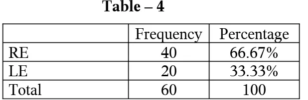
[image:48.595.168.477.345.446.2]66.67% of the study group showed involvement of right eye compared to 33.33% which showed involvement of left eye.
Table – 4
Frequency Percentage
RE 40 66.67%
LE 20 33.33%
Total 60 100
Presentation :
Pain of more than 10 days duration in the affected eye was the chief presentation in 72% of cases
PAIN :
Table – 5
Duration Frequency Percentage
5-10 days 17 28.33%
11-20 days 20 33.33% 21-30 days 13 21.66% > 30 days 10 16.66%
Defective Vision
Table – 6
Frequency Percentage 5 – 10 days 5 8.3 11-20 days 10 16.66 21-30 days 20 33.33 > 30 days 25 41.66
Total 60 100
Maximum number of patients presented with symptoms of defective vision of more than 30 days and symptoms of pain of 11-20 days.
Visual Acuity in the affected eye :
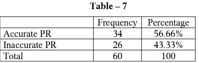
Patients were grouped either as having accurate or inaccurate projection of light rays.
Table – 7
Frequency Percentage Accurate PR 34 56.66% Inaccurate PR 26 43.33%
Total 60 100
Pupils :
[image:49.595.137.472.502.609.2]Table – 8
Frequency Percentage
Acting 16 26.67
Not Acting 44 67.50
Total 60 100
Preoperative IOP levels :
Patients presents with following preoperative IOP
Table – 9
IOP in mm Hg Frequency Percentage
20 – 25 2 3.33
26-30 4 6.67
31-35 5 8.33
36-40 6 10%
41-45 10 16.67
46-50 20 33.33
> 50 13 21.66
Total 60 100
The IOP ranged from 20 to more than 50 mm Hg. Maximum number of eyes had an IOP of more than 45 mm Hg.
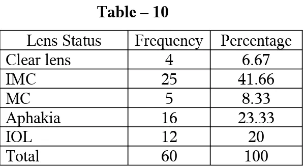
Other eye lens status :
Table – 10
Lens Status Frequency Percentage Clear lens 4 6.67
IMC 25 41.66
MC 5 8.33
Aphakia 16 23.33
IOL 12 20
Maximum number of patients had Immature cataract in the other eye.
Other eye Visual Acuity
Table – 11
Visual Acuity Frequency Percentage 6/6 – 6/60 30 50% 6/60 – 3/60 20 33.33% < 3/60 10 16.66%
Total 60 100
Examination by subgroups
Phacomorphic paitents – Other eye status : Table – 12
Lens Status Frequency Percentage Clear lens 2 5.5
IMC 20 55.55
MC 2 5.5
Aphakia 6 16.67
IOL 6 16.67
Total 36 100
Phacolytic Patients – Other eye status
Table – 13
Lens Status Frequency Percentage Clear lens 2 8.3
IMC 5 20.83
MC 3 12.50
Aphakia 10 41.66
IOL 4 16.66
Incase of phacomorphic glaucomas 55.55% of fellow eye showed immature cataracts
In phacolytic patients majority of the fellow eye was aphakia 41.66%
[image:52.595.71.537.506.688.2]Comparison between angle width in phacomorphic glaucoma cases of fellow eye.
Table – 14
Frequency Percentage AC shallow 14 58.33 AC – Normal depth 10 41.66
Total 24 100
None of the fellow phakic eyes of phacolytic glaucomas had shallow anterior chamber and all those eyes had a wide open angle by gonioscopy.
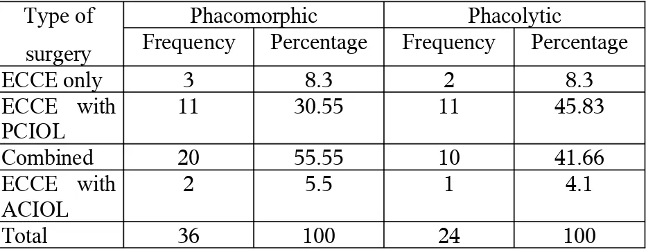
Comparison between the type of surgery performed based on subgroup
Table - 15
Type of surgery
Phacomorphic Phacolytic
Frequency Percentage Frequency Percentage
ECCE only 3 8.3 2 8.3
ECCE with PCIOL
11 30.55 11 45.83 Combined 20 55.55 10 41.66 ECCE with
ACIOL 2 5.5 1 4.1
Total 36 100 24 100
Table – 16
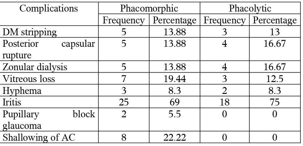
Intraoperative and postoperative complications
Complications Phacomorphic Phacolytic
Frequency Percentage Frequency Percentage DM stripping 5 13.88 3 13 Posterior capsular
rupture
5 13.88 4 16.67 Zonular dialysis 5 13.88 4 16.67 Vitreous loss 7 19.44 3 12.5
Hyphema 3 8.3 2 8.3
Iritis 25 69 18 75
Pupillary block glaucoma
2 5.5 0 0
Shallowing of AC 8 22.22 0 0
Complications were more in lens induced glaucoma BCVA at last follow up by subgroup
Table - 17
Visual Acuity
Phacomorphic Phacolytic
Frequency Percentage Frequency Percentage 6/6 – 6/12 20 55.55 14 58.33 6/18 – 6/60 10 27.77 6 25 < 6/60 6 16.66 4 16.66
Total 36 100 24 100
[image:53.595.66.541.452.579.2]Table – 18
Comparison between type of surgery and visual acuity
Type of surgery Final Visual Acuity Total
6/6 - 6/12 6/18 – 6/60 < 6/60
Simple cataract surgery
18 (60%) 7 (23.33%) 5 (16.66%) 30
Combined surgery 16 (53.33%) 9 (30%) 5 (16.67%) 30
Total 34 (56.67%) 16 (26.67%) 10 (16.67%) 60
[image:54.595.73.545.379.531.2]Visual acuity between simple cataract surgery and combined sugery does n’t showed any significant difference.
Table - 19
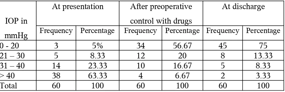
Comparison between IOP in 60 eyes with Lens induced glaucoma
IOP in mmHg
At presentation After preoperative control with drugs
At discharge
Frequency Percentage Frequency Percentage Frequency Percentage
0 - 20 3 5% 34 56.67 45 75
21 – 30 5 8.33 12 20 8 13.33
31 – 40 14 23.33 10 16.67 5 8.33
> 40 38 63.33 4 6.67 2 3.33
Total 60 100 60 100 60 100
At presentation more than 60% had IOP above 40 mm Hg. After treatment with drugs only 6% had IOP above 40 mm Hg.
Table - 20
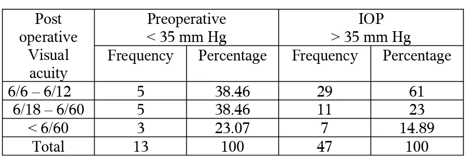
Comparison between BCVA at last follow up by preoperative IOP
Post operative
Visual acuity
Preoperative
< 35 mm Hg > 35 mm HgIOP
Frequency Percentage Frequency Percentage 6/6 – 6/12 5 38.46 29 61
6/18 – 6/60 5 38.46 11 23 < 6/60 3 23.07 7 14.89
Total 13 100 47 100
There was no significant difference between good visual acuity and preoperative IOP.
Table – 21
Relation between duration in days with post operative Visual Acuity
Duration of pain in days
Post operative Visual Acuity
6/6 - 6/12 6/18 – 6/60 < 6/60
5 – 10 days 15 (25%) 2 (3.3%) 0
11 – 20 days 13 (21.67%) 6 (10%) 1 (1.6%)
21-30 days 5 (8.3%) 4 (6.6%) 4 (6.6%)
> 30 days 1 (1.6%) 4 (6.6%) 5 (8.3%)
25% of patients who presented with in 5-10 days have a visual acuity of more than 6/12. But those who presented more than 30 days after only 1.6% had good visual acuity.
Visual Acuity < 6/60 is considered as poor visual acuity
Cause Frequency Percentage Optic Atrophy 3 30%
Uveitis 2 20%
Corneal edema 2 20% Retinal detachment 1 10% Vitreous Haemorrage 2 20%
Total 10 100%
DISCUSSION
Lens induced glaucomas are a common occurrence in India, hardly surprising in a situation where the incident of cataract cases far exceeds the total number of surgeries performed currently. Though these are clinically distinct entities, they have certain common factors in that they are lens induced, they compromise the function of the optic nerve due to rise of intraocular pressure, cataract surgery is curative in these cases, and finally they uniformly share a guarded prognosis.
This study was undertaken to outline the different characteristics of glaucomas, to determine the post operative visual results following simple cataract extraction of combined surgery and to evaluate any risk factors which may play a role in the determination of final post operative visual acuity.
Study was done in 60 eyes with lens induced glaucomas.
series. Velayutham et al in their study reported more number of phacolytic than phacomorphic cases.
The age range for both types of lens induced Glaucomas in this series was 40-80 years with 40% of the cases occurring in patients under the age of 60. More than 60% occurred in patients more than 60. Findings were consistent with study from Nepal and study at Aravind Eye hospital.
Study of GOH reported 99% of cases were in the age group between 51-60. Lens induced Glaucomas was more common in females. (35 eyes ; 58.33%) compared to males (25 eyes ; 41.66%)
In all studies females seemed to have significant increased risk of having these glaucomas. Thought it is possible that these entities are more common in females because of socio economic constraints, we also have to consider the fact that the prevalence of cataract itself is more common in females than males.
The IOP at presentation ranged from 20 to more than 50 mm Hg. Maximum numbers of patients had an IOP of more than 45 mm Hg.
We also documented the status of the lens in the fellow eyes of these patients to see if there was any difference between the subgroups.
Out of the 60 patients majority 41.66% had immature cataract in the other eye, 23.33% had aphakia, 20% had other eye IOL, 8.33% had mature cataract and 6.67 % had clear lens in the other eye.
However examination by subgroups revealed that in cases with phacomorphic glaucomas the status of the fellow eye was predominantly immature cataracts (about 55%) while aphakia contributed (16.67%). Analysis of the fellow eyes of patients with phacolytic glaucoma showed that 41.66% of cases showed aphakia and immature cataracts contributed to about 20% of cases.
Fifty eight percent of the fellow phakic eye of phacomorphic had shallow anterior chamber. None of the fellow phakic eyes of phacolytic glaucomas had shallow anterior chamber.
RESULTS OF SURGERY
Out of the 36 patients with phacomorphic glaucoma 20 patients (55.55%) underwent combined surgery, 11(30.55%) had ECCE with PCIOL. 3 patients ECCE only (8.33%) and 2 (5.5%) patients had ECCE with ACIOL.
In case of phacolytic glaucoma out of 24 patients 11 patients (45.83%) had ECCE with PCIOL, 10 patients (41.66%) had combined surgery, 2(8.3%) had ECCE only and 1 (4.1%) underwent ECCE with ACIOL.
In the Nepal study all cases underwent scleral tunnel sutureless cataract surgery
In the study by Prajana et al done at Aravind Eye hospital all the 93 patients underwent planned extra capsular cataract extraction (ECCE).
Intra operative and post operative complication were more in lens induced glaucomas compared to other types.
stormy in these cases and they were managed by more frequent application of topical steroids and antibiotics.
In our study 69% of patients with phacomorphic glaucoma and 75% of patients with phacolytic glaucoma had iritis.
Posterior capsular rupture was present in 13.88% of phacomorphic glaucoma and 16.67% of phacolytic glaucoma ; zonular dialysis was present in 13.88% of phacomorphic glaucoma and 16.67% of phacolytic, glaucoma. Vitreous loss was present in 19.44% of phacomorphic glaucoma and 12.5% of phacolytic glaucoma.
Pupillary block glaucoma was present in 5.5% of cases of phacomorphic glaucoma. None of the cases of phacolytic glaucoma had this complication. Shallowing of anterior chamber due to positive vitreous pressure was present in 22.22%. Descements membrane stripping, hyphema were equal in phacomorphic and phacolytic glaucomas 13% and 8.3% respectively.
Nepal study also showed increased incidence of subluxation of lens and post operative inflammation which were more in phacolytic glaucoma.
Follow up of the patients were done at one month and 3 months post operatively. The visual acuity at the last follow up was taken as the post operative visual acuity. A corrected visual acuity of 6/12 or better was taken as good visual acuity and accordingly 55.55% of phacomorphic glaucomas and 58.33% of phacolytic glaucomas recovered to a good visual acuity. Six patients (16.66%) with phacomorphic and 4 patients (16.66%) with phacolytic glaucoma had poor visual recovery (< 6/60). There was no significant difference between the two groups on final post operative visual recovery.
In the Nepal study 31.4% eyes achieved 6/18 or better with best correction 21% with visual acuity of less than 3/60.
Out of the total 60 cases in 30 cases simple cataract surgery was done in rest 30 cases combined surgery was done.
Good visual acuity of more than 6/12 was seen in 60% cases with simple cataract surgery and 53.3% cases with combined surgery.
Visual acuity of less than 6/60 was seen on 16.67% of simple cataract surgery and combined surgery.
In the case of phacomorphic glaucoma 55.55% had combined surgery compared to 41.66% in cases of phacolytic glaucoma. The increased incidence of combined surgery in phacomorphic glaucoma is due to gonioscopic evidence of angle closure and extensive peripheral anterior synechiae due to longer duration of attack.
Combined surgery in our cases did not provide significant advantage over simple cataract surgery in the management of Lens induced glaucoma.
In our analysis there was no relationship between height of Intra ocular pressure and final visual acuity.
Out of 13 patients who had IOP less than 35 mm Hg, 38.46% had good visual acuity. 61% of the patients had good visual acuity out of the 47 patients who had intra ocular pressure more than 35mm Hg.
Similar results were obtained in the study of velayutham et al and study by Prajana et al which showed that there was no statistically significant difference on the attainment of good post operative visual acuity and preoperative IOP.
In our study, we found a strong correlation between the duration of attack and find visual acuity. Those patients who presented with in 5-10 days of attack 25% had good visual acuity of more than 6/12.
Those who presented with in 20 days 21.67% had good visual acuity. But those who presented late ie. More than 30 days only 1.6% had good visual acuity and 8.3% had low visual acuity of less than 6/60. Among those who presented within 5-10 days no patients had visual acuity less than 6/60.
proportional to the duration of attack rather than the type of cataract and modality of surgery.
Among the 60 patients 10 had visual acuity less than < 6/60. 30% of patients had optic atrophy. 20% of patients had uveitis, corneal oedema and vitreous haemorrhage.
In the study by Prajana et al also 23% of cases had poor visual acuity less than 6/60. Causes included glaucomatous optic atrophy, severe persistent post operative uveitis and resultant cystoid changes in the macula.
CONCLUSION
In our study mean age of presentation is 60 years Female : Male ratio is 1.4 : 1
Higher incidence of phacomorphic than phacolytic glaucoma
There is no relation between final visual acuity and type of surgery. Increased incidence of complications in both groups.
Visual prognosis is almost similar in both groups after surgery
Post operative visual status and duration of attack had significant association and not the height of IOP.
Preoperative faulty projection of light need not defer definitive cataract surgery.
BIBLIOGRAPHY
1. National program, for control of blindness, vision 2020 Right to Sight ophthalmology Blindness control section. Directorate General of Health Service. Ministry of Health and family welfare, Govt. of India, New Delhi.
2. Diagnosis and therapy of glaucoma 7th edition Becker-Schaffer 3. Glaucomas associated with disorders of the lens – Shield’s Text
book of Glaucoma 5th edition.
4. Glaucoma secondary to lens intumesence and dislocation Ritch R, Shields MB, Krupin T.
5. Duke – Elder System of ophthalmology, Volume XI Diseases of Lens and vitreous, Glaucoma
6. Text book, Albert Jackobie, Principle and practice of ophthalmology 2nd edition.
7. Epstein DL, Diagnosis and Management of Lens induced glaucoma, 1952.
9. Halasa AH, The basic aspects of the Glaucoma, Spring field, Charles C. Thomas 1972.
10. Jain IS, GuptaA, Dogea MR, Gangwae DN, Dhir SP. Pharmorphic glaucoma Management and visual prognosis Indian Journal ophthalmol 31 : 648 – 653, 1983.
11. Murphy GE, Acute phacolytic glaucoma with primary intraocular lens implantation after intra capsular cataract extraction. Am Intraocular implant, SOC J7 : 266-267, 1981.
12. Lazae M, Bracha R, Nemet P, Cataract extraction during acute attack of phacolytic glaucoma. Trans Am, Acad ophthal 1976, 81 : 183-184.
13. Gross KA, Pearce JL. Phacolytic glaucoma with ECCE and Primary IOL implantation. Cataract 11 : 22-23, 1984.
14. Lane SS, Kopietz LA, Lindquist TD, Leavenworth N, Treatment of phacolytic glaucoma with extracapsular cataract extraction ophthalmology 95 : 749 – 753, 988.
16. Leske MC. The epidemiology of open angle glaucoma a review. Am J Epidermoid 118 : 166-191, 1983.
17. Phacomorphic glaucoma – Management and visual prognosis I.S. Jain, Amod Gupta, MR Gogra, DN Gangwa, SP Dhir, Dept. of Ophthalmology, Post graduate Institute of Medical education and Research, Chandhigarh, India.
18. Hyams SW, Angl closure Glaucoma. Comprehensive Review of primary and secondary angle closure Glaucoma. Amsterdam, Kugler and Ghadini 1990, 98-9.
19. Tomey KF. Management of Primary Angle closure glaucoma, Practical consideration. In Reinecke RD ed, Ophthalmology, Annual 1988, Newyork.
PHACOMORPHIC GLAUCOMA
PHACOLYTIC GLAUCOMA
LENS PARTICLE GLAUCOMA
HISTOLOGY OF PHACOLYTIC GLAUCOMA
GLAUCOMA FILTRATION PROCEDURE
(TRABECULECTOMY)
TRABECULECTOMY SHOWING BLEB
PHACOLYTIC GLAUCOMA PATHOLOGY
SUBLUXATED LENS
MASTER CHART
S. No.
Name Sex
A
ge IP No. Type of Glau coma Symptoms Pre op BC VA Pre op IOP Preop. Examination Other eye lens status Gonio other eye
Type of Sx Post op. BC VA Post Op. IOP mmHg Pain
days DV-days AC depth Lens 1
.
Mundiammal F 55 140102 PM 5-10 5-10 PR+ 33 Shallow Intu IMC Closed Combined 6/9 20.6
2 .
Balamani F 58 140548 PM 5-10 11-20 PR+ 45.8 Shallow Intu IMC Closed ECCE with PCIOL
6/36 22.4
3
. Eswari F 65 140308 PL 11-20 11-20 PR- 26.6 Deep HMC IMC Open ECCE with PCIOL 6/12 18.9
4 .
Ayyavu M 75 28533 PL 11-20 5-10 PR+ 54.7 Deep MC Aphakia Open Combined 6/12 22.4
5 .
Periyakarupan M 78 28275 PM >30 21-30 PR+ 49.8 Shallow Intu MC Open Combined 5/60 29.0
6 .
Meenammal F 62 140306 PL 5-10 5-10 PR+ 21.9 Deep MC IMC Open ECCE with PCIOL
6/9 14.6
7 .
Maruthaya F 49 30150 PM 11-20 5-10 PR+ 23.8 Shallow Intu IMC Open ECCE with PCIOL
6/24 17.3
8 .
Pitchaiammal F 72 140333 PM 11-20 5-10 PR- 27.4 Shallow Intu IMC Shallow ECCE with PCIOL
6/36 20.6
9 .
1 0 .
Karuppaiya M 51 140256 PM 21-30 11-20 PR+ 45.8 Shallow Intu IMC Shallow Combined 6/9 14.6
1 1 .
Chinnammal F 57 30403 PM >30 11-20 PR- 49.8 Shallow Intu Clearlen Open Combined 6/24 23.8
1 2 .
Valli F 76 27116 PL 5-10 5-10 PR+ 29 Deep HMC IMC Open ECCE only 6/12 20.6
1 3 .
Gurusamy M 75 30147 PL 21-30 11-20 PR+ 46.9 Deep MC Aphakia Open Combined 6/12 18.9
1 4 .
Perumayee F 71 35412 PL 21-30 21-30 PR- 59.1 Deep HMC Aphakia Open ECCE with PCIOL
6/9 12.2
1 5 .
Krishnammal F 80 32083 PL 5-10 5-10 PR+ 29.0 Deep HMC Aphakia Open ECCE with PCIOL
6/9 16.5
1 6 .
1 7 .
Vellayammal F 52 30149 PM 21-30 11-20 PR+ 46.9 Shallow Intu Aphakia Open Combined 6/12 18.9
1 8 .
Poochammal F 55 28311 PM 21-30 11-20 PR+ 75.1 Shallow Intu IMC Shallow Combined 6/9 18.9
1 9 .
Muthumari F 55 34431 PM 21-30 5-10 PR+ 42.1 Shallow Intu IMC Shallow Combined 6/9 20.6
2 0 .
Duraisamy M 63 30150 PL >30 21-30 PR- 54.7 Deep MC Aphakia Open combined 5/60 34.5
2 1 .
Pappu F 78 34887 PL 11-20 21-30 PR- 34.4 Deep HMC MC Open ECCE with PCIOL
6/9 12
2 2 .
Vellaisamy M 64 14507 PL >30 21-30 PR- 59.1 Deep HMC Aphakia Open ECCE with PCIOL
6/60 18.9
2 3 .
2 4 .
Kaliappan M 61 140280 PM >30 >30 PR+ 49.8 Shallow Intu IMC Shallow ECCE with PCIOL
6/36 17.3
2 5 .
Subbulakshmi F 53 140215 PM 21-30 11-20 PR- 49.8 Shallow Intu Clearlens Open ECCE with PCIOL
6/36 22.4
2 6 .
Veerammal F 58 140524 PM 21-30 >30 PR- 42.1 Shallow Intu IMC Shallow ECCE with PCIOL
6/12 20.6
2 7 .
Gandhiammal F 59 140936 PM >30 11-20 PR- 46.9 Shallow Intu MC Open Combined 6/9 25.0
2 8 .
Nagammal F 65 5402 PL >30 >30 PR- 46.9 Deep MC IOL Open ECCE with PCIOL
6/12 12.2
2 9 .
Chellammal F 79 30142 PL 11-20 >30 PR- 37.6 Deep MC Clearlens Open combined 6/12 14.6
3 0 .
Nagammal F 65 7095 PL >30 >30 PR- 45.8 Deep MC IOL Open ECCE with PCIOL
6/12 12
3 1 .
Thangammal F 62 30404 PM >30 11-20 PR- 75.1 Shallow Intu IMC Open ECCE with PCIOL
3 2 .
Mookusamy M 54 7044 PM 21-30 11-20 PR- 69.3 Shallow Intu IMC Shallow Combined 6/9 14.6
3 3 .
Veeranar M 70 20423 PM >30 >30 PR+ 69.3 Shallow Intu IMC Open Combined 4/60 49.8
3 4 .
Andi M 74 15354 PL 21-30 11-20 PR- 46.9 Deep MC MC Open ECCE with PCIOL
6/12 16.5
3 5 .
Saraswathi F 74 15235 PM 11-20 5-10 PR+ 31.6 Shallow Intu IOL Open ECCE with PCIOL
6/12 20.6
3 6 .
Veeran M 52 20035 PM 21-30 5-10 PR+ 37.8 Shallow Intu IMC Shallow ECCE with PCIOL
6/9 12.0
3 7 .
Kandasamy M 73 15750 PL 21-30 11-20 PR- 46.9 Deep HMC IOL Open Combined 6/9 14.6
3 8 .
3 9 .
Chinnaponnu F 61 17708 PM >30 11-20 PR- 69.3 Shallow Intu Aphakia Open ECCE with PCIOL
6/12 17.3
4 0 .
Saurabeevi F 63 19040 PM >30 21-30 PR- 42.1 Shallow Intu IMC Shallow Combined 6/9 18.9
4 1 .
Petchi F 67 22769 PL >30 >30 PR+ 69.3 Deep HMC Aphakia Open ECCE with PCIOL
4/60 34.5
4 2 .
Arumugam M 76 19056 PL 21-30 11-20 PR+ 37.8 Deep HMC IOL Open ECCE with PCIOL
4/60 38.8
4 3 .
Lakshmi F 68 30776 PL >30 >30 PR- 81.7 Deep HMC MC Open ECCE with PCIOL
6/60 20.6
4 4 .
Mariappan M 64 32081 PM >30 21-30 PR+ 45.8 Deep Intu IMC Open Combined 6/12 20.6
4 5 .
Ganesan M 56 33766 PM 21-30 11-20 PR+ 49.8 Shallow Intu IMC Open Combined 6/12 15.9
4 6 .
Krishnan M 65 20834 PM >30 21-30 PR+ 69.3 Shallow Intu IMC Shallow ECCE with PCIOL
4 7 .
Alagammal F 59 22720 PM 21-30 5-10 PR- 45.8 Shallow Intu Aphakia Open Combined 6/9 14.6
4 8 .
Muthiya M 67 208411 PM >30 >30 PR+ 46.9 Shallow Intu Aphakia Open Combined 4/60 49.8
4 9 .
Ganapathy M 45 32077 PM 11-20 5-10 PR- 33.0 Shallow Intu IOL Open Combined 6/12 18.9
5 0 .
Abdulkhaddar M 43 33758 PM 11-20 5-10 PR+ 37.8 Shallow Intu IMC Open ECCE with PCIOL
6/9 17.3
5 1 .
Karuppayee F 60 28798 PM 21-30 5-10 PR- 45.8 Shallow Intu aphakia Open ECCE with PCIOL
6/9 17.3
5 2 .
Ayyappan M 68 24451 PM >30 >30 PR+ 42.1 Shallow Intu IOL Open Combined 5/60 31.6
5 3 .
Pidaram M 57 140196 PM 21-30 11-20 PR+ 49.8 Shallow Intu IOL Open ECCE with PCIOL
5 4 .
Murugesan M 72 140996 PL 21-30 11-20 PR- 45.8 Deep HMC Aphakia Open Combined 6/12 12.0
5 5 .
Vellathai F 69 33777 PL >30 21-30 PR- 42.1 Deep MC Aphakia Open ECCE with PCIOL
6/24 29.0
5 6 .
Muniyammal F 55 31265 PM 21-30 11-20 PR- 42.1 Shallow Intu IMC shallow Combined 6/36 18.9
5
7
.
Dhanalakshmi F 64 31862 PL >30 21-30 PR+ 45.8 Deep HMC IMC Open Combined 5/60 18.0
5 8 .
Chinakarupan M 56 32879 PM 21-30 11-20 PR+ 69.3 Shallow Intu IOL Open ECCE with PCIOL
6/24 18.9
5 9 .
Ramayee F 70 35109 PM >30 21-30 PR+ 45.8 Deep HMC IOL Open Combined 6/36 14.6
6 0 .
Muthulakshmi F 63 16490 PL >30 21-30 PR+ 46.9 Deep HMC Clear lens
ABBREVATIONS
PM - Phacomorphic glaucoma PL - Phacolytic glaucoma PR - Projection of Light HMC- Hypermature cataract Intu - Intumescent cataract MC - Mature cataract