ANONYMOUS HIV INCIDENCE STUDY IN ROUTINE
AUTOPSY CASES
Dissertation submitted in partial fulfillment of the requirements for
the degree
M.D. (Forensic Medicine) BRANCH - XIV
INSTITUTE OF FORENSIC MEDICINE
MADRAS MEDICAL COLLEGE
CHENNAI – 600 003
THE TAMIL NADU DR. M.G.R. MEDICAL UNIVERSITY,
CHENNAI
BONAFIDE CERTIFICATE
This is to certify that the work embodied in this dissertation entitled
“ANONYMOUS HIV INCIDENCE STUDY IN ROUTINE
AUTOPSY CASES” has been carried out by Dr. R. SANGEETHA,
M.B.B.S., a Post Graduate student, under my supervision and guidance
for her study leading to Branch XIV M.D. Degree in Forensic Medicine
during the period from May 2010 to April 2013
DEAN Director and Professor
Madras Medical College & Institute of Forensic Medicine
Rajiv Gandhi Govt. General Hospital Madras Medical College
Chennai-3 Chennai-3
Date: Date:
DECLARATION
I, Dr. R. Sangeetha., solemnly declare that this dissertation titled
“ANONYMOUS HIV INCIDENCE STUDY IN ROUTINE AUTOPSY
CASES” is the bonafide work done by me under the expert guidance and
supervision of Capt. Dr. B. Santhakumar M.Sc., MD., DipNB (FM),
P.G.D.M.L.E, Director and Professor, Institute of Forensic Medicine,
Madras Medical College, Chennai – 3. This dissertation is submitted to The
Tamil Nadu Dr. M.G.R Medical University towards partial fulfillment of
requirement for the award of M.D., Degree (Branch XIV) in Forensic
Medicine.
DR .R.SANGEETHA
Place:
ACKNOWLEDGEMENT
I would like to express heartfelt gratitude to my esteemed
supervisor, Capt. Dr. B. Santhakumar M.Sc., MD., DipNB (FM),
P.G.D.M.L.E, Director and Professor, Institute of Forensic Medicine,
Madras Medical College, Chennai-3 for his expert guidance in bringing
out this dissertation for my MD examination.
I am greatly obliged to the Dean, Dr. Kanagasabai MD, Madras
Medical College and Rajiv Gandhi Government General hospital,
Chennai-3 for supporting me to conduct this study.
I am especially thankful to Assistant Professors Dr. M. N.
Rajamani Bheem Rao MD, and Dr. T. Vedanayagam MD., DO, for their
interest and encouragement.
I thank all my colleagues for helping, in collecting materials for my
study.
I would like to pay my gratitude to my parents, spouse and my
CONTENTS
S.NO TOPIC PAGE NO
1 INTRODUCTION 1
2 AIM AND OBJECTIVES 4
3 REVIEW OF THE LITERATURE 5
4 HISTORY AND DISCOVERY OF AIDS 15
5 MATERIALS AND METHODS 56
6 ANALYSIS AND RESULTS 63
7 DISCUSSION 68
8 CONCLUSION 73
ABBREVIATIONS
AIDS – Acquired Immune Deficiency Syndrome
CAEV – Caprine Arthritis Encephalitis Virus
CDC – Centres for Disease Control and Prevention
CCR – Chemokine Co-Receptor
CMV – Cytomegalovirus
CRF – Circulating Recombinant Forms
DNA – Deoxy-ribo Nucleic Acid
EBV – Epstein Barr Virus
ELISA – Enzyme Linked Immunosorbant Assay
FIV – Feline Immunodeficiency Virus
GRID – Gay Related Immune Deficiency
HAART – Highly Active Antiretroviral Therapy
HBV – Hepatitis B Virus
HCV – Hepatitis C virus
HIV – Human Immunodeficiency Virus
KS – Kaposi Sarcoma
LAV – Lymphadenopathy Associated Virus
NAT – Nucleic Acid Amplification Test
NNRTI – Non- Nucleoside Reverse Transcriptase Inhibitor
NRTI – Nucleoside Reverse Transcriptase Inhibitor
PCP – Pneumocystis Cariini Pneumonia
PCR – Polymerase Chain Reaction
PEP – Post Exposure Prophylaxis
PGL – Persistent Generalized Lymphadenopathy
PI – Protease Inhibitor
RIBA – Recombinant Immunoblot Assay
RNA – Ribonucleic Acid
SIV – Simian Immunodeficiency Virus
WB – Western Blot
ABSTRACT
Acquired Immunodeficiency Syndrome (AIDS) caused by Human
Immunodeficiency Virus (HIV). The postmortem examination room is
the place where infection can spread to persons who are performing
autopsy. These personnels are having high risk of various injuries,
infections. Forensic personnels who are involved in examination of
homosexuals and drug abusers, are having great chance of being infected
by HIV than common people. We conducted the Study to find the
incidence of HIV in routine autopsy cases.
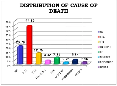
Materials and methods: The study sample consists of 486 routine
autopsy cases at Rajiv Gandhi Government General Hospital. The
samples were tested using an enzyme immunoassay using SD Bioline
HIV1/2 3.0 Rapid test kits to detect the presence of HIV-1 and HIV-2
antibodies. Samples yielding reactive results were confirmed by Alere
DetermineTM HIV 1/2 method which detects HIV-1, HIV-2 antibodies.
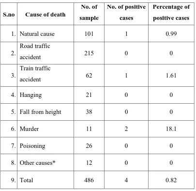
Conclusion: Out of 486 subjects in this study group, 4 cases were
positive for HIV and all were previously unknown seropositive cases.
This shows that HIV screening is of great importance among the
community where the crime rate is high. Homicide victims in our study
showed a relatively higher prevalence of HIV-1, infections compared
INTRODUCTION
Acquired Immunodeficiency Syndrome (AIDS) caused by Human
Immunodeficiency Virus (HIV). HIV is a disorder of the immune system
in which there is a breakdown of defense mechanism against infection,
which leaves the person susceptible to the life-threatening infectious
diseases including unusual malignancies.1
It is estimated that about 14,000 HIV infections occur every day
around the world approximately and out of these, 90% of these are in
developing countries2. HIV is of two types namely HIV-1 and HIV-2,
both of these viruses can cause manifestations, which cannot be
distinguished from each other. The difference remains in the onset of the
disease; HIV-2 has a delay in onset of disease3. Universally, infections
rates are maximum in sub-Saharan Africa and are also are high in
southeast Asia.4
The postmortem examination room is the place where infection can
spread to persons who are performing autopsy. These personnels are
having high risk of various injuries, infections.5,6 Forensic personnels
who are involved in examination of homosexuals and drug abusers, are
having great chance of being infected by HIV than common people.
Forensic experts are exposed not only to scalpels, needles but also to the
Autopsy safety was not of much attention till 1980, it was taken in
to consideration only when HIV infection appeared8. Testing for HIV in
the autopsy room has not been done routinely because samples to be
tested need a special equipped laboratory, well trained technician and also
more time is needed to obtain the results. Therefore a simple, reliable and
rapid means for detecting the antibodies to HIV infection is needed and
this can be useful for screening in the autopsy rooms9. Since there are no clear strategies as when a deceased body should no longer be considered
infectious.9
Only limited data is available regarding the occupational
transmission of the HIV from the corpses to the persons who engaged
directly or indirectly in the autopsy procedures and other individuals who
handle the dead bodies of various stages of decomposition10. The purpose
of this study is to determine whether autopsies of the corpses, which have
been presumed to be low risk groups, are safe or not. Thus, we conducted
a study to identify the seroprevalence of these viruses in a low risk
forensic autopsy population.
Testing HIV antibodies during autopsies has been useful not only
for assessing the risk to the mortuary workers but also useful for the
epidemiological studies11. Many simple, rapid and reliable tests for
hazards in the mortuary, because the HIV may survive for several days in
the postmortem samples. The applicability of the tests for detecting
antibodies to HIV in autopsy samples depends on the postmortem
stability of the antibodies as well as on the sensitivity of the screening
tests12.
In a population, where there is a low HIV prevalence, refusals from
screening for HIV tests by the people with risk behavior may occur and
this strongly bias the seroprevalence. The social habits, human ethical
issues and religious customs becomes an obstacle to obtain the
information about the dead bodies13,14.
Autopsy screening efficiently reveals the true incidence in groups
where refusal may be common. This study enables to identify HIV status
of the deceased whose HIV status was unknown previously because it is
not practical or cost-effective to take full universal precaution with every
AIMS AND OBJECTIVES
1. To Study the incidence of HIV in routine autopsy cases.
2. To create awareness among the doctors doing postmortem and the
mortuary workers.
3. To diagnose the clinically undetected HIV cases.
4. To determine whether postmortem of deadbodies which are
thought to be at low risk groups, are safe or not.
5. To formulate guidelines for a policy concerning HIV antibody
screening in forensic autopsy cases
6. To determine how much autopsies are safe in relation to
pre-autopsy testing for HIV.
REVIEW OF LITERATURE
The situation of Forensic Pathologists to perform autopsies in cases
whose HIV status is not known prior to autopsy. A person infected with
HIV may die of an unrelated cause, in those individuals there is no
clear-cut evidence of HIV status. HIV incidence is the measure of new cases in
a given period of time. This occurrence of new cases helps us to supervise
HIV epidemic in the country. This information also helps us to create or
modify the awareness programme to educate the general population and
high risk community who are vulnerable to HIV infection.
HIV antibodies testing in autopsies have been valuable for
epidemiological purposes15, 16. Consistent tests may also be needed to
reduce the occupational health hazards in the mortuary, because the HIV
may survive for several days in postmortem samples17.
The applicability of the tests for detecting HIV antibodies in
autopsy samples depends on the postmortem stability of the antibodies as
well as on the sensitivity of the screening test. The globulins that contain
antibodies are not affected by postmortem autolysis, or bacterial
contamination 18.
Medico legal autopsies include fatal traffic accidents and sudden
the living population. Moreover, homosexuals and drug addicts with a
high HIV-infection risk often commit suicide or otherwise die violently
and are thus subjected to medico legal autopsy19. Hence, HIV-testing in
medico legal autopsies would be a valuable method in searching for
'hidden' HIV carriers 20, 21.
When the forensic pathologists conduct postmortem on
dead-bodies that are in various stages of decomposition, a thorough autopsy of
the soft tissue and skeleton is necessary to come to a conclusion regarding
the identity and cause of death especially in suspicious cases22.
Irrespective of the stage of decomposition, the chance of exposure to
various infectious organisms in the body fluids and tissues is very high.
An exposure to HCW (Health care worker) at risk of HIV is defined as a
needle-stick or cut in the connective tissue by sharp instruments,
exposure to mucosa or contact with intact or cut skin when the period of
exposure is extended23.
The studies conducted prospectively have assessed that
approximate risk of infection by HIV after a percutaneous needle injury is
at a rate of 0.3%24. In the context of an autopsy it is important that not only blood but also other body fluids that are highly infectious like
amniotic fluid, pleural fluid, seminal fluid, pericardial fluid, peritoneal
Weston and Lober et al. have recognized that rupture of the
surgical glove occurs in about 9% of autopsy. About 33% of glove
puncture goes undetected by the pathology personnels, which can expose
the previous injury in the hand to come in contact with infected blood for
a long time25.
Coleman, D.L. Luce et al (1986)26 conducted a study to detect
antibodies to the Retrovirus in presumably healthy San Franciscans who
died unexpectedly. Of the 121 samples tested for antibodies to HIV, 23
cases were positive for HIV which was very high than other voluntary
screening programs, which shows that abstaining the high risk individual
from testing may cause serious errors in all kinds of voluntary
surveillance programs.
Schoub, B. D., Johnson, et al (1989)27 conducted a study to detect
the HIV seropositivity in sera from medico legal autopsies in
Johannesburg, out of the 745 samples tested 6 (0.8%) were positive for
HIV antibodies.
Dupon, M., Bonnici, et al (1989)28 in his study Screening for HIV
in Necropsies, of the 114 samples tested for HIV antibodies in the city of
Klatt et al (1989) assessed the reliability of postmortem enzyme
immunoassay testing for antibodies to HIV and determined that vitreous
humor is reliable for testing antibodies to HIV up to 34 hours postmortem
and blood atleast up to 58 days were frequently positive for HIV. He
concluded that Postmortem enzyme immunoassay testing of vitreous
humor blood and may be valuable to screen for screening HIV infection
in high-risk groups.40
Little and Ferris et al (1990)29 Tested for the presence of
antibodies to HIV in Forensic autopsies at Vancouver, using the
recombinant immunoblot assay (RIBA) technique. 207 forensic autopsy
cases were tested for the HIV antibodies. Out of which, 172 persons were
without any known history of HIV. In this only 2 cases were positive for
HIV, but the results were not confirmed by further testing. The RIBA
HIV test system has 100% sensitivity and 98.5% specificity. However
this procedure is time consuming and this requires special equipment so it
is not suitable for routine diagnosis for autopsy screening.
Karhunen, et al (1992)14 Detected the HIV antibodies in medico
legal autopsies using an enzyme immunoassay on postmortem sera was
conducted for a period of 5 years. This study consist medicolegal
autopsies of all deaths under the age of 65 years and the study sample was
were previously known case of HIV and remaining 2 were unknown. He
concluded that testing of HIV in medico legal autopsies helps in
identifying the new cases of HIV. These HIV testing have no ethical
issues and may be sensitive to early changes in epidemiology.14
David Sadler et al (1992)30 studied the prevalence of HIV
antibodies using ELISA in the 500 postmortem samples to detect the
antibodies to both HIV1 and HIV2, the positive results were confirmed
by second ELISA. He classified the individual in to risk and no known
risk individual based on the history from police, practitioner and from
hospital records. He concluded that autopsy population contains high risk
individual, therefore testing for HIV antibodies in these individuals is
useful epidemiologically.
Kringsholm, et al (1993)31 Studied the incidence of HIV-1
antibodies among 389 individuals who are addicted to drugs. The study
was conducted in a medical institution at Copenhagen. The incidence of
HIV 1 was almost equal in both male and female. In around one third of
the HIV-1-positive cases, the HIV status was not known previously.
Therefore he concluded that in autopsied drug addicts the diagnosis of
HIV-1-infection is significant for epidemiological data and also for safety
Li, Zhang, Constantine et al (1993)32Studied the Seroprevalence of HBV, HCV, HIV-1 in forensic autopsy, using ELISA to analyze the
risk factors connected with it. A total of 414 serum samples were tested
for HBV, HCV, HIV-1, HTLV-I and HTLV-II antibodies in autopsy
cases successively. Of the 414 cases, about 6% were positive for HIV-1,
19.1% for HCV, 23.2% for HBV, and 1.0% for HTLV-I and HTLV-II.
He established that the overall HIV 1 prevalence was greater than the
general population at Maryland. Routine testing only for HIV-1 may miss
other infections like hepatitis C virus or hepatitis B virus. This study
recommends universal precautions for all autopsy cases.
Lockemann et al (1993)33 studied the HIV-status of 3999 autopsy
cases from 1989-1992 at Hamburg. All the autopsy cases included in the
study were with unnatural death and the cause of death was not known.
Primarily HIV was tested by Enzyme Linked Immuno Sorbant Assay and
confirmed by Western-blot. Confirmation was done in 55 positive cases
using Western-blot. The seroprevalence of HIV was 1.4%. He concluded
that Serological HIV-testing of drug associated deaths helps in
epidemiology screening among high-risk group.
Douceron and Deforges et al (1993)34 conducted the study to
carry out the postmortem safely without the risk of infection by HIV. In
interval. Viable HIV was isolated from the blood sample on 17th day of autopsy, pleural fluid on 14th day of autopsy, and pericardial fluid on 16th day of autopsy. He concluded that there is no time period when autopsy
can be performed without risk of infection to HIV. Therefore delay in
performing postmortem will not abolish the risk of HIV infection.
Henrikki Brummer et al (1994)35 studied the stability of
antibodies to HIV from serum samples and bilious fluid collected from 8
HIV-positive postmortem cases. The postmortem interval of the autopsy
was on an average of 5 days. Serum and bilious fluid samples were stored
at 27°C for 50 to 310 days for testing. Detecting antibodies to HIV in
postmortem samples depends on the stability of the antibodies and
sensitivity of the screening tests. He concluded that HIV antibodies can
be detected for weeks to months in postmortem specimens, even if stored
at room temperature. Postmortem testing for antibodies to HIV in
autopsies thus appears to be a reliable for monitoring the prevalence of
asymptomatic carriers in autopsy series and screening for safety purposes.
Zehner et al (1995)36 conducted the study to detect HIV antibodies
in postmortem blood samples. Before commencing the autopsy, totally
456 samples were collected and was tested using the HIV 1/HIV 2 Test
pack. The samples showing positive result for HIV were confirmed by
positive and it was confirmed by Western blot. The study concludes that
this HIV-Test pack gives proper results to HIV antibodies in whole blood.
Cattaneo et al (1999)37 studied the Prevalence of antibodies to
HIV among the blood samples from the medico legal cases. This study
was conducted in 397 individuals aged between 16 to 50 years. These
cases were tested for HCV and HIV antibodies. Out of 134 individuals
who were tested positive overall, 20 individuals were found positive for
HIV antibodies alone, 69 individuals were found positive for HCV
antibodies alone, and 45 individuals were found positive for both HIV
and HCV antibodies. Cattaneo et al concluded that significant population
had risk of dangerous infection in medico legal autopsies.
Tjotta et al38 demonstrated that HIV survived in dried blood at
room temperature for up to five or six days if the optimum pH level is
maintained. The HIV infectivity in the blood does not appear to be
affected by drying.
Hossein Sanaei-Zadeh (2002)39 studied the seroprevalence of
HBV, HCV and HIV in autopsies, at Tehran. Postmortem blood samples
were collected in 173 cases for a period of 1 year, out of which 8 were
positive for Hbs Ag, and 7 cases were also positive for Hbs Ag and
anti-HCV. No case was positive for anti-HIV 1 and HIV 2. He concluded that
low risk, must be considered as highly and dangerously infectious.
Therefore suitable precaution must be carried out during necropsies.
Kaplan JC, Allan JD, Groopman JE et al40 isolated the viable
HIV from cranial bone, cerebrospinal fluid and brain up to five days after
death.
Nyberg et al41 Studied the viability of HIV in post mortem
samples and concluded that HIV-infected patients should apparently be
considered potentially infectious for at least one or two weeks
postmortem. HIV was cultured from tissue specimens at autopsy up to 6
days post mortem and from spleen specimens stored for 14 days. He
suggested precautions, including for bone, during autopsy of
HIV-infected patients even after extended postmortem intervals.
Johnson et al42 documented occupational transmission of HIV to a
forensic pathologist in the US who sustained injury with scalpel.
Eriksen MB et al (2009)43 reported the Postmortem Detection of
HBV, HCV and HIV genomes in the blood samples among deaths in drug
addicts in Denmark with rapid kits. He collected the blood samples from
autopsy cases and screened for HIV antibodies and hepatitis antibodies.
The screening of viral genome was performed by polymerase chain
40% of samples. Hepatitis B virus genome was detected in about 20% of
anti-HBc – positive/anti-HBs-negative samples.
Sonia Mehta et al (2012)8 conducted Pre-testing Screening for
HIV antibodies before Conducting Post-mortem. A total of 328 samples
of blood were collected prior to the autopsy and tested for antibodies to
HIV for a period of three years. The study was routine confidential and
anonymous testing for HIV antibody. Only 2 samples (0.6%) were found
to be HIV reactive, average postmortem interval was about 24 hours.
None of the cases had a known HIV status. This study concluded that
universal precautions as suggested in autopsy cases not practically
possible in a developing country like ours, therefore such pretesting may
HISTORY AND DISCOVERY OF AIDS
DISCOVERY OF HIV:
AIDS was first recognized in the year 198144, with reports of a
sudden unexplained outbreak of two very rare diseases, Pneumocystis
cariini pneumonia (PCP), and Kaposi sarcoma (KS). It is usually seen
among drug addicts and homosexuals. They lost their immunity and
becoming more prone to many opportunistic infections and other
secondary neoplasms. Many cases of PCP and KS emerged in the year
1981 and it alerts the CDC and this made the CDC to observe the
outbreak45.
Initially, the CDC has not coined official name for AIDS. It was in
the year 1981, the term GRID was used by the general press, which
stands for gay related immune deficiency 46. Later the CDC had coined
the name as "the 4H disease" as it mean for Haitans,
Homosexuals, Haemophiliacs and Heroin users47. Later it was determined
that AIDS was not only related to the gay community alone. It was
recognized that the name GRID was deceptive, therefore the
name AIDS in the year 1982. Then CDC uses the name Acquired
In 1983, Robert Gallo in United States and the French scientist Luc
Montagnier from the Pasteur Institute of Paris independently stated that a
retrovirus is the cause of AIDS. Gallo claimed that a virus isolated from
an AIDS patient was similar to other (HTLVs). Robert Gallo gave the
name for this newly isolated virus as HTLV-III49.
Montagnier's et al isolated a virus from a individual presenting
with, classic symptoms of AIDS. Montagnier revealed that core proteins
of the isolated virus were immunologically different from those of
HTLV-I and named that isolated virus as lymphadenopathy-associated
virus (LAV)45. During the commencement of 20th century HIV appeared
first in humans in Africa as a result of SIV infection.
Serological tests were available for the anti-HIV antibodies
detection. The estimation of the extent of HIV infection is made more
realistic only after the serological tests available. The generic name
human deficiency virus (HIV) 3 was finalized in 1986 by international
committee on Virus Nomenclature.
HIV-1 and HIV-2 are the result of several cross-species
transmissions of simian immunodeficiency viruses (SIVs) naturally
infecting African primates. Both HIV-1 and HIV-2 seemed to have
known as zoonosis; it was transferred to humans in the early 20th century50.
The origin of HIV-1 is through the evolution of SIV, a simian
immunodeficiency virus in southern cameroon that infects chimpanzees51,
52
. SIV is the closest relative of HIV-2, a virus of the sooty mangabey
residing in West Africa53. HIV-1 is believed to have skipped the species
barrier on three distinct occasions, giving rise to the three groups of the
virus namely, M, N, and O53.
It is believed that human beings who take part in chopping meat of
monkeys, either as vendors or hunters, usually get SIV54. Simian
immunodeficiency virus needs several transmissions from one person to
another person in rapid succession to give sufficient time for mutation to
HIV55.
The earliest well recognized case of HIV in human being dates
back to 1959 in the Congo56. It have been existing in the United States as early in 196657, but the infections outside the Africa can be found back to a single person who got infected with HIV in Haiti and then
transported the infection to the United States around57.
Among HIV-1 and HIV-2, HIV-1 is closely related to virus that
sub-species Pantroglodytes have been established to be the natural reservoir of the HIV-1 M (Major) and N (New) groups. The HIV-1 O
(Outlier) group is related to the viruses that originate in Cameroonian
gorillas58.
The M group consists of nine subtypes, named as A, B, C, D, F, G,
H, J, and K and circulating recombinant forms (CRFs) 59. By infection of
an individual with two different subtypes to create a new virus with a
STRUCTURE OF HIV:
HIV belongs to a retrovirus class of viruses. Within this class, it
comes under the subgroup called lentivirus. Some other lentiviruses
include Simian Immunodeficiency Virus (SIV), Feline Immunodeficiency
Virus (FIV), Visna and (CAEV) Caprine Arthritis Encephalitis Virus.
Mostly viruses store their genome as long strands of DNA but
Retroviruses are composed of RNA. HIV is an enveloped, spherical,
icosohedral virus about 90-120 nm in size. The outer envelope consists of
lipid bilayer with regularly arranged spikes or knobs which are 72 in
number. The outer envelope of HIV-1 has gp120/gp140, whereas the
outer envelope of HIV2 has gp41/gp36.Both are responsible for HIV
infection.
The virus core contains
(1) capsid protein p24;
(2) nucleocapsid protein p7 / p9-Regulation of gene expression
(3) 2 copies of genomic RNA; and
(4) 3 enzymes (protease, reverse transcriptase, and integrase) for
viral replication and maturation.
The viral antigen p24 is used for the diagnosis of HIV infection in
viral is bounded by a matrix protein called p17, which lies underneath the
envelope.
The diploid genome composed of 2 identical single-stranded,
positive sense RNA copies. When the virus infect the cell ,viral RNA is
transcribed by the enzyme reverse transcriptase , into single stranded
DNA and then to double stranded DNA (Pro virus) which is integrated
into the host cell. This provirus remains latent for long period and in
response to viral promoters it commences the viral replication by the
synthesis of viral RNA.
The HIV-1 RNA genome contains the gag, pol, and env genes,
translated primarily into large precursor proteins and then cleaved by the
viral enzyme protease to produce the fully matured proteins. HIV
comprises numerous accessory genes such as vif, nef, tat, rev, vpr, and
vpu, which control the synthesis and assembly of infectious viral particles and its pathogenicity.
TYPES:
HIV is a highly inconsistent virus which readily mutates according
to the necessity. There are different strains of HIV in different individuals
and many strains of HIV can be seen within the body of the same
individual. HIV can be classified in to two types genetically and
antigenically. On the basis of genetic similarity, HIV strains can be
classified further into groups and subtypes.
HIV-1 is worldwide predominant while HIV-2 is found mainly in
West Africa but rarely elsewhere. Both types are transmitted in the same
way, and they cause clinically indistinguishable AIDS. The transmission
of HIV-2 is less compared to HIV-1, and the incubation period which is
between the time of infection to appearance of symptoms of illness for
SUB-GROUPS AND SUB-TYPES:
The HIV-1 strains can be categorized into four Subgroups: the
major group M, the outlier group O and two new groups, N the new and
another strain P. The strain P was found in 2009 closely linked to gorilla
simian immunodeficiency virus.
Group M constitutes greater than 90 percent of HIV-1 infections
and are responsible for world pandemic. In group M there are nine
genetically distinct subtypes. Based on nucleotide sequence analyses of
the env and gag genes, within the M group of HIV-1 there are also at least
10 different subtypes they are designated from A to K. 60
Type C is the predominant form not only in India but caused the
most infections worldwide. The Subtype B is most prevalent in the
Americas and Europe, but globally subtypes C accounts for half of all
strains62. The reason for high prevalence of subtype C is due to the
predominance of it in Africa and India. Subtype D is more virulent due to
its effective binding to immune cells61. The Type O HIV-1 is often found
in Cameroon and Gabon and the rare sub-group N is also found in
Cameroon.
HIV-2 infection differs from HIV-1 in being inherently resistant to
have low viral load, lower rates of vertical transmission, slower decline in
CD4 count and several fold slower progression to AIDS. Rarely, many
subtypes can meet in the cell of an infected person and combine their
genetic material to generate a "circulating recombinant forms" orCRFs is
MODES OF TRANSMISSION:
HIV infection is a communicable disease and can be transmitted
from person to person, most commonly by having unprotected sexual
intercourse. HIV is present in blood, semen and other body fluids such as
breast milk, urine, saliva, sweat, tears, pre-ejaculated fluid and vaginal
fluid and can be transmitted by various methods. Worldwide,
heterosexual transmission being the major route (>75%) and vertical
transmission (5-10%)63.
SEXUAL CONTACT:
HIV is a sexually transmitted disease (STD) and it seems to be
transmitted from female to male with greater effectiveness than from
male to female. It can be transmitted through unprotected sexual act from
an infected individual. The risk of becoming infected with an act of
unprotected sexual practice depends on the amount of virus in the body
fluids, the presence of other sexually transmitted diseases.
VERTICAL TRANSMISSION:
HIV positive women can transmit HIV to her child either during
pregnancy or during the process of delivery or by the route of mother’s
milk. The HIV infected pregnant woman can transmit to fetus or newborn
with effective interventions. HIV is thought to be transmitted during the
last weeks of pregnancy or during delivery63.
OCCUPATIONAL EXPOSURE:
An occupational exposure may place a worker at risk of HIV
infection through injuries such as those involving a potentially
contaminated needle or sharp instrument or chapped, abraded skin or
contact with mucous membranes. The risks for occupational transmission
of HIV differ with the type and severity of exposure. Seroconversion is
defined as a situation in which seronegative health care worker sustains
an injury with a device soiled with blood or body fluids from a HIV
seropositive or unknown source and seroconverts within the succeeding 6
months.
The average risk for HIV transmission after percutaneous exposure
to HIV-infected blood has been expected to be about 0.23%64. Average
risk after a mucous membrane exposure is estimated to be approximately
0.09% Factors associated with an increased risk of transmission
comprises: deep injury, injury produced by a device that penetrates a
blood vessel, hollow-bore needle injury, and a source with high viral
NEEDLE SHARING:
Injectable-drug users share the same needles where the blood
contaminated needle of one person come into direct contact with other
individual. Sharing contaminated needles with individuals infected by
HIV will infect the new individual, due to the direct contact of blood of
infected individual. Accidental needle prick injuries are common in
health care workers but the risk of HIV transmission through needle prick
injury is less than 1 percent.
DIFFERENCES IN TRANSMISSION AMONG SUBTYPES:
Specific subtypes or CRFs of HIV are specifically related to
specific mode of transmission1. The Subtype C and CRF A/E are spread
mostly by heterosexual contact, while subtype B is spread mostly by
homosexual contact and drug addiction.
Percentage of risk in different modes:
• Sexually – about 0.5%
• Parenterally – about 90%
• Vertical transmission – 15-40 %
• Drug addiction - 0.5-1.0%
PATHOPHYSIOLOGY OF HIV:
After the entry of virus inside the human body there is a
rapid replication of virus resulting in a great quantity of virus in the
peripheral blood. There are several million virus particles per milliliter of
blood during primary infection. It is accompanied by reduction in the
circulating CD4+ T cells. This is followed by the activation of CD8 T
cells, which kill HIV-infected cells, and subsequently produces the
antibody, known as sero conversion period. When the CD8+ T cell
response is good, the disease progression is slowed down and a better
prognosis is obtained.
HIV causes AIDS by depleting CD4+ T helper lymphocytes which
weakens the defense mechanism and makes the individual susceptible to
opportunistic infection.
In the acute phase HIV induces lysis of cells and killing of infected
cells by activation of cytotoxic T cells occurs which accounts for
depletion of CD4 T cell. In the chronic phase, the generalized activation
of defense mechanism with the gradual loss of the capacity of the body to
generate new T cells results in slow decline in CD4+ T cell numbers.
The characteristic symptoms of AIDS does not appear for many
first few weeks of infection, particularly in the mucosa of the intestine,
which harbors most of lymphocytes present in body. HIV lodges and
abolishes CCR5 expressing CD4+ T cells during acute infection.
NATURAL HISTORY OF HIV/AIDS:
HIV infection has 4 stages:
1) Primary infection
2) Early immune deficiency
3) Intermediate immune deficiency
4) Advance immune deficiency
1) Primary infection
There is a fast proliferation of the virus in the lymph nodes and
blood following HIV infection. In this stage there is a sero conversion
illness which resolves within weeks. There is a rapid declination of the
CD4 count before the virus is controlled by the immune system.
2) Early immune deficiency
In this stage, the immune system has controlled the virus
particularly the lymphoid tissue and people who are infected with HIV
3) Intermediate immune deficiency
There is a rapid replication of virus and CD4cell turn over and
CD4 cell count will be around 200-500 count/microlitre, Signs and
symptoms begin to appear in this stage.
4) Advance immune deficiency
The virus proliferates all over the body and overcomes the defense
system. Oppurtunistic infections and chance of malignancies are common
CLINICAL FEATURES OF HIV INFECTION:
The WHO system uses the following classifications:
STAGE 1 : Primary infection,
STAGE 2 : Clinically asymptomatic stage,
STAGE 3: AIDS-related complex or persistent generalized
lymphadenopathy,
STAGE 4 : AIDS.
STAGE 1: Primary HIV infection
This primary HIV infection lasts for a few weeks and is frequently
accompanied by a flu-like illness for a short period. This is seen in 10%
of individuals and matches with seroconversion. They present with
mononucleosis like illness which includes sore throat, fever, lymph node
enlargement, joint pain, skin rash, and malaise.
In this stage, the defense mechanism of the body responds to the
virus by producing antibodies to HIV a process called seroconversion.
Large amount of virus will be present in the peripheral blood in this
STAGE 2: Clinically asymptomatic stage
As the name indicates, this stage is without major symptoms, and it
lasts approximately for ten years. In the peripheral blood, the HIV falls to
low levels but still the people remain infectious. Antibodies to HIV may
be detected in blood, showing positive result. Children will have a shorter
incubation period. Only lymph node enlargement may be present in this
stage.
STAGE 3: AIDS-related complex
In this stage the immune system becomes impaired severely. This
is due to:
¾ Impairment of the lymph nodes.
¾ HIV mutation, leading to T helper cell impairment.
¾ Failure to replace T helper cells that are lost.
This stage is usually characterized by multi-system disorder which
comprises of immunology, dermatology, hematology and nervous system.
Constitutional symptoms, such as weight loss, fever and night sweats,
STAGE 4: AIDS
The period of clinical latency differs in time from 1 to 2 years to
more than 15 years .The transition from stage 3 to full-blown AIDS may
happen quickly or slowly. The progression of the disease is predisposed
by stress, cofactors, and genetic factors. The poor prognostic factors
include decrease in the CD4 lymphocyte count and the reappearance of
HIV antigen in the blood. The individual may develop gradually severe
opportunistic infections as the immune system becomes severely
damaged, leading eventually to a full blown disease. In this stage,
opportunistic infections such as protozoa, fungi, bacterial and viral
diseases are seen in patients with AIDS.68
OPPORTUNISTIC INFECTIONS
PROTOZOAL
Toxoplasmosis
Pneumocystis Carinii
Cryptosporidiosis
BACTERIAL
Mycobacterium avium complex
Atypical mycobacterial disease
Salmonella septicemia
Multiple or recurrent pyogenic bacterial infection
FUNGAL
Candidiasis
Crytococcosis
Histoplasmosis
Coccidiodomycosis
VIRAL
Cytomegalovirus
Herpes Simplex Virus
Varicella Zoster Virus
Opportunistic infection is caused by bacteria, protozoa, fungi, and
viruses in patients with AIDS. Pneumocystis carinii causes pneumonia is
the most common opportunistic infection in patient with AIDS. The
classic symptoms are breathlessness, cough and fever. Diagnosis includes
bronchoscopy and the detection of Pneumocystis in the lavage.
Toxoplasmosis is dangerous because it affects brain. The treatment
is a pyrimethamine and sulfonamide combination. Cryptosporidium often
causes persistent diarrhea.
The second most common opportunistic infection in a patient with
AIDS is candida albicans. Oesophageal candidiasis is most commonly
seen in patients with pre-AIDS. Treatment is with amphotericin or
ketaconazole. Cryptococcus neoformans infection is fatal, if untreated.
Diagnosis may be made earlier by the demonstration of the fungal antigen
or fungus in the cerebrospinal fluid.
The commonest viral infections during HIV are caused by Herpes
Simplex Virus, Cytomegalovirus, Varicella Zoster Virus. CMV causes
retinitis and pneumonitis. HSV and VZV infections can be treated by
acyclovir.
OPPORTUNISTIC TUMOURS
The common tumour is Kaposi's sarcoma which is vascular in
nature is seen in 20% of patients with AIDS. In an average, 35% of
patients have diseases of the mucosal membrane of the oral cavity,
larynx, and most commonly the oesophagus, stomach and the intestines
are affected. Excision and local management with vincristine are the
Lymphomas of the HIV-infected individuals differ from other
known lymphoma individuals by their site, degree of malignancy, and
response to therapy. HIV-associated lymphomas are often found outside
the lymphatic system, mostly in the brain, GI tract, bone marrow, and
skin. Their response to treatment is less compared to that of classical
lymphomas.
NEUROLOGICAL MANIFESTATIONS
Neurological changes present in HIV-infected patients ranges from
mild neuropsychological symptoms such as memory loss, mood
disturbances and behavior abnormalities to organic psychosis and
dementia. The most common neurological disorder is subacute
encephalitis (AIDS dementia complex and AIDS encephalopathy) which
is present in 33% of cases. Other manifestations are peripheral
neuropathy, aseptic meningitis, and acute meningoencephalitis.
DERMATOLOGICAL MANIFESTATIONS
Skin manifestations are seen in all stages of HIV infection which
can be the first manifestation which makes the patient to give medical
attention. Oral hairy leukoplakia, Seborrhoeic eczema and pruritis with
maculopapular eruption are frequently observed in AIDS. Acne and
molluscum contagiosum, herpes zoster, condylomata acuminatum are
also frequently present.
GASTROINTESTINAL MANIFESTATIONS
Persistent diarrhoea which is profuse is a recurrent problem in
AIDS patients. Shigella, Salmonella, Giardia lamblia, Entamoeba
histolytica, and Campylobacter are responsible for the persistent diarrhea.
Other organisms such as Cryptosporadium, Mycobacterium avium or
Karposi's sarcoma may also present with persistent diarrhea in patients
COURSE OF HIV INFECTION:
Typical progressors
Rapid progressors
Longterm – nonprogressors (LTNP s)
Typical progressors :
Typical progressors constitute an average of 85% of HIV infected
individuals with a survival time of about 10 years. It has three phase’s
namely primary infection, clinical latency and clinically apparent disease.
Progression of HIV to full blown AIDS occurs in period of 8-10 years
approximately.
Rapid progressors:
Rapid progressors constitute about 10% of HIV infected
individuals with a median survival time of about 3-4 years after
seroconversion. Progression of HIV is rapid in these individuals because
of defective immune response and CD8 cell mediated suppression.
Longterm-nonprogressors(LTNP s):
About 5% of the HIV infected individuals are called Longterm –
non progressors (LTNPs). These individuals have stable CD4 cell count
is usually more than 500. They have a high titre of neutralizing antibodies
to HIV. Cell mediated immune response and humoral mediated immune
responses are high in these persons. Viral load and replication of virus is
four fold higher in these individuals in the lymph node and blood.61
REPLICATION CYCLE OF HIV:
The HIV life cycle includes the series of events that starts from the
attachment of virus to the CD4 receptors on the host cell surface, and
ends with the release of new viruses that bud off from the new host cell.
HIV can infect several cells in our body, but its target is the CD4
lymphocyte. When a CD4 lymphocytic cell is infected with HIV, it
undergoes many steps to replicate and generate many new viruses.
The replication of HIV can occur inside the human cells only. The
replication process begins when a virus particle enter into a cell that
carries on its surface a special protein called CD4. The virus particle
contains spikes on its surface which stick to the CD4 and permit the viral
envelope to fuse with the cell membrane. The HIV genomic particle then
gets released into the cell, and leaves the envelope on the surface of the
Binding and Fusion:
The surface membrane of living cells consists of complex protein
structures called receptors. The HIV life cycle begins when the HIV
binds to the CD4 receptor and its co-receptors on the membrane surface
of a CD4 T-lymphocyte. Then it fuses with the host cell, thereby
releasing RNA into the host cell. The firm attachment of the viral
particle to the CD4 receptor helps in fusion of HIV with the cell
membrane and release of its genome into the host cell.
Reverse Transcription:
The enzyme called reverse transcriptase which reads the sequence
of viral RNA nucleic acid and converts the single – stranded HIV RNA to
double-stranded HIV DNA.
Integration:
After the reverse transcription of viral RNA, the HIV DNA enters
the nucleus of the host cell; the enzyme integrase hides the HIV DNA.
The integrated HIV DNA is called provirus which becomes inactive for
many years, making copies of HIV66.
The transcription occurs only when the lymphocyte is activated.
After receiving a signal, the provirus utilizes the host enzyme RNA
polymerase, to make copies of the HIV RNA, and also shorter strands of
RNA named messenger RNA. The mRNA acts as a blueprint to create
long chains of HIV proteins.
Assembly:
The Protease enzyme splits up the long chain of HIV proteins into
small proteins. When the smaller HIV proteins and the HIV genomic
materialcome together, a new virus particle is formed.
Budding:
The virus which is formed newly will be pushed out of the host cell
which is called as budding. The new formed virus takes a portion of the
cell's outer envelope which is covered with protein and sugar
combinations called HIV glycoproteins. These glycoproteins are essential
for the virus to bind CD4 and co receptors. The new HIV copies can
infect other cells.
HIV replication is related with a high mutation rate because reverse
transcription does not permit for rectification of errors in nucleotide
evolution thereby making tough for the immune system to identify and
efficiently combat.67
LABORATORY DIAGNOSIS OF HIV:
HIV diagnostic testing has become available for the detection of
antibodies to HIV only in the 1980s. To detect antibody as early as
possible after infection, the enzyme immunoassays are sensitive. To
confirm positive antibody screens, a variety of other assays are available.
Purpose of HIV testing69
• For prophylaxis and management.
• For blood safety and donation safety.
• For monitoring the epidemic (sentinel surveillance)
• Identification of asymptomatic individuals.
• To encourage for behavior modification through counselling.
• To diagnose clinically suspected cases.
• Voluntary testing after counseling
SEROLOGICAL TEST INCLUDES:
a) Antibody tests
(a) Antibody tests:
For screening of blood samples for antibody to HIV Enzyme
Linked Immunosorbent Assay (ELISA) is the most commonly performed
method. The sensitivity and specificity of the present systems reaches
100% but false positive and false negative results cannot be excluded.
Other antibody tests are Western blots, immunofluorescence, passive
particle agglutination, and RIPA bioassays. Western blots are considered
as the gold standard and seropositivity is diagnosed when antibodies
against both the env and the gag proteins are detected69.
(b) Antigen tests:
Before the appearance of antibody to HIV; antigen can be detected
early in the course of HIV infection. Antigen is not detectable during the
latent period of HIV infection but become it becomes detectable during
the last stages of the infection.
Demonstration of viral nucleic acid:
This can be accomplished by probes or by PCR techniques. The
Polymerase chain reaction may be exceptionally useful because of its
Methods to detect specific HIV antibodies:
i. Screening tests
ii. Supplemental tests
SCREENING TESTS:
The Enzyme-Linked Immunosorbent Assay (ELISA) usually takes
2-3 hours to yield the results. Rapid screening tests give results within
minutes and include visual assays like dot blot tests, particles (gelatin,
RBC, latex, micro beads) agglutination, HIV spot and comb test and
fluorometric micro particle technologies. These tests are easy to perform,
rapid, do not require sophisticated equipment, technical expertise and are
mostly cost-effective. They also distinguish HIV-1 and HIV-2
antibodies.70
ELISA
The Enzyme-Linked Immunosorbent Assay is the most commonly
performed screening test which is easy to carry out for a large sample
size. It is highly sensitive, specific and cost-effective. The
Enzyme-Linked Immunosorbent Assay is a technique used to detect the presence
of an antigen/antibody in a specimen sample. It utilizes two antibodies,
one is specific to the antigen and the other is coupled to an enzyme. The
produce a signal. It is a device for determining serum antibody as well as
the antigen. ELISA based on indirect method is used in most of the kits.70
SUPPLEMENTAL TESTS:
The samples which are reactive in the screening tests were
confirmed with the help of supplemental tests for the confirmation of
diagnosis.
When the sample is reactive by any one of the screening tests, it is
tested by another different system to confirm the diagnosis. If a specimen
is reactive in 2 different systems, it has to be tested again using one of the
supplemental tests which may be a third ELISA Enzyme-Linked
Immunosorbent Assay or Rapid test or a Western Blot test (WB).
WESTERN BLOT
For detecting HIV antibodies, the Western blot is the most
commonly performing confirmatory assay. It is considered as the gold
standard for confirmation of HIV infection. This technique is based on
electrophoresis which denatures the proteins of the viral components and
imparts negative charge to the antigens, and separates the antigens on the
basis of their molecular weight. The separation of antigens permits the
RAPID ASSAYS:
¾ Particle agglutination
¾ Dotblot assays
¾ Immunochromatography
¾ HIV spot and comb tests
¾ Dipstick and comb assays
There are many rapid assays which work on the principle of
agglutination and Enzyme-Linked Immunosorbent Assay has been
developed for quick results and easy performance. These rapid assays
usually require 30 minutes or lesser time for performing the test and does
not require any special equipment’s.70
DOT BLOT ASSAYS
This rapid assay is easy to perform, and it can generally distinguish
between HIV-1 and HIV-2 and do not require complicated equipments.
The results are indicated by development of color. One of the drawbacks
of the test is the high cost. Sensitivity and specificity of these assays are
comparable with ELISA. These assays are beneficial for single test
application (e.g: autopsy room and blood banks, emergency).
The immune chromatographic assays are 1-step rapid assay test kit,
or paper. Blood, serum or other body fluid is placed at the tip of the
device and are allowed to diffuse along a strip that is impregnated with
chemicals mostly colloidal gold that bind and permit visual detection of
HIV antibodies some use third-generation. These tests can be completed
in less than 10 minutes and even less than two minutes, no need for
addition of reagents, and has control reagent to reduce technical errors.
These can be stored at a different temperature and are transported simply
without any consequences.
Based on type of HIV antigen, various types of HIV kits are
available. First generation kits utilises antigens derived from viruses
grown in human lymphocytes. Second generation kits utilise artificially
derived recombinant antigens from fungi or bacteria. Third generation
kits use chemically manufactured oligopeptides.
Laboratory diagnosis during window period:
Window period is a period of seronegativity during which an
infected person do not give a positive result to either Western blot or the
Enzyme-Linked Immunosorbent Assay, even though the viral load is
high. These individuals may show certain symptoms and this period can
During window period HIV infection can be detected by viral
demonstration and viral components by:
• Polymerase Chain Reaction
• p 24 antigen test
• Culture of virus69
• Nucleic acid amplification test (NAT) which detects viral nucleic
acid.
Other laboratory findings for HIV infection may include
leukopenia, anaemia and thrombocytopenia. Increased ESR, increased
blood gammaglobulin, and decreased blood cholesterol level. Post
exposure testing is suggested at six weeks, three months, and then after
six months.
The important target of HIV replication is CD4 cell which results
in depletion of CD4 cells. Therefore CD4 cell count is a marker for
staging the HIV infection and monitoring the disease progression .The
rate of HIV replication is also reflected by increase in plasma viral RNA
load and is considered as one of the most specific and sensitive test to
VIRAL LOAD TEST:
Viral load test is defined as the number of copies of HIV in 1ml of
blood specimen. The viral load tests determines the amount of HIV RNA
in a small quantity of blood .The viral load test is valuable for managing
therapy response, evaluation of newly diagnosed HIV infection,
surveillance of patients not receiving drug therapy. It will help to predict
TREATMENT
There is no cure for AIDS. The target of antiretroviral treatment is
to bring the viral load in the body to a low level by suppressing the
replication of the HIV virus in the body. This helps to reduce the
weakening of the immune system and allows it to recover from damage
that HIV might have caused previously. Combination of three or more
than three anti-HIV drugs is referred to as Highly Active Antiretroviral
Therapy (HAART). The most common drug combination comprises of
two nucleoside reverse transcriptase inhibitor with either a
non-nucleoside reverse transcriptase inhibitor or a protease inhibitor71.
Highly Active Antiretroviral Therapy (HAART) has improved life
expectancy and prognosis in patients with HIV infection. There has been
80% decrease in mortality after its introduction. Sometimes there may be
resistant to one combination of Highly Active Antiretroviral Therapy
(HAART), especially in patients who skip their medications.
Antiretroviral drugs in general fall into the following groups72:
• Nucleoside analogues – Inhibits the reverse transcriptase enzyme
thereby preventing replication
• Protease inhibitors – Prevent viral protein processing.
• Fusion inhibitor – Inhibit the fusion of viral and cellular membranes.
Lifestyle factors such as exercise, diet, and stopping smoking, and
recreational drug use are also important in HIV management.
POST EXPOSURE PROPHYLAXIS OF HIV:
Post-exposure prophylaxis refers to the management to reduce the
risk of infection following exposure to blood-borne infectious organisms.
At present there are only two ways to diminish the risk of developing
HIV infection following exposure to HIV post-exposure prophylaxis
(PEP) and interventions to prevent transmission from mother-to-child.74
Post-exposure prophylaxis (PEP) comprises taking anti-HIV drugs
as soon as possible following exposure to HIV to decrease the chance of
becoming HIV positive. The aim of post-exposure prophylaxis (PEP) is
to decrease or prevent local viral replication prior to spread of infection,
so that the infection can be abandoned.
There are two categories of post-exposure prophylaxis (PEP):
(1) Occupational PEP, (“oPEP”)
Occupational exposure (oPEP) is said to have occurred when a
person working in a medical setup is exposed to materials which is
infected with HIV. nPEP is when a person is exposed to HIV in some
other situations like (condom breakage, sexual assault, etc.) To be
effective, PEP must begin within 72 hours of exposure, before the viral
replication. PEP consists of 2-3 antiretroviral medications and should be
taken for 28 days. In addition counseling should be provided because of
the psychosocial issues.74
POST EXPOSURE PROPHYLAXIS REGIMEN73:
Basic two drug regimen (For low risk)
Zidovudine 300 mg + Lamivudine 150 mg - Twice daily for 4 weeks
Expanded three drug regimen (For high risk)
Zidovudine 300 mg + Lamivudine 150 mg - Twice daily for 4 weeks
+ Indinavir 800 mg (or another PI) - Thrice daily for 4 weeks
LOW RISK:
¾ When the source of infection is positive for HIV, but asymptomatic
with low HIV-RNA titre and high CD4 cell count.
¾ When the exposure is through the mucous membrane, or
HIGH RISK:
¾ When the source is symptomatic AIDS patient with high HIV
-RNA titre or low CD4 count.
¾ Exposure is through major splash or large area contact of longer
duration with mucous membrane or abraded skin or through small
gauge needle, puncture wound which is deep, and patient’s blood
on the needle.
UNIVERSAL PRECAUTIONS DURING AUTOPSY83:
Universal precautions are intended to follow for blood, vaginal
secretions and semen as well as to peritoneal, pericardial cerebrospinal,
synovial, pleura fluids, and it is believed that tears, nasal secretions,
sweat, sputum, urine, faeces and vomitus and amniotic fluids are not
infectious unless they contain visible blood.
Only experts and health care professionals who are skilled in
handling the infected material are allowed to enter in to the postmortem
examination room. Only these experienced professionals should conduct
the autopsy examination because it is proved that the risk of accidental
infection occurs more common in inexperienced persons. Studies
reported that laceration occurred in 1 in every 11 autopsies conducted by
who have open wounds or skin lesions like dermatitis should not involve
in postmortem examination.
The postmortem examination room should be sufficient without
overcrowding and proper ventilation should be provided. Protective
measures such as gloves, headwear, masks, eyewear, shoes and
full-covered gown should be used by the mortuary staff while conducting the
postmortem examination. Gloves should be checked for leakage
frequently for cuts and puncture. Glove perforation occurs commonly,
double gloving in the dissection room is suggested to reduce the risks of
cutaneous blood contacts.
Hand washing is mandatory after handling blood and body fluids
contamination and also after the removal of gloves. Contamination with
blood and/or other body fluids must not be moved from gloved hands to
other surfaces which may subsequently be touched by ungloved hands
like door handles, telephone receiver, table, chair, etc. No hand-to-hand
passing of Sharp instruments while performing postmortem examination
and it should be handled with great care. Disposable syringes, needles
and other sharps should be retained in puncture resistant containers for
PREVENTION OF PERINATAL TRANSMISSION OF HIV
The rate of HIV transmission can be reduced to two-thirds by
administration of zidovudine to the women during pregnancy, labor, and
delivery and also to their newborns. Zidovudine treatment is more
effective when it is administered during labour or administered to the
infant, if the treatment is begun in less than 48 hours after birth.74
If zidovudine resistance is suspected, combination antiretroviral
treatment is recommended. A regimen of oral zidovudine between 14
and 34 weeks of gestation (100 mg five times a day), intravenous
zidovudine during labor, and zidovudine syrup from birth through 6
weeks of age has been shown to reduce the rate of vertical
(mother-to-newborn) transmission of HIV by up to 23%.
PREVENTION:
There is no effective vaccine against HIV. The only way to prevent
infection is to avoid certain risk factors, such as sharing needles or
unprotected sex.
Primary Prevention
Factors that determine the prevention of HIV infection:
• HIV prophylaxis in perinatal period,
• Blood products screening,
• Infection control in medical profession.
Secondary Prevention
The disease progression can be significantly reduced with the
presently available effective treatment. Along the current effective
treatment, available prophylactic regimens to opportunistic infections
MATERIAL AND METHODS
The present study was conducted at the Institute of Forensic
Medicine, Chennai-3. The study sample consists of 486 routine autopsy
cases at Rajiv Gandhi Government General Hospital. Before getting into
the study, Ethical clearance was obtained from the Ethical committee.
Blood samples were collected from a sum of 486 cases autopsied at
Rajiv Gandhi Government General Hospital. The samples were collected
via cardiac chamber or femoral vessel at the time of autopsy. The samples
were tested blindly that the identity of the individual was unknown.
None of the cases had a previous or known HIV status. The positive
results were confirmed by another rapid assay test.
The samples were tested using an enzyme immunoassay using SD
Bioline HIV1/2 3.0 Rapid test kits to detect the presence of HIV-1 and
HIV-2 antibodies. The test procedure was performed according to the
protocols supplied by the manufacturers. Samples yielding reactive
results were confirmed by Alere DetermineTM HIV 1/2 method which
detects HIV-1, HIV-2 antibodies. The present study was carried out for a
period of 1 year on a dead bodies submitted for medicolegal autopsies at
The tested samples are anonymous, and it is collected without
informed consent. Samples are anonymous, if it is impossible under any
situation for anyone to identify the source of the sample. It is an
epidemiological testing for the HIV prevalence in a particular population
with the minimum of participation bias.
The individual’s data such as demographics, cause of death,
postmortem interval and positivity for the type of HIV (HIV-1
and/orHIV-2) are recorded. SD BIOLINE HIV1/2 3.0 Rapid test kit is an
immunochromatographic test for the detecting antibodies to all isotypes
(IgM, IgG) specific to HIV-1 and HIV-2 concurrently in human serum,
plasma or whole blood qualitatively.
SD BIOLINE HIV1/2 3.0 SPECIFICATIONS:
The SD BIOLINE HIV1/2 3.0 Rapid test has a membrane strip
which has two test band regions. Test band 1 has precoated recombinant
HIV 1 capture antigen (gp24, p41). Test band 2 has precoated
recombinant HIV2 capture antigen (gp36). The recombinant HIV1/2
antigen (gp24, p41 and gp36) gold conjugate and the sample move
chromatograpically to the test region (T) and develop a visible line as the
antigen – antibody-antigen gold particle complex with a high degree of
(HIV-1), letter 2 for test line 2(HIV-2) and C representing control line
respectively on the surface. Test lines and control line are not visible
before applying any sample. The control line is signifies the procedural
control. Control should appear if test procedure is performed properly.
PROCEDURE
After opening the foil pouch the test device is placed on a dry,
clean, bright, flat surface. With the help of capillary pipette 20µl of blood
specimen which is taken directly from the cardiac vessel or femoral
vessel puncture is added into the sample well (It is indicated with the
letter ‘S’) in the test device, then four drops of assay diluent is added in to
the sample well .If the test is in progress, the purple colour moves across
the result window which is in the center of the test device. Interpretation
of the test results was done within 5-20 minutes.
INTERPRETATION
A colour band that appears in the left section of the test device
signifies that the test is properly working. This band is control line (C).
Colour bands that appear in the right and middle section of the
result window are the test lines. The bands are test line 1(1) and test line 2
NEGATIVE RESULT
The presence of only control line (C) in the test device designates a
negative result.
POSITIVE RESULT
The presence of two lines - control line(C) and test line 1 (1) within
the test device designates a positive result for HIV 1. The presence of two
lines as control line(C) and test line 2 (2) within the test device designates
a positive result for HIV 2. The occurrence of three lines as control
line(C) and test line 1 (1) and test line 2 (2) within the test device
designates a positive result for both HIV-1and HIV-2.
INVALID RESULT
Absence of control line (C) within the result window specifies an
invalid result. The instructions are followed incorrectly or the test sample
may have been deteriorated. Therefore the specimen should be re-tested.
LIMITATIONS OF THE TEST
To diagnose AIDS, Immunochromatographic testing alone cannot
be used even if the antibodies against HIV-1 and/or HIV-2 are present in