0022-538X/96/$04.0010
Copyrightq1996, American Society for Microbiology
An African Swine Fever Virus Bcl-2 Homolog, 5-HL,
Suppresses Apoptotic Cell Death
C. L. AFONSO, J. G. NEILAN, G. F. KUTISH,ANDD. L. ROCK*
Plum Island Animal Disease Center, Agricultural Research Service, U.S. Department of Agriculture, Greenport, New York 11944-0848
Received 20 February 1996/Accepted 12 April 1996
Here, we show that the African swine fever virus 5-HL gene is a highly conserved viral gene and contains all known protein domains associated with Bcl-2 activity, including those involved with dimerization, mediating cell death, and protein-binding functions, and that its protein product, p21, suppresses apoptotic cell death in the mammalian lymphoid cell line FL5.12. Thus, 5-HL is a true functional viral member of the Bcl-2 gene family.
African swine fever virus (ASFV), the causative agent of ASF, is a unique and complex DNA virus; it is the sole member of an unnamed family of animal viruses and is the only known DNA arbovirus (3, 10, 54). ASFV, a large icosahedral virus with a linear double-stranded DNA genome of 170 to 190 kbp, replicates in the cell cytoplasm (10, 54). Like poxviruses, the ASFV genome possesses terminal inverted repeat regions, ter-minal cross-links, a central conserved region, and variable re-gions at each end of the genome (10, 54).
In nature, the perpetuation and transmission of this virus involve the cycling of virus between Ornithodoros ticks and wild pig populations (warthogs and bushpigs) in sub-Saharan Africa (43, 49, 57). An important aspect of this natural virus-vector-host interaction is persistent infection; virus persists in both ticks and pigs after infection (4, 11, 12, 45, 49). In domestic pigs, long-term persistent infection is the natural sequel to infection with ASFV, with monocytes/macrophages harboring viral DNA during the persistent phase of infection (4).
ASF occurs in several disease forms, ranging from highly lethal to subclinical infections, depending on contributing viral and host factors (8, 35). Hemostatic and hemodynamic changes (hemorrhage, edema, ascites, and shock) resulting from intravascular activation of coagulation are observed in dying pigs infected with highly virulent strains of this virus (51–53). ASFV infects cells of the reticuloendothelial system, including fixed tissue macrophages and specific lineages of reticular cells; affected tissues show extensive damage after infection with highly virulent viral strains (9, 27, 28, 36, 37). Moderately virulent ASFV strains also appear to infect these cell types, but the degree of tissue involvement and the result-ing tissue damage are much less severe (23, 35, 36). The abil-ities of ASFV to replicate and induce marked cytopathology in these cell types in vivo appear to be critical factors in ASFV virulence.
Previously, we described an ASFV gene, 5-HL, with se-quence similarities to the human cell survival gene bcl-2 (24, 50) and the Epstein-Barr virus (EBV) gene bhrfl (34) and showed that it encodes a 21-kDa protein which is expressed throughout the infection cycle (38). Since then, the Bcl-2 gene family has grown extensively, with approximately 45 entries in genetic databases. Three conserved functional domains have
been identified in these proteins (6, 60), and it is known that family members can either prevent or promote apoptotic cell death in a variety of vertebrate and invertebrate cell types (2, 7, 22, 24, 25, 42, 50, 59).
The Bcl-2 family includes Bcl-2, Bax, Mcl-1, A1, Bak, Bad, Bcl-xL, and Bcl-xS and the viral proteins BHRF1 of EBV and the E1B 19-kDa protein of adenovirus (2, 5, 7, 17, 22, 24, 25, 29, 30, 42, 50, 59). Most of these regulate cell death and have sequence homology that is principally, but not exclusively, clus-tered within three conserved regions, Bcl-2 homology domains 1, 2, and 3 (BH1, BH2, and BH3, respectively), (6, 60). To compare the 5-HL amino acid sequence with those of members of the Bcl-2 family, global sequence alignments were con-structed with the MSA (31) and hidden Markov model (15) computer programs. Regions of local similarity with statistical significance were located with the GIBBS (40), ASSET (39), CAP (48), and MACAW (46) computer programs. Predicted protein structures and other protein characterizations were computed with the Genetics Computer Group computer pro-grams (13).
We found that 5-HL contains all the Bcl-2 protein domains. At the 5-HL amino terminus, there is an SEH domain (resi-dues 14 to 50) which matches the GD(D/E) region of the BH3 domain (mediating cell death with protein binding) found in vertebrate Bcl-2 proteins (Fig. 1). The goodness of fit for the
BH3 region alignment is high (x25207.6) compared with that
for a random multiple alignment, and the 5-HL sequence adds
a significant contribution (x2 5 13.84; P 5 0.0002) to the
overall BH3 region compared with a random sequence. Even though the number of matches between 5-HL residues and residues in the conserved BH3 region is small, there is signif-icant similarity when a data-dependent and structural prior information comparison is done on this region. In fact, there is enough sequence conservation in 5-HL to retain the predicted Bcl-2 alpha-helix hydrophilic-hydrophobic transition character. Open reading frame (ORF) 5-HL also contains the two dimer-ization domains, BH1 and BH2, found in all Bcl-2 family mem-bers (Fig. 1). The 5-HL BH1 domain is highly conserved, compared with those of other Bcl-2 proteins, including the important Gly-85 (Gly-145 in Bcl-2) and the predicted alpha-helix turn beta sheet structure. The 5-HL BH2 domain, con-taining the important Trp-126 (Trp-188 in Bcl-2) and a pre-dicted alpha-helix turn beta sheet structure, is also highly conserved. The carboxy terminus of 5-HL, as in other Bcl-2 family members, contains a stretch of 18 residues with a
pre-* Corresponding author. Mailing address: Plum Island Animal Dis-ease Center, P.O. Box 848, Greenport, NY 11944-0848. Phone: (516) 323-2500, ext. 330. Fax: (516) 323-2507.
4858
on November 9, 2019 by guest
http://jvi.asm.org/
FIG. 1. ASFV 5-HL protein domains resemble the functional domains in other Bcl-2 family members. Alignment of the BH1 (A) and BH2 (B) dimerization domains and the BH3 homology domains (C) in 5-HL, EBV BHRF1 (GenBank accession no. X59988), C. elegans CED-9 (GenBank accession no. L26545), mouse A1 (GenBank accession no. Q07440), human BAX (GenBank accession no. L22473), MCL-1 (GenBank accession no. L08246), BAK (GenBank accession no. U16811), BCL-2 (GenBank accession no. L16462), and BCL-X (GenBank accession no. Z23116). Important conserved residues are marked with asterisks. Amino acid identity is shown in black, and conservative substitutions are shaded.
on November 9, 2019 by guest
http://jvi.asm.org/
dicted hydrophobic beta sheet structure and several terminal basic residues which form a predicted membrane anchor re-gion.
Over the entire sequence and within the BH1 and BH2 dimerization domains, the BH3 domain, and the carboxy ter-minus, 5-HL is most similar to two vertebrate Bcl-2 family members, mouse A1 (24% identity and 47% similarity over 189 amino acids) and human Bax (20% identity and 49% similarity over 186 residues), which are expressed in the hemopoietic system. The mouse A1 gene is an early response gene which is expressed in several hemopoietic cell lineages, such as macro-phages, neutrophils, and T-helper lymphocytes (30), and Bax is a negative regulator of apoptosis expressed in a variety of tissues, including the thymus, lymph nodes, bone marrow, and spleen (42). 5-HL is most distinct from the Caenorhabditis elegans, CED-9 (12% identity and 43% similarity over 211 residues), and EBV, BHRF1 (16% identity and 45% similarity over 187 residues), Bcl-2 homologs.
ORF 5-HL is highly conserved in all the ASFV isolates examined, both at the nucleotide and the amino acid level. The sequence of the avirulent MS44 variant, derived from the vir-ulent European E70 isolate passaged 44 times in MS cells, is 97% identical at both the nucleotide and amino acid levels to that of 5-HL, with the six nonidentical amino acids being con-servative substitutions (38). Recently, the complete sequence of the ASFV BA71V strain was published (58). At the 5-HL locus, the MS44 isolate is identical to BA71V ORF A179L.
To further assess the degree of gene conservation, 16 viral isolates representing pathogenic and cell-culture-adapted vari-ants of African, European, and Caribbean origins were exam-ined for p21 expression. Primary swine macrophages and Vero cells were prepared as previously described (1, 19), infected
(multiplicity of infection520) with African isolates (Malawi
Lil 20/1, Uganda 61, Tengani, Cameroon, and Kerita), Euro-pean isolates (Lisbon 60, E70, MS44, MS81, and Madrid) and Caribbean isolates (Haiti NHV-811, Haiti HT411, DRI, DRII, and Brazil 2) (14, 21, 26), and labeled at 3 to 6 h postinfection
with L-[35S]methionine (250 mCi/ml) in methionine-deficient
RPMI media. Immunoprecipitation of radiolabeled proteins with monospecific antibodies to p21 (38) was performed as previously described (1). Abundant p21 expression was de-tected in cells infected with all isolates (Fig. 2 and data not shown). The high degree of 5-HL sequence conservation ob-served between pathogenic and cell-culture-adapted viruses, together with observations indicating conservation of this gene even in highly cell-culture-adapted viruses, suggests a signifi-cant and perhaps essential function for it in virus replication. Members of the Bcl-2 gene family either prevent or promote cellular apoptosis. Given the structural similarities between ASFV 5-HL and Bcl-2 gene family members, we examined the ability of p21 to modulate apoptotic cell death in the interleu-kin-3 (IL-3)-dependent murine pro-B-lymphocytic cell line FL5.12. This cell line, in which IL-3 removal leads to apoptotic cell death, has been used extensively to functionally character-ize the effects of other Bcl-2 gene family members on the control of apoptosis (41, 42). ASFV 5-HL was amplified by
PCR with primers 59 TATAGGGTGCCGCCATGGAGG
GAG 39 and 59 CCCGTCGACATGCTATATCAAATT 39
and cloned into a TA vector (Invitrogen, San Diego, Calif.). Subsequently, this gene was recloned into the EcoRI sites of expression plasmids PSFFV-neo (18) and pcDNAIII (Invitro-gen) under the control of the spleen focus-forming virus long terminal repeat promoter (PSFFV/5-HL) and the immediate-early gene promoter of human cytomegalovirus (pcDNA/5-HL), respectively. A control ASFV gene, 23-NL, was amplified
by PCR with primers 59 ACTATCCATGGCGAGGAG
AAATAAA 39 and 59 GAAAAACGTCGACC-CGCCCC 39
and cloned into PSFFV-neo (PSFFV/23-NL) as described above. The PSFFV-neo plasmid containing the human bcl-2 gene (PSFFV/Bcl-2) and one with no insert (PSFFV-neo) were the generous gifts of D. Hockenbery. All constructed expres-sion plasmids were partially sequenced to verify ORF amplifi-cation and orientation. FL5.12 cells were transfected with
var-ious plasmids by electroporation (200 V, 960mF), and cell lines
were selected for the acquisition of neomycin resistance by using G418 (1 mg/ml). Bulk transfectants originating from two to five independent transfections of each plasmid were main-tained in media supplemented with IL-3 as previously de-scribed (41) and used in the experiments dede-scribed below. The expression of p21 and Bcl-2 in transfected cell lines was con-firmed by immunoprecipitation using a monospecific antibody to 5-HL (38) or a monoclonal antibody to Bcl-2 (Dako Corp., Carpinteria, Calif.), followed by Western blot (immunoblot) analysis as previously described (1). Proteins with the expected molecular masses of 21 and 27 kDa were specifically immuno-precipitated from cell lines containing PSFFV/5-HL and PSFFV/Bcl-2, respectively.
To examine the role of p21 in modulating apoptotic cell
death, cell lines were grown at 2 3 105 cells per ml in the
presence of IL-3 for 20 to 24 h, washed three times with RPMI
1640 to remove residual IL-3, and plated at 43105cells per
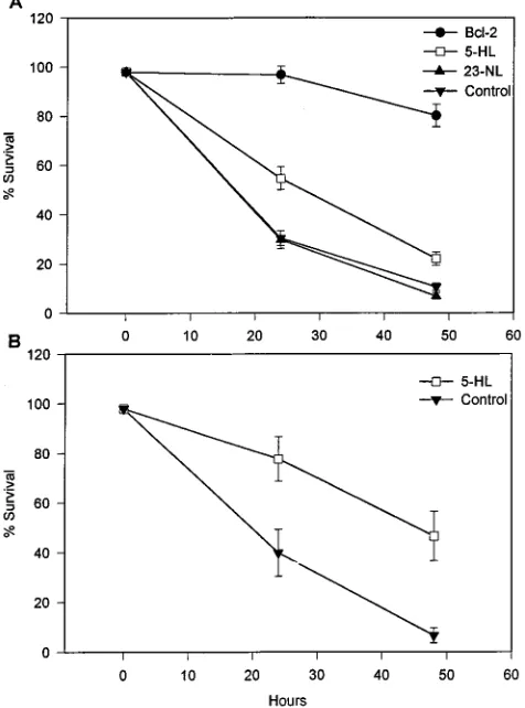
[image:3.612.116.237.74.253.2]well on 12-well plates. Cell survival was assessed and scored at 0, 24, and 48 h after IL-3 removal by phase-contrast micros-copy and trypan blue dye exclusion. Cytopathic changes asso-ciated with apoptotic cell death, which included nuclear con-densation and fragmentation and increased plasma membrane blebbing (Fig. 3) as well as loss of cell viability, were observed first in control cell lines (PSFFV-neo and PSFFV/23-NL) as early as 8 h after growth factor removal, with the number of apoptotic cells increasing significantly at 24 and 48 h (Fig. 4A). In contrast, cell lines expressing Bcl-2 and ASFV p21 showed significant increases in cell survival when compared with these controls. In 12 independent experiments, a significant increase
FIG. 2. Expression of p21 in cells infected with pathogenic and cell-culture-adapted ASFV. (A) Extracts of swine macrophages infected with various ASFV isolates were immunoprecipitated with a preimmune serum (lane 1) or an anti-p21 monospecific serum (lanes 2 to 5). Lanes 1 and 2, DRI; lane 3, Malawi Lil 20/1; lane 4, Haiti NHV-811; lane 5, E70. (B) Extracts of Vero cells infected with ASFV cell-culture-adapted variants were immunoprecipitated with a preimmune serum (lane 1) or an anti-p21 monospecific serum (lanes 2 to 9). Lanes 1 and 2, MS44; lane 3; MS81; lane 4, Lisbon 60; lane 5, NHV-811; lane 6, Haiti HT411; lane 7, DRII, lane 8, Brazil 2; lane 9, Uganda 61. The positions of molecular mass markers (in kilodaltons) are given on the left.
on November 9, 2019 by guest
http://jvi.asm.org/
of 20 to 30% in cell viability was observed for p21-expressing
cell lines at 24 and 48 h (P50.001) (Fig. 4A), with the number
of surviving cells being three- to fivefold higher in PSFFV/ 5-HL cell lines at 48 h. Interestingly, this p21-mediated repres-sion of cell death was approximately one-half of that observed for Bcl-2-expressing cell lines, in which increases of approxi-mately 70% in cell survival were routinely observed (Fig. 4A). Similar results were observed for FL5.12 cell lines transformed with pcDNA/5-HL; in five independent experiments, a signif-icant 40% increase in cell survival over that of control cells (those transformed with pcDNAIII containing no insert) was
detected at 24 and 48 h after IL-3 removal (P50.05) (Fig. 4B).
These data demonstrate that ASFV p21 has apoptotic-death-repressing activity when expressed in the mammalian cell line FL5.12. This activity occurs in the absence of other
[image:4.612.58.299.72.511.2]viral proteins, indicating that p21 acts directly on cellular tar-gets. The death-repressing effect of p21 in these cells is not as pronounced as that produced by human Bcl-2. Plausible expla-nations for this difference include the functioning of p21 in the FL5.12 cellular context and/or the level of p21 expression in these cell lines. Bcl-2 proteins function as either homo- or heterodimers (42); thus, the reduced death-repressing activity of p21 could be due to reduced affinity of dimerization between p21 and other mouse Bcl-2 family members. In spite of the protein similarities discussed above, there are also clear differ-ences in the BH2 domains of 5-HL and Bcl-2. These corre-spond to human Bcl-2 amino acids 190 to 192 (QDN) and 200 (E), which are conserved in most family members as Q(D/ E)(N/Q) and (E/D/S) but not in 5-HL. The 5-HL amino acids corresponding to human Bcl-2 amino acids 190 to 192 and 200 are ISH and A, respectively. When the same human Bcl-2 amino acids were replaced with residues AAA and A, respec-tively, the altered protein displayed about one-half the death-repressing activity of wild-type Bcl-2 in transformed FL5.12 cells but still retained the capacity to heterodimerize with Bax (60). This reduced level of death-repressing activity is similar to the level seen when 5-HL is expressed in FL5.12 cells. Alternatively, although p21 is clearly detectable in FL5.12 cells by using the expression plasmids described here, it may not be present at levels optimal for repressing FL5.12 cell death.
FIG. 3. Phase-contrast microscopy of transformed FL5.12 cells at 40 h after IL-3 removal. Cells were transformed with PSFFV/Bcl-2 (A), PSFFV/5-HL (B), and PSFFV-neo (control) (C). Note the extensive nuclear condensation with fragmentation (arrowhead) in control cells.
FIG. 4. Survival of transformed FL5.12 cells after IL-3 removal. Cell survival was assessed at 0, 24, and 48 h after IL-3 removal by phase-contrast microscopy and trypan blue dye exclusion. (A) Survival of cells transformed with PSFFV expression plasmids. Data are the means6standard errors of 12 experiments. (B) Survival of cells transformed with pcDNAIII expression plasmids. Data are the means6standard errors of five experiments.
on November 9, 2019 by guest
http://jvi.asm.org/
[image:4.612.315.553.355.676.2]A number of viruses have evolved antiapoptotic mechanisms to promote infected-cell survival, either to ensure efficient pro-ductive viral replication or to promote long-term survival of virus-infected cells (for a review, see reference 44). In addition to ASFV, viral members of the Bcl-2 gene family in gamma-herpesviruses and adenovirus have been described (5, 22, 34). The EBV protein BHRF1 provides an alternative cellular Bcl-2-independent means of enhancing infected B-cell survival (22). ORFs with sequence similarities to Bcl-2 family members have also been identified in two additional gammaherpesvi-ruses, herpesvirus saimiri and bovine herpesvirus 4 (32, 47). As yet, these genes have not been functionally characterized. The E1B 19-kDa protein of adenovirus has been shown to prolong infected-cell survival by inhibiting apoptosis induced by ade-novirus E1A protein, tumor necrosis factor alpha, and Fas antigen (5, 55, 56). Similarly, ASFV 5-HL may suppress apo-ptosis in ASFV-infected cells, thus promoting the survival of host cells during productive and/or persistent infection of ei-ther the pig or tick host. Certainly, this gene is highly conserved and present in even highly cell-culture-adapted viruses (Fig. 2B); our recent unsuccessful attempts to delete it from the viral genomes of pathogenic isolates suggest that it is essential for replication in swine macrophages (37a). ASFV infection in-duces apoptosis in primary swine macrophages in vitro as early as 16 h postinfection, a time at which viral replication has already occurred in these cells (37a). Thus, it is possible that p21, which is expressed throughout the infection cycle, includ-ing early time points, transiently modulates infected-macro-phage survival, allowing productive viral replication to occur. Cytopathological changes consistent with apoptotic cell death, including karyorrhexis and chromatin condensation, have been observed in mononuclear cells (lymphocytes and monocytes/ macrophages) in tissues of pigs infected with highly virulent ASFV isolates (9, 16, 20, 27, 33, 36, 37). Therefore, suppres-sion of apoptosis may be of significance to aspects of viral pathogenesis and virulence and p21 may mediate this effect. Additionally, because ASFV-swine monocyte/macrophage in-teractions result in either lytic or latent infection (3a, 4), p21 could conceivably have a role in promoting the survival of latently infected mononuclear cells.
We thank E. Kramer and F. Lyburt for excellent technical assistance and L. Zsak for helpful manuscript review comments.
REFERENCES
1. Afonso, C. L., C. Alcaraz, A. Brun, M. D. Sussman, D. V. Onisk, J. M. Escribano, and D. L. Rock.1992. Characterization of P30, a highly antigenic membrane and secreted protein of African swine fever virus. Virology 189: 368–373.
2. Boise, L. H., M. Gonzalez-Garcia, C. E. Postema, L. Ding, T. Lindsten, L. A. Turka, X. Mao, G. Nunez, and C. B. Thompson.1993. bcl-x, a bcl-2-related gene that functions as a dominant regulator of apoptotic cell death. Cell 74:597–608.
3. Brown, F. 1986. The classification and nomenclature of viruses: summary of results of meetings of the International Committee on Taxonomy of Viruses in Sendai, September 1984. Intervirology 25:141–143.
3a.Carrillo, C., et al. Unpublished data.
4. Carrillo, C., M. V. Borca, C. L. Afonso, D. V. Onisk, and D. L. Rock. 1994. Long-term persistent infection of swine monocytes/macrophages with Afri-can swine fever virus. J. Virol. 68:580–583.
5. Chiou, S.-K., C.-C. Tseng, L. Rao, and E. White. 1994. Functional comple-mentation of the adenovirus E1B 19-kilodalton protein with Bcl-2 in the inhibition of apoptosis in infected cells. J. Virol. 68:6553–6566.
6. Chittenden, T., C. Flemington, A. B. Houghton, R. G. Ebb, G. J. Gallo, B. Elangovan, G. Chinnadural, and R. J. Lutz.1995. A conserved domain in Bak, distinct from BH1 and BH2, mediates cell death and protein binding functions. EMBO J. 14:5589–5596.
7. Chittenden, T., E. A. Harrington, R. O’Connor, C. Flemington, R. J. Lutz, G. I. Evan, and B. C. Guild. 1995. Induction of apoptosis by the Bcl-2 homologue Bak. Nature (London) 374:733–736.
8. Coggins, L. 1974. African swine fever virus: pathogenesis. Prog. Med. Virol. 18:48–63.
9. Colgrove, G. S., E. O. Haelterman, and L. Coggins. 1969. Pathogenesis of African swine fever in young pigs. Am. J. Vet. Res. 30:1343–1359. 10. Costa, J. V. 1990. African swine fever virus, p. 247–270. In G. Darai (ed.),
Molecular biology of iridoviruses. Kluwer Academic Publishers, Norwell, Mass. 11. DeKock, G., E. M. Robinson, and J. J. G. Keppel. 1994. Swine fever in
South-Africa. Onderstepoort J. Vet. Sci. Anim. Ind. 14:31–93.
12. DeTray, D. E. 1957. Persistence of viremia and immunity in African swine fever. Am. J. Vet. Res. 18:811–816.
13. Devereux, J., P. Haeberli, and O. Smithies. 1984. A comprehensive set of sequence analysis programs for the VAX. Nucleic Acids Res. 12:387–395. 14. Dixon, L. K. 1988. Molecular cloning and restriction enzyme mapping of an
African swine fever virus isolate from Malawi. J. Gen. Virol. 69:1683–1694. 15. Eddy, S., G. Mitchison, and R. Durbin. 1995. Maximum discrimination
hidden Markov models of sequence consensus. J. Comput. Biol. 2:9–23. 16. Enjuanes, L., I. Cubero, and E. Vinuela. 1977. Sensitivity of macrophages
from different species to African swine fever virus. J. Gen. Virol. 34:455–463. 17. Farrow, S. N., J. H. M. White, I. Martinou, T. Raven, K.-T. Pun, C. J. Grinham, J.-C. Martinou, and R. Brown.1995. Cloning of a bcl-2 homologue by interaction with adenovirus E1B 19K. Nature (London) 374:731–733. 18. Fuhlbrigge, R. C., S. M. Fine, E. R. Unanue, and D. D. Chaplin. 1988.
Expression of membrane interleukin-1 by fibroblasts transfected with murine pro-interleukin 1acDNA. Proc. Natl. Acad. Sci. USA 85:5649–5653. 19. Genovesi, E. V., F. Villinger, D. J. Gerstner, T. C. Whyard, and R. C.
Knudsen. 1990. Effect of macrophage-specific colony-stimulating factor (CSF-1) on swine monocyte/macrophage susceptibility to in vitro infection by African swine fever virus. Vet. Microbiol. 25:153–176.
20. Gomez-Villamandos, J. C., J. Hervas, A. Mendez, L. Carrasco, J. M. de las Mulas, C. J. Villeda, P. J. Wilkinson, and M. A. Sierra.1995. Experimental African swine fever: apoptosis of lymphocytes and virus replication in other cells. J. Gen. Virol. 76:2399–2405.
21. Hamdy, F. H., and A. H. Dardiri. 1984. Clinical and immunologic responses of pigs to African swine fever virus isolated from the Western hemisphere. Am. J. Vet. Res. 45:711–714.
22. Henderson, S., D. Huen, M. Rowe, C. Dawson, G. Johnson, and A. Rickin-son.1993. Epstein-Barr virus-coded BHRF1 protein, a viral homologue of Bcl-2, protects human B cells from programmed cell death. Proc. Natl. Acad. Sci. USA 90:8479–8483.
23. Hess, W. R. 1982. African swine fever: a reassessment. Adv. Vet. Sci. Comp. Med. 25:39–69.
24. Hockenbery, D., G. Nunez, C. Milliman, R. D. Schreiber, and S. J. Kors-meyer.1990. Bcl-2 is an inner mitochondrial membrane protein that blocks programmed cell death. Nature (London) 348:334–336.
25. Kiefer, M. C., M. J. Brauer, V. C. Powers, J. J. Wu, S. R. Umansky, L. D. Tomei, and P. J. Barr.1995. Modulation of apoptosis by the widely distrib-uted Bcl-2 homologue Bak. Nature (London) 374:736–739.
26. Knudsen, R. C., and E. V. Genovesi. 1987. In vivo and in vitro effects of moderately virulent African swine fever virus on mitogenesis of pig lympho-cytes. Vet. Immunol. Immunopathol. 15:323–336.
27. Konno, S., W. D. Taylor, and A. H. Dardiri. 1971. Acute African swine fever. Proliferative phase in lymphoreticular tissue and the reticuloendothelial system. Cornell Vet. 61:71–84.
28. Konno, S., W. D. Taylor, W. R. Hess, and W. P. Heuschele. 1971. Liver pathology in African swine fever. Cornell Vet. 61:125–150.
29. Kozopas, K. M., T. Yang, H. L. Buchan, P. Zhou, and R. W. Craig. 1993. MCL1, a gene expressed in programmed myeloid cell differentiation, has sequence similarity to BCL2. Proc. Natl. Acad. Sci. USA 90:3516–3520. 30. Lin, E. Y., A. Orlofsky, M. S. Berger, and M. B. Prystowsky. 1993.
Charac-terization of A1, a novel hemopoietic-specific early-response gene with se-quence similarity to bcl-2. J. Immunol. 151:1979–1988.
31. Lipman, D. J., S. F. Altschul, and J. D. Kececioglu. 1989. A tool for multiple sequence alignment. Proc. Natl. Acad. Sci. USA 86:4412–4415.
32. Lomonte, P., M. Bublot, V. van Santen, G. M. Keil, P.-P. Pastoret, and E. Thiry. 1995. Analysis of bovine herpesvirus 4 genomic regions located outside the conserved gammaherpesvirus gene blocks. J. Gen. Virol. 76:1835–1841. 33. Malmquist, W. A., and D. Hay. 1960. Hemadsorption and cytopathic effect
produced by African swine fever virus in swine bone marrow and buffy coat cultures. Am. J. Vet. Res. 21:104–108.
34. Marchini, A., B. Tomkinson, J. I. Cohen, and E. Kieff. 1991. BHRF1, the Epstein-Barr virus gene with homology to Bcl2, is dispensable for B-lym-phocyte transformation and virus replication. J. Virol. 65:5991–6000. 35. Mebus, C. A. 1988. African swine fever. Adv. Virus Res. 35:251–269. 36. Mebus, C. A., J. W. McVicar, and A. H. Dardiri. 1981. Comparison of the
pathology of high and low virulence African swine fever virus infections, p. 183–194. In P. J. Wilkinson (ed.), Proceedings of CEC/FAO expert consul-tation in African swine fever research, Sardinia, Italy, September 1981. Commission of the European Communities, Luxembourg, Belgium. 37. Moulton, J., and L. Coggins. 1968. Comparison of lesions in acute and
chronic African swine fever. Cornell Vet. 58:364–388. 37a.Neilan, J. G., et al. Unpublished data.
38. Neilan, J. G., Z. Lu, C. L. Afonso, G. F. Kutish, M. D. Sussman, and D. L.
on November 9, 2019 by guest
http://jvi.asm.org/
Rock.1993. An African swine fever virus gene with similarity to the proto-oncogene bcl-2 and the Epstein-Barr virus gene BHRF1. J. Virol. 67:4391– 4394.
39. Neuwald, A. F., and P. Green. 1994. Detecting patterns in protein sequences. J. Mol. Biol. 239:698–712.
40. Neuwald, A. F., J. S. Liu, and C. E. Lawrence. 1995. Gibbs motif sampling-detection of bacterial outer membrane protein repeats. Protein Sci. 4:1618– 1632.
41. Nunez, G., L. London, D. Hockenbery, M. Alexander, J. P. McKearn, and S. J. Korsmeyer.1990. Deregulated Bcl-2 gene expression selectively pro-longs survival of growth factor-deprived hematopoietic cell lines. J. Immu-nol. 144:3602–3610.
42. Oltvai, Z. N., C. L. Milliman, and S. J. Korsmeyer. 1993. Bcl-2 heterodimer-izes in vivo with a conserved homolog, Bax, that accelerates programmed cell death. Cell 74:609–619.
43. Plowright, W., J. Parker, and M. A. Pierce. 1969. The epizootiology of African swine fever in Africa. Vet. Rec. 85:668–674.
44. Razvi, E. S., and R. M. Welsh. 1995. Apoptosis in viral infections. Adv. Virus Res. 45:1–60.
45. Sanchez-Botija, C. 1963. Reservorios del virus de la peste porcina africana. Investigacion del virus de la P.P.A. en los artropodos mediante la prueba de la hemoadsorcion. Bull. Off. Int. Epizoot. 60:895–899.
46. Schuler, G. D., S. F. Altschul, and D. J. Lipman. 1991. A workbench for multiple alignment construction and analysis. Proteins Struct. Funct. Genet. 9:180–190.
47. Smith, C. A. 1995. A novel viral homologue of Bcl-2 and Ced-9. Trends Cell Biol. 5:344.
48. Tatusov, R. L., S. F. Altschul, and E. V. Koonin. 1994. Detection of con-served segments in proteins: interactive scanning of sequence databases with alignment blocks. Proc. Natl. Acad. Sci. USA 91:12091–12095.
49. Thomson, G. R., M. Gainaru, A. Lewis, H. Biggs, E. Nevill, M. Van Der Pypekamp, L. Gerbes, J. Esterhuysen, R. Bengis, D. Bezuidenhout, and J. Condy.1983. The relationship between ASFV, the warthog and Ornithodo-ros species in southern Africa, p. 85–100. In P. J. Wilkinson (ed.), ASF, EUR 8466 EN, Proceedings of CEC/FAO Research Seminar, Sardinia, Italy,
Sep-tember 1981. Commission of the European Communities, Luxembourg, Bel-gium.
50. Vaux, D. L., S. Cory, and J. M. Adams. 1988. bcl-2 gene promotes hemo-poietic cell survival and cooperates with c-myc to immortalize pre-B cells. Nature (London) 335:440–442.
51. Villeda, C. J., C. Gomez-Villamandos, S. M. Williams, J. Hervas, P. J. Wilkinson, and E. Vinuela.1995. The role of fibrinolysis in the pathogenesis of the haemorrhagic syndrome produced by virulent isolates of ASFV. Thromb. Haemostasis 73:112–117.
52. Villeda, C. J., S. M. Williams, P. J. Wilkinson, and E. Vinuela. 1993. Hae-mostatic abnormalities in ASF: a comparison of two virus strains of different virulence (Dominican Republic ’78 and Malta ’78). Arch. Virol. 130:71–83. 53. Villeda, C. J., S. M. Williams, P. J. Wilkinson, and E. Vinuela. 1993. Con-sumption coagulopathy associated with shock in acute ASF. Arch. Virol. 133:467–475.
54. Vinuela, E. 1985. African swine fever virus. Curr. Top. Microbiol. Immunol. 116:151–170.
55. White, E., R. Cipriani, P. Sabbatini, and A. Denton. 1991. Adenovirus E1B 19-kilodalton protein overcomes the cytotoxicity of E1A proteins. J. Virol. 65:2968–2978.
56. White, E., P. Sabbatini, M. Debbas, W. S. M. Wold, D. I. Kusher, and L. Gooding.1992. The 19-kilodalton adenovirus E1B transforming protein inhibits programmed cell death and prevents cytolysis by tumor necrosis factora. Mol. Cell. Biol. 12:2570–2580.
57. Wilkinson, P. J. 1989. African swine fever virus, p. 17–35. In M. B. Pensaert (ed.), Virus infections of porcines. Elsevier Science Publishers, Amsterdam. 58. Yanez, R. J., J. M. Rodriguez, M. L. Nogal, L. Yuste, C. Enriquez, J. F. Rodriguez, and E. Vinuela.1995. Analysis of the complete nucleotide se-quence of African swine fever virus. Virology 208:249–278.
59. Yang, E., J. Zha, J. Jockel, L. H. Boise, C. B. Thompson, and S. J. Kors-meyer.1995. Bad, a heterodimeric partner for Bcl-xL and Bcl-2, displaces Bax and promotes cell death. Cell 80:285–291.
60. Yin, X.-M., Z. N. Oltvai, and S. J. Korsmeyer. 1994. BH1 and BH2 domains of Bcl-2 are required for inhibition of apoptosis and heterodimerization with Bax. Nature (London) 369:321–323.