PROTECTIVE EFFECT OF
Desmostachya
bipinnata
(L.) Stapf
AGAINST
PARACETAMOL-INDUCED HEPATIC DAMAGE
Dissertation submitted to
The Tamil Nadu Dr. M. G. R. Medical University,
Chennai
in partial fulfillment of the award of degree of
MASTER OF PHARMACY
(PHARMACOLOGY)
MARCH – 2010
COLLEGE OF PHARMACY
SRI RAMAKRISHNA INSTITUTE OF PARAMEDICAL SCIENCES
Certificate
This is to certify that the dissertation entitled “PROTECTIVE EFFECT OF Desmostachya bipinnata (L.) Stapf AGAINST PARACETAMOL-INDUCED HEPATIC DAMAGE” being submitted to The Tamil Nadu Dr.M.G.R. Medical University, Chennai in partial fulfillment of the Master of Pharmacy programme in Pharmacology, carried out by INDRASENAN. R, in the Department of Pharmacology, College of Pharmacy, SRIPMS, Coimbatore, under my direct guidance and supervision to my fullest satisfaction.
Dr. M. UMA MAHESWARI, M.Pharm., Ph. D., Assistant Professor, Dept In-Charge, Department of Pharmacology, College of Pharmacy, SRIPMS, Coimbatore – 44 Place: Coimbatore
Certificate
This is to certify that the dissertation entitled “PROTECTIVE EFFECT OF Desmostachya bipinnata (L.) Stapf AGAINST PARACETAMOL-INDUCED HEPATIC DAMAGE" was carried out by
INDRASENAN.R, in the Department of Pharmacology, College of Pharmacy, Sri Ramakrishna Institute of Paramedical Sciences, Coimbatore, which is affiliated to The Tamil Nadu Dr.M.G. R. Medical University, Chennai, under supervision and direct guidance of Dr.M.UMA MAHESWARI, M.Pharm.,Ph.D.,
Department of Pharmacology, College of Pharmacy, SRIPMS, Coimbatore – 44.
Dr. T. K. RAVI, M. Pharm., Ph. D., FAGE., Principal, College of Pharmacy, SRIPMS,
Coimbatore – 44
Place: Coimbatore
Acknowledgement
I take this opportunity to render my profound sense of gratitude, indebtedness and respectful regards to my esteemed teacher and guide Dr. M. Uma Maheswari, M. Pharm. Ph.D.,
Assistant Professor, Department of Pharmacology for her excellent suggestions, kind encouragement and able guidance throughout the course of my work.
I submit my sincere thanks and respect to our Managing Trustees, Sevaratna Dr.R.Venkatesalu Naidu and Mr.C. Soundararaj for all the facilities provided in our institution.
I am overwhelmed by the timely guidance at each and every step of my work and enthusiastic encouragement offered by our beloved principal Dr. T. K. Ravi, M. Pharm., Ph. D., FAGE,
throughout the course of investigation and the successful completion of this work.
It gives me immense pleasure to thank Dr. K. Asok Kumar, M. Pharm., Ph. D., Head of the Department of Pharmacology, for his valuable guidance and help.
It is my privilege to express my sincere gratitude to
Dr. S. Krishnan M. Pharm., Ph.D., Department of Pharmaceutical Biotechnology and , Dr. M. Gandhimathi, M. Pharm., PGDMM, Ph.D.,, Department of Pharmaceutical Analysis for lending me all the facilities to carryout the analytical work.
Mrs. V. Subadra Devi, M.Pharm., Mr. Jagannathan, M. Pharm
lecturers, for their support, timely help and suggestions.
I would like to express my sincere thanks to Mr. H. John, Mrs. Karpagam and Mrs.Sguna who directly or indirectly gave a helping hand to perform this study.
My special thanks to the Library Staffs of SRIPMS for their valuable help.
This project would not be a resplendent one without the constant love, prayer and moral support by My Ever-Loving Family
and my sister Chandralekha and I take this opportunity to acknowledge them with thanks.
I extend my deep sense of gratitude to my friends and
Ravindran, Anju, Austin, Avinash, Christy, Muthiah, Usha, Lalitha, Jobin, Arun, Jinto, Manoj, Niyas, Lukose, Anil, Sachin and juniors for their valuable suggestions, co-operations and encouragement.
My sincere thanks are extended to Saraswathi Computer Centre for helping me to complete my project in right time.
Above all, allow me to proclaim the over powering presence of
The Almighty who is the source of all wisdom and knowledge for the successful completion of this dissertation.
CONTENTS
S. NO TITLE PAGE NO
1 Introduction 1
2. Plant Profile 30
3 Review of Literature 32
4. Objectives & Plan of work 51
5. Materials and Methods 54
6. Results 71
7. Discussion and Conclusion 83
References
LIST OF ABBREVIATIONS
ALP : Alkaline phosphatase
ALT : Alanine transaminase
AST : Aspartate transaminase
CAT : Catalase
CF : Chloroform fraction
EAF : Ethyl acetate fraction
GSSH : Glutathione reductase
GSH : Reduced glutathione
GPx : Glutathione peroxidase
LH : Lipid hydroperoxides
LDH : Lactate Dehydrogenase.
MDA : Malondialdehyde
mg/dl : milligram/deciliter
MPT : Mitochondrial permeability transition
PcmL : Paracetamol
PEF : Petroleum ether fraction
Px : Peroxidase
RNS : Reactive Nitrogen Species
ROS : Reactive Oxygen Species
rpm : Rotation per minute
SGOT : Serum oxaloacetate transaminase
SGPT : Serum glutamate pyruvate transaminase
SOD : Superoxide dismutase
TBA : Thiobarbituric acid
TBARS : Thiobarbituric acid reactive substances
TCA : Trichloroacetic acid
TP : Total Protein
U/L : Units/liter
1. INTRODUCTION
LIVER
Liver is the largest gland in the body weighing
between 1 to 2.3 kg. It is situated in the upper part of the
abdominal cavity occupying the greater part of the right
hypochondriac region. The liver consists of 4 lobes, a large
right lobe and the smaller wedge shaped left lobe. The
other two, the caudate and quadrate lobes are areas on
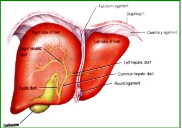
Fig. 1 Anterior view of liver (Tortora and Derrickson, 2006)
Organs associated with liver
Superiorly & anteriorly : Diaphragm and abdominal
wall.
Inferiorly : Stomach, bile ducts,
Posteriorly : Oesophagus, inferior
venecava, aorta, gall bladder.
Laterally : Lower ribs and
diaphragm.
Anatomy of the liver
The liver is almost completely covered by visceral
peritoneum and by a dense irregular connective tissue
layer that lies deep to the peritoneum. The right and left
lobes of the liver are separated by the falciform
ligament-a fold of peritoneum. The fligament-alciform ligligament-ament extends from
the under surface of the diaphragm between the two
principal lobes of the liver to the superior surface of the
liver helping to suspend the liver in the abdominal cavity.
The right and left coronary ligaments are narrow extension
of the parietal peritoneum that suspends the liver from
Histology of the liver
The lobes of the liver are made up of many
functional units called lobules. A lobule is typically a six
sided (hexagon) structure that consists of specialized
epithelial cells called hepatocytes, arranged in irregular,
branching, inter connected plate around a central vein.
In addition the liver lobule contains highly permeable
capillaries called sinusoids through which blood passes.
The fixed phagocytes called stellate reticulo endothelial
cells (Kupfer cells) are also present in the sinusoids which
destroy worn out white blood cells and red blood cell
bacteria and other foreign matter in the blood draining
from gastrointestinal tract (Tortora and Derrickson, 2006).
Blood supply to the liver
The liver receives blood from two sources. From the
hepatic portal vein it receives deoxygenated blood
containing newly absorbed nutrients, drugs, and possibly
microbes and toxins from the gastrointestinal tract (Tortora
and Derrickson, 2006).
Secretions of liver
Bile is both a product of secretion as well as an
excretion of the liver (Chaterjee, 2004).
Composition of bile
Total quantity : 500-1000 ml daily.
Colour : Yellowish green.
Taste : Bitter.
Bile salts : Sodium taurocholate and sodium
glycocholate.
Bile pigments : Bilirubin and biliverdin.
Functions of the liver (Waugh and Grant, 2001).
i) Deaminates amino acids
ii) Converts glucose to glycogen
iii) Desaturates fat
iv) Produce heat
v) Secretes bile
vi) Synthesize vitamin A, non essential amino acids,
plasma proteins and blood clotting factors.
vii) Detoxicates drugs and noxious substances
viii) Metabolizes ethanol
ix) Inactivates hormones
x) Stores fat-soluble and water- soluble vitamins, iron
and copper.
Liver diseases
and chronic,
a) Acute liver disease
This is usually a self-limiting episode of liver cell
(hepatocyte) inflammation or damage, which in most
cases resolves without clinical sequel.
b) Chronic liver disease
This occurs when permanent structural changes
within the liver occur secondary to long- standing cell
damage, with the consequent loss of normal liver
architecture. In most cases, this progress to cirrhosis and
leads to liver failure.
Causes of liver diseases
i) Viral infections-Hepatitis A, B, C, D, E
Fig. 2. Stages of liver damage
The ingestion of ethanol in alcoholic beverages is the
commonest cause of liver cirrhosis. Prolonged and
excessive exposure to alcohol induces inflammatory
activity within the liver tissue and the hepatocytes
accumulate large droplets of fat as inclusion bodies within
the swollen cells. A fine network of collagen fibers develop
around the liver cells near the hepatic venule, and the
extent of fibrosis increases which eventually leads to liver
failure.
Autoimmune diseases occurs when the immune
system develops autoantibodies within the body. e.g.
Autoimmune hepatitis.
iv) Vascular abnormalities
The Budd-Chiare syndrome occurs when obstruction
of the major hepatic veins leads to cell destruction and
cirrhosis. Veno-occlusive disease occurs when the smaller
veins of liver become obliterated as a result of exposure to
toxins, irradiation or cytotoxicity.
MARKERS IN LIVER DISEASE
Alanine Transaminase (ALT)
ALT is an enzyme produced in the hepatocytes. This
enzyme functions normally to transfer the amino group
from an aminoacid alanine to a keto acid producing
pyruvate. The activities of ALT outside the liver are low and
hepatocellular damage. As cells are damaged, ALT leaks
out into the bloodstream and the levels are elevated in
cases of hepatitis, shock or drug toxicity. It is a measure of
integrity of cell death or inflammation (Harrison, 1990).
Aspartate Transaminase (AST)
It is an enzyme similar to ALT and is also produced in
the muscle. This enzyme transfers the amino group from
the amino acid aspartate to a keto acid producing
oxaloacetate. It can be elevated in other conditions like
heart attack. In case of liver inflammation, the ALT and
AST activities are elevated in the ratio of 1:1. In certain
conditions like alcoholic or shock liver, the level of AST is
more elevated than ALT.
Alkaline Phosphatase (ALP)
Alkaline phosphatase is a group of enzymes that are
capable of hydrolyzing phosphate esters at alkaline pH
activities in the liver, GIT, bone and placenta. These
enzymes are found in greatest concentration in
membranes associated with absorption and secretory
functions. In the liver they are localized in the sinusoidal
and biliary canalicular membrane.
Gama glutamyl transferase (GGT)
It is a microsomal enzyme found in many cells and
tissues of the body. The largest concentrations are found
in the liver, localized in the hepatocytes and epithelium of
the small bile ducts.GGT functions normally to transfer
glutamyl groups from gama glutamyl peptides to other
peptides and amino acids.
Lactate dehydrogenase (LDH)
Lactate dehydrogenase is an enzyme catalyzing
oxidation of lactic acid to pyruvic acid. In blood serum
there are five physically distinct iso enzymes of this
enzyme. They are known as LDH 1-5. All these iso enzymes
oxidation of lactic acid to pyruvic acid. If LDH4 and LDH5
are increased it is a case of liver disease.
Bilirubin
Bilirubin is the breakdown product from the
destruction of old red lood cells. The level of bilirubin is
elevated in the blood either by increased production,
decreased uptake by the liver, decreased conjugation
and decreased secretion from liver or blockage of the bile
ducts. Increased production produces unconjugated or
indirect bilirubin and decreased production produces
conjugated or direct bilirubin.
HEPATOTOXICITY
Liver toxicity is a major health problem of worldwide
proportions. It is influenced by various physiological,
nutritional and therapeutic factors. It can be modulated
when exposure occurs in the presence of enzyme
pathways,
1) Direct hepatotoxicity
2) Adverse immune reactions
In most cases, it is initiated by the activation of drugs
to chemically reactive metabolites, which have the ability
to interact with cellular macromolecules like proteins, lipids
and nucleic acids causing protein dysfunction, lipid
peroxidation, DNA damage and oxidative stress, thus
leading to the impairment of cellular function which can
culminate in liver failure and cell death (Zhang, 2002).
CLASSIFICATION OF HEPATOTOXIC AGENTS
The hepatotoxic agents can be broadly classified
into two major categories (Zhang, 2002).
1) True, intrinsic or predictable hepatotoxins:
Their hepatotoxicity is a fundamental property to
2) Non-predictable or idiosyncratic hepatotoxins:
Consist of agents that produce hepatic injury only in
unusually susceptible humans i.e., their toxic effect results
[image:22.612.118.513.311.681.2]from the special vulnerability of the affected individual.
Table 1. CLASSIFICATION OF HEPATOTOXIC AGENTS
Category of
Agents Mechanism
Histological
Lesion Examples
I)Intrinsic toxins a) Direct Direct physiochemic al disortion and
destruction of structural basis cell
metabolism
Necrosis ( zonal ) and /or steatosis
CCl4,CHCl3,
phosphorous, tetrachloroet hane
b) Indirect
i) Cytotoxic
hepatotoxins ii) Cholestatic Interfere with hepatic excretory pathways leading to cholestasis
Drug induced liver injury (DILI)
Drug induced liver injury is intiated by direct
hepatotoxic effect of a drug or a reactive metabolite of a
drug. Parenchymal cell injury indicates activation of
innate and or adaptive immune cells which in turn
produce pro inflammatory and tissue hepatotoxic
mediators and or mount immune reactions against drug
associated antigens. DILI accounts for more than 50% of
acute liver failure including hepatotoxicity caused by over
dose of acetaminophen (APAP) 39% and idiosyncratic
liver injury triggered by other drugs 13% (Holt, 2006).
MECHANISM OF DRUG INDUCED LIVER INJURY
Drug induced direct hepatotoxicity
In most instances DILI is intiated by the bioactivation
of drugs to chemically reactive metabolite which have
the ability to interact with cellular macro molecules such
dysfunction, lipid peroxidation, DNA damage and
oxidative stress. Additionally these reactive metabolite
may induce disruptions of ionic gradients and intra cellular
calcium stores resulting in mitochondrial dysfunction and
loss of energy production. This impairement of cellular
function can culminate in cell death and possible liver
failure. DILI can affect both parenchymal and non
parenchymal cells of the liver, leading to a wide variety of
pathological conditions.
Role of innate immunity in DILI
Drug induced stress or damage of hepatocytes may
trigger activation of inflammatory responses of the innate
immune system with in liver. Evidence to support this idea
has been mainly obtained from studies of liver injury
induced by over dose of APAP. The intial NAPQI induced
hepatocyte damage may lead to activation of innate
immune cells with in the liver there by stimulating hepatic
innate immune system produce a range of inflammatory
mediators including cytokines, chemokines, and reactive
oxygen and nitrogen species that contribute to the
[image:26.612.169.461.255.421.2]progression of liver injury.
Fig. 3. Mechanism of Drug induced liver injury
The innate immune cells reported to participate in
APAP hepatotoxicity include natural killer (NK), and
natural killer T (NKT) cells, macrophages, and neutrophills.
The depletion of NK and NKT cells protected mice from
APAP induced liver injury. This protective mechanism
decreasing neutrophil accumulation with in the liver.
APAP hepatotoxicity has also been attributed in part
to the activation of Kupfer Cells (KC) secondary to
hepatocyte damage. KC activation results in the release
of a wide range of pro inflammatory mediators such as
TNF α which may directly induce tissue damage and IL-12 and IL-18 which are important activation of NK and NKT
cells.
KC may play a protective role in addition to their pro
toxicant effect as KC are the pre dominant source of IL-10
and IL-16 which are important in counter acting
inflammatory responses or stimulating liver regeneration.
PARACETAMOL (PcmL)
It is the active metabolite of phenacetin. PcmL is an
effective alternative to aspirin as an analgesic and
antipyretic, but a weak anti-inflammatory agent. Lesions
produced by PcmL are due to glutathione depletion,
and oxidative stress. During PcmL intoxication both
covalent binding to cellular macromolecules and lipid
peroxidation occurs
(Arnaiz et al., 1995).
The active metabolite capable of binding covalently
to proteins is found to be N-acetyl-p-benzoquinoneimine.
It binds to proteins and also other nucleophiles. It
contributes to overall process leading to necrosis (Gupta
et al., 2004).
PcmL is a mild analgesic that is harmless in
therapeutic doses and potent a hepatotoxin in overdose.
It has been proved to be an elegant model for the
exploration of the mechanisms of hepatotoxicity (Sumioka
et al., 2004). PcmL is largely converted to conjugates of
glucuronate and sulfate (Gupta et al., 2004). A minor
amount, about 25%, is converted to the active metabolite
29
compound being converted to mercaptopuric acid and
cysteine. When the amount of active molecule formed
exceeds the GSH available for binding, and then it causes
necrosis.
PcmL causes hepatic necrosis only when the dose is
sufficiently large to deplete hepatic GSH by 85% or more.
Susceptibility to hepatotoxic effects relates to the rate of
conversion of Pcml to an active metabolite, presumably a
reactive imidoquinone derived from
N-hydroxyl-N-acetyl-p-hydroxyaniline.
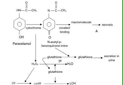
Fig. 4Mechanisms involved in the hepatotoxic action of paracetamol
OH
HN C O
CH3
cytochrome
O
N C O CH3 covalent binding macromolecule necrosis A Paracetamol N-acetyl-p- benzoquinone imine glutathione glutathione j t excretion in urine H2O2 H2O
[image:29.612.122.516.513.784.2]OXIDATIVE STRESS AND FREE RADICAL GENERATION
Oxidative stress occurs by acute paracetamol
administration in mice liver. Paracetamol hepatotoxicity
appears to be critically dependent on the depletion of
cellular glutathione. A relatively high reduction in the
intracellular level of reduced glutathione leads to
oxidative stress. The one electron oxidation of
paracetamol by P450 stepwise may generate reactive
through oxidation may lead to an alteration in calcium
homeostasis and cause hepatotoxicity. It is suggested that
an alkylating paracetamol metabolite cause Ca2+
de-regulation in the nucleus, leading to an activation of Ca2+
-sensitive endonuclease, fragmentation of DNA and cell
death (Arnaiz et al., 1995). Hydrogen peroxide and
superoxide anion radical are produced during metabolic
activation of paracetamol in the mixed function oxidase
system (Sumioka et al., 2004). Peroxynitrite is normally
detoxified by GSH, which is depleted in paracetamol
toxicity. NO synthesis (serum nitrate plus nitrite) was
[image:31.612.192.420.563.713.2]dramatically increased following acetaminophen activity .
Fig. 5 Role of oxidative stress in acetaminophen toxicity
NO•+ O2• H2O2
ONOO•
Metabolic Activation of Acetaminophen
Dr. Gillette’s laboratory firmly studied the importance
of metabolism in acetaminophen toxicity.
Acetaminophen is metabolically activated by
cytochrome P450 to form a reactive metabolite that
covalently binds to protein. The reactive metabolite was
found to be N-acetyl-p-benzoquinoneimine (NAPQI),
which is formed by a direct two-electron oxidation. They
also revealed that NAPQI is detoxified by glutathione
(GSH) to form an acetaminophen-GSH conjugate. After a
toxic dose of acetaminophen, total hepatic GSH is
depleted by as much as 90%, and as a result, the
metabolite covalently binds to cysteine groups on protein,
forming acetaminophen-protein adducts. This mechanism
Superoxide Formation and Mitochondrial Dysfunction in
Acetaminophen Toxicity
Superoxide may be formed from the CYP-450 and
other enzymes. Investigation is carried out on the
importance of activation of kuffer cells, macrophages, or
neutrophils (the so-called respiratory burst) in
acetaminophen toxicity. Finally, it was concluded that
superoxide anion is released from increased activity of
enzyme, NADPH-oxidase. Mitochondrial dysfunction may
be formed via occurring the MPT (mitochondrial transition
permeability) with formation of superoxide anion radicals
which leads to peroxynitrite and tyrosine nitration. The
toxicity was mediated by oxidants such as peroxides,
peroxynitrite, increased calcium, Pi promote the onset of
MPT, mg2+, ADP, low pH, high membrane potential, ROS
and decreased ATP .
acetaminophen toxicity
DIFFERENT EXPERIMENTAL MODELS USED TO EVALUATE
HEPATOPROTECTIVE ACTIVITY
Both in vitro and in vivo models are available for the
evaluation of hepatoprotective activity.
Acetaminophen P-450
NAPQI MPT O2 •
GSH APAP-SG
H2O2 Oxidative
Stress Mitochondrial
Dysfunction
Ca++ ATP O2•
+ NO
ONOO
Protein Nitration
I) In vitro liver models
The most frequently used isolated liver preparations
include isolated perfused organs, precision cut liver
slices, sub-cellular fractions and isolated and cultured liver
cells (Groneberg, 2002).
i) Isolated perfused organ
The isolated perfused organ displays an approach
towards the assessment of organ physiology and
morphology and represent the closest model to the in vivo
situation. The major advantages are that the preservation
of the 3-dimensional organ structure with all its cell-to-cell
interactions and the possibility of real-time bile collection
and analysis. This model allows the study of hemodynamic
parameters if blood is used as a perfusate. The different
models of isolated perfused livers proved to be very
complex in keeping organ function within physiological
maintained over a prolonged period. Also the
establishment of these models is very expensive.
ii) Precision cut liver slices
The precision-cut liver slice model can be used to
examine the cellular aspects of liver toxicology in a
tissue-specific background. The liver slice was one of the first
preparations used to study in vitro liver metabolism. This
model helps to retain tissue organization and cell-to-cell
matrix interactions such as perfused organs. The main
advantages are represented by the preservation of
lobular structures in contrast to cell cultures and the
possible application of biochemical and molecular
biological methods in contrast to organ perfusions. This
system can be used for a period of 2 to 3 days as a valid
model to study hepatotoxicity and even human tissues
can be used after surgical or needle biopsy removal.
These models can be obtained from animals
pretreated by inducers and are useful to study the
mechanisms of hepatotoxicity. Microsomes are widely
used to identify drug metabolic pathways, covalent
binding and lipid peroxidation induced by hepatotoxins.
Isolated mitochondria are widely used in the study of the
effect of toxicants on cellular energy transformation.
iv) Liver cells
a) Isolated and cultured hepatocytes
The liver cell culture model can be applied to
examine the effects of drugs/toxins on isolated
hepatocytes on the cellular level. Berry and Friend
established the basic protocol involving a two-step
perfusion of the liver, first with calcium free buffer, followed
by calcium-supplemented buffer containing collaginase.
are the homogenicity of the suspension, functional
similarities with the in vivo state and the ability to analyze
multiple parameters from a single cell suspension. To
guarantee the survival of hepatocytes isolated from
individual donors, cryopreservation or cold storage
techniques can be applied that lead to an indefinite or 48
h extension respectively. The viability of stored cells is
much lower than that of freshly isolated hepatocytes and
dependent on factors such as initial cell integrity, ice
crystal formation, and hypoxia during freezing and toxicity
of cryopreservation substances. Hepatocytes have the
capacity of biotransformation which is crucial for
toxicological studies. A significant disadvantage in the
hepatocyte culture is the absence of organ-specific
cell-to-cell interactions.
b) Liver cell lines
transfected hepatocytes. Hepatoma cell lines are difficult
to establish from primary carcinomas and exhibits only
some of the characteristics of normal liver.
II) In vivo models
Whole animal as experimental model
The studies performed during the past 100 years
have employed a variety of species, the most popular
being mice because of their size and relatively low cost.
Accordingly, most of the accumulated information
bearing on experimental hepatotoxicity and on modifier
of susceptibility, such as age, sex, stage of development,
diet and exposure to other toxic substance, applies to the
mice. The general employment of relatively uniform
experimental model permits comparison of results
obtained in widely separated laboratories. To a varying
degree, rat, hamsters, guinea pigs, rabbits, dogs, cats,
cattle, horse, sheep, and several species of birds have
chemical may include any of these or other species.
During recent years, primates have come to use, for the
obvious reason of the greater presumed relevance to the
disease of humans.
PARAMETERS OF INJURY
Measure of hepatic injury includes lethality,
histological changes seen by light electron microscopy,
chemical changes seen in the liver and biochemical tests
that measure the functional status or that reflect the type
or intensity of hepatic injury.
1) Lethality
Death as a measure of hepatotoxic potency is
applicable mainly to known hepatotoxins. Employment of
the LD50 or other measures of lethal potency permits
comparison of hepatotoxic agents.
2) Histology
microscopy and scanning electron microscopy.
a) Light microscopy
LM is the traditional method for demonstrating toxic
hepatic injury and categorizing its type. It provides
yardstick against which other abnormalities can be
measured. However, LM provides only a crude estimation
for the quantitation of the degree of injury.
b) Electron microscopy
It provides a much earlier demonstration of
hepatocyte injury and permits the recognition of damage
too subtle to be appreciated by LM. It is also useful in
differentiating lesions that appear to be similar in LM. For
e.g. hepatic injury induced in mice by galactosamine
resembles that of viral hepatitis when examined by LM,
but not when examined by EM. It provides evidence
earlier than LM and may yield clues to the mechanisms of
c) Scanning electron microscopy
This approach to ultra structural studies appears to
have added new dimensions to the study of structural
changes induced by toxic agents. Studies of cholestatic
effects of hepatotoxins utilizing the scanning technique
provided the database for a new hypothesis for the
development of cholestasis.
Enzymatic antioxidants
The term antioxidant has been defined by Halliwell
and Gutteridge as any substance that delays or inhibits
oxidative damage to a target molecule. The first line of
defense against superoxide anion (O2.-) and hydrogen
peroxide (H2O2) mediated injury are antioxidants enzyme
like superoxide dismutase (SOD), glutathione peroxides
Lipid peroxidation (LPO)
Malondialdehyde (MDA) is the major reactive
aldehyde resulting from the peroxidation of biological
membrane polyunsaturated fatty acids (PUFA). MDA, a
secondary product of LPO, is used as an indicator of tissue
damage by a series of chain reactions. MDA is also a
byproduct of prostaglandin biosynthesis. It reacts with
thiobarbituric acid and produces a red-coloured product.
MDA is a mutagenic and genotoxic agent that may
contribute to the development of human cancer (Halliwell
et al., 1993).
Superoxide dismutase (SOD)
SODs are a family of metalloenzymes that convert
O2. - to H2O2 according to the following reaction,
2H
SOD
SOD is the most important enzyme because it is
found virtually in all aerobic organisms. There are four
families of SOD, namely Cu-SOD, Cu-Zn-SOD, Mn-SOD,
and Fe-SOD. The transition metal of the enzyme reacts
with O2. - taking its electron. Superoxide anion is the only
known substrate for SOD.
Cu-Zn-SOD is found in the cytosol of most eukaryotic
cells. A different form of Cu-Zn-SOD is found in extra
cellular fluids, where it is called EC-SOD. Mn-SOD is located
in the mitochondrial matrix and bacteria, while Fe-SOD is
present in many aerobic bacteria. Cu-Zn-SOD is sensitive
to cyanide, but is destroyed by the treatment with
chloroform plus ethanol. SOD is considered to be a stress
protein, which is synthesized in response to oxidative stress.
and organisms, and is thought to protect the cell from
damage caused by O2.- and OH radicals generated from
the metal-catalysed interaction of O2 with H2O2 (Halliwell
et al., 1993).
Glutathione (GSH)
Glutathione is a major antioxidant, critical to the
protection of tissues from free radical injury. GSH is a
ubiquitous tri-peptide formed from three aminoacids
glutamate, glycine and cysteine and synthesized by two
ATP-dependent enzymatic reactions. GSH is a major
intracellular antioxidant molecule. It plays a critical role in
detoxification of peroxides and electrophilic toxins as a
substrate for GSH peroxidase and GSH transferase. It was
shown that depletion of GSH enhances cerebral ischemic
injury in rats.
Glutathione peroxidase (GPx)
line defense against oxidative stress, which in turn requires
glutathione as a co-factor. GPx catalyses the oxidation of
GSH to GSSG (oxidized glutathione) at the expense of
H2O2. By its selenium dependency, GPx can be divided in
two forms, Se-dependent GPx and Se-independent GPx.
The former is a tetramer of MW 84000 with very high
activity towards both H2O2 and organic hydroperoxides. It
is found in both cytosol (70%) and mitochondria (30%) of
various tissues. Since selenium is an integral component of
GPx, the measurement of this enzyme has been used as a
functional index of selenium level. GPx activity is reduced
in selenium deficiency (Halliwell et al., 1993).
Catalase (CAT)
Catalase is an enzyme, which is present in most cells
and catalyses the decomposition of hydrogen peroxide to
water and oxygen. CAT is a heme containing protein. The
CAT
2H2O2 2H2O + O2
CAT is found to act 104 times faster than peroxidase.
It is localized mainly in the mitochondria and in sub-cellular
respiratory organelles. CAT is present in peroxisomes (80%)
and cytosol (20%). It has a molecular weight of about
2,40,000 and consists of four protein subunits, each
containing a heme Fe(III)-protoporphyrin group bound to
its active site. GPx and CAT were found to be important in
the inactivation of many environmental mutagens
(Halliwell et al., 1993).
HEPATOPROTECTION
Liver diseases constitute a major health problem of
worldwide populations. Liver injury is induced by various
hepatotoxicants, etc. Herbal medicines derived from
plant extracts are being increasingly utilized to treat a
wide variety of clinical diseases. There is a growing interest
in the pharmacological evaluation of various plants used
in Indian traditional system of medicine. Many research’s
have been directed towards the provision of empirical
proof to back up the use of many plants in traditional
medical practice. However, there still exist a vast number
of plants with tremendous medicinal potential but with no
empirical proof to support claims of efficacy (Halliwell et
2. PLANT
PROFILE
Desmostachya bipinnata (L.)Stapf
Family : Poaceae
Vernalcular names
English : Sacrificial grass
Hindi : Davoli
Malayalam : Balidarbha
Sanskrit : Darbhah
Tamil : Darbhaibhul
Telugu :
Aswalayana,Darbha,Kushadarbha.
Perennial grass, found throughout India in hot and dry places.
Description
Perennial grass, tall, branched from the base, root stock stout, creeping, stolons very stout, covered with shining sheathes, stems 30-90cms high, tufted, smooth, erect, stout. leaves many, the basal fascicled, reaching sometimes 50cm long and 1cm broad at the base, rigid, acuminate, with filiform tips and hispid margins, sheath glabrous, ligule a hairy line. Panicles erect, narrowly pyramidal clothed from the base with sessile imbricating spikelets, grain 0.5-0.6mm long, obliquely ovoid, laterally compressed.
Parts used
Whole plant is used.
Constituents
Flavonoids, glycosides, saponins, tannins, carbohydrate .
Properties
Medicinal uses
[image:53.612.187.439.295.608.2]It is useful in thirst, asthma. Jaundice, disease of the blood, disease of the bladder, skin eruption, dysentery, uropathy, strangury, vesical calculi. (Krithikar and Basu, 1987).
3.
REVIEW OF LITERATURE
Akare et al., (2009) reported that the ethanol and aqueous
extracts of Acacia ferruginea leaves were tested for their efficacy
against carbon tetrachloride (CCL4) induced hepatotoxicity in Wistar
albino rats. The different groups of animals were administered with
CCL4 (1ml/kg, s.c.). The ethanol and aqueous extracts at the dose of
200mg/kg were administered to CCL4 treated rats.The result of present
study demonstrated that ethanol extract significantly decreases the
level of alanine aminotransferase, aspartase aminotransferase, total
bilirubin and direct bilirubin in blood, as compare to aqueous extract.
The phytochemical screening revealed the presence of active
hepatoprotection. The present work support the traditional claim of
plant in the treatment of liver injury, may provide a new drug against a
war with liver diseases.
Satyanarayana et al., (2009) evaluated the
hepatoprotective effect of the alcohol extract of
Capparis sepiaria Linn. (Capparaceae) stem against
carbon tetrachloride (CCl4)-induced toxicity in albino rats.
The rats were given daily pretreatment with alcohol
extract of C. sepiaria (100 mg/kg) and the standard
silymarin (25 mg/kg) orally for 7 days. The toxicant used on
7th day was CCl4 at a dose of 1.25 ml/kg as 1:1 mixture
with olive oil. The extract produced significant (p<0.01)
reduction in the elevated levels of aspartate transaminase
(AST), alanine transaminase (ALT), total bilirubin (TB) and
rise of decreased total protein level when compared with
the toxic control.
Vadivu et al., (2009) reported the hepatoprotective and in-vitro
Linn. Hepatoprotective activity was studied by carbon tetrachloride -
induced hepatotoxicity in rats and the in-vitro cytotoxic activity is
carried out by tryphane blue exclusion method using EAC cell lines.
The degree of protection in hepatoprotective activity has been
measured by using biochemical parameters such as serum glutamate
oxalate transaminase (SGOT) and serum glutamate pyruvate
transaminase (SGPT), alkaline phosphatase (ALP), bilirubin and total
protein. The results suggest that the alcoholic extract at the dose level
of 250mg/kg has produced significant (p<0.001) hepatoprotection by
decreasing the activity of serum enzymes, bilirubin, and lipid
peroxidation which is comparable to that of standard drug silymarin.
The alcoholic extract also does exhibit the IC50 value of 75μg/ml
which indicates the significant in-vitro cytotoxic activity of the
extract. It is concluded that alcoholic extract of leaves of Premna
serratifolia Linn is not only an effective hepatoprotective agent, but
also possesses significant antitumor activity.
Awaad et al., (2008) isolated the five main flavonoid
glycosides from the ethanol extract of Desmostachia
kaempferol(1), quercetin(2), quercetin-3-glucoside(3),
trycin(4) and trycin-7-glucoside(5). The structure
elucidation was based on UV, Electrospray ionization mass
spectrometry (ESIMS), 1H and 13C NMR, proton- proton
correlation spectroscopy (1H-1H Cosy), distortionless
enhancement by polarization transfer (DEPT),
heteronuclear single quantum coherence (HSQC), and
heteronuclear multiple bond correlations spectrum
(HMBC). The total extract (200 and 300 mg/kg) and two of
the isolated compounds (trycin and trycin-7-glucoside.100
mg/kg each) showed a very promising antiulcerogenic
activity.
Donfack et al., (2008) subjected the 40%
hydroethanolic stem bark extract (HE40) from Erythrina
senegalensis was subjected to purification by repeated
column chromatography. Three diprenylated
isoflavonoids were isolated and identified as 2,
(3). These compounds were tested for hepatoprotective
activities against in vitro CCl4-induced hepatitis in rat liver
slices. The following four model systems were used to
measure the antioxidant activity of these three
isoflavones: 2,4-dinitrophenyl-1-picrylhydrazyl (DPPH)
radical scavenging activities, β- Carotene-Linoleic Acid
Model System (β-CLAMS), Ferric-Reducing Antioxidant
Power (FRAP) assay and microsomal lipid peroxidation. By
comparison to compound (2) and (3), compound (1)
showed significant antioxidant effect with EC50 values of
41.28±1.2, 31.27± 2.14, 19.17±1.2 and 15.99±0.49 μg/ml respectively for the radical-scavenging action, inhibition
of microsomal lipid peroxidation, β-CLAMS and FRAP
assays. The hepatoprotective activity of silymarin used as
reference compound was lower than the activities of
isolated compounds. The results obtained provide
promising baseline information for the potential use of this
crude extract as well as some of the isolated compounds
also worth noting that these results validate, by in vitro
tests, the therapeutic use of the plant in traditional
medicine.
Mohammed et al., (2008) reported the
hepatoprotective effect of alcoholic and water extract of
Annona squamosa (custard apple). These extracts were
used to study the Hepatoprotective effect in isoniazid +
rifampicin induced hepatotoxic model. There was a
significant decrease in total bilirubin accompanied by
significant increase in the level of total protein and also
significant decrease in ALP, AST, ALT and γ-GT in treatment group as compared to the hepatotoxic group. In the
histopathological study the hepatotoxic group showed
hepatocytic necrosis and inflammation in the centrilobular
region with portal triaditis. The treatment group showed
minimal inflammation with moderate portal triaditis and
their lobular architecture was normal. It should be
not able to revert completely hepatic injury induced by
isoniazid + rifampicin, but it could limit the effect of these
drugs in liver. The effect of extracts compared with
standard drug silymarin.
Manokaran et al., (2008) evaluated the
hepatoprotective activity of hydroalcoholic extract of
Aerva lanata against paracetamol induced liver damage
in rats. The hydroalcoholic extract of Aerva lanata
(600mg/kg) was administered orally to the animals with
hepatotoxicity induced by paracetamol (3gm/kg).
Silymarin (25mg/kg) was given as reference standard. All
the test drugs were administered orally by suspending in
0.5% Carboxy methyl cellulose solution. The plant extract
was effective in protecting the liver against the injury
induced by paracetamol in rats. This was evident from
significant reduction in serum enzymes alanine
aminotransferase (ALT), aspartate aminotransferase (AST),
from the result that the hydroalcoholic extract of Aerva
lanata possesses hepatoprotective activity against
paracetamol induced hepatotoxicity in rats.
Shi et al., (2008) reported the hepatoprotective
effect of Ganoderma lucidum peptides (GLP) against
D-galactosamine (D-GalN)-induced liver injury in mice.
Ganoderma lucidum is a traditional Chinese medicinal
mushroom. GLP was administered orally for 2 weeks daily
at doses of 60, 120 and 180 mg/kg. Control groups were
given same amount of saline. After 2 weeks GLP-treated
groups were treated with D-GalN (750 mg/kg) suspended
in normal saline by intraperitoneal injection. D-GalN
induced hepatic injury was manifested by a significant
increase in the activities of marker enzymes (AST, ALT) in
serum and MDA levels in liver and by a significant
decrease in activity of SOD and GSH level in liver. Pre
treatment of mice with GLP reversed these altered
effects of GLP were observed after treatment with the
dose of 180 mg/kg.
Asha et al., (2007) evaluated the hepatoprotecttive
activity of Phyllanthus maderaspatensis against
experimentally induced liver injury in rats. In this study the
hexane extract of Phyllanthus maderaspatensis (200 and
100 mg/kg) showed significant hepatoprotection on
carbon tetrachloride and thioacetamide induced liver
damage in rats. The protective effect was evident from
serum biochemical parameters and histopathological
analysis. Rats treated with P.maderaspatensis remarkably
prevented the elevation of serum AST, ALT and LDH and
liver lipid peroxides in carbon tetrachloride and
thioacetamide treated rats. Hepatic glutathione levels
significantly increased by the treatment with the extracts.
The activity of the extract was comparable to that of
silymarin, the reference hepatoprotective drug.
Pramyothin et al., (2007) reported the in vitro and in
Schum extract in ethanol treated animals. In the in vitro
study, Phyllanthus amarus (PA) (1-4 mg/ml) increased %
MTT reduction assay and decreased the release of
transaminases (ALT and AST) in rat primary cultured
hepatocytes being treated with ethanol. Treatment of rats
with PA (75 mg/kg day, p.o) or silymarin (5 mg/kg, p.o ) for
7 days after 21 days with ethanol (4 g/kg day,p.o)
enhanced liver cell recovery by bringing the levels of AST,
ALT, HTG back to normal. Histopathological studies also
confirmed the beneficial role of PA.
Setty et al., (2007) evaluated hepatoprotective
activity of Calotropis procera flowers against
paracetamol-induced hepatic injury in rats. In their study
hydroethanolic extract of Calotropis procera was
prepared and tested for its hepatoprotective effect.
Alteration in the levels of biochemical markers of hepatic
damage like SGPT, SGOT, ALP, HDL, bilirubin, cholesterol
groups. Paracetamol (2 g/kg) has enhanced the SGPT,
SGOT, ALP, bilirubin and cholesterol levels and reduced
the serum levels of HDL and tissue level of GSH. Treatment
with hydroethanollic extract of C.procera flowers (200
mg/kg and 400 mg/kg) has brought back the altered
levels of biochemical markers to the normal in the dose
dependent manner.
Gupta et al., (2006) reported the hepatoprotective
activity of aqueous ethanolic extract of Chamomile
recutita capitula in paracetamol intoxicated albino rats. In
this study the effect of aqueous ethanolic extract of
Chamomile recutita capitula on blood and liver
glutathione, Na+ K+ -ATPase activity, serum marker
enzymes, serum bilirubin, glycogen and thiobarbituric acid
reactive substances against paracetamol induced
damage in rats have been studied to find out the possible
mechanism of hepatoprotection. Albino rats were
p.o and capitula extract at a fixed dose of 400 mg/kg p.o.
The results suggested that the hepatoprotective activity of
chamomile may be due to normalization of impaired
membrane function activity.
Kumar et al., (2006) reported the protective effect of
root extract of Operculina turpenthum Linn against
paracetamol-induced hepatotoxicity in rats. The ethanolic
extract obtained from roots of Operculina turpenthum
were evaluated for hepatoprotective activity in rats by
inducing liver damage by paracetamol. The ethanol
extract at an oral dose of 200 mg/kg exhibited a
significant protective effect by lowering serum levels of
glutamate oxaloacetate transaminase, glutamate
pyruvate transaminase, alkaline phosphatase, and total
bilirubin. These biochemical observations were
supplemented by histopathological examination of liver
sections. Hepatotoxicity was developed by inducing
as the reference drug and was administered at a dose of
200 mg/kg orally.
Manjunatha et al., (2006) evaluated the
hepatoprotective activity of crude aqueous and ethanol
bark extracts of Pterocarpus santalinus (Fabaceae)
using CCl4 induced hepatic damage in male Wistar rats.
The hepatoprotective activity was assessed using various
biochemical parameters like serum albumin, protein,
alanine transaminase and alkaline phosphatase along
with histopathological studies of liver tissue. There was
significant increase in serum levels of billurubin, alanine
transaminase and alkaline phosphatase with a decrease
in total protein levels in CCl4 treated animals. The ethanol
and aqueous stem bark extract of P.santalinus afforded
significant protection against CCl4 induced hepatocellular
Yen et al., (2006) reported the hepatoprotective and
antioxidant effects of Cuscuta chinensis against
acetaminophen-induced hepatotoxicity in rats. They
evaluated and compared the hepatoprotective and
antioxidant activities of the aqueous and ethanolic
extract of C.chinensis on APAP induced hepatotoxicity in
rats. The ethanolic extract of C chinensis at an oral dose of
125 and 250 mg/kg showed a significant
hepatoprotective activity by reducing levels of glutamate
oxaloacetate transaminase (GOT), glutamate pyruvate
transaminase (GPT), and alkaline phosphatase (ALP) and
it also showed a significant antioxidant activity by
increasing levels of super oxide dismutase (SOD), catalase
(CAT), and glutathione peroxidase (GPx) and by reducing
malondialdehyde (MDA) levels. The same dose of
aqueous extract did not showed any hepatoprotective
effect as seen in ethanolic extract. The results suggest that
injuries from APAP-induced hepatotoxicity in rats and this
likely mediated through its antioxidant activities.
Murugesh et al., (2005) evaluated the
hepatoprotective and antioxidant role of Berberis tinctoria
Lesch leaves on paracetamol - induced hepatic damage
in rats. In this method the methanol extract of Berberis
tinctoria Lesch leaves (MEBT) was investigated for its
hepatoprotective and antioxidant effects on
paracetamol (750 mg/kg) induced acute liver damage in
Wistar albino rats. The MEBT at the doses of 150 mg/kg and
300 mg/kg produced significant hepatoprotective effect
by decreasing the activity of serum enzymes, bilirubin, and
lipid peroxidation, while it significantly increased the levels
of glutathione (GSH), catalase (CAT), and super oxide
dismutase (SOD) in a dose dependent manner.
Pramyothin et al., (2005) evaluated the in vitro and in
vivo hepatoprotective activities of Thunbergia laurifolia
primary cultures of rat hepatocyte and rats were used as
the in vitro and in vivo models to evaluate the
hepatoprotective activity of aqueous extract from
Thunbergia laurifolia (TLE). Ethanol was used as the
hepatotoxin and silymarin as the reference
hepatoprotective agent. In the in vitro study MTT
reduction assay and the release of transaminases (ALT
and AST) were the criteria for cell viability. Primary cultures
of rat hepatocyte (24 h culturing) were treated with
ethanol (96 µl/ml) and various concentrations of TLE or
silymarin for 2 h. Ethanol decreased MTT (%) nearly by half.
Both TLE and SL increased MTT reduction and brought MTT
(%) back to normal. Ethanol induced release of ALT and
AST was also reduced by TLE (2.5 and 5.0 mg/ml) and SL (1
mg/ml). In the in vivo study TLE at 25 mg/kg day po and
silymarin 5 mg/kg day po, for 7 days after ethanol
enhanced liver cell recovery by bringing HTg, ALT and AST
back to normal. The results suggested that TLE and SL
induced liver injury in both primary cultures of rat
hepatocyte and rats.
Umamaheswari and Rao, (2005) assesed the
hepatoprotective effect of Grape seed oil (GSO) against
paracetamol. The hepatoprotective activity was
evaluated on the basis of biochemical and
histopathological studies. The serum enzyme levels (AST,
ALT and ALP) were estimated by standard biochemical
procedures using an auto analyzer. Grape seed oil
reversed the biochemical and histopathological changes
in the liver induced by paracetamol. GSO was shown to
cause an increase in glutathione and total protein levels
and a decrease in lipid peroxidation in paracetamol
induced hepatic damage in rats. The above studies
suggest that Grape seed oil offers vast possibilities in the
treatment of various liver disorders. This may be due to the
high level of anti-oxidant vitamin E, which was claimed to
Gupta et al., (2004) reported the antioxidant and
hepatoprotective effects of Bauhinia racemosa against
paracetamol and carbon tetrachloride - induced liver
damage in rats. In this study different groups of animals
were administered with paracetamol {500 mg/kg p.o}
once in a day for 7 days and carbon tetrachloride {30 % (1
ml/kg b.wt). in liquid paraffin 3 doses (i.p) at 72 h interval}.
The methanolic extract of Bauhinia racemosa (MEBR) was
administered at the doses of 50, 100, and 200 mg/kg and
silymarin 25 mg/kg were administered to the animals. The
MEBR and silymarin produced significant
hepatoprotective effect by decreasing the activity of
serum enzymes, bilirubin, and lipid peroxidation and
increased the levels of GSH, SOD, CAT, and protein in a
dose dependent manner. MEBR also showed antioxidant
effects on FeCl2-ascorbate-induced lipid peroxidation in
rat liver homogenate and on superoxide scavenging
Mankani et al., (2004) reported the hepatoprotective
activity of Pterocarpus marsupium stem bark extracts
against carbon tetrachloride (CCl4)-induced
hepatotoxicity. Hepatotoxicity was induced in male wistar
rats by intraperitoneal injection of CCl4 (0.1 ml/kg/day for
10 days). Methanol and aqueous extracts of P. marsupium
stem bark were administered to the experimental rats (25
mg/kg/day, p.o. for 14 days). The hepatoprotective effect
of these extracts was evaluated by the assay of liver
function biochemical parameters (total bilirubin, serum
protein, alanine aminotransaminase, aspartate
aminotransaminase, and alkaline phosphatase activities)
and histopathological studies of the liver. In methanol
extract-treated animals, the toxic effect of CCl4 was
controlled significantly by restoration of the levels of serum
bilirubin, protein and enzymes as compared to the normal
and the standard drug silymarin-treated groups. Histology
extracts showed the presence of normal hepatic cords,
absence
of necrosis and fatty infiltration, which further evidenced
the hepatoprotective activity.
Naik et al., (2004) reported the protection of liver
cells from ethanol cytotoxicity by curcumin in liver slice
culture in vitro. In this study liver slice culture model was
used to demonstrate hepatoprotective activity of
curcumin in vitro. Ethanol was used as the hepatotoxin
and the cytotoxicity of ethanol was estimated by
quantitating the release of LDH. Ethanol induces 3.5 times
more release of LDH from the liver cells and twice the
amount of lipid peroxidation as compared to the cells
from untreated liver tissue and this was significantly
reduced in presence of curcumin (5 µM). The activity of
antioxidant enzymes like superoxide dismutase, catalase,
and peroxidase were measured and found that in ethanol
elevated. But when curcumin was added along with
ethanol their levels were kept low.
Pandian et al., (2004) reported the hepatoprotective
activity of Trianthema portulacastrum L. against
paracetamol and thioacetamide intoxication in albino
rats. The ethanolic extract of Trianthema portulacastrum L.
showed a significant dose dependent (100 mg, 200 mg/kg
p.o) protective effect. Paracetamol was administered at
a dose of 3 g/kg p.o and thioacetamide induced
hepatotoxicity was done by injecting thioacetamide (100
mg/kg s.c) as a 2 % w/v solution in distilled water. The
degree of protection was measured by using biochemical
parameters like serum glutamate oxaloacetate
transaminase (SGOT), serum glutamate pyruvate
transaminase (SGPT), alkaline phosphatase (ALP), bilirubin
and total protein (TP). The plant extract completely
prevented the toxic effects of paracetamol and
Raza et al., (2003) performed a comparison of
hepatoprotective activities of aminoguanidine (AG) and
N-acetylcysteine (NAC) in rat against the toxic damage
induced by azathioprine (AZA). The rationale behind this
study was the proven efficacy of N-acetylcysteine, and
reports on the antioxidant potential of aminoguanidine
that might be useful to protect against the toxic
implications of AZA. AG (100 mg/kg i.p) or NAC (100
mg/kg i.p) were administered to the Wistar male rats for 7
days and after that AZA (15 mg/kg i.p) was given as a
single dose. This caused an increase in the activity of
hepatic aminotransferases (AST and ALT) in the serum 24 h
after AZA treatment and also caused an increase in rat
liver lipid peroxides and a lowering of reduced glutathione
(GSH). Pretreatment with NAC prevented any change in
the activities of both the amino transferases after AZA. This
also resulted in a significant decline in the contents of lipid
peroxides and a significant elevation in GSH level was
pretreatment the activities of AST and ALT did not increase
significantly after AZA when compared to control.
However the lipid peroxides and GSH levels did not have
any significant difference when compare to AZA group.
These indicate that the improvement in the GSH levels by
NAC is the most significant protective mechanism. The
protective effect of AG against the enzyme leakage
seems to be through the liver cell membrane permeability
restoration and is independent of any effects on liver GSH
contents.
Czinner et al., (2001) evaluated the in vitro effect of
Helichrysi flos on microsomal lipid peroxidation. The aim of
this study was to verify the antioxidant properties of
lyophilized water extracts with different polyphenol and
flavanoid contents from inflorescences. The effects of
natural extracts on microsomal fraction of rat liver were
examined. Enzymatically induced lipid peroxidation and
microsomes were measured by spectrophotometric
methods. Results were compared with the activity of
silibinin flavanoid , the main agent of well-known milk
thistle. The natural plant extracts diminish the
enzymatically induced lipid peroxidation in a
concentration-dependent manner and reduce the
cytochrome c dose dependently. The lyophilized Helichrysi
flos extracts proved to be more effective compared to
silibinin in examined concentrations.
Reen et al., (2001) reported the screening of various
Swertia species extracts in primary monolayer cultures of
rat hepatocytes against carbon tetrachloride and
paracetamol-induced toxicity. In this study eight species
of Swertia were collected and was extracted into
methanol, the aqueous extract of which was sequentially
extracted into hexane, chloroform and butanol extracts.
The extracts were screened for their anti-hepatotoxic
(AAP) toxicity in primary monolayer cultures of rat
hepatocytes. The primary cultures, 2.5×10⁶ cells/ 3 ml
medium/ 60 mm collagen coated plates, were exposed
to 2.5 mM CCl4 or 12 mM paracetamol in the presence or
absence of plant extracts (100 µg/ml). Cells and medium
were harvested after 22 h of treatment for the assay of
cellular reduced glutathione (GSH) content and leakage
of lactate dehydrogenase as biological end points of
toxicity. Both CCl4 and AAP at the indicated
concentrations reduced GSH by almost 50 and 80%, while
the enzyme leakage was almost 15 % above the
untreated control. Hexane and methanol extracts offered
4 OBJECTIVE & PLAN OF WORK
Liver is the largest gland in the body and is an
extremely active organ. Many drugs undergo chemical
changes in the liver before excretion in bile or by other
organs. They may damage the liver cells in their original
form or while in various intermediate stages. Paracetamol
(also known as Acetaminophen) is a widely used
analgesic and antipyretic drug available as an over the
counter medication. It is generally harmless at therapeutic
doses, but large doses of acetaminophen causes acute
centrilobular hepatic necrosis in mice, rats and man.
Paracetamol toxicity in hepatocytes initiates a sequence
of events that eventually leads to cell death. Toxic doses
glutathione (GSH) followed by covalent binding of the
reactive metabolite N-acetyl–p–benzoquinoneimine
(NAPQI) formed during the biotransformation reaction by
microsomal cytochrome P450 mixed function oxidase to
tissue proteins. When GSH levels are low, the reactive
metabolite fails to be detoxified by conjugation leading to
the accumulation of NAPQI, which plays a major role in
hepatic oxidative stress. Antioxidants can inhibit these
events suggesting that deleterious oxidative changes are
involved.
In recent years, many researchers have examined
the effect of plants used traditionally by indigenous
people to support liver function and plants used
traditionally by indigenous people to support liver function
and treat disease of the liver. In most cases, research has
confirmed traditional experience by discovering the
mechanism and mode of action of these plants.
Poaceae is a grass grown abundantly in various parts of
India. The plant contains carbohydrates, flavonoids,
glycosides, saponins and tannins as constituents. The
different parts of the plant are used to treat ulcers, skin
eruption, dysentery, liver disorders, asthma.
The objective of the present study is to evaluate the
hepatoprotective activity of the leaves of Desmostachya
bipinnata (L.) Stapf against paracetamol-induced
hepatotoxicity using in vitro models.
PLAN OF WORK
The work involved the following steps,
Collection and authentification of the leaves of
Preparation of ethanolic extract and fractions.
Phytochemical screening.
In vitro studies using liver slice culture method.
Estimation of lactate dehydrogenase.
Estimation of tissue protein, malondialdehyde and
lipid hydroperoxides in experimental animals.
Estimation of liver enzymatic antioxidants like
superoxide dismutase, catalase, glutathione
peroxidase, peroxidase, glutathione reductase in
experimental animals.
Estimation of liver non-enzymatic antioxidant like
reduced glutathione.
Histopathological studies of the liver.
5 MATERIALS AND METHODS
Plant Material
The plant material consists of dried powdered leaves
of Desmostachya bipinnata (L.) Stapf belonging to the
family Poaceae. Aerial parts of the plant sample was
air-dried in shade, reduced to fine powder, packed in tightly
closed container and stored for phytochemical and