A PROSPECTIVE STUDY TO EVALUATE THE
IMPACT OF HIGHLY ACTIVE ANTIRETROVIRAL
THERAPY ON CD
4T CELL COUNT
Dissertation submitted to
The Tamilnadu Dr. M.G.R. Medical University
Chennai - 600 032.
In partial fulfillment of the
regulations for the award of the Degree of
MD (General Medicine)
Branch I, Part II
Kilpauk Medical College
Chennai - 600 010.
CERTIFICATE
This is to certify that Dr. V. MADHAVAN, post graduate student
(April 2004 to March 2007) in the Department of Medicine, Kilpauk Medical
College, Chennai - 600 010, has done this dissertation " A PROSPECTIVE
STUDY TO EVALUATE THE IMPACT OF HIGHLY ACTIVE
ANTIRETROVIRAL THERAPY ON CD4 T CELL COUNT" under my
guidance and supervision in fulfillment of the regulation laid down by THE
TAMILNADU DR. M.G.R. MEDICAL UNIVERSITY for the award of MD
Degree in General Medicine. (Branch I, Part II)
Prof. S.R. Sakuntala, MD.,
Professor and HOD Department of Medicine Govt. Kilpauk Medical College Chennai.
Prof. Dr. Thiagavalli Kirubakaran, MD.
Dean, Govt. Kilpauk Medical College, Chennai.
Date :
ACKNOWLEDGEMENT
I owe my thanks to the Dean, Govt. Kilpauk Medical College and
Hospital. Dr. Thiagavalli Kirubakaran, MD for allowing me to avail the
facilities needed for my dissertation work.
I am extremely grateful to Prof. Dr. S.R. Sakuntala, MD Professor
and Head of the Department of Medicine, Govt. Kilpauk Medical College
and Hospital for permitting me to do the study and for her constant
encouragement and guidance.
I express my gratitude to the Medical Registrar Dr. Raghunandhan,
MD and Assistant Professors Dr. R. Kulothungan MD, Dr. Gunasekaran
MD, for their encouragement and guidance.
I am also thankful to Dr. E. Suresh DCH ART Centre Medical
Officer, KMCH, for his cooperation in this study.
Last, but not the least, my sincere thanks to all the patients who
cooperated for this study, without whom this study could not have been
CONTENTS
S. No Title Page No
1. INTRODUCTION 1
2. AIM 3
3. REVIEW OF LITERATURE 4
4. PATIENTS AND METHODS 35
5. RESULTS 39
6. DISCUSSION 49
7. CONCLUSION 54
8. BIBLIOGRAPHY
9. ANNEXURES
1) ABBREVIATIONS
2) PROFORMA
INTRODUCTION
Human Immunodeficiency Virus, the cause of AIDS, continues to
spread, being described as a global health emergency by the world health
organization. HIV type 1 is the etiologic agent of most cases of AIDS1,2. The
epidemic of HIV-1 infection continues to expand globally, with more than 40
million humans currently infected by the virus3. HIV disease has claimed
more than 20 million lives worldwide4. The recent estimate done by National
AIDS Control organization reports 5.1 million HIV infected people in India.
India has the second highest HIV / AIDS burden in the world next to
south Africa. It is a disease that is acquired, for which no permanent cure has
been found till date, and consequently has a great impact on the quality of life
of a patient. HIV/AIDS infection results in a wide range of clinical
consequences from asymptomatic carriage to life threatening opportunistic
diseases. In persons infected with HIV, ongoing viral replication produces a
sequential decline in and ablation of cell mediated immunity, giving rise to
diverse manifestations of opportunistic disease. The acquired immuno
deficiency syndrome is the most advanced stage of this illness, in which the
infected host can no longer control opportunistic organisms or malignancies,
that rarely cause illness in immuno competent individuals. The interactions
between the human immuno deficiency virus and the human immune system
are extraordinarily complex, as evidenced by the highly variable rates of
HIV subverts the immune system by infecting CD4+ T cells that
normally orchestrate immune responses and by activating the immune system
and inducing a cytokine milieu that the virus uses to its own replicative
advantage. The discovery that certain chemokine receptors function as HIV
Co-receptors of HIV entry into target cells has expanded the scope of host
factors that play a role in the pathogenesis of HIV-induced disease. The lack
of recognizable correlates of protective immunity in HIV infection continues
to hamper vaccine development and immunotherapeutic approaches. It
remains unclear why the vast majority of HIV infected patients experience
inexorable immunodeficiency and disease progression despite the presence of
these robust antiviral immune responses.
The progress that has been made to date in understanding the
pathogenesis of HIV infection is unparalleled. The recent availability of
effective combination antiretroviral therapy has had extraordinary clinical
benefits for patients and has also provided important insights into the
immunologic and virologic factors associated with control of HIV infection
and disease progression. It is clear that HIV induces dysfunction of nearly all
elements of the immune system and that the pathogenesis of HIV disease is
multifactorial, causing CD4+ T cell depletion and dysfunction. And the
prevalence and morbidity due to opportunistic infections can be controlled by
improving the general condition and immune status of the individual.
In this study the impact of antiretroviral therapy on CD4+ cell count
was evaluated in patients suffering from HIV / AIDS at Govt. Kilpauk
AIM OF THE STUDY
AIM:
The aim of the study which was conducted on patients attending
antiretroviral therapy centre (ART Centre) of Govt. Kilpauk Medical College
Hospital was to evaluate the impact of highly active antiretroviral therapy on
REVIEW OF LITERATURE
DEFINITION:
HIV belongs to the lentivirus group of the retrovirus family. There are
at least two types, HIV - 1 and HIV - 2. AIDS definition has varied widely,
the most widely used being the Centre for Disease Control (CDC)
classification of 1993. Since 1993, the definition of AIDS has differed
between USA and Europe. The USA definition includes individuals with CD4
count below 200/µl or CD4 percentage of total lymphocyte count of <14 % in
addition to the clinical classification based on the presence of specific
indicator diagnosis called as AIDS - defining conditions. In Europe, the
definition remain based on the diagnoses of specific clinical conditions with
no inclusion of CD4 lymphocyte count.
HIV-1 is an enveloped retrovirus with a plus - stranded ribonucleic
acid (RNA) genome that contains genes for proteins with structural,
enzymatic, and regulatory functions.
PREVALENCE AND CLASSIFICATION:
The global epidemiologic pattern of human immuno deficiency virus
has changed dramatically from North America and Western Europe to
Sub-Saharan Africa and Asia, that includes countries like Nigeria, Ethiopia, India,
Russia and China. The disease has evolved into an epidemic of mainly
heterosexual transmission, disproportionately affecting those most socially
and economically vulnerable sections, resembling the 'classic' infectious
diseases. It is estimated that one third of those currently living with
HIV/AIDS are between of the ages of 15 and 24. In India, the epidemic
seems to be following the so called 'type 4' pattern, where the epidemic shifts
from the highest risk group (commercial sex workers, drug users) to bridge
population (clients of sex workers, STD patients and partners of drug users)
and then to general population.
Estimate at the national level is that there are about 5.1 million people
suffering from HIV infection at the end of 2004. States such as Tamilnadu,
Andhra Pradesh, Manipur, Maharashtra, Nagaland and Karnataka are
CLASSIFICATION OF AIDS:
1993 AIDS SURVEILLANCE CASE DEFINITION:
Bacterial infections, multiple or recurrent.
Candidiasis of bronchi, trachea, or lungs
Candidiasis, esophageal
Cervical cancer, invasive
Coccidioidomycosis, disseminated or extrapulmonary
Cryptococcosis, extrapulmonary
Cryptosporidiosis, chronic intestinal (>1 month's duration)
Cytomegalovirus disease (other than liver, spleen or nodes)
Cytomegalovirus retinitis (with loss of vision)
Encephalopathy, HIV related
Herpes simplex, chronic ulcer (>1 month's duration) or bronchitis,
pneumonitis, or esophagitis
Histoplasmosis, disseminated or extrapulmonary
Kaposis sarcoma
Lymphoid interstitial pneumonia and/or pulmonary lymphoid
hyperplasia
Lymphoma, Burkitt's (or equivalent term)
Lymphoma, immunoblastic (or equivalent term)
Lymphoma, primary of brain
Mycobacterium tuberculosis, any site (pulmonary or extrapulmonary)
Mycobacterium avium - intracellulare complex or Mycobacterium
kansasii (disseminated or extrapulmonary)
Pnemocystis jirovecii pneumonia
Pneumonia, recurrent
Progressive multifocal leukoencephalopathy
Salmonella septicemia, recurrent
Toxoplasmosis of brain
1993 REVISED CLASSIFICATION SYSTEM FOR HIV INFECTION
AND EXPANDED AIDS SURVEILLANCE CASE DEFINITION FOR
ADOLESCENTS AND ADULTS5
CLINICAL CATEGORIES
A B C
CD4+ T Cell
Categories
Asymptomatic,
Acute (Primary)
HIV, or PGL
Symptomatic,
Not A or C
Conditions
AIDS-Indicator
Conditions
1. ≥500/µL A1 B1 C1
2. 200-499/µL A2 B2 C2
3. <200/µL AIDS
indicator T-cell count
A3 B3 C3
Clinical conditions in category C are listed in the 1993 Aids
CLASSIFICATION OF HUMAN IMMUNODEFICIENCY VIRUS
INFECTION (WHO CLINICAL STAGING SYSTEM)
Clinical Stage 1
Asymptomatic
Persistent generalized Lymphadenopathy (PGL)
Performance scale 1 : asymptomatic, normal activity
Clinical Stage 2
Weight loss, < 10% of body weight
Minor mucocutaneous manifestations
Herpes zoster, within the last 5 years
Recurrent upper respiratory tract infections (e.g bacterial sinusitis)
And/or performance scale 2 : symptomatic, normal activity
Clinical stage 3
Weight loss, >10% of body weight
Unexplained chronic diarrhea, > 1 month
Oral candidiasis (thrush)
Oral hairy leukoplakia
Pulmonary tuberculosis, within the past year
Severe bacterial infections (e.g pneumonia, pyomyositis)
And/or performance scale 3 : bedridden, >50% of the day during the last
month
Clinical stage 4
HIV wasting syndrome,
Pneumocystis jirovecii pneumonia
Toxoplasmosis of the brain
Cryptosporidiosis with diarrhea, > 1 month
Cryptococcosis, extrapulmonary
Cytomegalovirus (CMV) disease of an organ other than liver, spleen, or
lymph nodes
Herpes simplex virus (HSV) infection, mucocutaneous >1 month, or visceral
any duration.
Any disseminated endemic mycosis (e.g histoplasmosis, coccidioidomycosis)
Candidiasis of the esophagus, trachea, bronchi, or lungs
Nontyphoid Salmonella septicemia
Extrapulmonary tuberculosis
Atypical mycobacteriosis, disseminated
Lymphoma
Kaposis' sarcoma (KS)
HIV encephalopathy
And/or performance scale 4 : bedridden, >50% of the day during the last
month.
MODES OF TRANSMISSION:
Worldwide, HIV infection is basically a sexually transmitted infection.
Unprotected heterosexual intercourse accounts for the large majority of cases
of HIV infection in the developing world. The low efficiency of penile
vaginal intercourse for transmission of HIV has now been well documented
especially for transmission from women to men. Factors that may enhance the
efficiency of heterosexual transmission of HIV include higher viremia or
sex during menses and presence of other Sexually Transmitted Diseases. Viral
load of HIV has been shown to be the primary determinant of heterosexual
transmission in HIV - discordant couples6. Other factors that may increase the
risk of heterosexual transmission are traumatic sexual intercourse, cervical
ectopy etc7.
FACTORS INFLUENCING THE SPREAD OF THE HUMAN
IMMUNODEFICENCY VIRUS:
Sexual behaviour is undoubtedly the most important determinant of
HIV spread. In general men have more partners than women. The behaviour
of one's partner is as relevant for the risk of HIV infection as is one's own
behaviour. Sexual practices in particular the frequency of anal intercourse,
which is the most efficient mode of sexual transmission of HIV. Last, but not
least, the rate of condom use plays a major role in the extent of HIV spread.
NATURAL HISTORY OF HUMAN IMMUNODEFICIENCY VIRUS
INFECTION:
The clinical spectrum of HIV infection includes primary infection (the
acute retroviral syndrome), asymptomatic infection, early symptomatic
infection, and advanced immunodeficiency with opportunistic complications.
Viral load or viremia is monitored by measurement of HIV RNA in
Plasma viremia declines precipitously with antibody seroconversion and the
development of an anti-HIV immune response usually reaching a steady-state
level within 6 to 12 months8,9.
In most untreated asymptomatic patients, the CD4 cell count declines
gradually over several years. The slope of decline is a function of the plasma
viral load. Plasma viremia increases, accompanied by a more rapid decline in
CD4 cell count before the onset of symptomatic disease. As the viral load
increases and CD4 cell count falls the risk of opportunistic infections,
malignancies, wasting, neurologic complications, and death increases
substantially.
There is considerable variation in the progression of HIV disease, with
some individuals progressing from infection to AIDS in less than 5 years10
and so-called long-term nonprogressors remaining asymptomatic without
treatment or evidence of immunologic decline for many years11,12.
A number of laboratory tests have been correlated with progressive
immunodeficiency, the development of AIDS and mortality. Taken together,
however, the CD4 lymphocyte count and plasma viral load are the best
prognostic markers for subsequent disease course in an HIV-infected
individual. The CD4 lymphocyte count, a specific test for cellular
immunocompetence, is a sensitive predictor of the development of
immunologic capacity13. Conversely the plasma viral load (HIV-1 RNA) is an
extremely useful predictor of disease course over a more extended period of
time and is strongly associated with the rate of subsequent CD4 cell count
decline. A more rapid decline in CD4 count, faster clinical progression and
decreased survival are all associated with a higher baseline viral load.
Baseline plasma viral load was a stronger predictor of progression and
mortality than CD4 count.
In addition the average annual decline in the CD4 count of HIV
infected men varied according to their initial viral load, decreasing by 36 CD4
cells/year among men with baseline HIV-1 RNA less than 500 copies/mL,
and by 77 CD4+ cells/year among men with baseline HIV-1 RNA greater than
30,000 copies/mL14. Using the viral load and CD4+ count together, however,
gives the best prognostic estimate of subsequent clinical course.
Put in the context of HIV pathogenesis, the viral load measures the
replicative rate of the infection and its destructive potential for the cellular
immune system, and the CD4+ count gauges the extent of immune
compromise and the present risk of opportunistic disease. Subsequent studies
revealed that survival after diagnosis of AIDS was directly related to the CD4
cell level at diagnosis. In most studies before the availability of combination
antiretroviral therapy, median survival after the diagnosis of AIDS was
estimated to be between 12 and 18 months15. The mean survival time after a
Other markers of HIV disease progression that have been validated in
clinical studies include the HIV p24 antigen, serum β2-microglobulin,
neopterin, acid-labile interferon-α, anti-p24 antibody, and soluble CD8. These
so-called surrogate markers are measures of either viral markers or host
immune responses to HIV. Many of these measures do not provide prognostic
information independent of the viral load. However the heat-denatured p24
antigen assay does provide prognostic information independent of HIV-1
RNA and could also be used in lieu of viral load or CD4+ lymphocytes as a
marker of subsequent disease progression18,19.
Other low-cost predictors of disease progression include total
lymphocyte count and hemoglobin20,21,22. The probability of a HIV- infected
individual developing opportunistic disease is influenced by several factors.
First, immunocompetence is a critical determinant of whether an infected
individual can contain a potential pathogen. As discussed later, the CD4+ cell
count appears to be the most clinically useful measure of host cellular
immunocompetence and plays a central role in the staging of HIV disease.
Second, exposure to potential pathogens is required before disease can result.
Third, the relative virulence of the pathogen.
Although the range of CD4+ cell counts for some conditions is broad,
most patients with truly opportunistic infections had CD4+ counts less than
is also endemic in increased frequency and severity in HIV-infected persons,
particularly during pregnancy.
Clinical findings may also predict disease progression in seropositive
subjects. Oral candidiasis and oral hairy leukoplakia are early clinical markers
of immunosuppression and herald the development of AIDS in many
patients24. Generalized lymphadenopathy is also a clinical marker of HIV
infection but does not predict progression to AIDS. Most opportunistic
diseases increase the risk of death independently of the CD4+ cell count25.
NATURAL HISTORY OF HUMAN IMMUNODEFICIENCY
CLINICAL PRESENTATION OF HUMAN IMMUNODEFICIENCY
VIRUS INFECTION:
HIV infection causes disease manifestations that include, an acute viral
illness seen in the initial weeks of infection and associated with a high viral
load and an intense host immune response, immunologically mediated process
and opportunistic disease resulting from impaired host responses as the
cellular immune system is damaged or ablated.
Potent antiretroviral therapy has added two new categories of clinical
manifestations that may be commonly encountered in patients with HIV
infection namely immune reconstitution syndromes with exacerbations of
previously silent or adequately treated infections, especially mycobacterial
infections, and a syndrome of lipodystrophy with fat loss and redistribution,
elevated serum triglycerides and cholesterol, and insulin resistance seen in
patients receiving HAART, especially with protease inhibitors.
CLINICAL FINDINGS:
Acute Retroviral Syndrome
The initial manifestation of HIV infection in one half to two thirds of
recently infected individuals is a mononucleosis like illness referred to as the
acute retroviral syndrome. The incidence of the acute retroviral syndrome is
clinical features of the acute retroviral syndrome are nonspecific and variable.
The onset of the illness ranges from 1 to 6 weeks after exposure to the virus
but peaks at 3 weeks. Fever, sweats, malaise, myalgias, anorexia, nausea,
diarrhea, and a nonexudative pharyngitis are prominent symptoms26.
Physical examination frequently reveals cervical, occipital, or axillary
lymphadenopathy, rash and less commonly hepatosplenomegaly. Their
occurrence is probably due to the depression of the CD4+ cell count that
generally accompanies acute HIV infection.
Laboratory evaluation of patients with the syndrome reveals a reduced
total lymphocyte count, elevated sedimentation rate, negative heterophile
antibody test, and elevated transaminase and alkaline phosphatase levels26.
Initially, the total lymphocyte count, including both CD4+ and CD8+
cells decreases with a normal ratio of CD4+ to CD8+ cells. Within several
weeks, both the CD4+ and CD8+ cell populations begin to increase. The rise
in CD8+ cell numbers is relatively greater than that in CD4+ cells, and the
CD4/CD8 ratio is inverted. In the weeks that follow, the CD8+ cell population
increase rather markedly because of HIV specific CD8+ Tlymphocytes. The
ratio of CD4+ to CD8+ cells usually remains inverted as the acute illness
resolves (primarily because of excess numbers of CD8+ cells)
HIV p24 antigen may be detected in the serum and cerebrospinal fluid
exposure. The most sensitive marker for acute HIV infection, however, is
plasma HIV RNA, which is markedly elevated in most patients27. Typical
RNA levels range from 105 to more than 106 copies/mL of plasma. High level
viremia is virtually diagnostic of acute infection in the absence of anti-HIV
antibodies.
There is increasing interest in treating acute HIV with combination
antiretroviral therapy, as there is evidence that this may both lower the viral
setpoint and lead to enhanced CD4+ and CD8+ HIV-specific responses28.
However early treatment does not appear to prevent establishment of
reservoirs of latently infected resting CD4+ cells and may not provide any
long term benefit29.
PERSISTENT GENERALIZED LYMPHADENOPATHY:
The pathogenesis of generalized lymphadenopathy is related to the
rapid infection of CD4+ cells in lymph nodes by HIV after initial infection.
The syndrome of PGL is defined as the presence of two or more extrainguinal
sites of lymphadenopathy for a minimum of 3 to 6 months for which no other
explanation can be found. The most frequently involved node groups are the
posterior and anterior cervical, submandibular, occipital, and axillary chains.
Epitrochlear and femoral nodes may also be enlarged. Physical examination
usually reveals symmetrical, mobile, rubbery lymph nodes ranging from
0.5 to 2 cm. Pain and tenderness are uncommon. Mediastinal and hilar
adenopathy is not characteristic of the syndrome.
The natural history of HIV infection in individuals with PGL does not
differ significantly from that of HIV infection without PGL. In patients
treated with HAART, previously involuted lymph nodes may again enlarge
as HIV-specific and other T cells are replenished. In addition, focal
lymphadenitis with constitutional symptoms may occur in patients with
previously silent mycobacterial infections, 1 to 2 months after starting
HAART. These 'reversal reactions' or immune reconstitution syndromes are
reminiscent of reversal reactions seen in multibacillary forms of leprosy,
CONSTITUTIONAL DISEASE AND WASTING:
Severe wasting with loss of more than 10% of body weight is generally
a finding of advanced HIV disease. The exact incidence of constitutional
symptoms, fatigue, and weight loss is not known, and the etiology is varied
and often multifactorial. Elevated levels of myostatin-immunoreactive
protein, a muscle catabolic agent, have been found in men with HIV and
wasting30. Weight loss has remained an important predictor of mortality even
in the era of HAART31. In patients with more advanced HIV disease with
high viral loads and severe depletion of CD4+ cells, constitutional disease
(fatigue, weight loss, malaise, fever) usually heralds the onset of
opportunistic infections or malignancies.
The definition of wasting syndrome in the United States is the presence
of unexplained constitutional disease for more than 1 month with a
temperature greater than 38.3oC, diarrhea, and loss of more than 10% of
baseline body weight.
IMMUNE RECONSTITUTION SYNDROMES:
HAART (regimens that include a HIV protease inhibitor or non
nucleoside reverse transciptase inhibitor together with nucleoside reverse
transciptase inhibitors) is associated with dramatic reductions in HIV-1 RNA
and increase in CD4 lymphocyte counts. They are sometimes associated with
patient who has immunologic improvement with antiretroviral therapy
experience paradoxical worsening. But it has been noted in Mycobacterium
tuberculosis, Mycobacterium avium Complex Disease, Cytomegalovirus,
Varicella-Zoster virus, Viral Hepatitis.
THE IMMUNOLOGY OF HUMAN IMMUNODEFICIENCY
VIRUS INFECTION:
HIV subverts the immune system by infecting CD4+ T cells. It is clear
that HIV induces dysfunction of nearly all elements of the immune system
and that the pathogenesis of HIV disease is multifactorial33.
HUMAN IMMUNODEFICIENCY VIRUS ENTRY:
CD4 was identified as the major cellular receptor for HIV fusion and
entry in 198434. Transfection of the CD4 gene into CD-4 negative (CD-4)
human cells rendered them infectable with HIV35. However, transfection of
the human CD4 gene into murine cell lines did not render these cells
susceptible to HIV infection despite glycoprotein (gp) 120 binding to CD4,
suggesting that other factors were necessary for HIV fusion and entry. The
protein, called fusin, together with CD4, was required for T-tropic envelope
DISSEMINATION OF HUMAN IMMUNODEFICIENCY VIRUS
INFECTION:
It remains unclear which cell type in the blood, lymphoid tissue,
spleen, or mucosa is the first to actually become infected with HIV. However
in studies of macaques exposed to SIV intravaginally, bone marrow-derived
dendritic cells (DCs) in the vaginal mucosa are the first cells to contain SIV
DNA which is detectable 2 days after exposure.
Recently, it has been shown that dendritic cell-specific intracellular
adhesion molecule (ICAM) 3-grabbing nonintegrin (DC-SIGN)36,37, a protein
expressed on DCs in the T-cell area of tonsils, lymph nodes, spleen and in the
lamina propria of mucosal tissues, may be important in the attachment of HIV
to DCs and may be an important factor in the transmission of HIV from DCs
to T cells. It is likely that DCs carry HIV from tissues in which the initial
rounds of viral replication occur to the regional lymph nodes, where CD4+ T
cells become infected after contact with DCs. This leads to subsequent
rounds of virus replication and spread in the absence of HIV-specific immune
responses. Thus, lymphoid tissue plays a key role in the initiation and
HUMORAL IMMUNE RESPONSES:
Antibodies that bind HIV proteins, including the viral surface envelope
glycoprotein, can be detected in the plasma within weeks of HIV infection
coincident with the decline of plasma viremia38,39.
Both the CD4 and co-receptor binding sites are well conserved among
known viral isolates and are not glycosylated. For these reasons they are
thought to be important targets of neutralizing antibodies.
CELLULAR IMMUNE RESPONSES:
Cytotoxic T-Lymphocytes
MHC class I-restricted, HIV-specific CD8+ cytotoxic T-lymphocyte
(CTL) responses are found in the peripheral blood within the first few months
of HIV infection and are detected during the chronic phase of infection in the
majority of HIV infected individuals40. First the temporal association of the
peak of the HIV-specific CTL response with the decline of viremia during
acute infections is thought to represent the effect of virus-specific CTL in
restricting HIV replication in humans40,41. HIV-specific CD8+ T cells falls due
CD-4+ T-Cell Responses
Unlike most other infections of humans, HIV infection is characterized
by the absence of HIV-specific CD4+ T-cell proliferative responses in the vast
majority of untreated patients43. Because HIV infects CD4+ T-cell, it was
believed that the early loss of HIV-specific proliferative responses may be the
result of infection and deletion of HIV-specific CD4+ T cells in the lymphoid
tissues on encountering the virus. Thus, there is now general agreement that
HIV-specific CD4+ T cells persist in patients with progressive disease.
RESERVOIRS OF HUMAN IMMUNODEFICIENCY VIRUS
INFECTION:
There is unequivocal evidence from several lines of investigation that
there is ongoing HIV replication despite effective ART. The most powerful
demonstration of the inability of ART to eradicate HIV infection comes from
in vivo studies of individuals who began ART during the chronic stage of
HIV infection, achieved and maintained suppression of plasma HIV RNA for
up to 2 years and subsequently interrupted therapy. Interruption of ART
resulted in a rapid rebound of plasma viremia in 95% of individuals44. It has
been demonstrated that an evolution in HIV envelope and protease genes
occurs in individuals who have been effectively treated with ART, indicating
persistent HIV replication despite adequate therapy45. Finally it has been
3 copies/mL that many individuals with "undetectable" (<50 copies/mL)
plasma HIV by standard assays have persistent low-level plasma viremia46.
Thus there are reservoirs of ongoing HIV replication that persist in the
presence of effective ART. Important HIV reservoir sites include lymphoid
tissue and resting CD4+ T cells that circulate in the blood.
RESTING CD4 T CELLS:
It has been clearly demonstrated that the pool of resting CD4+ T cells
that carry replication-competent HIV persisted in essentially all infected
individuals who were receiving ART29, 47,48. This HIV reservoir is established
during the earliest stages of HIV infection. The initiation of ART as early as
10 days following infection with HIV does not prevent the establishment of
the resting CD4+ T-cell reservoir of HIV. Therefore, the pool of HIV-infected
resting CD4+ T cells is a clinically relevant reservoir of HIV.
LYMPHOID TISSUE:
Lymphoid tissue is a major site of HIV replication and plays a role in
the progression of disease throughout all stages of infection. The significant
role of lymphoid tissue in ongoing HIV replication during all stages of disease
in the absence of ART suggests that this compartment may play a significant
role in ongoing HIV replication in the presence of ART. There is a rapid
decrease in lymph node viral burden following the initiation of ART,
hybridization is eliminated and it is uncommon to detect HIV RNA in the
germinal centers49. There is a commensurate decrease in HIV RNA as
quantified by reverse transcription - polymerase chain reaction per gram of
tissue or in isolated lymph node mononuclear cells. Lymphoid tissue other
than lymph nodes may serve as important reservoirs of HIV infection.
Gastrointestinal lymphoid tissue harbors HIV that is not completely cleared
with ART. However, the precise contribution of non-lymph node lymphoid
tissue to persistent HIV replication in the presence of ART remains unknown.
MECHANISMS OF CD4+ T-CELL DEPLETION :
Effective ART has provided fundamental insights into the
understanding of the potential contributions of increased destruction,
decreased production, and redistribution as mechanisms for CD4+ T-cell
depletion in HIV-infected individuals.
a) Increased Destruction
1. Direct Infection : The observations that CD4+ T cells are the principal
targets of HIV infection in vivo34 and that HIV infection of CD4+ T cells
in vitro causes cytopathicity50,51 led to a reasonable assumption that direct
infection of CD4+ T cells in vivo results in their depletion. However
quantitative studies of the frequency of HIV infected cells in vivo suggest
that single cell killing by direct infection with HIV may not be the
2. Apoptosis : Apoptotic cell death is characterized by plasma
membrane blebbing, nuclear condensation, DNA fragmentation and
release of cellular contents in the form of small, dense apoptotic
bodies. Ingestion of apoptotic bodies by phagocytes completes the
apoptotic death process without the inflammation associated with
spillage of cellular contents that occurs in nonphysiologic necrotic cell
death. Perhaps the most compelling evidence that apoptosis may play a
role in HIV pathogenesis is that an increased frequency of apoptosis in
CD4+ T cells is seen in HIV infected humans52.
3. Autoimmune phenomena : may contribute to CD4+ T cells depletion
in HIV infected individuals.
4. Bystander Phenomena : HIV-uninfected cells as a contributory
mechanism to the loss of CD4+ T cells during the course of infection.
b. Decreased Production
Decreased production of CD4+ T cells could occur by disruption of the
thymic microenvironment53 and by HIV induced depletion of thymocytes.
c. Redistribution
Data from HIV infections indicate that there is significant trafficking
of CD4+ T cells from the peripheral blood to lymphoid tissue in acute and
CD8+ T Cells
Dysregulation of CD8+ T cell numbers and function is evident through
out the course of HIV disease. After acute primary infection, CD8+ T cell
counts usually rebound to supranormal levels and may remain elevated for
prolonged periods. Increases in CD8+ T cells during all but the late stages of
disease may in part reflect the expansion of HIV - specific CD8+ cytotoxic T
lymphocytes. In addition to cytotoxic T lymphocyte activity, other CD8+ T
cell functions are impaired during HIV disease progression, including loss of
noncytolytic non-MHC restricted CD8+ T cell derived HIV suppression.
B - lymphocytes :
Dysregulation of B - cell activation and the decreased ability of these
cells to respond to antigen are likely responsible in part for the increase in
certain bacterial infections seen in advanced HIV disease in adults. The
number of circulating B cells may be decreased in primary HIV infection,
However this is usually a transient phenomenon and likely reflects, at least in
part, a redistribution of cells into lymphoid tissues. Soon after the resolution
of acute HIV infection, hypergammaglobulinemia and B - lymphocyte
hyperactivation are noted. The increase in immunoglobulins occurs for all
NATURAL KILLER CELLS
Abnormalities of NK cells are observed throughout the course of HIV
disease and these abnormalities increase with disease progression. Most
studies report that NK cells are normal in numbers and phenotype in
HIV-infected individuals. However, decreases in numbers of the CD16+/CD56+
subpopulation of NK cells with an associated increases in activation markers
have been reported55. NK cells from HIV-infected individuals are defective in
their ability to kill typical NK target cells as well as gp 160-expressing cells.
In addition, it has recently been demonstrated that HIV viremia is inversely
correlated with the ability of NK cells and NK-derived cell supernatants to
suppress virus replication. Thus, NK cells, like CD8+ T cells, may inhibit HIV
replication by cell-mediated killing, as well as by secretion of soluble HIV
inhibitory factors.
NEUTROPHILS
Dysregulation of neutrophil function occurs at all stages of
HIV-infection. The oxidative capacity of neutrophils after priming with
granulocyte macrophage colony-stimulating factor is also increased in
HIV-infected individuals. The opsonizing activity of neutrophils is significantly
impaired in HIV infection and the degree of impairment correlates with
MONOCYTE - MACROPHAGES
Cells of the monocyte-macrophage lineage play key roles in the
immunopathogenesis of HIV disease. These cells serve as reservoirs of viral
infection and are responsible for a variety of tissue-specific pathologic
processes. Dysfunction of these cells contributes to CD4+ T cell dysfunction
and to impaired host defense against intracellular pathogens33. These cells are
central to the pathogenesis of HIV-induced central nervous system disease.
As a consequence of these HIV-induced functional abnormalities,
monocyte-macrophages exhibit poor intracellular killing of Histoplasma capsulatum56
and others.
DENDRITIC CELLS
Dendritic Cells are among the first cells to encounter HIV after
mucosal exposure and are probably responsible for transporting the virus to
lymphoid organs thus facilitating infection of CD4+ T cells and viral
dissemination.
HIGHLY ACTIVE ANTIRETROVIRAL TREATMENT:
Studies of HIV kinetics showed rapid replication of HIV throughout
the course of the illness, and 99% of viral production is from recently infected
cells. And the average patient, in the absence of treatment, progresses to
AIDS defining diagnosis during 9-10 years after viral transmission and has an
superior combination of drugs reduces viral burden, CD4+ Cell slope (the rate
of decline of the number of CD4+ cells) and rates of progression.
The commonly used combinations are usually among Nucleoside
Analogs like Zidovudine, Didanosine, Zalcitabine, Stavudine, lamivudine,
Abacavir, Tenofovir and among Nonnucleoside reverse transcriptase
inhibitors like Nevirapine, Delavirdine, Efavirenz and among protease
inhibitors like Indinavir, Ritonavir, Saquinavir, Nelfinavir, Amprenavir,
Lopinavir.
The HAART treatment required atleast 3 drugs to achieve maximum
viral suppression but adherence was challenging and critical57,58. Despite
hundreds of studies with thousand of patients, there was no evidence of
long-term benefit with therapy started before the CD4 count was 200 / mm3. There
were large cohort studies that showed that treatment initiated with a CD4
count of less than 200 / mm3 was beneficial and possibly too late. The result
of these observations is the recommendation to start therapy when the CD4
count is 350 / mm3 but the current NACO guidelines recommend the
threshold of 200 / mm3.
WHAT TO START:
Once the decision is made to initiate treatment, the regimen used
should provide maximum viral suppression. This is best achieved with one of
the following59,60. Generally recommended combination are Two nucleosides
and a Protease Inhibitors, Two nucleosides and an NNRTI, Two nucleosides
WHEN TO CHANGE:
The goal is maximal viral suppression, which is generally defined as a
viral load of less than 20 to 50 copies / mL after at least 6 months of
treatment. The rationale for this goal is that maximal viral suppression means
minimal viral replication with evolution of resistance mutations. Nevertheless,
it should be acknowledged that (a) opportunistic infections are infrequent with
viral loads less than 5,000 copies / mL61, (b) the threshold for risk of
resistance is unclear62,63, (c) there is benefit to treatment even in the absence
of a demonstrable antiviral effect attributed to viral fitness64, (d) for many
patients the goal of a viral load of less than 20 to 50 copies / mL is unrealistic,
so changes based on this threshold could result in the rapid loss of therapeutic
options65. The conclusion is that the goal of therapy should ideally be "no
detectable virus" using an assay with a threshold of 20 to 50 copies / mL, but
that several additional considerations include the need to preserve therapeutic
options and the benefit of partial suppression66.
MONITORING:
The major method to determine response to therapy is sequential viral
load measurements. Expectations with HAART for treatment-naive patients is
a viral load decrease of 1 to 2 log10 copies / mL at 1 to 2 months, viral load
less than 400 copies / mL at 12 weeks, and less than 50 copies / mL at 16 to
24 weeks66. The CD4 count is another method to monitor response to therapy
and is the most critical measurement for determining vulnerability to
the CD4 cell response and the viral load response is seen in up to 30% of
patients, about 15% showing a CD4 response with minimal viral response,
and 15% showing a good virologic response with no CD4 response67.
THE IMPACT OF HAART ON THE CLINICAL MANIFESTATIONS
OF HUMAN IMMUNODEFICIENCY VIRUS :
Changes in plasma viral and CD4+ cell counts resulting from
antiretroviral drug treatment have been shown to be strong predictors of
clinical progression (or regression) of HIV disease68,69. A study from
Switzerland indicates that patients receiving effective antiretroviral treatment
have a risk of death that is similar to that in patients with cured cancer70.
Effective therapy has not only decreased the incidence of new
opportunistic infections but also led to resolution of preexisting conditions;
HAART represent atleast a partial immune reconstitution, although the
recovery of antigen-specific immunity appears to lag behind CD4+ cell count
increases71,72,73,74. The incidence of new opportunistic infections in patients
who have had satisfactory virologic and immunologic responses to HAART is
extremely low, even when primary prophylaxis has been discontinued75,76.
The clinical course of HIV disease in individuals receiving combination
PATIENTS AND METHODS
Patients who are confirmed to have HIV/AIDS and attending the ART
CLINIC were taken up for the study in the period between December 2005 to
September 2006. Only adults above the age of 12 years among both males
and females were selected. Total of 58 number of patients were analysed.
INCLUSION CRITERIA:
¾ All confirmed HIV/AIDS patients whose CD4 cell count was less than
200/µl were only taken up for highly active antiretroviral therapy.
¾ Patient above the age of 12 years.
EXCLUSION CRITERIA:
¾ Patients who were confirmed as HIV/AIDS positive patients whose CD4
cell count was more than 200 / µl were excluded from the study.
¾ Pt who were in the pediatric age group of less than 12 years were also
excluded.
In this study, patients irrespective of the clinical stage, whose CD4 count
CONFIRMATION OF HIV/AIDS
Screening:
All patients who had high risk behaviour were screened. High risk
behaviour was defined as premarital sex / Extramarital sex / Multiple partners /
Tuberculosis / Intravenous Drug Abuse / Men having Sex with Men /
Commercial Sex Workers were screened and confirmed in a three stage process.
Stage I: (HIV 1+2 Immunodot Test Kit) Dot immunoassay employs
the same principle as Enzyme immunoassay whereby the immobilised antigen
antibody complex is visualized by means of colour producing (chromogenic)
reaction.
PROCEDURE:
All kit components and samples to be tested should be brought to room
temperature before starting the test. Add 2 drops (o.1ml) into micro test wells
and diluted.
Antigen coated comb is labeled and incubated in diluted samples
for 10 min.
Wash solution concentrate is diluted
Comb is incubated for 10 min at room temperature.
Then comb is washed with wash buffers.
Results are read visually and a magenta red spot is a positive indication
of HIV 1/ and or 2 antibodies in the sample.
STAGE 2/3:
If stage 1 is positive, than a colloidal gold enhanced rapid immuno
chromotographic assay for the qualitative detection of antibodies was done.
PROCEDURE :
Bring all reagent and specimens to room temperature.
Dispense 3 drops (100 µl) of the specimen or control into the sample
well on the card.
Interpret in 15 mts.
The appearing of T1 test line indicates HIV 1 positive result.
The appearing of T2 test line indicates HIV 2 positive result.
The appearance of Both test lines indicates both HIV 1 and 2 as
And another spot test to detect HIV 1 & 2 antibodies in plasma / serum
is done to detect the bound antibodies. They are visualised by reacting with
protein A Gold conjugate which binds to the HIV antibodies giving a distinct
red spot. If two red spot appears, the specimen was taken as positive.
If both the 2nd and 3rd test were positive, the case was confirmed as
HIV positive.
CD4 COUNT ASSAY:
Blood was collected in heparinized bottles for flow cytometry analysis.
Blood was drawn in the morning and heparinized and was sent to Madras
Medical College for analysis of CD3, CD4 and CD8 counts by flow
cytometry. Flow cytometry is used in the phenotyping of T cell subsets
for monitoring of HIV pts77.
PROCEDURE:
The Heparinized blood of about 100 µl of whole blood is
simultaneously stained and analysed for CD3, CD4 and CD878 by FACS
RESULTS
[image:43.612.127.495.427.666.2]Total number of 58 patients were analysed. Both the initial CD4 count and CD4 count after 6 months of highly active antiretroviral therapy were obtained as follows:
TABLE - 1: AGE DISTRIBUTION - ANALYSIS
Age Group No of Cases Percentage
< 30 y 9 15.52
31-40 y 36 62.07
41-50 y 9 15.52
51-60 y 4 06.90
Mean ± SD
37.03448 ± 6.965996
Interpretation:
Among the 58 patients studied the age incidence was highest in the 31-40 year age group (62-07%). This was followed by 15.52% in both under 30 and between 41-50 years age group.
AGE CONFIGURATION 15.52% 62.07% 15.52% 6.90% 0.00% 10.00% 20.00% 30.00% 40.00% 50.00% 60.00% 70.00%
< 30 Y 31-40 Y 41-50 Y 51-60 y
AGE IN YEARS
PERCENT
A
G
E
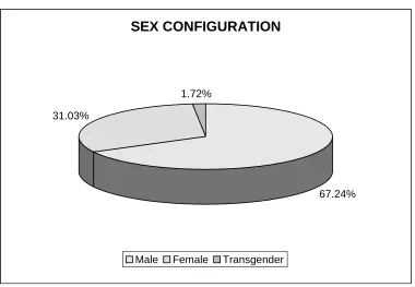
TABLE - 2: SEX DISTRIBUTION – ANALYSIS
Sex No of Cases %
Male 39 67.24
Female 18 31.03
Transgender 1 1.72
Total 58 100
Interpretation:
Among the 58 patients studied the males where more commonly
affected (67.24%) when compared to females 31.03% and transgender of
(1.72%).
SEX CONFIGURATION
67.24% 31.03%
1.72%
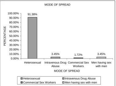
TABLE - 3: MODE OF SPREAD
Mode No of Cases %
Heterosexual (1) 53 91.38
Intravenous Drug abuse (2) 2 3.45
Commercial sex workers (3) 1 1.72
Men having sex with men (4) 2 3.45
Total 58 100
Interpretation:
Among the 58 patients analysed it is the heterosexual transmission that
is most common mode of transmission (91.38%), With men having sex with
men constituting 3.45% and intravenous drug abuse constituting 3.45% and
commercial sex workers constituting 1.72%.
MODE OF SPREAD
91.38%
3.45% 1.72% 3.45%
0.00% 10.00% 20.00% 30.00% 40.00% 50.00% 60.00% 70.00% 80.00% 90.00% 100.00%
Heterosexual Intravenous Drug Abuse
Commercial Sex Workers
Men having sex with men
PE
RCE
NT
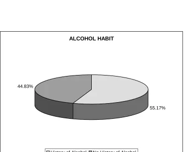
TABLE - 4: ALCOHOLISM - CORRELATION
TO SEXUAL BEHAVIOUR
History of Alcohol Intake No of Cases %
Yes 32 55.17
No 26 44.83
ALCOHOL HABIT
55.17% 44.83%
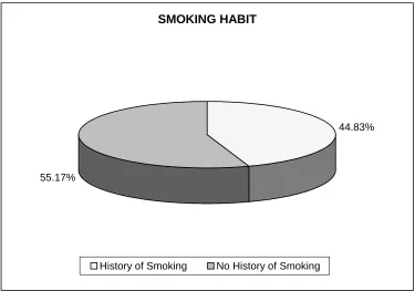
TABLE - 5: SMOKING - CORRELATION TO SEXUAL BEHAVIOUR
History of Smoking No of Cases %
Yes 26 44.83
No 32 55.17
Interpretation:
Among the 58 patients studied 32 cases (55.17%) have history of
Alcohol intake and 26 cases have history of smoking. Overall 18 patients out
of 58 patients were both alcoholic and smoker. So it seems that smoking and
alcohol influence sexual behaviour.
SMOKING HABIT
44.83%
55.17%
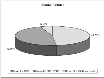
TABLE: 6 - INCOME ANALYSIS
Group No of Cases %
Group I (<1500 Rs.) 28 48.28
Group II (1500 – 2500 Rs.) 27 46.55
Group III (>2500 Rs.) 3 5.17
Total 58 100
Interpretation
Among the 58 patients studied 94.83% of the patients were earning
lessthan Rs.2500 per month. So its seems that lower socio economic status is
playing a role in altering sexual behaviour patterns. But this could also be
because of a sampling bias, as most people who come to Government
Hospital are poor patients.
INCOME CHART
48.28%
46.55%
5.17%
TABLE - 7: ASSOCIATED ILLNESS / DISEASE
Group Associated Illness No of Cases %
1. Yes 6 10.34 %
2. No 52 89.66 %
TABLE - 8: DISEASE CONFIGURATION [ N = 6]
Disease No of Cases %
Pul.TB 4 66.67
Extrapul.TB 1 16.67
Jaundice 1 16.67
Total 6 100
Interpretation:
Among the 58 patients studied, 6 patients had co-existing illness.
Pulmonary tuberculosis was present in 4 patients, extra pulmonary
tuberculosis in one patient and jaundice in one patient. So tuberculosis was
the most common opportunistic infection in the study population. The
associated illness was diagnosed at the time of the initial diagnosis of HIV
and no patient developed any opportunistic infection during HAART so its
TABLE - 9: BMI COMPARISON [ N = 58]
BMI Mean ± SD
Pre HAART BMI 18.84483 ± 2.867248
During HAART BMI (after 6 months ART) 20.77586 ± 3.068191
Interpretation
Among the 58 patients when the initial BMI was analysed it showed a
mean BMI of 18.84483 ± 2.867248, which showed the high incidence of
wasting in the HIV/AIDS patients. The follow up BMI showed a mean BMI
of 20.77586 ± 3.068191%. When analysed for the statistical significance
using paired 't' test its showed 'p' value of 0.000660, which seems to show that
TABLE - 10: CD4 COUNT ANALYSIS [N = 58]
Pre HAART CD4
Count
No of Patients Percentage
Group I 0 - 99 17 29.31
Group II 100 - 149 16 27.59
Group III 150 - 199 25 43.10
Total 58 100
Interpretation
Among the 58 patients, the maximum no. of patients who were started
on HAART had a CD4 count between 150-199 (43.10%) followed by 17
patients in Group I having CD4 count between 0-99 (29.31%) followed by 16
TABLE - 11: CD4 CELL COUNT COMPARISON [N = 27]
CD4 Cell Count Mean ± SD
Pre HAART CD4 127.4444 ± 53.08218
During HAART CD4 (after 6 months follow up) 332.1482 ± 158.08
Interpretation
Among the 27 patients analysed for the impact of HAART on CD4, the
mean increase of 205 cells / mm3 was noted after six months of HAART,
which was also statistically significant when analysed by paired 't' test which
DISCUSSION
HIV/AIDS is an evolving health problem world wide, with more than
40 million people already infected3.
It is important to view untreated HIV infection as a chronic ultimately
fatal process that is punctuated by various manifestations, which are
influenced by multiple factors like route of HIV infection, size of inoculum,
gender, medical intervention etc. In India it is commonly acquired through
Heterosexual contact. In the study the most common mode of transmission
was found to be heterosexual with 91.38% acquiring the disease through this
route.
Once acquiring the infection, one to 6 weeks later patients experience a
nonspecific illness called as "acute retroviral syndrome"26. During this
episode, counts of total lymphocyte (both CD4 and CD8) characteristically
fall, followed by an increase of CD879. CD4 count have been reported to be
between 244 - 1055 cell / mm3 in one review within the first 4 weeks after
acquisition of HIV80. CD4 count may recover for some patients but most
patients demonstrate a decrease of 100 to 200 cells in the first 6 months after
In one review of 318 seroconverters mean CD4+ cell count in the
initial 12 month after seroconversion fell from 999 to 673/ mm81. In this study
the mean initial CD4 count of all 58 patients who were started on HAART
was 130 cells / mm3 ± 53.95.
Studies suggest that early intervention with HAART can slow the
decline of CD4 and reduce the no of clinical events during the initial several
years of infection79,82. Barring one patient who died during the study no
patient developed any clinical illness during the study period. And the natural
history of illness have been dramatically altered by HAART. The likelihood
of an initial AIDS defining condition developing in an untreated person who
is HIV positive average about 4 to 10 percent per year after acquisition of
HIV infection83.
In terms of laboratory parameters the absolute peripheral CD4
lymphocyte count and percentage of peripheral cells that are CD4+, both
correlate with the likelihood of development of AIDS. And also retrospective
and prospective studies show that lower the absolute CD4 count more likely is
the patient to develop opportunistic infections like Cytomegalovirus,
Pneumocystis Carinii pneumonia. Hence the relationship of the CD4 count to
the development of opportunistic infectious complications of HIV is
First, HIV infection implies that unless an effective therapeutic
intervention is administered the immune function inexonerably declines and
infectious complication occur. Second, the monitoring of the immunologic
decline primes the clinician to do the measures in anticipation of the
complication. Third the immunologic state as measured by CD4 cell count
provides guidance regarding the benefit of HAART. When the CD4 count
falls below 200 cells /µl, effective HAART can clearly improve survival. At
higher levels, HAART may improve survival. Fourth, a rise of CD4 in
response to HAART predicts clinical benefit of therapeutic intervention.
During potent antiretroviral therapy, immune recovery is characterized
by suppression of HIV - 1 replication and increasing CD4+ T Cell count84. In
our study group, the CD4 cell count improved by a mean of 205 cells / mm3.
Control of HIV - 1 replication reduces CD4 T cell loss resulting from
direct cytolysis85,86 and may partially restore T cell homeostasis by promoting
decreased T cell proliferation87.88. Redistribution of T cells into peripheral
circulation73,89 and improved thymic output90. Although many patients
continue to have CD4 T cell recovery for several years after receiving
HAART91, the degree of immune recovery achieved during viral suppression
is highly variable. In some individuals increases in the CD4 cell count appears
to plateau after the first few months of HAART92,93,94,95,96. This suboptimal
CD4 T cell response during therapy otherwise known as 'immunologic
discordance' can have detrimental clinical consequences67. At present there is
In general, reconstitution of CD4 T cells during viral suppression
follows a biphasic pattern97. During the first three months of HAART the
number of CD4 T cells typically increase by 50 to 120 cells per mm3, 92,98,99.
This burst is followed by a, second slower phase of T cell repopulation with
an average rate of increase of 2 to 7 cells mm3 per month92,98,99,100.
In our study population, the CD4 count seems to have risen to a greater
degree of about 205 cells / mm3, which could be both due to the smaller
sample size but also could be because of the nutritional counselling that is
given to our patients at the ART centre and also because of the
supplementation of micro and macro nutrients and monthly monitoring of
body weight and height.
The extent of early immune recovery may be a function of prior T
cell destruction, because lower CD4 T cell nadirs have been associated with
limited immune recovery during therapy101. Furthermore because viral
replication is incompletely suppressed by HAART102. In our study the patient
on Group I and Group II showed a lesser increase when compared to Group
III which showed the most statistically significant increase. This increase in
Group III patients during HAART follow up showed a mean increase of 194
cell / mm3, which when analysed for statistical significance by using paired 't'
test showed a 'p' value of 0.000506, which is highly significant, in
concordance with the previous studies. The intent of the present study was to
In this study there were 58 patients with CD4 cell count of less than
200 / µl, who were started on highly active antiretroviral therapy and followed
up for 6 months. Of these 58 patients 17 patients had a CD4 count between
0-99, who were classified a Group I, 16 patients had a CD4 cell count between
100-150, who were classified as Group II and, 25 patients had a CD4 cell
count between 150-199, who were classified as Group III. But, of these 58
patients only 27 patients could be followed up for 6 months and a repeat CD4
count could be done. These 27 patients' 6 month follow up CD4 count was
analysed and it showed an improvement by a mean of 205 cells / mm3, which
was also statistically significant when analysed by paired 't' test, that showed
'p' value of 0.0000. When the groups were analyzed individually, the 3 groups
of group I, group II, group III showed a 'p' value of 0.006, 0.008 and 0.0005
respectively. But it is Group III, with the higher initial CD4 count of 150 –
199 that showed the most statistically significant improvement when
compared to the other two groups. When the improvement in BMI was
assessed after 6 months by using paired 't' test it also showed a statistically
significant improvement, with a 'P' value of 0.000660. From the study it is
clear, when HAART is started, with the CD4 count at a higher level (Group
III), the improvement in CD4 count as well as the general condition
CONCLUSION
1. In this study there were 58 patients with CD4 cell count of less than
200 / µl who were started on highly active antiretroviral therapy and
followed up for six months. But only 27 patients came back after 6
months of HAART, whose follow up CD4 count was done and
analysed to evaluate the impact of HAART on CD4 cell count.
2. It is important to do CD4 cell count in all the patients who are
confirmed as HIV/AIDS, irrespective of the clinical stage, since the
clinical stage and the CD4 count do not correlate.
3. Patients were classified into three groups as per the initial CD4 cell
count. (N = 27)
Initial CD4 Cell Count No of Patients followed
up for 6 months
Group I 0-99 8
Group II 100-149 8
Group III 150-199 11
Total 27
4. These 27 patients 6 month follow up CD4 count was analysed and it
showed an improvement by a mean of 205 cells / mm3 which was also
statistically significant when analysed by using the paired 't' test, that
showed a 'p' value of 0.0000. When the groups were analysed
a 'P' value of 0.006, 0.008, and 0.0005 respectively. Among these 3
groups, it is group III, that showed the most statistically significant
improvement. So HAART has a significant improvement on CD4 cell
count when HAART is started with the CD4 count at a higher level as
in Group III.
5. HAART has improved the BMI and thereby improving the general
condition and well being of the patients. This could also be attributed
to the micronutrients and the macronutrients that were provided to the
patients at the ART centre, KMCH.
6. HAART decreases the incidence of opportunistic infections.
7. Tuberculosis was the most common opportunistic infection.
8. CD4 cell count monitoring is very important and could be done every
3 months, but for resource constraints it is being done every 6 months.
9. Limitation of the study:
(a) small sample size,
(b) four combinations of HAART regimens where used in these 27
patients and the individual effect of each combination on CD4
BIBLIOGRAPHY
1. Barre - Sinousse, F. J.C. Chermann, F.Rey, mt Nugeyre, S Chamaret, j. Gruest, C. Dauguet, Caxler Blin, F. Vezient Brun, C. Rouzioox, W. Rozen Baum and L. Montagnier 1983. Isolation of a t - Lymphotropic retrovirus from a patient at risk for AIDS. Science 220 : 868-871.
2. Gallo R.C; SZ. Salahuddin M. Popovic; G.M. Shearer, M. Kaplan; B.F. Haynes; T.J. Palker; R. Redfield J. Oleske; B. Safai et.,al. 1984. Frequent detection and isolation of cytopathic retroviruses (HTLV - III) from patients with AIDS and at risk for AIDS. Science 224 : 500-503.
3. Joint united nations programme on HIV / AIDS 2004. 2004 report on the global HIV / AIDS epidemic : 4th global report.
4. Centers for Disease Control and prevention. HIV / AIDS Surveillance Reports, 1989 - 2003 - Atlanta, GA.
5. From Centers for Disease Control and Prevention. 1993 Revised classification system for HIV infection and expanded surveillance case definition for AIDS among adolescents and adults. MMWR Morb Mortal Wkly Rep. 1992-41:1-19.
6. QUINN TC. Wawer MJ. Sewan Kambo. N et al. Viral load and
Heterosexual transmission of HIV type 1 NEJM. 2000; 342 : 921-929.
7. Clemetson DBA. Moss Gm, Willer ford DM et.,al. Detection of HIV DNA in cervical and vaginal secretions : prevalence and correlates among women in Nairobi, Kenya. JAMA, 1993; 269 : 2860-2864.
8. Havlir DV, Richman DD. Viral dynamics of HIV : Implications for drug development and therapeutic strategies. Ann Intern Med. 1996; 124 : 984-989.
9. Henrard DR, Phillips JF, Muenz LR, et al. Natural history of HIV-1 cell-free viremia. JAMA. 1995 : 274 : 554-558.
10. Phair J, Jacobson L., Detels R, et al. Acquired immune deficiency syndrome occuring within 5 years of infection with human immunodeficiency virus type 1: The Multicenter AIDS Cohort Study.
11. Cao Y, Qin L, Zhang L, et al. Virologic and immunologic characterization of long-term survivors of human immunodeficiency virus type 1 infection. N Engl J Med. 1995; 332 : 201-208.
12. Sheppard HW, Lang W, Ascher MS, et al. The characteristics of non-progressors : Long term HIV-1 infection with stable CD4+ T cell levels. AIDS. 1993; 7: 1159-1166.
13. Fahey JH, Taylor JM, Detels R, et al. The prognostic value of cellular and serologic markers in infection with human immunodeficiency virus type 1. N Engl J Med. 1990 : 322 : 166-172.
14. Mellors JW, Munoz A, Giorgi JV, et al. Plasma viral load and CD4+ lymphocytes as prognostic markers of HIV-1 infection. Ann Intern Med. 1997; 126 : 946-954.
15. Mocroft A, Johnson MA, Phillips An. Factors affecting survival in patients with AIDS. AIDS. 1996; 10 : 1057-1065.
16. Osmond D, Harlebois E, Lang W, et al. Changes in AIDS survival time in two San Francisco cohorts of homosexual men, 1983 to 1993.
JAMA. 1994 : 271 : 1083-1087.
17. Saravolatz L, Neaton J, Sacks L, et al. CD4+ lymphocyte counts and patterns of mortality among patients infected with human immunodeficiency virus who were enrolled in community programs for clinical research on AIDS. Clin Infect Dis. 1996; 11 : 513-520.
18. Schubach J, Flepp M, Pontelli D, et al. Heat-mediated immune complex dissociation and enzyme-linked immunosorbent assay signal amplification render p24 antigen detection in plasma as sensitive as HIV-1 RNA detection by polymerase chain reaction. AIDS. 1996; 10 : 1085-1090.
19. Sterling TR, Hoover Dr, Astemborski J, et al. Heat-denatured human immunodeficiency virus type 1 protein 24 antigen : Prognostic value in adults with early-stage disease. J Infect Dis. 2002; 186 : 1181-1185.
21. Spacek LA, Griswold M, Quinn TC, Moore RD. Total lymphocyte count and hemoglobin combined in an algorithm to initiate the use of highly active antiretroviral therapy in resource-limited settings, AIDS. 2003; 17 : 1311-1317.
22. Lau B, Gange SJ, Phair JP, et al. Rapid declines in total lymphocyte counts and hemoglobin concentration prior to AIDS among HIV-1 infected men. AIDS. 2003; 17 : 2035-2044.
23. Corbett EL, Wat CJ, Walker N, et al. The growing burden of tuberculosis: global trends and interactions with the HIV epidemic.
Arch Intern Med. 2003; 163 : 1009-1021.
24. Greenspan D, Greenspan JS, Hearst NG, et al. Relation of oral hairy Leukoplakia to infection with the human immunodeficiency virus and risk of developing AIDS. J Infect Dis. 1987; 155 : 475-481.
25. Chaisson RE, Gallant JE, Keruly J, Moore RD. Impact of opportunistic disease on survival in patients with HIV infection. AIDS. 1998; 12: 29-33.
26. Miu MT, Stein DS, Schnittman SM. Primary human immunodeficiency virus type 1 infection Review of pathogenesis and early treatment intervention in human and animal retrovirus infections.
J Infect Dis. 1993; 168 : 1490-1501.
27. Henrard DR, Phillips J, Windsor I, et al. Detection of human immunodeficiency virus type 1 p24 antigen and plasma RNA : Relevance to indeterminant serologic tests Transfusion. 1994; 34 : 376-380.
28. Rosenberg ES, Billingsley JM, caliendo AM, et al. Vigorous HIV-1 specific CD4+ T cell responses associated with control of viremia.
Science. 1997; 278 : 1447-1450.
29. Finzi D, Hermankova M, Pierson T, et al. Identification of a reservoir for HIV-1 in patients on highly active antiretroviral therapy. Science. 1997; 278 : 1295 - 1300.


![TABLE - 9: BMI COMPARISON [ N = 58]](https://thumb-us.123doks.com/thumbv2/123dok_us/1258082.78119/50.612.116.506.129.225/table-bmi-comparison-n.webp)
![TABLE - 10: CD4 COUNT ANALYSIS [N = 58]](https://thumb-us.123doks.com/thumbv2/123dok_us/1258082.78119/51.612.121.504.128.313/table-cd-count-analysis-n.webp)
![TABLE - 11: CD4 CELL COUNT COMPARISON [N = 27]](https://thumb-us.123doks.com/thumbv2/123dok_us/1258082.78119/52.612.117.503.117.198/table-cd-cell-count-comparison.webp)