A DISSERTATION ON THE COMPARATIVE
STUDY OF THE EFFICACY OF VARIOUS
TOPICAL TREATMENT MODALITIES IN
PALMOPLANTAR PSORIASIS
Dissertation Submitted to
THE TAMIL NADU DR. M.G.R. MEDICAL UNIVERSITY in partial fulfillment of the regulations
for the award of the degree of
MD DEGREE IN
Dermatology, Venereology and Leprology
(BRANCH XII A)
MADRAS MEDICAL COLLEGE
THE TAMIL NADU DR. M.G.R. MEDICAL UNIVERSITY
CHENNAI
CERTIFICATE
Certified that this dissertation entitled “Comparative Study of the efficacy of various topical treatment modalities in Palmoplantar
psoriasis” is a bonafide work done by DR. V.RENUKA, Post graduate student of the Department of Dermatology and Leprology and Institute
of Venereology, Madras Medical College, Chennai- 3, during the
academic year 2004 – 2007. This work has not previously formed the basis for the award of any degree or diploma.
Prof. Dr. B. PARVEEN, M.D., D.D., Professor and Head of the Department,
Department of Dermatology and Leprology, Madras Medical College,
Chennai- 3.
Prof. Dr .KALAVATHI PONNIRAIVAN, B. Sc., M.D.,
Declaration
I, Dr. V.RENUKA, solemnly declare that dissertation
titled, “COMPARATIVE STUDY OF THE EFFICACY OF VARIOUS TOPICAL
TREATMENT MODALITIES IN PALMOPLANTAR PSORIASIS”
is a bonafide work done by me at Madras Medical College
during 2004-2007 under the guidance and supervision of
Prof. Dr. B. PARVEEN, M.D.,D.D., Professor and Head, Department of
Dermatology, Madras Medical College, Chennai-600 003.
The dissertation is submitted to The Tamilnadu, Dr. M.G.R.
Medical University, towards partial fulfillment of requirement for the
award of M.D. Degree in Dermatology, Venereology and Leprology
(BRANCH – XII A).
Place : Chennai.
Date :
SPECIAL ACKNOWLEDGMENT
My sincere thanks to Prof.Dr.KALAVATHI
PONNIRAIVAN, B. Sc., M.D., The DEAN, Madras Medical
College for allowing me to do this Dissertation and utilize the
ACKNOWLEDGEMENTS
I am gratefully indebted to Prof. Dr. B. Parveen M.D., D.D.,
Professor and Head of Department of Dermatology and Leprology for her
invaluable guidance, motivation and help though out the study. I would like
to express my sincere and heartfelt gratitude to Prof. Dr. V.S. Dorairaj,
M.D., D.V., Director in charge, Institute of Venereology.
I wish to thank Dr. N. Gomathy M.D., D.D., former Professor,
Department of Dermatology and Dr. N. Usman M.D., D.V., Ph.D., former
Director, Institute of Venereology for their constant support and motivation.
I am very grateful to Dr. S. Jayakumar M.D., D.D., Additional
Professor, Department of Dermatology for his invaluable guidance and help.
I sincerely thank Dr. C. Janaki M.D., D.D., Reader of Dermatology
(Mycology) for her priceless support.
I express my earnest gratefulness to Dr. D. Prabavathy M.D., D.D.,
Professor and Head of Department of Occupational Dermatology and
Contact Dermatitis for her constant motivation and guidance. I thank
Dr. V. Somasundaram M.D., D.D., Additional Professor, Department of
Occupational Dermatology and Contact Dermatitis for his benevolent help
I express my sincere gratitude to Dr. K. Rathinavelu M.D., D.D.,
Professor of Leprosy and Dr. R. Arunadevi M.D., D.D., Lecturer/Registrar,
Department of Dermatology for their support.
I incline to thank Dr. R. Priyavathani M.D., D.D., D.N.B.,
Dr. V. Anandan M.D.,(Derm), D.C.H., D.N.B.,(Paed) and
Dr. G.K. Tharini M.D., Dr. M.Vijay Anand M.D.,(Derm),
Assistant Professors, Department of Dermatology for their kind support and
encouragement.
I thank Dr. A. Hameedullah M.D., D.D., Dr. S. Kumaravelu M.D.,
D.D., Dr. J. Manjula M.D., D.N.B., (Derm) and Dr. Aftab Jameela Wahab
M.D., D.D., Assistant Professors, Department of Occupational Dermatology
and Contact Dermatitis for their support and help.
My sincere thanks to Dr. S. Mohan M.D, D.V. former Registrar,
Dr. V. Thirunavukkarasu M.D., D.V., Dr. K. Venkateswaran M.D., D.V.,
Dr. P. Elangovan M.D., D.V., Dr. D. Ramachandra Reddy M.D., D.V.,
Dr. S. Thilagavathy M.D., D.V., Dr. P. Mohan M.D., D.V.,
Dr. S. Arunkumar M.D., D.V., and Dr. S. Kalaivani M.D., D.V.,
I am also thankful to Dr. K. Manoharan M.D., D.D., and
Dr. V. Sampath M.D., D.D., for their continuing guidance and support.
I duly acknowledge the paramedical staff and my colleagues for their
help and favour.
Last but not least I am profoundly grateful to all patients for their
CONTENTS
Sl.No Title Page No
1
INTRODUCTION 12
REVIEW OF LITERATURE 53
AIM OF THE STUDY 414
MATERIALS AND METHODS 425
OBSERVATIONS AND RESULTS 456
DISCUSSION 497
CONCLUSION 528
REFERENCES9
PROFORMAINTRODUCTION
Psoriasis is a common, genetically determined, inflammatory and
proliferative disease of the skin. The most characteristic lesions consist of
red, scaly, sharply demarcated, indurated plaques present particularly on the
elbows, knees, lowerback, extensor surfaces and scalp.
The first recognisable description of psoriasis is attributed to Celsus
(25BC-45AD) in his de re medica nearly 2000 years ago. The disease was
described under the heading of impetigo from the Latin word impeto which
means "to attack or rush on" Galen was the first to use the word psoriasis
from the Greek work 'psora' which means 'to itch'. Psoriasis and Leprosy
were grouped together for centuries. Willan was the first to accurately
describe psoriasis and its various manifestations in 1809, but he did not
separate it with certainty from Leprosy. In 1841, Hebra definitively
distinguished the clinical picture of psoriasis from that of Hansen's disease.
Eventhough a number of treatment modalities are available, psoriasis
continues to be a therapeutic challenge in spite of our growing knowledge
DIFFERENT CLINICAL TYPES OF PSORIASIS
Classical types Psoriasis vulgaris
Guttate psoriasis Pustular psoriasis Arthropathic psoriasis Erythrodermic psoriasis
Special types Rupioid
Ostraceous Elephantine
Atypical forms Follicular
Verrucous
Lichenoid variety Linear
Zonal
Seborrhoeic Mucosal lesions Ocular lesions
According to site Scalp
Penis Flexural Nail
Palmo plantar psoriasis is one of the type of psoriasis which can occur
alone or along with the involvement of other areas.
In most cases the lesions are well defined but they are less scaly and
the surface often shows fissures. It may be pustular or non pustular
Three forms of lesions can occur in palms and soles
1. Diffuse hyperkeratotic plaques
2. Erythematous patches or plaques studded with minute superficial pustules
3. Discrete scaly plaques or patches
4. Rarely Rupioid Lesions can occur on the soles with characteristic limpet
like scales.
Palmo plantar psoriasis can also be classified as
1. Pustular - Acute pustular bacterid
Chronic pustular bacterid
Palmo plantar psoriasis with pustules
2. Non pustular - Diffuse
Annular
Delling or crateriform
In this study various topical modalities of treatment are used for
palmoplantar psoriasis like
0.1% Betamethasone valerate ointment
0.05% Tazarotene gel
Topical PUVA using methoxypsoralen solution 1%
Short contact compound dithranol oint (dithranol 1.15%, salicyclic acid
1.15%, coal tar 5.3% in white soft paraffin)
Liquid paraffin.
There are numerous topical therapies available like coaltar, anthralin,
methotrexate, vitamin D analogues tacrolimus, salicylic acid 2 - 10%.
LITERATURE REVIEW
INTRODUCTION
Palmo plantar psoriasis has various therapeutic options both topical
and systemic. The topical modalities of treatment generally used are
1. Coal tar
2. Dithranol
3. Topical corticosteroids
4. Vitamin D analogues
a. Calcitriol
b. Calcipotriol
c. Tacalcitol
d. Maxacalcitol
5. Topical psoralen
6. Topical retinoid (Tazarotene)
7. Topical cytostatic therapy
a. Mechlorethamine (Nitrogen mustard)
b. Thiotepa
c. 5 - Flurouracil
d. Lomustine
8. Tacrolimus (FK 506)
9. Emollients
10. Salicylic acid 2 - 10%
11. Arachidonic acid 0.5 - 2%
12. Topical allantoin
COAL TAR
Tar has been used in Topical therapy for more than a century
DIFFERENT TYPES OF TAR [1]
a. Shale tar
Icthammol
b. Wood tar
Juniper tar
Pine tar
Pix liquida
c. Coal Tar
Coal tar solution
Coal tar is a complex mixture of thousands of substances produced by
primary condensation during the carbonization of coal[2]. Some 400 known
substances comprise 55% of Tar by weight.
PREPARATIONS
Coal tar is available as ointment, liquid, alcohol extract, gel, shampoo
and soap. Each has its own used in specific areas. It is also available in
combination with salicylic acid.
MECHANISM OF ACTION
1. Anti-Mitotic
Coal Tar has been found to depress mitosis and DNA synthesis by the
production of DNA adducts and oxidation induced DNA damage in
human mammary epithelial cells [3].
2. Phototoxicity
Goeckerman in 1925, described the enhancement of the therapeutic
effect of tar after UV light exposure. The following components of
coal tar act as photosensitizers, anthracene, fluoranthrene,
Ultraviolet fluorescence microscopy has proved that the hair follicles
and the sebaceous glands may be an important route of penetration of
the coal tar into the skin.
3. Sebostatic effect
It has been found that a 10% coal tar distillate reduces the size of the
sebaceous glands and the number of mitoses. It is possible that this
will lead to a subsequent reduction of the secretion of sebum [4].
4. Antifungal effect[5]
It has been demonstrated that coal tar has an antifungal potential on
Malassezia furfur in vitro. This could, in part, be responsible for the
response of seborrhoeic dermatitis and pityriasis capitis to coal tar [5].
INDICATIONS
Apart from its use in psoriasis, coal tar can also be used in pityriasis
capitis and seborrhoeic dermatitis [4]. It has also been used in eczemas,
ADVERSE EFFECTS
Irritation folliculitis and tar acne are the most frequent adverse effects
of tar treatment. Phototoxic reactions are also common. Allergic contact
dermatitis does occur but is rare.
Kennaway identified two carcinogens in coal tar, 3,4 benzpyrene and
1, 2-benzpyrene. A 2.4 fold increased risk of developing skin carcinomas
was found in patients with high rate of exposure to tar and UV light [7].
PREPARATIONS
Coal tar is available in the form of ointments, creams, gels, scalp
shampoos, solutions, lotions and suspensions.
COMBINATION THERAPIES
Coal tar can be combined with various other drugs for better efficacy.
1. Goeckermans regimen [8]
It consists of daily application of 2 - 5% crude tar, combined with a
regimen have been advocated using alcoholic extracts of tar in cream
or ointment bases, tar gels, etc. which are easier to handle.
2. Coal tar and Dithranol [9]
A combination of 5% crude coal tar and dithranol was found to be as
effective as dithranol alone when used in short contact treatment of
psoriasis and was also less irritant.
3. Coal tar and topical steroids [10]
This combination reduces the irritation caused by coal tar.
4. Coal tar and salicylic acid.
Used in treatment of psoriasis
PRECAUTIONS
1. Coal tar application should be avoided over face, genitalia and flexures.
2. Coal tar therapy is contraindicated in erythrodermic and generalised
pustular psoriasis.
3. Pre existing folliculities or severe acne are also possible contraindications.
DITHRANOL
In history of medicine there are a few accidents proved to be good for
obtained from araroba tree. The word araroba meaning ‘tawny coloured’.
This is the active ingredient of Goa powder, imported by Portuguese from
Brazil to India [11]. Following misdiagnosis it was accidentally used in
psoriasis and found to be effective . It was called "Cignolin" in Germany
and "Anthraline” in North America.
Structural Chemistry[13]
OH O HO
H H
Anthralin has an unstable chemistry with 2 focal points: phenolic
hydroxyl groups at carbon atoms 1 and 8 and two reactive hydrogen atoms
on carbon ten. It undergoes auto-oxidation, which is both pH and light
MECHANISM OF ACTION
1. Antiproliferative effect
It induces a decrease in epidermal growth factor binding in a dose
dependent manner. This may contribute to the anti psoriatic action of
dithranol [14]
Dithranol also causes a marked decrease in transforming growth
factor α MRNA expression on cultured human keratinocytes [15].
2. Action of anthralin on mitochondria
It interacts with the electron transport chain on the inner
mitochondrial membrane resulting in a reduction of ATP Synthesis [13]
This loss of energy supply in keratinocytes including reduction of
glucose 6 phosphate dehydrogenase could explain, at least in part, the
therapeutic efficiency of anthralin in psoriasis.
3. Inhibition of biosynthesis of polyamine by anthralin
Ornithine decarboxylase catalyses the production of putrescine from
ornithine and is associated with cellular proliferation. In psoriatic
epidermis, polyamine levels are abnormally elevated, so anthralin
4.Effect of anthralin on cyclic nucleotides
Anthralin has also been shown to reduce the elevated levels of cyclic
guanosine mono-phosphate (cGMP) that were present in involved
skin. Although the precise mechanism is not known, there is much
evidence to suggest that it acts on various cell regulatory systems [13].
5. Anti chemotactic activity of anthralin.
It is a potent inhibitor of leukotriene production and LTB4 - omega
oxidation by human neutrophils thereby reducing the LTB4 - induced
intra epidermal accumulation of polymorphonuclear leucocytes [16].
Also significantly lowers IFN - gamma and TNF alpha levels in
psoriatic patients [17].
Anthralin has been found to cause significant fall in epidermal
calmodulin levels in the psoriatic lesions associated with clearance of
psoriatic lesions [18].
Anthralin also down regulates 12(S) hydroxy ecosotetraenoic acid
(12 (S) HETE) receptors on epidermal cells which may contribute to
its antipsoriatic action [19].
INDICATIONS
1. Psoriasis - both as monotherapy and in combination with various other
2. Facial seborrhoeic dermatitis in low-dose preparations [20]. At higher
concentrations shown to inhibit growth of pityrosporum ovale [21].
3. Inflammatory linear verrucous epidermal naevus [22].
4. 2% use in verruca vulgaris [23]
5. Alopecia areata [24]
PREPARATIONS
Anthralin is available as paste, ointment, cream and sticks.
MODIFICATIONS OF ANTHRALIN THERAPY
1. Short contact therapy [11,12]
With the hope of avoiding the disadvantages, higher strength of
dithranol is kept in contact with lesion for a short period.
Concentrations ranging from 0.5 to 3% and even upto 8% have been
tried as short contact therapy for 10 -20 minutes and have been found
to be successful [25].
2. Ingram regimen
First the patient soaks and washes thoroughly in 90 litre (20 gallon)
bath to which 120 ml of alcoholic solution of coal tar and 30ml of
teepol is added. After drying, small doses of UVB are given,
increasing gradually to suberythmogenic dose. Then the lesion is
lassars paste, which is powdered with talc and covered with stockinet
dressing. The patient then dresses and pursues his work, returning for
treatment after 24 hours. After 24 hours, the paste being removed
with olive oil prior to bathing the following day [13].
Disadvantages of this treatment
1. Requires hospitalization
2. Staining of the lesion and dress
3. Irritation at the site
3. Liposomal delivery of dithranol.
There were no reports of lesional or perilesional irritation and only
one patient showed faint brown staining of the skin [26].
4. Micanol
In this dithranol is micro encapsulated in crystalline monoglyceride.
This preparation is easy to wash off and staining and irritation are
inconspicuocs. Hence, it can be used in out patient setting [27].
COMBINATION THERAPIES
Anthralin can be used in combination with
a. PUVA
b. Narrowband UVB
d. Topical steroids
e. Calcipotriol
f. Tazarotene
g. Oral cyclosporine
h. Oral retinoids
ADVERSE EFFECTS
1. Anthralin erythema is due to release of prostaglandin and superoxide
radicals released from C1O methylene group of anthralin and anthralin
radicals produced in the skin [28].
2. Staining and pigmentation is due to formation of anthraquinone oxidation
products.
3. Rarely skin cancer
4. Ocular damage
5. Allergic contact dermatitis
PRECAUTIONS
1. Should be applied only to psoriatic plaques and not to normal skin
2. Better avoided in face.
3. Contra-indicated in acute, unstable, generalised pustular and
CONCLUSION
Anthralin has been available for many years and has been an useful
anti-psoriatic agent. It must be used with care and instructions for its use are
required for both physicians and patients. The introduction of short contact
therapies may make this compound more useful in treatment of palmoplantar
psoriasis.
TAZAROTENE
Tazarotene is a third generation receptor selective polyaromatic,
synthetic retinoid also known as Arotenoid. It is a prodrug of a water
soluble active metabolite called tazarotenic acid. The conversion takes place
in the skin by the help of an enzyme called esterase.
It has high affinity for RAR βγ than for RAR α. But no affinity for
RXR.
MECHANISM OF ACTION
Down regulates abnormal expression of keratinocyte transglutaminase
(T gase 1), epidermal growth factor receptor and hyper proliferative keratin
K6/K16.
Blocks the induction of ornithine decarboxylase activity, which is
associated with cell proliferation and hyperplasia.
Decreases migration inhibitory factor related protein (MRP-8) a
marker of inflammation in psoriasis[29].
Up regulates tazarotene inducible genes (TG 1, 2, 3) and has
antiproliferative properties
PHARMACO KINETICS
Systemic absorbtion is negligible because of rapid metabolism (less than 20 minutes) to hydrophilic metabolite and limited percutaneous penetration. Onset of action is within two weeks and elimination half life is 17 to 18 hrs.
Excreted through skin, urine and faeces. It is available as cream or gel (0.05% and 0.1%)
PRECAUTIONS
Simultaneous use of tazarotene should be avoided with abrasive or medicated soap, products containing high concentration of alcohol, astringents, medications or cosmetics having strong drying effects
INDICATIONS
Mild to moderate psorasis involving 10% - 20% of body surface area[30] Acne vulgaris
Cutaneous T cell lymphoma
Disorders of keratinization (Dariers disease, pityriasis rubra pilaris, ichthyosis)
Treat and prevent skin cancer (Basal cell carcinoma, Xeroderma pigmentosum) [31]
CONTRAINDICATION
Pregnancy
Lactation
SIDE EFFECTS
Skin irritation, which is characterized by erythema, peeling,
desquamation, dryness, burning and pruritus[32]
Hypo or hyper pigmentation
Koebnerization of psoriasis
TOPICAL CORTICOSTEROIDS
INTRODUCTION
The revolution brought about in dermatological therapy by the
introduction of topical corticosteroids, which started with compound F or
hydrocortisone in 1952, is well known. Topical steroids play an important
role in the treatment of psorasis particularly when combined with other
STRUCTURE
Hydrocortisone is the parent compound of modern glucocortico
steroid derivatives. It has a cyclopentanoperhydrophenanthrene nucleus,
which has been chemically modified over the years to enhance
glucocorticosteroid activity and minimise mineralocorticoid effects[33].
Factors affecting the potency of topical corticosteroids are
lipophilicity of the molecule, additional double bond at 1, 2 position,
addition of halide at 9, 6, or 21 position and the state of the epidermal
barrier. Halogenation of the 9 position will significantly increase
glucocorticoid activity.
CLASSIFICATION[34]
The introduction of the vasoconstrictor assay has proved of great
value in the classification of topical corticosteroids. The assay depends
upon the property of corticosteroids to produce transient vasoconstriction.
The corticosteroids are classified into the following groups based on
their potency.
1. Very potent : Clobetasol propionate 0.05%
Halcinonide 0.5%
Beclomethasone dipropionate 0.05%
2. Potent : Beclomethasone dipropionate 0.025%
Betamethasone valerate 0.1%
Fluocinolone acetonide 0.05%
Fluocinonide 0.05%
Fluticasone propionate 0.05%
Momentasone furoate 0.1%
3. Moderately Potent : Triamcinolone acetonide 0.1%
Hydrocortisone butyrate 0.1%
Desonide 0.5%
Flurandrenolone 0.05%
4. Mildly Potent : Clobetasole butyrate 0.05%
Fluocortolone hexonate 0.1%
5. Weak : Hydrocortisone 0.1 - 2.5%
MECHANISM OF ACTION
Corticosteroids have anti-inflammatory, immunosuppressive and
antimitogenic activities due to their ability to exert multiple effects on the
various functions of leucocytes, epidermal and dermal cells[35].
Corticosteroids diffuse through the stratum corneum barrier and
through the cell membranes reach the cytoplasm. In the cytoplasm they bind
with glucocorticoid receptor α (GRα) and then enters the nuclear
compartment and interacts with glucocorticoid responsive elements on the
genome.
In addition, the ligand bound receptor can inhibit directly or
indirectly, the activity of other transcription factors like NFKB, AP-1 and
NFAT[36].
These interactions leads to suppression of the production of
inflammatory cytokines, inhibition of T cell activation, changes in the
function of endothelial cells, granulocytes, mastcells, fibroblast and
They induce synthesis of lipocortin that regulate the activity of
phospholipase A2. This enzyme affects the production of arachidonic acid,
the precursor for leukotrienes and prostaglandins[37].
There is one more receptor in the cytoplasm called the GRβ, which is
an endogenous inhibitor of glucocorticoid action[38]. Staphylococcal
superantigen can upregulate expression of GRβ, providing a potential
mechanism by which these bacteria might induce corticosteroid
resistance[39].
THERAPEUTIC USES
Papulosquamous disorder.
1. Psoriasis
2. Lichen planus[40]
3. Reiters disease
Eczemas
1. Atopic dermatitis[41]
2. Seborrhoeic dermatitis[42]
4. Photocontact dermatitis
5. Lichen simplex chronicus
6. Actinic reticuloid[43]
7. Pompholyx[42]
8. Asteatotic eczema
Bullous disorders
1. Pemphigus group[43]
2. Bullous pemphigoid[46]
3. Epidermolysis bullosa
4. Sub corneal pustular dermatosis
Collagen vascular diseases
1. Lupus erythematosus
2. Dermatomyositis
Miscellaneous
1. Alopecia areata
2. Hemangiomas
3. Mycosis fungoides[44]
5. Vitiligo
6. Cutaneous amyloidosis
7. Keloid / Hypertrophic scar
8. Aphthous stomatitis
9. Mastocytosis and Urticaria pigmentosa
10. Acne rosacea
11. Vasculitis, etc.
PREPARATIONS
They are available as ointments, creams, lotions, gels, foams, sprays
and tapes[46].
COMBINATION THERAPIES
Topical corticosteroids have been used in combination with various
other topical and systemic agents in the treatment of psoriasis. They are
1. Coal tar[47]
2. Anthralin
3. Calcipotriol[48]
4. Tazarotene[49]
ADVERSE EFFECTS
Epidermal : Thinning of epidermis
Reduction in the size of epidermal cells
Reduction in the metabolic activity
Reduction in the number of cell layers
Inhibition of melanocyte function with hypopigmentation
Dermal : Resorption of mucopolysaccharide ground substance with atrophy[51]
Fragility of skin
Suppression of collagen synthesis, so striae
Vasculature : Loss of connective tissue support leading to
Erythema
Telangiectasia[52]
Miscellaneous
Perioral dermatitis[52]
Acneiform eruption
Acne rosacea
Glaucoma
Cataracts and retinal detachment[53] Allergic contact dermatitis[54]
Hypertrichosis
Infantile gluteal granulomas
Delayed wound healing
Cutaneous lymphangiectases[55] Tachyphylaxis.
SYSTEMIC
Suppression of the pitutary - adrenal axis can occur with virtually any
topical steroid especially potent steroids, leading to cushing syndrome[56]
PRECAUTIONS
No more than 45 gm per week of moderate to potent steroids in adults
and 15gm per week in young children and infants are recommended for
topical use.
Corticosteroids on continued use exhibit tachyphylaxis, the
Sudden withdrawal of topical steroids in psoriasis can precipitate
pustular psoriasis.
Topical steroids are to be used with caution over the face.
CONCLUSION
Topical steroids play an important role in the treatment of psoriasis.
They should be used judiciously because of the possibility of rebound
phenomenon when they are discontinued as well as the potential
development of topical and systemic adverse effects. They can play an
important role as an adjunct to other forms of therapy in the treatment of
psoriasis.
SALICYLIC ACID
INTRODUCTION
Willow bark (Salix alba) is an ancient herbal remedy. The chemical
structure of its active ingredient, salicylic acid (2-hydroxy-benzoic acid) was
identified in 1838 [57]
Salicylic acid is a crystalline powder. It is lipid soluble and hence
To obtain significant keratolytic effect salicylic acid must be
formulated at a proper pH so that there will be enough of free acid.
STRUCTURE
COOH
OH
MECHANISM OF ACTION
Reduction of corneocyte adhesion, perhaps by an action on the cement
substance. This results in enhanced shedding of corneocytes and has no
effect on mitotic activity[58].
Topical Salicylic Acid
Corneocytes desquamate
Corneocyte pH of Stratum
Intercellular bonding Corneum
Corneocytes
Salicylic acid thus can cause enhancement of penetration of other
chemicals like topical corticosteroids.
Salicylic acid can be photoprotective[59].
It also has bacteriostatic and bactericidal activity against yeast, gram
negative and gram positive bacteria[57]
INDICATIONS
Psoriasis[60]
Seborrhoeic dermatitis[58]
Ichthiosis[58]
Warts[61]
Corns and calluses[58]
Acne[58]
Pityriasis rubra pilaris[62]
Keratodermas[58]
Dermatophyte infections[58]
Avulsion of toe nails[58]
Sunscreens[63]
PREPARATIONS
Salicylic acid is available in
Collodion - based paints and gels
Shampoos
Ointments
Paste (Lassars paste contains salicylic acid and zinc oxide)
Creams
COMBINATION THERAPIES
Salicylic acid is used commonly in combination with various other
topical therapeutic agents like,
Corticosteroids[65] Coal tar
Other Keratolytics Lactic acid
Propylene glycol
Urea[66] Sulphur
Benzoic acid ( Whitfield ointment) Zinc oxide (Lassars paste)
ADVERSE EFFECTS
Irritation and maceration at high concentrations
Contact sensitization
Systemic toxicity (Salicylism) [68]
The manifestations include nausea, vomiting, confusion, dizziness,
delerium, psychosis, stupor, coma and death. Can also cause marked hyper
ventilation and respiratory alkalosis.
PRECAUTIONS
Increases the toxicity of other topical agents because of enhanced
absorption and hence should be used with caution.
Prolonged use should be avoided to prevent salicylism.
Used with caution in patients with hepatic and renal impairment
especially young children.
CONCLUSION
Salicylic acid, especially when used cautiously, is an effective and
safe antipsoriatic agent.
TOPICAL PUVA THERAPY
Psoralen photochemotherapy is a combination of psoralen (P) and
long wave UVR (PUVA) that brings about a therapeutically beneficial result
not produced by either the drug or radiation alone [70,71].
Application of 8-MOP in creams, ointments or lotions followed by
UVR is effective in clearing psoriasis but has several disadvantages.
PSORALEN PHOTO CHEMISTRY
Ground state psoralen molecules are activated to the excited singlet
state by absorption of photons in the UVA wave band. The peak of the
absorption spectrum is around 320 – 330 nm. Two types of photo chemical
reaction occur[72].
Type 1 – (Direct) resulting in mono functional adducts in DNA
Type-2 – (Indirect) reactions resulting in production of reactive O2
species and free-radicals that cause damage to cell membrane and
cytoplasmic constituents[73].
MECHANISM OF ACTION
Suppersion of DNA Synthesis, through formation of mono and bi
functional adducts [74], which occurs immediately. Subsequently cell
proliferation is also inhibited in patients with psoriasis who receive repeated
PUVA treatment.
IMMUNOLOGIC EFFECTS
PUVA down regulate certain lymphocytes and antigen presenting
cells and influences adhesion molecule expression [75].
Selective cytotoxicity through production of free radicals
Stimulation of melanocyte proliferation and increased tyrosinase
INDICATIONS [76-79]
Psoriasis[80,81] Vitiligo
Mycosis fungoides Lymphomatoid papulosis Pityriasis lichenoides
Langerhans cell histiocytosis Atopic dermatitis[82]
Seborrhoeic dermatitis Pityriasis rubra pilaris Lichen nitidus
Alopecia areata Urticaria pigmentosa Actinic prurigo[83]
MODE OF ADMINISTRATION
Applied in the form of lotion and creams. A lotion containing 0.1 to
1.0 % 8 methoxypsoralen is used. Then the area should be exposed to UVA.
This treatment is given twice weekly with UVA dose being gradually
The hands should be immediately washed and topical emollients
should be applied and further photo protection from sunlight insisted using
sun screeners.
LIGHT SOURCES FOR UVA
UVA radiation spectrum ranges from 320 to 400 nm. The PUVA
action spectrum is defined as the effectiveness of clearing psoralen
sensitized psoriasis as a function of wave length. Currently broad band high
intensity UVA sources ranging from 320 to 400 nm with a peak emission at
352 nm are typically used in PUVA.
However recent advances demonstrate the peak action spectrum of
psoriasis to be around 330 nm.
The commonly used lamps are [84]
1. Fluorescent lamps
PHOTO SENSITIVE EFFECTS OF PUVA
PUVA produces an inflammatory response that manifest as
1. Delayed phototoxic erythema [85,86]
2. Intense pruritus
3. Pigmentation
These reactions are related to the dose of the drug and of UVA and to
the individual sensitivity to phototoxic reaction.
TREATMENT PROCEDURE
Before starting a patient on PUVA therapy contra indication to it have
to be excluded.
CONTRA INDICATION [87]
ABSOLUTE
1. Xeroderma pigmentosum, Blooms syndrome and other congenital
photo sensitivity disorders
2. Lupus erythematosus
3. Pemphigus, pemphigoid [88]
RELATIVE
1. Concurrent photo sensitizing medication
2. Prior exposure to ionizing radiation or arsenic[89]
3. History of skin cancer/chronic actinic damage[89]
4. History or family history or melanomas
5. Pregnancy, lactation [90]
6. Age less than 18 years
7. Significant renal, hepatic or cardiac disfunction
8. Cataract
ADVERSE EFFECT[77]
SHORT TERM
A. Photo toxic reactions
1. Erythema
2. Pruritus
3. Pain
4. Blistering on hands and feet
5. Photonycholysis
B. Due to psoralen
1. Nausea, vomiting
2. Head ache
3. Dizziness
4. Broncho constriction
5. Hepato toxictity
6. Drug fever
7. Exanthems
C. Unsual side effect
1. Hypertrichosis of face
2. Subungual haemorrhage of finger nails
3. Acneiform eruptions
4. Induction of bullous pemphigoid
LONG TERM ADVERSE EFFECT
1. Chronic actinic damage
2. Carcinogenesis – Squamous cell carcinoma [91,92]
- Melanomas
3. Cataract
Advantages
No systemic side effects such as nausea and no carcinogenic effects.
Disadvantages
1. Topical PUVA is laborious and time consuming if every lesion has
to be treated individually
2. The formation of erythema and blisters is more common with
topical psoralen application
3. Intense irregular pigmentation may be seen at the site of treated
AIM OF THE STUDY
BACKGROUND
Palmoplantar psoriasis is a chronic disease with remissions and
exacerbations. Most of the topical therapies currently available for psoriasis
are either suited for short term therapy or long term maintenance therapy.
Furthermore topical corticosteroids commonly used for palmplantar
psoriasis, show diminished response on continuous use due to tachypylaxis
and more incidence of recurrence.
OBJECTIVE
To compare the efficacy of various topical therapies like
Short contact compound dithranol ointment (dithranol 1.15%, salicylic acid
1.15%, coal tar solution 5.3% in white soft paraffin)
Topical 0.1% Betamethasone valerate ointment
Topical tazarotene 0.05% gel
Topical PUVA using -1% methoxypsoralen solution
MATERIALS AND METHODS
100 patients who attended the psoriasis out patient clinic at the
department of Dermatology, Government General Hospital, Chennai from
September 2004 to September 2006 were included in the study. The
diagnosis was made on clinical grounds and histopathological examination.
INCLUSION CRITERIA
Patients with stable palmoplantar psoriasis
Patients who has not used other forms of topical therapy during the previous
4 weeks.
For topical PUVA therapy age more than 18years was considered.
EXCLUSION CRITERIA
Palmoplantar psoriasis along with other body surface area involvement.
Pregnancy and lactation
Unwillingness to give consent for return for weekly evaluation.
PATIENT EVALUATION
History
General Examination Systemic Examination
Dermatological Examination Laboratory Investigations
a. Baseline Haemogram b. Urinary Analysis
c. Biopsy in doubtful cases
TREATMENT PROTOCOL
100 patients were randomly allocated to 5 groups (20 patients each) to
receive topical 0.1% betamethasone valerate, 0.05% tazarotene, short
contact compound dithranol, topical PUVA and liquid paraffin respectively.
Topical 0.1% betamethasone valerate was applied twice daily.
0.05% tazarotene was applied once a day at bed time.
Short contact compound dithranol was used for 20 minutes once a day.
Topical PUVA was given thrice weekly after applying 1% methoxypsoralen.
TREATMENT EVALUATION
Severity and extent of psoriasis were evaluated at pretreatment baseline (Day 0) and then at weekly intervals for 4 weeks, followed fortnightly for 12 weeks, followed by monthly intervals up to 24 weeks by means of "Psoriasis Area and Severity Index" (PASI).
Severity of erythema (E), Desquamation (D) and Induration (I) was recorded on a 5-point scale as follows
0 - Nil 1 - Mild 2 - Moderate 3 - Severe 4 - Very severe
Area of involvement for palms and soles was taken as 1 i.e less than 10%
involvement
PASI was calculated as
PASI = 0.2 (EU + IU + DU) AU + 0.4 (EL + IL + DL) AL
U - Palms (Upperlimb)
L - Soles (Lower limb)
OBSERVATIONS AND RESULTS
AGE DISTRIBUTION
The mean age in this study was 43.35. The range was from 10 to 85
years. The age distribution is depicted in Fig.1
SEX DISTRIBUTION
In patients chosen for steroids and dithranol, the males and females
were equal, where as for placebo and tazarotene males outnumbered females
and for PUVA, females outnumbered males. (Fig. 2)
The overall male to female ratio was 52 :48. (Fig. 3)
DURATION OF ILLNESS
The mean duration of psoriasis among the patients was in the range
between 39.11 and 25.65 months. (Fig. 4)
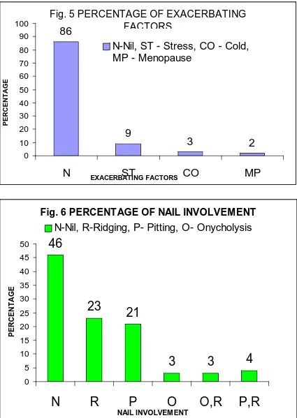
EXACERBATING FACTORS
There was no exacerbating factor for 86% of the study group. Among
the most common exacerbating factor, stress, cold and menopause, 9% was
Fig. 1 AGE DISTRIBUTION 0.00 10.00 20.00 30.00 40.00 50.00 60.00
PUV STE TAZ PLA DIT
TREATMENT MODALITIES
PERCENTAGE
YEARS
Fig. 2 SEX DISTRIBUTION
45 50 55 60 50 55 50 45 40 50 0 10 20 30 40 50 60 70
PUV STE TAZ PLA DIT
PERCENTAGE
Fig. 3 SEX RATIO
MALE 52%
FEMALE 48%
39.10 35.00
29.00 33.35 25.65
0.00 10.00 20.00 30.00 40.00
PUV STE TAZ PLA DIT
MONTHS
TREATMENT MODALITIES
Fig. 4 DURATION OF PSORIASIS
Fig. 5 PERCENTAGE OF EXACERBATING FACTORS
86
9
3
2
0 10 20 30 40 50 60 70 80 90 100N
EXACERBATING FACTORSST
CO
MP
PERCENTAG
E
N-Nil, ST - Stress, CO - Cold,
MP - Menopause
Fig. 6 PERCENTAGE OF NAIL INVOLVEMENT
46
23
21
3
3
4
0 5 10 15 20 25 30 35 40 45 50
N
R
P
O
O,R
P,R
NAIL INVOLVEM ENT
PERCENTAG
E
FAMILY HISTORY
Family history of psoriasis was present among 3% of total patients,
1% each in placebo, tazarotene, dithranol groups and none in the PUVA and
steroid groups.
NAIL CHANGES
46% had no nail changes. Among the others 23% had ridging, 21%
had pitting, 3% had onycholysis, 4% pitting and ridging and 3% had
onycholysis and ridging. (Fig. 6)
FOCAL SEPSIS
17% had evidence of focal sepsis in the ear, nose and throat and dental
sepsis in the form of gingivitis, which was treated before the onset of
therapy.
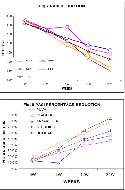
PASI REDUCTION
Fig. 7 depicts the reduction in the PASI scores obtained in the 5
Fig.7 PASI REDUCTION 0.00 0.50 1.00 1.50 2.00 2.50 3.00 3.50
0 W 4 W 6 W 12 W 24 W
WEEKS
PASI SCORE
PUV STE
TAZ PLA
DIT
Fig. 8 PASI PERCENTAGE REDUCTION
0.0% 10.0% 20.0% 30.0% 40.0% 50.0% 60.0% 70.0% 80.0% 90.0%
4W 8W 12W 24W
From the graph, the mean baseline PASI scores in the PUVA, steroid,
tazarotene, placebo and dithranol were 3.3, 3.2, 3.3, 3.1 and 3.2 respectively.
At 4 weeks of treatment, the mean PASI scores in 5 groups were 2.6, 2.8,
2.8, 2.6 and 2.7 respectively. At the end of 6th and 12th week there was
substantial reduction in the mean PASI scores for all the groups. In the
steroid group there was increase in PASI score between 4th and 6th week and
thereafter reduction was observed up to the end of 24th week. At the end of
24th week the tazarotene and PUVA groups showed a sustained reduction in
PASI score to 0.51 & 0.69 respectively. The other three groups showed a
moderate reduction in PASI score between 1.14 and 1.67.
PERCENTAGE REDUCTION IN PASI
Fig. 8 depicts the percentage reduction in PASI scores in all the 5
groups at 4,8, 12 & 24th week. At the end of 24th week there was 84.66%
reduction in PASI for tazarotene group followed by PUVA with 79.17%,
dithranol with 63.46%, steroid with 55.38% and placebo with 46.94%.
ADVERSE EFFECTS
Adverse effects were found in all the groups except placebo. During
4th to 6th week of treatment, 25% of patients in steroid group showed
showed in the form of dryness and pruritus. 15% of patients of PUVA group
showed in the form of erythema, polymorphic light eruption, burning
sensation. 15% of patients in dithranol group showed in the form of burning
TOPICAL TAZAROTENE GROUP
Before therapy
TOPICAL TAZAROTENE GROUP
Before therapy
TOPICAL PUVA GROUP
Before therapy
TOPICAL PUVA GROUP
Before therapy
TOPICAL DITHRANOL GROUP
Before therapy
TOPICAL DITHRANOL GROUP
Before therapy
TOPICAL STEROID GROUP
Before therapy
TOPICAL STEROID GROUP
Before therapy
TOPICAL LIQUID PARAFFIN GROUP
Before therapy
ADVERSE EFFECTS
Erythema after PUVA therapy
DISCUSSION
The study has shown that the mean age of the patients was 43.35 ranging from 10 to 85 years. The sex ratio was 52% male and 48% female. The various exacerbating factors in this study in descending order were stress, cold weather and menopause. Topical therapies like short contact compound dithranol, 0.1 % Betamethasone valerate, topical PUVA, 0.05 % tazarotene and liquid paraffin were used and reduction in PASI was assessed at every 4th , 6th , 12th & 24th weeks.
TAZAROTENE GROUP
Previous study on the topic “Management of topical modalities of psoriasis with special reference to Tazarotene by Dr.Susmit Haldar reported 50% of improvement by applying Tazarotene gel 0.05% twice daily at the end of 6weeks[30].
In this study, treatment made with the same 0.05% of Tazarotene gel once a day showed a maximum percentage reduction in PASI at the end of 8th, 12th and 24th week was 34.14%, 64.81% and 84.66% respectively. Maximum improvement was observed between 12th and 24th week.
PUVA GROUP (TOPICAL)
In this study PUVA group showed the second best efficacy after
tazarotene group showing a maximum reduction in PASI 79.17% at the end
of 24 weeks as evidenced by flattening of plaques, decreased scaling and
erythema[80,81]. This was consistent with the previous study conducted with
topical application of methoxypsoralen plus UVA in which 67% of patients
responded with considerable improvement.
15% of the patient had adverse effects in the form of polymorphic
light eruption, erythema and burning sensation at the exposed areas. Except
4 defaulters, there was good compliance among the patients.
DITHRANOL GROUP
In this group, the percentage improvement in PASI was found to be
63.46% at the end of 24 weeks[12].
The study was consistent with the study conducted in UK by
Gisslen H, Nordin P in which complete clearance of lesions was reported in
75% of the patients at the end of 24 weeks.
There was also good compliance in this group with no defaulters,
however some adverse effects in the form of burning sensation and
pigmentation were seen. However when the contact period was reduced to
STEROID GROUP (0.1% BETAMETHASONE VALERATE GROUP)
Steroid group showed moderate efficacy with 55.38% improvement in
PASI at the end of 24 weeks. However, there was no specific study reported
using 0.1% betamethasonevalerate. There was good compliance among the
patients with no defaulters. Few side effects like erythema and exacerbation
were observed in some patients.
PLACEBO GROUP (LIQUID PARAFFIN)
In this group application of liquid paraffin showed 46.94% reduction
of PASI at the end of 24weeks. There was good compliance among this
group with no defaulters and no adverse effects.
The previous study has reported that use of liquid paraffin in palmo
plantar psoriasis relieved feeling of dryness and pruritus[93]. There was no
CONCLUSION
Topical therapies are the first line therapeutic strategy in the treatment
of localized palmoplantar psoriasis and can be made effective when the
appropriate drugs were used judiciously.
Among the five modalities compared in this study, tazarotene (0.05%)
gel may be considered as an initial treatment of choice.
Topical PUVA is as effective as tazarotene except for the limiting
factors for PUVA therapy such as availability of PUVA unit, patient
compliance and long term side effects.
Topical dithranol is as effective as topical PUVA when used as
20minutes short contact therapy.
Topical 0.1% Betamethasone valerate was moderately effective with
frequent exacerbation.
Liquid paraffin was the least effective with no adverse effects, no
exacerbation and remissions. However it can be used as an adjunct with
REFERENCES
1. Polano M.K. Topical Skin therapeutics 1989 Churchill Livingstone, New York 94-96
2. Gruber M.Klein R. Foxx M. Chemical Standardization and quality assurance of whole crude Coal tar USP utilizing GLC procedures J.Pharmacent Sei 1970: 59: 830
3. Leadon SA Sumeral J.Minton TA. Tischler A; Coal tar residues produce both DNA adducts and oxidative DNA damage in human mammary epithelial cells. Carcinogenesis 1995 Dec. 16(12): 3021-6
4. Niels Hjorth, Metle, Jacobsen; Coal Tar; Seminars in Dermatology 1983 Dec.; 2(4); 281-285
5. Nenoff P, Hanustein UF, Fidler A; The antifungal activity of a Coal tar gel on Malassezia furfur in vitro; Dermatology 1995 : 191 (4) 311-4
6. Vander valk PG, Snater E, Verbeek, Gijsbers W, Duller P, Van de Kerkhof PC: Out-patient treatment of atopic dermatitis with crude coal tar: Dermatology 1996: 193 (1); 41-4
7. Marks R: Topical therapy for psoriasis: General principles; Dermatology clinics Jul 1984 (2):3
8. Silverman A, Menter A, Hairston JL; Tars and anthralins; Dermatol Clin 1995 Oct; 13(4)
9. Wemmer U, Schulze HJ, Mahrle G. Steigleder GK effect of various kinds of tar and tar concentration on anthralin erythema; Z Hautler 1986 Jun 15 61(12); 849 – 52
11. Seville RH: Dithranol based therapies, in text book of Psoriasis Ed.Mier PD, Vandekerkhol PCM 1986 Churchill Livingstone Edinburgh- pg 178-189
12. Kar PK, Jha PK, Snchips Anthralin, Short contact therapy in psoriasis IJDVL 1990:56 (193-95)
13. Nicholas J. Lowe M.D. MRCD, FACP, Richard Ashton, M.B. MRCP, Anthralin and Coal tar therapy for psoriasis; Dermatology Clinics; Vol.2, No.3 July 1984; 389-393
14. Kemeny L. Michel G, Arenberger P, Ruzicka T: Down regulation of epidermal growth factors receptors by dithranol: Acta Derm Venereol 1993 Feb: 73(1): 37-40
15. Gottlieb AB Khandke L, Krane JF Staia-Coico L, Ashinoff R Krueger JG; Anthralin decreases keratinocytes TGFα expression and EGF receptor binding in vitro: J invest Dermatol 1992 May; 98(5) : 690-5
16. Schroder JN: Anthralin (1,8,dihydroxy anthrone) is a potent inhibitor of leukotriene production J invest Dermatol 1986 Nov; 87(5): 624-9
17. Chodorwska G; Plasma concentration of IFN γ and TNFα in psoriatic patients before and after local treatment with dithranol ointment : J EUR Acad Dermatol Venereol 1998 Mar 10.(2): 147-151
18. Kaur I, Kaur & Vaishnav C, Ganguly NK, Garg J Kohli M; Epidermal calmodulin levels in psoriasis before and after therapy: Indian J Med REs 1991 April 94: 130-3
19. Kemeny L, Gross E, ArenBerger P, Ruzicka 7: Arch Darmatol Res 1991 : 283 (5); 333-6
20. Wolbling KH, Schofer H, Melbrodt R: Treatment of Seborrhoeic dermatitis: Hautarzt 1985 Sep; 36 (9) 529-30
22. Rulo HF, Vancle Kerkhof PC : Treatment of inflammatory linear verrucous epid Naevus; Dermatologica 1991 : 182(2) : 112-4
23. Flindt - Hasen H, Tikjob G, Brandrup F; Wart treatment with anthralin : Acta Derm Venereol 1984: 84 (2) : 177-9
24. Nelson DA, Spielvogel RL; Anthralin therapy for alopecia areata; Int J. Dermatol 1985 Nov; 24 (9): 606-7
25. Marsden JR, Coburn PR, Marks J, Shuster S; Measurement of the response of psoriasis to short term application of anthralin Br J Dermatol 1983 Aug. 109 (2) ; 202-18
26. Agarwal R, Saraswat A, Kaur I, etal . A Novel liposomal formation of dithranol for psoriasis. Preliminary results J. Dermatol 2002: 29:529-532
27. Vander Vleuten CJ Gerritsen MJ. Dejong EM etal: A Novel dithranol formulation (Micanol): Acta Derm Venereol 1996;76;887-91
28. Lange RW Germolec DR, Foley JF, Luster MI; Antioxidants alternate anthralin - induced skin inflammation in 'BALB/C mice; role of specific proinflammatory cytokines; J. Leukoc Bio 1998 Aug; 64(2): 170-6
29. Duvic M Nagpal S, Asano AT etal Molecularmechanism of tazorotene action in psorasis. JAM Acad Dermatol 1997; 37; S18-24
30. Management of Topical modalities of psoriasis with special refernce
to Tazarotene (Dr. Susmit Haldar) Gerald G. etal Arch Dermatol 1998 134 : 57 – 60
31. Peris K, Fargnoli MC, Chimenti S. Preliminary observation on the use of topical tazarotene to treat BCC N Engl Med. 1999; 341: 1767-8
32. Prystowskly J. Topical Retinoids Jn : SeW (ed) comprehensive Dermatologic Drug therapy Philadelphias Saundres – 2001
34. SRINIVAS CR, Satish Pai B, RaisKumar BC; Principle of Topical therapy in Dermatology; IADVL text book and atlas of dermatology; II edition (2001) Vol 2: 1245-1264
35. Francis C. Practical application of local corticosteroid therapy: Topical Corticosteroids: Rev. Prat 1990 Feb 21: 40 (6) 527-30
36. Almawi WY. Melemedjian OK. Molecular Mechanism of glucocorticoid antiproliferative effects. Antagonism of transcription factor activity by glucocorticoid receptor. J Leukoc Biol 2002 71:9-15
37. Blackwell GJ. Canuccio R. Di Rosa Mebal. Glucocorticoid in inflammatory proliferative skin disease reduces arachidonic and HETE acids. Science 1977 : 197; 994-5
38. Bamberger CM, Bamberger AM, decastro M, Chrousos GP Glucocorticoid receptor β, a potential endogenous inhibitor of glucocorticoid action in humans . J Clin livest 1995; 95: 2435-41
39. Hauk PJ, Hamid QA, Chrousos GP, Induction of corticosteroid insensitivity in human PBMCS by microbial superantigens J Allergy Clin Immunol 2000; 105 782-7
40. Carbone M, Carrozo M, Conrotto D; Topical treatment of atrophic / erosive oral LP with clobetasol in bio adhesive gel, Minerva stomatol 1997 Jul-Aug; 46(7-8) : 423-8
41. Yawalkar N, Karlen S, Egli F; Down regulation of IL-12 by topical corticosteriods in Chronic atopic dermatitis: J allergy clinic Immunol 2000 Nov: 106 (5) 941-7
42. Harper J: Topical corticosteroids for skin disorders in infants and children; Drugs 1988 : 36(5) 34-7
43. Ortonne JP: Clinical potential of Topical corticosteriods: Drugs 1988: 36 (5) : 38-42
45. Basta-Guzbasic A, Dabsic I; The effect of lcoal administration of corticosteroids on the course and therapy of Rosacea; Lyec Vjesn 1980 Mar; 111 (3) 89-93
46. Mark Lebwohl, Suad Ali Treatment of psoriasis - Topical therapy and phototherapy, J AM Acad Dermatol 45(4):487-498
47. Horwitz SN, Johnson RA, Sefton J.Frost P, Addition of a Topically applied corticosteroid to a modified Goeckerman regimen for treatment of psoriasis J AM Acad Dermatol 1985, Nov; 13(5); 784-791
48. Vander Vleuter CJ, Vande Kerkhof PC; Management of scalp psoriasis, Guidelines of corticosteroid use in combination treatment, Drugs 2001; 61(11): 1593-8
49. Lebwohl M, Poulin Y, Tazarotene in combination with Topical Corticosteroids, J AM Acad Dermatol 1998 Oct; 39 (4p+2), 139-43
50. Meola T. Soter NA Lim H: Are topical corticosteroids useful adjunctive therapy for the treatment of psoriasis with UVR; Arch Dermatol 1991 Nov; 127 1708-13
51. Takeda K. Arase S, Takahashi S: Side effects of Topical Corticosteroids and their prevention: Drugs 1988:36(5); 15-23
52. Wells K.Brodell RT: Topical corticosteroids addiction - a cause of perioral dermatitis: Postgrad Med 1993 Apr; 93(5): 225-30
53. Taniguchi H, Ohki O, Yokozeki H, Katayama I, Tanaka A Kiyosawa M; Cataract and retinal detachment in patients with severe atopic dermatitis who were withdrawn from the use of topical corticosteroid; J Dermatol 1999 Oct; 26 (10) : 658-65
55. Pena JM, Ford MJ; Cutaneous Lymphangiectasis associated with severe photo-aging and topical corticosteroid application ; J Cutan Pathol 1996 Apr: 23(2) ; 175-81
56. Patel L, Clayton PE, Addison GM, Price DA, David TJ; Adrenal function following topical steroid treatment in children with atopic dermatitis : Br. J Dermatol 1995 Jun; 132 (6); 950-5
57. Mark Lebwohl: The role of salicylic acid in the treatment of psoriasis: International Journal of Dermatology 1999(3): 16-24
58. Andrew N.Lin, Thomas Nakatsui: Salicylic acid revisited : International Journal of Dermatology 1998 (37): 335-342
59. Madhu AP, Thomas BF, Paul N, David SA, Sun protective agents: Formulations, effects and side effects; Fitzpatricks Dermatology in General Medicine: V Edition (1999) Vol 2: 2742-2763
60. Vande Kerkhof PC, Franssen ME; Psoriasis of the scalp-Diagnosis and management: AM J Clin Dermatol 2001;2(3):159-65
61. VanBrederode RL, Engel DD: combined cryotherapy 70% salicylic acid and treatment for plantar verrucae J Foot ankle Surg 2001 Jan-Feb: 40(I):36-41
62. Pavithran K ; Disorders of keratinization : IADVL text book and atlas of Dermatology; II Edition (2001) ; Vol II : 799-846
63. Gladstone HB, Nguyen SL, Williams R, Ottomeyer T, Wortzman M, Efficacy of hydroquinone Cream used alone or in combination with salicylic acid peels in improving photodamage on the neck and upper chest: Dermatol Surg 2000 Apr:26 (4):333-7
64. Abadjieva TI; Treatment of androgenic alopecia in females in reproductive age with Topical estradiol benzoate, prednisolone and Salicylic A Folia Med 2000; 42(3) 26-9
treatment of moderate to severe psoriasis ; a multi center study ; clin Ther 1998 Mar-Apr ; 20(2) : 283-91
66. Gloor M, Fluhr J, Wasik B, Gehring W; Clinical effect of salicylic and high dose urea applied according to the Standardized New German Formulary ; Pharmazic 2001 Oct. 56 (10) 810-4
67. Thaci D, Daiber W, Kaufmann R; Calcipotriol Solution for the treatment of scalp psoriasis evaluation of efficacy, safety and acceptance in 3,396 patients : Dermatology 2001 : 203 (2) : 153-6
68. Maune S, Frese KA, Mrowietz U, Reker U; Toxic innner ear damage in yopical treatment of psoriasis with Salicylates ; Laryngorhinootalogie 1977 Jun ; 76(6) : 368-70
69. Lebwohl M; Martinez J, Weber P, De Luca R; Effects of topical preparations on the erythemogenecity of UVB; implications for psoriasis in phototherapy; JAM Acad Dermatol 1995 Mar:32(3) : 469-71
70. Christophers E, Mrowietz U, Psoriasis in Freedbarg IM, Eisen Az, Wolff K, et al (eds). Fitz Patricks Dermatology in General Medicine 6th Edition McGraw Hill New York ; 2003, p.407-27
71. Dawe RS - Cameron H Yule S, et al UV-B phototherapy clears psoriasis through local effects Arch Dermatol 2002, 138 : 1071-6
72. Pramod Kumar, Advances in phototherapy. Ind J DVL 2001 : 67 :172- 176
73. Marita A, Werfel I, Stege Hetal. Evidence that singlet oxygen induced human T helpercell apoptosis is the basic mechanism of UVA phototherapy J Exp Med 1987; 186; 1783 – 1768
75. Krulmann J. Therapeutic photo immunology : photo immunological mechanism in photochemo therapy J photochem, Photobiol 1998 : 44 : 159-64
76. Herbert Honigsmann, Markus Szeimies, Robert Knobler Photochemotherapy and photodynamic therapy; Fitzpatricks Dermatology in General Medicine; 5th edition 1999; Vol.2; 2880 – 2900
77. Lindelof B. Sigurgeirsson. PUVA treatment in Sweden. Acta Derm Venereol 1992; 19;35-65
78. Tzan D, Kowk YK ; Goti CL. A retrospective review of PUVA therapy at the National skin center of Singapore. Photodermatol photoimmunol photo med 2001 Aug:17:164 – 7
79. Sedef Satin, Ugur H etal PUVA treatment of Vitiligo: a retrospective study of Turkish patients. Int J Dermatol 1999 July; 38 : 512 – 545.
80. Abel EA Gold berg LH Farber EM. Arch Dermatol 1980 Nov: 116(11): 1257 – 61
81. Wilkinson JD Ralfs IG, Harper JI Black MM Acta Derm Venereol Suppl (Stockh). 1979 : 59(85): 193 – 8
82. Farr PM, Diffey BL. PUVA treatment of psoriasis in the UK. Br.J Dermatol 1991 Apr: 124: 365 – 7
83. Farr DM: Diffey BL. Treatment of Actinic prurigo with PUVA: Mechanism of action, Br J Dermatol 1989 March: 120: 411-418
84. Henry H Roenigk J. Howard I Mainback. Psoriasis; III edition; 1998; Ch41: 543 – 557
85. Ibbotson SH, Farr PM. The time course of psoriasis ultraviolet A. (PUVA) erythema. J .Invest Dermatol 1999 Sep 113; 346 – 50
87. Bitsland DJ, Rhodes LE, Zaki I, Wilkinson S M etal psoriasis audit workshop of British Association of Dermatologists. Br J Dermatol 1985 Aug: 131:220-5
88. Thomsen K, Schmidt H : PUVA induced bullous pemphigoid. Br J Dermatol 1976: 95 : 568 –569
89. Van Praag MC. Tseng LN, Mommaas AM etal, The risk of PUVA treatment Drug Saf 1993 May; 8: 340-9
90. Stern RS Outcomes of pregnancies in women and partners of men with a history of exposure to PUVA for treatment of psoriasis Arch Dermatol 1991; 127: 347-350
91. Label E, Paver K, King R, et al. The relationship of skin cancer to PUVA therapy in Australia. Aus J Dermatol 1981 : 22 : 100-103
92. Chunag TV, Heinrich LA, Scvhultz MD, et al PUVA and skin cancer J. AM Acad Dermatol 1991 ; 124 : 49-55
COMPARATIVE STUDY OF THE EFFICACY OF VARIOUS TOPICAL TREATMENT MODALITIES IN PALMOPLANTAR PSORIASIS
PROFORMA
Date
Name: Age: Sex: Address:
Case No: Psoriasis clinic no: Marital Status: Occupation:
History
Duration Years Months
Itching Yes No
Exacerbation with
Cold Climate Sunlight Dialysis
Infection Trauma Mental stress
Puberty Pregnancy Menopause
Drugs
• Preceding sore throat Yes No
• Alcohol intake Yes No
• Systemic illness Diabetes Hypertension HIV
• Pregnancy Yes No
• Lactation Yes No
• Past history of Photosensitivity Cutaneous malignancies
• Family history of Psoriasis Mother Father
Sibilings Others
Drug Intake
Lithium NSAIDS Natural remedies
Steroid withdrawal Beta blockers Antimalarials
Amiodarone Digoxin Trazadone
Penicillin Terfenadine
Examination
General
Systemic: CVS RS Abdomen CNS
Dermatological
Auspitz sign No Yes
Clinical type Palmoplantar
Nail changes None Pitting Onycolysis
Subungual hyperkeratosis Ridges
Joint involvement Yes No
Focal sepsis ENT Dental Others
Surface area involved
Investigations Hb: TC: DC: ESR:
Urine albumin: Sugar: Deposits: Blood sugar: Urea: Creat:
Follow up
UPPER LIMB
PALMS LOWER LIMB SOLES Date No of
Wks E I D A E I D A
PASI SCORE
E – Erythema, I – Induration, D – Desquamation, A – Area,
Erythema / Induration / Desquamation scoring Area Scoring for Palms & Soles
0 – Nil 0 – Nil
1 – Mild 1 – Less than 10%
2 – Moderate
3 – Severe
4 – Very severe
MASTER CHART
S.NO AGE SEX DU EX FH NI FS GR PASI-0 PASI-4 PASI-8 PASI-12 PASI-24 AE
1 44 M 1 N N P Y TAZ 3.40 2.40 1.40 0.80 0.00 N
2 42 F 1 N N R N TAZ 4.40 4.60 3.60 2.60 1.60 N
3 63 F 0.5 ST N N Y TAZ 2.60 2.60 2.80 1.40 0.80 N
4 62 M 0.58 N N N N TAZ 3.00 2.20 1.80 0.80 0.00 N
5 29 F 3 ST N R N TAZ 2.60 3.40 2.20 0.60 0.00 Y
6 10 M 2 N N N N TAZ 4.00 3.00 1.80 1.60 1.20 N
7 38 M 4 N N P Y TAZ 2.40 3.00 3.20 1.60 0.80 N
8 55 M 3 N N N N TAZ 3.60 3.80 2.40 1.20 0.80 N
9 68 M 10 N N P N TAZ 3.20 2.60 2.00 0.80 0.60 Y
10 35 M 4 N N N N TAZ 4.60 2.80 2.20 1.00 0.40 N
11 28 M 2 N N O N TAZ 2.00 1.00 0.60 0.00 0.00 N
12 58 F 1 M N P N TAZ 3.50 3.30 3.00 1.60 0.60 N
13 49 M 2 N N R N TAZ 4.00 3.00 2.80 1.00 0.00 N
14 65 F 0.5 N N N N TAZ 2.20 1.40 1.20 0.60 0.00 N
15 50 F 1 N N P,R Y TAZ 3.40 2.80 2.20 1.00 0.80 Y
16 32 F 1.5 N N N N TAZ 3.20 2.20 1.60 1.00 0.00 N
17 27 M 0.67 N N P N TAZ 3.20 2.60 1.60 0.60 0.00 N
18 62 F 1 ST N P,R N TAZ 3.80 3.20 2.60 1.40 0.60 N
19 41 F 2 ST N N N TAZ 4.60 4.00 2.80 2.00 0.60 N
20 10 M 2 N Y R N TAZ 2.80 2.60 2.00 1.80 1.40 N
43.4 2.138 TAZ 3.33 2.83 2.19 1.17 0.51
21 14 F 0.58 N N R N PLA 2.60 1.80 1.40 0.60 0.20 N
22 68 F 0.08 N N O N PLA 2.80 1.80 2.00 1.60 2.20 N
23 53 M 0.5 N N N N PLA 1.80 1.60 2.00 2.40 1.80 N
24 38 M 0.75 N N N N PLA 3.80 2.20 1.80 2.00 2.40 N
25 63 M 15 ST Y R N PLA 2.80 2.60 2.00 1.60 1.40 N
26 47 M 0.25 N N P N PLA 3.60 2.80 2.40 2.00 1.80 N
27 40 M 4 N N N N PLA 2.60 2.40 2.20 2.60 1.80 N
28 23 M 13 N N P N PLA 4.80 4.40 3.60 3.20 3.40 N
29 30 M 3 CO N N N PLA 4.00 3.60 2.80 2.00 1.80 N
30 13 F 2 N N R Y PLA 2.00 1.80 1.40 0.80 0.40 N
31 36 M 0.25 N N N N PLA 2.60 2.00 1.80 0.60 0.20 N
32 49 F 0.67 N N O,R N PLA 3.20 2.60 2.00 1.40 0.80 N
33 51 M 2 N N N N PLA 2.80 2.60 2.80 2.20 2.60 N
34 13 M 1.5 N N N N PLA 3.20 2.20 1.60 1.47 0.80 N
35 50 F 5 N N P N PLA 4.80 4.60 3.80 4.20 4.00 N
36 85 F 0.5 N N N N PLA 3.60 3.40 3.00 2.80 2.60 N
37 12 M 3 N N P Y PLA 2.60 2.20 1.60 0.80 0.80 N
38 32 M 0.5 N N N N PLA 2.60 2.00 2.20 1.80 0.80 N
39 58 F 2 N N R N PLA 4..8 3.60 3.20 2.40 2.00 N
40 60 F 1 N N R N PLA 3.60 3.20 2.60 2.00 1.60 N
41.75 2.779 PLA 3.15 2.67 2.31 1.924 1.67
41 18 F 10 N N P N PUV 4.80 4.00 3.40 2.60 1.80 N
42 48 F 0.83 N N P N PUV 2.80 2.00 1.60 1.20 0.80 N
43 31 F 3 N N N Y PUV 1.80 1.60 0.60 0.60 0.40 N
44 35 F 0.17 N N P N PUV 2.80 2.20 1.80 - - Y
45 60 F 0.5 N N N Y PUV 4.20 3.20 2.80 1.80 0.80 N
46 45 M 20 N N O N PUV 4.80 3.80 2.60 2.00 1.20 Y
47 37 F 3 N N R N PUV 3.20 2.80 2.20 1.40 0.80 N
48 38 M 2 N N R N PUV 4.80 4.00 - - - -
49 60 F 0.25 ST N R N PUV 3.00 2.80 1.80 1.20 0.60 N