0022-538X/95/$04.0010
Copyrightq1995, American Society for Microbiology
Translation of the Human Papillomavirus Type 16 E7
Oncoprotein from Bicistronic mRNA Is Independent of Splicing
Events within the E6 Open Reading Frame
SIMON N. STACEY,1* DEBORAH JORDAN,1PETER J. F. SNIJDERS,2MICHAEL MACKETT,1
JAN M. M. WALBOOMERS,2ANDJOHN R. ARRAND1
Cancer Research Campaign, Department of Molecular Biology, Paterson Institute for Cancer Research, Christie
Hospital, Manchester M20 9BX, United Kingdom,1and Department of Pathology, Free University Hospital,
Amsterdam, The Netherlands2
Received 27 February 1995/Accepted 28 July 1995
In this study we investigated the translational capacities of bicistronic and spliced mRNAs originating from the E6 and E7 regions of the high-risk genital human papillomavirus type 16 (HPV-16) and the low-risk HPV-11. For HPV-16 it was found, unexpectedly, that E7 protein could be translated from full-length bicis-tronic E6-E7 mRNAs. E6*I and E6*II splicing events were not required for E7 synthesis, nor did splicing increase the efficiency of E7 translation significantly. In cells, E7 synthesis from all known naturally occurring mRNA structures was very inefficient compared with that from synthetic monocistronic controls, suggesting that HPV-16 employs translational mechanisms to restrict E7 protein levels. For HPV-11, only RNAs initiated at the P264promoter, located within the E6 open reading frame, were capable of providing an efficient template
for E7 synthesis. P264-initiated mRNAs were as efficient in vivo as monocistronic controls, suggesting that the
low-risk HPV-11 does not limit E7 synthesis by translational mechanisms. A detailed analysis of HPV-16 templates by using site-directed mutagenesis showed that the majority of ribosomes which ultimately translate E7 have not reinitiated after translating some or all of the upstream open reading frames. The data support a model in which the failure of 40S ribosomal initiation complexes to recognize the E6 AUG renders them capable of proceeding efficiently to translate E7.
Human papillomaviruses (HPVs) comprise a large group of small DNA viruses with type-specific tropisms for cutaneous or mucosal epithelia. Several types of HPV infect genital mucosal epithelia. One group of genital HPV types, exemplified by HPV type 6 (HPV-6) and HPV-11, is associated with condy-lomata acuminata and early stages of cervical intraepithelial neoplasia. Because lesions containing these HPV types seldom progress to high-grade cervical intraepithelial neoplasia and cervical carcinoma, they have been denoted low-risk genital HPV types. Lesions containing HPV-16, -18, -31, -33, and several other, less common types are thought to have a high propensity to progress to high-grade cervical intraepithelial neoplasia and cervical carcinoma. Accordingly, these have been designated high-risk genital HPV types (see references 57 and 59 for reviews).
Recent studies using nuclease protection, cDNA cloning, and reverse transcriptase PCR have allowed a very precise definition of the structures of early HPV mRNAs produced in HPV-11- and HPV-16-associated lesions. This analysis re-vealed that many of the transcripts appear to be bicistronic or polycistronic (8, 12, 34, 38, 40, 41, 46, 48, 49). However, rela-tively few attempts to determine which proteins are actually translated from these polycistronic mRNAs have been made. Nearly all eukaryotic mRNAs are monocistronic, and
trans-lation usually initiates at the AUG codon nearest the 59end of
the message (25). There are a few cellular eukaryotic mRNAs
in which the 59untranslated leader sequence contains one or
more short open reading frames (ORFs) or minicistrons.
Al-most 50% of these cellular RNAs are oncogene transcripts, and it is thought that the presence of upstream AUGs may have a role in the regulation of their translation (26). In a modified version of Kozak’s scanning hypothesis, ribosomal 40S subunit initiation complexes are thought to negotiate up-stream AUGs either by ignoring them (leaky scanning) or by translation of the minicistron followed by a resumption of scanning (termination-reinitiation) (29, 35).
In high-risk HPV types the two major transforming genes E6
and E7 (33, 56) are transcribed from the same promoter (P97
in HPV-16), producing a structurally bicistronic transcript. In the majority of transcripts, an intron contained within the E6 ORF is spliced out, forming the E6*I mRNA (49) (Fig. 1). A rare variant using the same splice donor but a different accep-tor has been designated E6*II. In the E6*I mRNA, the E6
ORF is thrown out of frame and terminates just 39of the splice
acceptor site. The truncated reading frame has been desig-nated the E6*I ORF.
It has been established previously with model systems that the efficiency of translational termination-reinitiation depends on the presence of an adequate run of RNA between the termination of one cistron and the start of another (28). This is thought to indicate a requirement for those 40S ribosomal subunits which resume scanning to regain competence to ini-tiate translation, perhaps because of the reassociation of ac-cessory factors with the scanning subunit. In a full-length bi-cistronic E6-E7 mRNA, the E7 AUG occurs just 2 bp downstream of the E6 stop codon. Such a configuration would be expected to preclude termination-reinitiation because of an insufficiency of intercistronic space. It has been proposed that the function of E6*I intron splicing is to increase the intercis-tronic space between the foreshortened E6*I ORF and the E7 AUG to allow efficient termination-reinitiation to occur (50).
* Corresponding author. Mailing address: Department of Molecular Biology, Paterson Institute for Cancer Research, Christie Hospital, Manchester M20 9BX, United Kingdom. Phone: 446-3186. Fax: 161-446-3121.
7023
on November 9, 2019 by guest
http://jvi.asm.org/
In this model, unspliced transcripts would express only E6 protein, whereas splicing would be necessary for efficient E7 translation.
In the low-risk HPV-11, the E7 mRNA is thought to arise by a different mechanism (48). No splicing has been observed
within the E6 ORF, and the E6 proximal promoter (P90) drives
only a full-length bicistronic message (Fig. 1). In HPV-11 the
E6 ORF terminates downstream (39) of the E7 AUG, which
may obviate termination-reinitiation. The E7 mRNA was pro-posed to be synthesized from transcripts originating from a
second promoter (P264) located within the E6 ORF. However,
the P264-initiated mRNA contains two small cistrons located
upstream of the E7 AUG which would need to be bypassed in order for E7 translation to occur.
In this study we investigated the translational capacities of cDNA structures from HPV-16 and HPV-11 by using in vitro and cellular translation systems. Contrary to our expectations, the HPV-16 E7 protein was found to be translated from full-length E6-E7 bicistronic mRNA structures, demonstrating that splicing was not required for E7 synthesis. Moreover, while the spliced structures E6*I and E6*II were also capable of acting as templates for E7 synthesis, the splicing events did not sig-nificantly alter the E7 translational efficiencies of these mRNAs. These observations are inconsistent with a simple termination-reinitiation model for E7 translation. Site-directed mutagenesis experiments showed that the majority of ribo-somes which ultimately translate the E7 ORF have ignored some or all of the ORFs present upstream of the E7 AUG. In particular, ribosomal 40S initiation complexes that scan past the E6 AUG have a high propensity to translate the E7 ORF.
MATERIALS AND METHODS
Cell lines.CV-1 and HeLa cells were obtained from the European Collection of Animal Cell Cultures (Porton Down, United Kingdom). SVD2 cells are simian virus 40-transformed human keratinocytes and were obtained from Jon Tinsley,
Cancer Research Campaign Department of Cancer Studies, Birmingham, United Kingdom.
Construction of recombinant clones and viruses.All HPV sequences were cloned by standard techniques as BamHI fragments into the BamHI site of transcription vector pET-3 (39). HPV-16 coordinates were taken from reference 45, and HPV-11 coordinates were taken from reference 9.
pHET7 (53) contains the HPV-16 PvuII(553)-to-PstI(879) E7 fragment. pHET6 contains HPV-16 sequences from nucleotide (nt) 97 to 654 cloned by using oligonucleotide adapters. pHET67 contains HPV-16 nt 97 to 879. For pHET*I and pHET*II, cDNA clones were isolated from SiHa cells by reverse transcriptase PCR by methods described previously (52). Spliced sequences from nt 97 to 875 were cloned into pET-3. In addition to confirming the appropriate spliced structures, DNA sequencing revealed that both cDNA clones contained an A3C transversion at nt 645, which is a known variant present in SiHa cells (21). The *I cDNA clone was found also to have an A3C transversion at nt 442, presumably caused by an amplification error. Neither of these mutations oc-curred in a position where it was likely to influence the results.
pCET6 contains HPV-11 sequences from nt 90 to 576. pCET7 (53) contains HPV-11 sequences from nt 534 to 1071 cloned into pET-3 with a favorable translation initiation codon in the form of HPV-16 nt 533 to 566 inserted upstream of the E7 AUG. pCET67 contains HPV-11 nt 90 to 1071. For pC6delta, HPV-11 sequences from nt 264 to 569 were cloned into pET-3 by PCR with adapter-primers and then sequenced.
For pMCD*I, the sequence of the MC1 and MC2 start codons . . .ATG TATG. . . was changed to . . .ATAGATC. . . by site-directed mutagenesis (Pro-mega). The insert was sequenced, recovered as an nt 97-to-875 BamHI fragment, and cloned into vaccinia virus insertion vector p1118 (see below). A fragment containing nt 499 to 875 with the above-described mutation was subcloned by using oligonucleotide adapters to produce pMCDTaq. An analogous fragment was subcloned from wild-type sequences to produce pH7Taq.
The E6 AUG was altered by oligonucleotide insertion to AGG in an E6*I cDNA context or a full-length bicistronic context to produce p*IATGDand p6ATGD, respectively. For pH6L, the E6 stop codon TAA was changed to CAA by site-directed mutagenesis, and the mutant sequence (nt 97 to 879) was se-quenced and cloned into p1118 (for pH6L) and pET-3 (for pHET-6L).
For T7 vectors containing the encephalomyocarditis virus (EMCV) leader sequence, the E6 AUG codon sequence was modified by oligonucleotide inser-tion to create an NcoI-compatible end, and the appropriate fragments were cloned as NcoI-BamHI fragments into pCITE2a (Novagen) to produce pCITE*I, pCITE67, and pCITE6L.
[image:2.612.68.552.74.298.2]Production of recombinant vaccinia viruses.For vaccinia virus recombinants, the BamHI fragments as described above were cloned into the BamHI site of vaccinia virus insertion vector p1118 (30a). Vector p1118 contains the T7 pro-moter-terminator fragment from pET-3 flanked by sequences from the vaccinia FIG. 1. Genomic organizations, mRNA structures, and cDNA clones of HPV-16 and HPV-11 early regions. The top line of each panel shows the locations of the E6 and E7 ORFs. The lines with arrows represent naturally occurring mRNA structures. The numbers refer to the locations of the promoters and splice junctions for each RNA. The names of the naturally occurring RNAs as used in the text are given at the right and are followed by the names of the corresponding T7-cDNA constructs used to express the RNAs. Control T7-cDNA constructs were produced in cases in which no corresponding natural mRNA is known, as indicated by N/A in the mRNA column. The DNA fragments used to produce these control constructs are represented as lines with bars.
on November 9, 2019 by guest
http://jvi.asm.org/
virus thymidine kinase locus. The vector also contained the Escherichia coli gpt gene under the control of the vaccinia virus 7.5K early-late promoter, transcrib-ing in the direction opposite to that of the T7 promoter. Recombinant vaccinia virus pools were produced by transfection of each p1118-based insertion vector into CV-1 cells infected with the WR strain of vaccinia virus, followed by three rounds of selection in medium containing 25mg of mycophenolic acid per ml, 250
mg of xanthine per ml, and 15mg of hypoxanthine per ml. Pools of.100 clones of each recombinant were maintained at all times. Stocks were titrated on CV-1 monolayers as described previously (31, 32). Recombinant vaccinia viruses were found by RNase protection to initiate transcription of the HPV inserts correctly at the T7 promoter, and expression of HPV proteins was found to be dependent on coinfection with vTF7-3 (data not shown).
In vitro transcription and translation.RNA was synthesized in vitro by using a Promega Ribo-Max kit according to the manufacturer’s instructions. To pro-duce capped RNA, m7
G(59)ppp(59)G was added to 30 mM and the GTP con-centration was limited to 0.75 mM for 30 min at 378C, and then the GTP concentration was increased to 7.5 mM for a further 60 min of incubation. Purified RNA (25mg/ml) was translated in the presence of [35
S]cysteine (22mCi per reaction) by using a rabbit reticulocyte system (Promega) with 2.0 mM Mg21 (final concentration) and 70 mM added KCl for maximum translation fidelity (3, 10). Products (50ml) were diluted 10-fold in radioimmunoprecipitation assay (RIPA) buffer (0.5% Nonidet P-40, 25 mM Tris-HCl, 150 mM NaCl, 100mM ZnCl2, 1 mM dithiothreitol, pH 7.8).
Cellular translation assays and metabolic labelling.For transfection-infection assays, 106
CV-1 cells in a 25-cm2
flask were infected with vTF7-3 (16) at a multiplicity of 10 PFU per cell for 60 min. The cells were then transfected with 5mg of T7-cDNA plasmid by using 30ml of DOTAP (Boehringer). At 24 h posttransfection, the cells were metabolically labelled with [35
S]cysteine (200mCi per 106
cells) for 90 min. The cells were then washed and lysed in 500ml of RIPA buffer and stored at2708C.
For dual-infection assays, 0.53106cells (CV-1, HeLa, or SVD2) were
in-fected with 5 PFU of vTF7-3 and 5 PFU of T7-cDNA virus per cell. The cells were labelled and harvested at 12 h (CV-1) or 16 h (HeLa and SVD2) postin-fection as described above.
Immunoprecipitation assays.Immunoprecipitations were carried out at 48C with 100-ml aliquots of labelled material. Samples were precleared with a 1-h incubation with 5ml of normal rabbit serum followed by a 1-h incubation with 10
ml of protein A-Sepharose beads. Ten microliters of specific antiserum was then added, and samples were incubated overnight. Immunoglobulin complexes were collected on 20ml of protein A-Sepharose beads and then washed three times with RIPA buffer containing 25 mM EDTA and no ZnCl2. Fifty microliters of
sodium dodecyl sulfate-polyacrylamide gel electrophoresis (SDS-PAGE) loading buffer was then added, and 25-ml samples were analyzed by SDS–14% PAGE followed by autoradiography or PhosphorImager analysis. Antibodies used for immunoprecipitation were as follows: for HPV-16 E6, antipeptide antibody 145-6R (53); for HPV-16 E7, polyclonal antibody RPaE7 (54); for HPV-11 E7, antipeptide A48-74-1 (53); and for HPV-11 E6, a rabbit polyclonal anti-pMAL-E6 fusion protein antiserum (kindly provided by Huw Davies and Ian Tarpey).
RNA isolation and blotting.For RNA analysis, templates were generated by transfection-infection or dual infection as described above, and then total cyto-plasmic RNA was isolated. Cytocyto-plasmic DNA (from the transfection and vac-cinia virus infection) was removed by treatment with DNase in the presence of RNasin. Twofold serial dilutions of RNA starting from 1mg were slot-blotted onto nylon membranes and hybridized to32
P-labelled HPV-16 E7 (nt 553 to 879) or HPV-11 E7 (nt 534 to 1071) antisense riboprobes.
PhosphorImager analysis.Quantitative determinations of E7 protein expres-sion from wild-type and mutant cDNA templates were done with a Molecular Dynamics (Chesham, United Kingdom) 425S PhosphorImager operating at a 176-mm resolution. Data were analyzed with ImageQuant software according to the manufacturer’s instructions.
RESULTS
In order to investigate the translational capacities of early HPV-16 and HPV-11 mRNAs, a number of cDNA constructs in which the cDNAs were inserted between the T7 promoter and the T7 terminator of the vector pET-3 were cloned (39) (Fig. 1). This inevitably led to a short sequence of T7 leader, which is required for stabilization of RNAs expressed in
eu-karyotic systems, being added to the 59end of each cDNA (15).
Constructs which contained the P97-initiated full-length
bicis-tronic transcript (pHET67) or the two spliced products E6*I (pHET*I) and E6*II (pHET*II) were produced. In addition, monocistronic control constructs which contained only the E6 ORF (pHET6) or the E7 ORF (pHET7) cloned directly down-stream of the T7 promoter were made. For the low-risk
HPV-11, the full-length bicistronic cDNA and the P264-initiated
cDNAs were cloned as pCET67 and pC6delta, respectively. The monocistronic HPV-11 E6 (pCET6) and E7 (pCET7) control constructs were also produced.
Translation of cDNA constructs in vitro. The various mRNAs were tested for translational capacities first in a rabbit reticulocyte lysate system. Transcripts were synthesized with or
without the addition of an m7G(59)ppp(59)G cap. Figure 2a
shows that the E7 protein was produced by the bicistronic vector pHET67 from both capped and uncapped transcripts. In addition, the two spliced forms pHET*I (Fig. 2a) and pHET*II (not shown) and the control construct pHET7 also produced E7 protein. Splicing of the mRNA structures caused little if any difference in the levels of E7 produced. In the capped versions the production of E7 protein by pHET67 and pHET*I was inefficient relative to that by the synthetic monocistronic control construct pHET7. This occurred because capping in-creased E7 synthesis by the monocistronic control but not by the spliced or full-length bicistronic RNA. The full-length bi-cistronic mRNA structure simultaneously produced E6 pro-tein, as did the monocistronic E6 control construct pHET6. Capping increased considerably the synthesis of E6 from both bicistronic and monocistronic constructs as shown in the lower panel of Fig. 2a. The increase in E6 synthesis due to capping of the bicistronic transcript was not reflected in an increase in E7 synthesis from the same mRNA. Therefore, E7 synthesis from this mRNA was independent of the level of translation occur-ring in the upstream ORF. Similarly, neither capping nor splic-ing increased the level of E7 synthesis from pHET*I, suggest-ing that E7 production from these transcripts is unaffected by the amount of translation of the E6*I ORF.
For HPV-11 (Fig. 2b) some E7 protein was produced by the full-length bicistronic mRNA (from pCET67). However, E7
protein was synthesized more efficiently by the P264-initiated
[image:3.612.318.550.72.284.2]mRNA (from pC6delta) and the monocistronic control. This
FIG. 2. In vitro translation of HPV-16 and HPV-11 mRNAs. Lane designa-tions refer to mRNA structures and cDNA constructs as shown in Fig. 1. (a) HPV-16 mRNA was synthesized in vitro with (1) or without (2) incorporation of an m7G(59)ppp(59)G cap (CAP) and translated in rabbit reticulocyte lysate
containing [35S]cysteine; this was followed by RIPA with RPaE7 polyclonal
anti-E7 antiserum (aE7) and 145-6R anti-16E6 antipeptide serum (aE6). (b) HPV-11 mRNAs were similarly synthesized, translated, and subjected to RIPA with A48-74-1 anti-11E7 antipeptide serum (aE7) and an anti-HPV-11 E6 E. coli fusion protein serum (aE6).
on November 9, 2019 by guest
http://jvi.asm.org/
difference in template efficiency was pronounced in the capped versions of the transcripts. E6 protein was simultaneously pro-duced by the bicistronic mRNA pCET67. E6 synthesis from the bicistronic transcript was increased by the presence of a cap, whereas E7 synthesis was not.
Translation of cDNA constructs in cells by using vaccinia virus T7 transfection-infection.In order to examine translation of the mRNA structures in vivo, a cellular translation system was employed (16). T7 promoter-driven cDNAs were trans-fected into CV-1 cells; this was followed by superinfection with recombinant vaccinia virus vTF7-3, which expresses T7 RNA polymerase. Transactivation of the T7 promoter in the cyto-plasm of recipient cells resulted in the production of high levels of mRNA from the cDNA clones.
The plasmids described in Fig. 1 were transfected into vTF7-3-infected CV-1 cells. At 24 h postinfection the cells were
metabolically labelled with [35S]cysteine and lysed, and the E6
and E7 proteins were detected by immunoprecipitation. RNA was extracted from parallel samples to obtain estimates of relative template abundances in order to reveal any variations in the rates of RNA synthesis and/or decay. This experiment was repeated at least three times for each construct, and typical results are presented in Fig. 3.
For HPV-16 it was found that the bicistronic pHET67 and the spliced pHET*I and pHET*II cDNA constructs were all capable of producing E7 protein and at roughly equal levels. Thus, splicing had no effect on the level of E7 translation in vivo. The efficiency of E7 production by these three cDNA constructs was substantially lower than that by the E7 mono-cistronic control construct pHET7. This suggested that all three known naturally occurring mRNAs containing the E7 ORF are translated inefficiently in cells, as they were from capped transcripts in vitro. RNA blotting (Fig. 3c) showed that the relative abundances of the mRNA structures did not vary significantly; therefore, the differences in E7 protein preva-lences between pHET7 and the three naturally occurring tran-scripts were most likely due to differences in translation effi-ciency. Immunoprecipitation with an E6 antipeptide serum showed that in addition to the E7 protein, the bicistronic pHET67 vector also produced abundant E6 protein. E6* pro-teins, which would result from translation of a spliced E6 ORF, were not detected, even though the E6 antipeptide antiserum is targeted on N-terminal sequences of E6 (53). This indicated that the E6* proteins, if they were synthesized, were rapidly degraded.
Strikingly, the HPV-11 full-length bicistronic vector pCET67 produced no detectable E7 protein in vivo (Fig. 3b), despite the fact that its mRNA was produced in amounts equivalent to those produced by the other constructs (Fig. 3c). This was in contrast to the in vitro translation experiments, in which pCET67 transcripts produced some E7, albeit inefficiently. The
E7 protein was produced efficiently in CV-1 cells by the P264
-initiated cDNA construct pC6delta, which appeared (when the results were corrected for RNA abundance) to be at least as efficient a template for translation as RNA from the control monocistronic E7 construct pCET7. The two small upstream minicistrons (between nt 346 and 369 and nt 382 and 396)
present on the P264-initiated HPV-11 mRNA must not,
there-fore, present a serious obstacle to scanning ribosomal 40S subunits, as they were bypassed without a substantial loss of template translational efficiency. Since the full-length bicis-tronic HPV-11 construct, on the other hand, did not appear to be capable of providing an E7 template in the more stringent
environment of live cells, we conclude that only the P264
-initi-ated transcript can serve as a template for HPV-11 E7
trans-lation. This is in agreement with the prediction of Smotkin et al. (48).
Production and analysis of site-directed mutants.Two ques-tions regarding the evident translation of HPV-16 E7 from structurally bicistronic transcripts became apparent. First, why did splicing of the E6*I intron and consequent truncation of the E6 ORF not increase the efficiency of E7 translation? Second, by what mechanism was E7 translated from unspliced (and spliced) bicistronic transcripts? In order to address these questions, a series of site-directed mutations were made in the cDNA constructs as depicted in Fig. 4. In order to assess the activities of mutated constructs quantitatively, a dual-infection protocol in which the mutant constructs (and controls) were incorporated into recombinant vaccinia viruses was used (14, 15). For each construct, recombinant viruses were selected for the presence of a coinserted gpt marker, and pools comprising
.100 clones of each recombinant were expanded and titrated
[image:4.612.326.546.68.398.2]before use. Each pool was assessed by dual infection with vTF7-3 followed by detection of E7 and E6 proteins by RIPA.
FIG. 3. Translation of HPV-16 and HPV-11 cDNA constructs in CV-1 cells by using the vaccinia virus-T7 transfection-infection system. CV-1 cells were infected with T7 RNA polymerase-expressing vaccinia virus recombinant vTF7-3 and then transfected with T7-cDNA constructs as indicated by the lane desig-nations. Numbers on the right of panels a and b indicate molecular masses in kilodaltons. (a) RIPA detection of HPV-16 E7 (top) and E6 (bottom) protein synthesis in transfected cells. (b) RIPA detection of HPV-11 E7 protein synthesis in cells transfected with HPV-11 T7-cDNA constructs. Lane M, marker proteins. (c) RNA was isolated from transfected cells, and RNA slot blots of twofold serial dilutions (starting from 1mg) were prepared. Slot blots were hybridized to
32P-labelled E7 riboprobes from HPV-16 (left) and HPV-11 (right). Reduced
signals are observed in HET-6- and CET-6-transfected cells because of limited overlap between the RNAs and the E7 riboprobes. MOCK, equivalent amounts of RNA isolated from mock-transfected cells.
on November 9, 2019 by guest
http://jvi.asm.org/
Sample results of a dual-infection experiment with associ-ated control results are shown in Fig. 5. A summary of results from dual-infection experiments with CV-1, HeLa, and SVD2 (simian virus 40-transformed human keratinocyte) cells is shown in Fig. 6. Endogenous HPV expression in HeLa cells
[image:5.612.63.294.69.454.2]was not detected under the conditions used. With all three lines it was found that full-length bicistronic transcripts pro-duced E7, typically at less than 10% efficiency relative to the monocistronic control. The E6*I spliced version was only mar-ginally (less than twofold) more efficient than the full-length bicistronic transcript, and this difference could be accounted for by slightly increased abundances of E7 RNA from this construct. Both the *I and *II spliced versions were very inef-ficient relative to monocistronic controls.
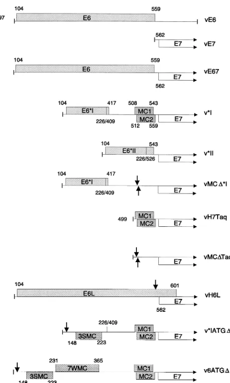
[image:5.612.332.526.78.536.2]FIG. 4. Scale diagram showing the structures of HPV-16 cDNA constructs and their mutated derivatives. Only the E6 region and the N-terminal region of E7 are shown. Stippled boxes depict potentially active ORFs in the E6 region. The open boxes with arrows represent the entire E7 ORF. Numbers indicate coordinates for ORF ends, splice junctions, and promoter-proximal 59ends. vE6 and vE7 are the control monocistronic constructs. vE67 is the full-length bicis-tronic cDNA construct. v*I has the E6*I intron between nt 226 and 409 removed, and the locations of the MC1 and MC2 minicistrons between the E6*I and E7 ORFs are shown. v*II has the E6*II intron between nt 226 and 526 removed. In vMCD*I, the MC1 and MC2 AUG codons have been removed by site-directed mutagenesis, as depicted by the arrows. In vH7Taq, the MC1 and MC2 minicis-trons were cloned directly upstream of the E7 ORF. The same fragment, but with the MC1 and MC2 start codons mutated (arrows), was cloned directly upstream of the E7 ORF to produce vMCDTaq. In vH6L, the E6 ORF termination codon was mutated (arrow), thereby extending the reading frame to the next in-frame termination codon at nt 601. In v*IATGD, the E6 ORF start codon was mutated (arrow), and the locations of the three strong-context minicistrons thereby ex-posed are indicated (3SMC). The location of the *I splice junction is also shown. In v6ATGD, the E6 ORF AUG was removed from a full-length bicistronic cDNA by site-directed mutagenesis (arrow). The locations of the 3SMC and seven weak-context minicistrons (7WMC) present in the E6 region of this con-struct are indicated. Each concon-struct was cloned downstream of the T7 promoter in vaccinia virus insertion vector p1118, and recombinant vaccinia viruses pro-ducing each cDNA were generated.
FIG. 5. Dual-infection experiment using mutant constructs. CV-1 cells were infected with 5 PFU of mutant virus stock and 5 PFU of vTF7-3 per cell. (a to c) After 12 h, cells were metabolically labelled with [35
S]cysteine, lysed, and immu-noprecipitated with E7 antibody (RPaE7), E6 antibody (145-6R), or pooled normal rabbit serum (NRS). The constructs used are described in Fig. 4, except E710, which is a vaccinia virus recombinant that utilizes the vaccinia virus 7.5K promoter for E7 expression (54). (d) RNA dot blot of twofold serial dilutions starting from 1mg of RNA taken from 12-h-infected CV-1 cells, hybridized to an HPV-16 E7 riboprobe.
on November 9, 2019 by guest
http://jvi.asm.org/
A close reexamination of the sequence of the E6*I cDNA indicated that the *I splice does not actually create an unin-terrupted intercistronic space. Rather, a ribosomal subunit which resumed scanning after termination at the E6*I stop codon at nt 417 would encounter two small ORFs running from nt 508 to 543 and nt 512 to 559. These two minicistrons have been designated MC1 and MC2, respectively, in Fig. 4. Although both of the start codons for these minicistrons are in unfavorable contexts for initiation (6, 27), if they were trans-lated, then termination would be brought within 18 nt (MC1) and 2 nt (MC2) of the E7 AUG. Therefore, it was possible that the E6*I spliced product did not represent an efficient E7 template because the two minicistrons negate the benefit of increased intercistronic space created by the splice.
In order to investigate the effects of MC1 and MC2, their start codons were removed by site-directed mutagenesis to
produce pMCD*I (Fig. 4). The effect of the minicistrons on
scanning 40S subunits was further examined by placing the two minicistrons directly upstream of the E7 ORF in a control monocistronic construct (to produce pH7Taq). The equivalent fragment, but with the MC1 and MC2 start codons removed, was also placed directly upstream of the E7 ORF to produce
pMCDTaq (Fig. 4). The latter two constructs were designed to
examine the effects of MC1 and MC2 on scanning initiation
complexes, whereas the pMCD*I construct would examine the
effects of MC1 and MC2 on scanning reinitiation complexes. The results are shown in Fig. 5 and 6. In all three cell lines tested, mutation of the MC1 and MC2 initiation codons in the context of an *I-spliced mRNA had little or no effect on E7
translation efficiency (compare *I with MCD*I). This
demon-strated that ribosomes which ultimately translate the E7 ORF do not approach the E7 AUG engaged in the translation of MC1 and MC2. Therefore, inhibition of scanning and reinitia-tion by MC1 and MC2 cannot account for the low efficiency of *I mRNAs as templates for E7 synthesis.
In CV-1 cells, placement of MC1 and MC2 directly up-stream of a monocistronic E7 ORF caused an inhibition of E7 translation which was alleviated by deleting the MC1 and MC2
start codons (compare E7, H7Taq, and MCDTaq in Fig. 6),
suggesting that MC1 and MC2 can inhibit 40S subunits
scan-ning in from a nearby 59end but not in the interior of an *I
mRNA. This effect was not observed in HeLa and SVD2 cells, however.
If E7 is translated from full-length bicistronic mRNA by a termination-reinitiation mechanism, then ribosomes destined to translate E7 should approach the E7 AUG while engaged in E6 translation. This point was examined by removing the E6 stop codon by site-directed mutagenesis to produce vH6L (Fig. 4). The resulting configuration resembled that of HPV-11 in that the extended E6 ORF overlapped the E7 ORF by about 30 bp. Such a configuration would be expected, in the cellular translation assays, to prevent any termination-reinitiation by ribosomes that had translated E6. However, in CV-1 cells, vH6L was found to produce E7 at rates similar to those for the full-length bicistronic construct (compare vH6L [7.9% efficien-cy] and vE67 [6.8% efficienefficien-cy] in Fig. 6). In HeLa and SVD2 cells, vH6L expressed E7 protein at levels which were lower but above the detection limits. This suggested that in the full-length bicistronic mRNA, most ribosomes which eventually translate E7 do not approach the E7 ORF engaged in trans-lation of E6.
The HPV-16 E6 AUG is located 8 nt downstream of the P97
transcription initiation site. Although the E6 AUG is in a favorable sequence context for translation initiation, its
prox-imity to the 59end of the mRNA is likely to cause substantial
leaky scanning of the E6 AUG (30, 47). It was possible to model the consequences of such leaky scanning simply by re-moving the start codons of the E6 ORF from full-length
bicis-tronic and *I spliced cDNA clones, to produce v6ATGDand
v*IATGD, respectively (Fig. 4). Scanning 40S subunits would
under these circumstances encounter several clusters of mini-cistrons, some in strong Kozak contexts, located out of frame
relative to E6 (12 minicistrons for v6ATGDand 5 minicistrons
for *IATGD), before encountering the E7 AUG (Fig. 4).
Anal-ysis of the E6 ATGDmutants in CV-1 cells showed that
dele-tion of the E6 AUG caused E7 expression to increase fivefold relative to those with the respective control constructs v*I and vE67. Similar effects were seen in HeLa and SVD2 cells (Fig. 6). Therefore, it appeared that ribosomal 40S subunits that failed to recognize the E6 start codon had a significantly higher propensity to initiate at the E7 AUG. This occurred regardless of whether the transcript was spliced or not and despite the presence of numerous upstream AUGs.
Similarly, by forcing ribosomal subunits to translate the E6 ORF efficiently, it was possible to test the extent of termina-tion-reinitiation from the various transcripts. We examined the effect of increasing translation of the E6 ORF by inserting the
EMCV 59untranslated leader sequence between the T7
pro-FIG. 6. Levels of E7 protein expression from wild-type and mutant templates in the CV-1, HeLa, and SVD2 cell lines. Cells were infected and analyzed for E7 expression as described in the legend to Fig. 5. Amounts of E7 produced were determined by PhosphorImager analysis and are expressed relative to the level of E7 produced by the monocistronic control construct vE7 (set to 100). Error bars show standard deviations for values obtained from independent experiments.
on November 9, 2019 by guest
http://jvi.asm.org/
[image:6.612.92.267.71.430.2]moter and the E6 AUG, to promote efficient, cap-independent translation of the E6 ORF (24, 36). Preliminary experiments showed that the EMCV sequence increased expression from a monocistronic construct approximately 10-fold, in agreement with previous observations (13, 15, 55). The effect of the EMCV-E6 ORF on translation of a downstream E7 ORF was tested in the context of full-length bicistronic and spliced cDNAs (Fig. 7). By using the transfection-infection method, it was found that addition of the EMCV leader reduced E7 synthesis three- to sixfold, again suggesting that termination-reinitiation of ribosomes that had translated E6 did not con-tribute significantly to E7 synthesis.
DISCUSSION
In a detailed study of HPV-16 early mRNA structures Smot-kin and Wettstein (49) noted that the majority of early tran-scripts were spliced from nt 226 to 409 in the E6 ORF. This splice causes a foreshortening of the E6 ORF and appeared to increase the intercistronic space between the E6 and E7 ORFs. In the absence of any evidence of a promoter specific for E7 mRNAs, it was proposed that E6*I splicing functioned to allow efficient E7 translation to occur by a termination-reinitiation mechanism (50). The original intention of the present work was to confirm that splicing of the E6*I intron was necessary to produce an efficient template for E7 translation. Unexpectedly,
we found that translation of E7 from P97-initiated mRNAs was
independent of E6* intron splicing. This was shown by using cDNA constructs expressed in vitro and in vivo. Quantitative analysis showed that in cells, both spliced and unspliced mRNAs produced E7 with similar efficiencies corresponding to about 10% of the efficiency of control monocistronic tran-scripts.
The implication of these observations is that the E6*I splice does not increase the template efficiency for E7 because the foreshortening of the E6 ORF does not act to facilitate E7 translation by a termination-reinitiation mechanism. The lack of a pronounced effect of deleting MC1 and MC2 in an E6*I context demonstrated that they are not responsible for the inefficiency of spliced mRNAs and implied that the two mini-cistrons are not translated. If termination-reinitiation were
op-erating, one might expect MC1 and MC2 to be inhibitory to E7 synthesis. Similarly, the observation that E7 synthesis was maintained by vH6L mutant templates, in which the E6 ORF had been extended to overlap E7, suggests that in full-length bicistronic transcripts E7 synthesis does not depend on reini-tiation by ribosomes that have translated E6. Moreover, cap-ping of mRNAs synthesized in vitro increased E6 expression from bicistronic transcripts but had no effect on E7 synthesis, again suggesting that E7 is translated independently of E6.
Removal of the E6 start codon from a full-length bicistronic cDNA resulted in an increase in E7 translation. Therefore, ribosomes that initiate at the E6 AUG must have a low pro-pensity to reinitiate at the E7 AUG. This is the case even where the E6*I splice has been carried out, since removal of the E6 start codon from an E6*I cDNA also resulted in an increase in E7 synthesis. Conversely, increasing the translation of the E6 ORF did not similarly increase translation of the E7 ORF. Taken together, the evidence presented in this report supports the conclusion that those ribosomes which ultimately translate E7 have not previously translated the E6, E6*I, MC1, or MC2 cistron. These observations are inconsistent with a termination-reinitiation hypothesis for E7 translation.
Conceivably, translation of E7 protein from spliced or un-spliced transcripts could occur by several routes. First, a frame-shift event (19, 23) could occur whereby a proportion of the ribosomes engaged in E6 translation shift into the E7 frame before encountering the E6 stop codon. Ribosomal frameshift-ing is inconsistent with the present data because, in vH6L, proteolytic processing of the E6-E7 fusion protein would pro-duce an E6 molecule of the correct size of 18 kDa (formed from frameshift events) in addition to the elongated form produced by this construct. With an E6-specific antipeptide antibody, no indication of any 18-kDa form of E6 derived from vH6L was observed (Fig. 5b).
Second, ribosomal 40S subunits could engage the E7 AUG by a scanning-independent mechanism such as internal entry. However, an internal entry site should be insensitive to
changes at the 59 end of the molecule, yet we observed that
mutations to the E6 AUG resulted in increased translation of E7. While these observations are inconsistent with an internal ribosomal entry event, it could be anticipated that translational activity of an upstream ORF could affect the efficiency of ribosome binding downstream. Our data cannot, therefore, exclude an internal entry hypothesis. Similarly, reduced
trans-lational activity in the E6 ORF of ATGDmutants might have
rendered the transcripts susceptible to RNA degradation, ex-posing the E7 ORF as the first reading frame encountered in a partially degraded RNA (29). This is an unlikely mechanism for E7 synthesis, since partially degraded RNA could not be translated efficiently (2) and RNA degradation could not easily account for the observed differences between HPV-16 and HPV-11 E7 syntheses from bicistronic transcripts.
Finally, the observations of the present work are also diffi-cult to reconcile with a simple version of the scanning hypoth-esis. In unspliced transcripts, ribosomal 40S subunits destined to translate E7 would be expected to miss the E6 AUG and then bypass 12 more out-of-frame (with respect to E6) AUGs before initiating at the E7 AUG. Four of these AUGs are in favorable contexts for initiation. In spliced E6*I transcripts, ribosomal 40S subunits which scanned past the E6 AUG would encounter five out-of-frame AUGs, with three of these being in strong initiation contexts, before reaching the E7 AUG. Our finding that mutation of the E6 AUG increases E7 translation suggests a model in which E7 is translated primarily by
ribo-somes which contact the 59end of the mRNA but ignore the
[image:7.612.62.286.79.225.2]E6 AUG and proceed efficiently past the 3SMC and 7WMC
FIG. 7. Effect on E7 expression of increasing the efficiency of translation from the E6 AUG. Vectors were constructed in which the E6 AUG was embed-ded in an EMCV 59untranslated leader sequence which allows efficient, cap-independent initiation of translation (CITE-67 and CITE*I). These vectors were tested in comparison with analogous constructs without the EMCV leader (HET-67 and HET*I) by using the transfection-infection protocol. E7 levels were quantified by RIPA and PhosphorImager analysis. Units are volume inte-grations (103).
on November 9, 2019 by guest
http://jvi.asm.org/
cistron clusters (Fig. 4) to initiate at the E7 AUG. The mech-anism by which ribosomal initiation complexes could bypass these AUGs merits further investigation.
The conditions used for the in vitro translation experiments have been shown previously to minimize ‘‘illegitimate’’ initia-tion (3, 10). Nevertheless, we observed some in vitro E7 syn-thesis from the full-length bicistronic HPV-11 (pCET67) tran-script, which was not capable of sponsoring E7 synthesis in vivo. Criticism of the authenticity of scanning behavior in in vitro systems (26, 29) leads to the expectation that the cellular translation systems should be the more authentic. Translation in the cellular system occurs within the context of a vaccinia virus infection. There are reports which show that vaccinia virus selectively translates its own mRNAs, and it is not known whether T7-generated RNAs would be seen as virus or host derived in this context (1, 4). These observations are balanced by reports which show that vaccinia virus does not modify eukaryotic initiation factors and acts rather to normalize trans-lation by preventing the activities of double-stranded-RNA-dependent and heme-double-stranded-RNA-dependent eukaryotic initiation factor 2 kinases (5, 7, 11, 17, 37, 43, 58). In this study, levels of E7 translation were measured relative to those with control mono-cistronic constructs in identical vector backgrounds and so would be insensitive to variations in overall translation efficiency.
The findings in this report contradict those of Sedman et al. (44), who found by using stable transfections of long-terminal-repeat-driven constructs that spliced cDNAs were more effi-cient in E7 synthesis than full-length bicistronic constructs containing splice donor mutations. It is possible that spliced mRNAs are processed and transported more efficiently than RNAs containing splice mutations. Alternatively, in our stud-ies the high levels of template generated by the T7 cellular transcription system may saturate cellular translational capac-ities and obscure differences in template behavior. In experi-ments using a monocistronic E6 preceded by an EMCV leader sequence (24, 36), we found that cap-dependent initiation of scanning is the limiting factor in this system, in agreement with other studies (13, 15, 55).
Although in the cellular system P264-initiated mRNA was
revealed to be an efficient template for HPV-11 E7 synthesis, all of the naturally observed HPV-16 transcript variants ap-peared to be inefficient, producing ca. 10% of the levels pro-duced by monocistronic controls. Thus, although HPV-16 E7 can be produced from either full-length bicistronic or spliced transcripts, the structures of the transcripts appear to disallow high-level synthesis of E7 protein. The structures of spliced HPV-18 and HPV-33 also seem to conspire to limit E7 expres-sion: in HPV-18 the E6* spliced product contains a minicistron in the space between the E6* and E7 ORFs (42), and in HPV-33 the E6*I ORF actually terminates closer to the E7 AUG than does the full-length E6 ORF (51). These structures suggest that, as with HPV-16, HPV-18 and -33 splicing does not function to increase the template efficiency for E7 translation.
The E7 proteins of low-risk HPV types have low levels of intrinsic biochemical transforming activity (18, 20). Neverthe-less, a considerable degree of cellular proliferation occurs in resulting lesions. Conversely, E7 proteins from high-risk HPV types have high levels of intrinsic transforming activities (33, 56) while causing comparatively little cellular proliferation in productive lesions. It is conceivable that high-risk HPV types have adopted an evolutionary strategy entailing very restricted synthesis of an E7 protein with high biological specific activity, while low-risk HPV types allow a more relaxed control over the synthesis of a lower-activity protein (22). In addition to
regu-lation by the weak but constitutive P97promoter, coordinate
expression of the two HPV-16 transforming proteins is further
regulated by splicing (to limit E6 production) and translational restrictions (to limit E7 production). The dependence upon coordinate E6 and E7 expression from the same promoter could and render the high-risk viruses susceptible to loss of control of oncogene expression by transcriptional, splicing-dependent, and translational mechanisms. The numerous lev-els at which HPV-16 appears to restrict E6 and E7 expression suggest that overexpression of these proteins has severely del-eterious consequences for both the virus and the host cell.
ACKNOWLEDGMENTS
We thank H. zur Hausen, E. M. DeVilliers, and H. Vennema for gifts of plasmid clones, D. H. Davies and I. Tarpey for antisera, and S. Pepper and R. Clarke for advice and technical assistance.
This work was supported in part by the Cancer Research Campaign. The research by P.J.F.S. has been made possible by a fellowship of the Royal Dutch Academy of Arts and Sciences.
REFERENCES
1. Bablanian, R., S. K. Goswami, M. Esteban, A. K. Banerjee, and W. C.
Merrick.1991. Mechanism of selective translation of vaccinia virus mRNAs: differential role of poly(A) and initiation factors in the translation of viral and cellular mRNAs. J. Virol. 65:4449–4460.
2. Banerjee, A. K. 1980. 59-terminal cap structure in eucaryotic messenger ribonucleic acids. Microbiol. Rev. 44:175–205.
3. Beckler, G. S. 1992. Optimization of in vitro translation reactions using the Salt Select lysate system. Promega Notes 25:5–10.
4. Cacoullos, N., and R. Bablanian. 1991. Polyadenylated RNA sequences produced in vaccinia virus-infected cells under aberrant conditions inhibit protein synthesis in vitro. Virology 184:747–751.
5. Carroll, K., O. Elroy-Stein, B. Moss, and R. Jagus. 1993. Recombinant vaccinia virus K3L gene product prevents activation of double-stranded RNA-dependent, initiation factor-2-alpha-specific protein kinase. J. Biol. Chem. 268:12837–12842.
6. Cavener, D. R., and S. C. Ray. 1989. Eukaryotic start and stop translation sites. Nucleic Acids Res. 19:3185–3192.
7. Chang, H. W., J. C. Watson, and B. L. Jacobs. 1992. The E3L gene of vaccinia virus encodes an inhibitor of the interferon-induced, double-stranded RNA-dependent protein kinase. Proc. Natl. Acad. Sci. USA 89: 4825–4829.
8. Chow, L. T., M. Nasseri, S. M. Wolinsky, and T. R. Broker. 1987. Human papillomavirus type 6 and 11 mRNAs from genital condylomata acuminata. J. Virol. 61:2581–2588.
9. Dartman, K., E. Schwarz, L. Gissman, and H. zur Hausen. 1986. The nucleotide sequence and genome organisation of human papillomavirus type 11. Virology 151:124–130.
10. Dasso, M. C., and R. J. Jackson. 1989. On the fidelity of mRNA translation in the nuclease-treated rabbit reticulocyte lysate system. Nucleic Acids Res.
17:3129–3144.
11. Davies, M. V., O. Elroy-Stein, R. Jagus, B. Moss, and R. J. Kaufman. 1992. The vaccinia virus K3L gene product potentiates translation by inhibiting double-stranded RNA-activated protein kinase and phosphorylation of the alpha subunit of eukaryotic initiation factor II. J. Virol. 66:1943–1950. 12. Doorbar, J., A. Parton, K. Hartley, L. Banks, T. Crook, M. Stanley, and L.
Crawford.1990. Detection of novel splicing patterns in a HPV16-containing keratinocyte cell line. Virology 178:254–262.
13. Elroy-Stein, O., and B. Moss. 1990. Cytoplasmic expression system based on constitutive synthesis of bacteriophage T7 RNA polymerase in mammalian cells. Proc. Natl. Acad. Sci. USA 87:6743–6747.
14. Fuerst, T. R., P. L. Earl, and B. Moss. 1987. Use of a hybrid vaccinia virus-T7 RNA polymerase system for expression of foreign genes. Mol. Cell. Biol.
7:2538–2544.
15. Fuerst, T. R., and B. Moss. 1989. Structure and stability of mRNA synthe-sized by vaccinia virus-encoded bacteriophage T7 RNA polymerase in mam-malian cells. J. Mol. Biol. 206:333–348.
16. Fuerst, T. R., E. G. Niles, F. W. Studier, and B. Moss. 1986. Eukaryotic transient-expression system based on recombinant vaccinia virus that syn-thesizes bacteriophage T7 RNA polymerase. Proc. Natl. Acad. Sci. USA
83:8122–8126.
17. Gierman, T. M., R. M. Frederickson, N. Sonenberg, and D. J. Pickup. 1992. The eukaryotic translation initiation factor-4E is not modified during the course of vaccinia virus replication. Virology 188:934–937.
18. Halbert, C. L., G. W. Demers, and D. A. Galloway. 1992. The E6 and E7 genes of human papillomavirus type 6 have weak immortalizing activity in human epithelial cells. J. Virol. 66:2125–2134.
19. Hatfield, D. L., J. G. Levin, A. Rein, and S. Oroszlan. 1992. Translational suppression in retroviral gene expression. Adv. Virus Res. 41:193–239. 20. Heck, D. V., C. L. Yee, P. M. Howley, and K. Munger. 1992. Efficiency of
on November 9, 2019 by guest
http://jvi.asm.org/
binding the retinoblastoma protein correlates with the transforming capacity of the E7 oncoproteins of the human papillomaviruses. Proc. Natl. Acad. Sci. USA 89:4442–4446.
21. Icenogle, J. P., P. Sathya, D. L. Miller, R. A. Tucker, and W. E. Rawls. 1991. Nucleotide and amino acid sequence in the L1 and E7 open reading frames of human papillomavirus type 6 and type 16. Virology 184:101–107. 22. Iftner, T., M. Oft, S. Bohm, S. P. Wilczynski, and H. Pfister. 1992.
Tran-scription of the E6 and E7 genes of human papillomavirus type 6 in ano-genital condylomata is restricted to undifferentiated cell layers of the epi-thelium. J. Virol. 66:4639–4646.
23. Jacks, T. 1990. Translational suppression in gene expression in retroviruses and retrotransposons. Curr. Top. Microbiol. Immunol. 157:93–124. 24. Jang, S. K., H.-G. Krausslich, M. Nicklin, G. Duke, A. C. Palmenberg, and
E. Wimmer.1988. A segment of the 59nontranslated region of encephalo-myocarditis virus RNA directs internal entry of ribosomes during in vitro translation. J. Virol. 62:2636–2643.
25. Kozak, M. 1983. Comparison of initiation of protein synthesis in prokaryotes, eukaryotes, and organelles. Microbiol. Rev. 47:1–45.
26. Kozak, M. 1986. Bifunctional messenger RNAs in eukaryotes. Cell 47:481–483. 27. Kozak, M. 1986. Point mutations define a sequence flanking the AUG initiator codon that modulates translation by eukaryotic ribosomes. Cell
44:283–292.
28. Kozak, M. 1987. Effects of intercistronic length on the efficiency of reinitia-tion by eukaryotic ribosomes. Mol. Cell. Biol. 7:3438–3445.
29. Kozak, M. 1989. The scanning model for translation: an update. J. Cell Biol.
108:229–241.
30. Kozak, M. 1991. A short leader sequence impairs the fidelity of initiation by eukaryotic ribosomes. Gene Expr. 1:111–115.
30a.Mackett, M., et al. Unpublished data.
31. Mackett, M., G. L. Smith, and B. Moss. 1984. General method for produc-tion and selecproduc-tion of infectious vaccinia virus recombinants expressing for-eign genes. J. Virol. 49:857–864.
32. Mackett, M., G. L. Smith, and B. Moss. 1985. The construction and char-acterisation of vaccinia virus recombinants expressing foreign genes, p. 191– 211. In D. M. Glover (ed.), DNA cloning: a practical approach, vol. II. IRL Press, Oxford.
33. Munger, K., M. Scheffner, J. M. Huibregtse, and P. M. Howley. 1992. Inter-actions of HPV E6 and E7 oncoproteins with tumour suppressor gene products. Cancer Surv. 12:197–217.
34. Nasseri, M., R. Hirochika, T. R. Broker, and L. T. Chow. 1987. A human papilloma virus type 11 transcript encoding an E1/E4 protein. Virology
159:433–439.
35. Peabody, D. S., and P. Berg. 1986. Termination-reinitiation occurs in the translation of mammalian cell mRNAs. Mol. Cell. Biol. 6:2695–2703. 36. Pelletier, J., and N. Sonenberg. 1988. Internal initiation of translation of
eukaryotic mRNA directed by a sequence derived from poliovirus RNA. Nature (London) 334:320–325.
37. Rice, A. P., and I. Kerr. 1984. The interferon-mediated double-stranded RNA-dependent protein kinase is inhibited in extracts from vaccinia virus-infected cells. J. Virol. 50:229–236.
38. Rohlfs, M., S. Winkenbach, S. Meyer, T. Rupp, and M. Durst. 1991. Viral transcription in human keratinocyte cell lines immortalized by human pap-illomavirus type-16. Virology 183:331–342.
39. Rosenberg, A. H., B. N. Lade, D. S. Chui, S. W. Lin, J. J. Dunn, and F. W.
Studier.1987. Vectors for selective expression of cloned DNAs by T7 RNA polymerase. Gene 56:125–135.
40. Rottenberg, M. O., C. M. Chiang, M. L. Ho, T. R. Broker, and L. T. Chow. 1989. Characterization of cDNAs of spliced HPV-11 E2 mRNA and other HPV mRNAs recovered via retrovirus-mediated gene transfer. Virology
172:468–477.
41. Rottenberg, M. O., L. T. Chow, and T. R. Broker. 1989. Characterization of rare human papillomavirus type 11 mRNAs coding for regulatory and struc-tural proteins, using the polymerase chain reaction. Virology 172:489–497. 42. Schneider-Gadicke, A., and E. Schwarz. 1986. Different human cervical
carcinoma cell lines show similar transcription patterns of human papillo-mavirus type 18 early genes. EMBO J. 5:2285–2292.
43. Schnierle, B. S., and B. Moss. 1992. Vaccinia virus-mediated inhibition of host protein-synthesis involves neither degradation nor underphosphoryla-tion of components of the cap-binding eukaryotic translaunderphosphoryla-tion initiaunderphosphoryla-tion- initiation-factor complex eIF-4F. Virology 188:931–933.
44. Sedman, S. A., M. S. Barbosa, W. C. Vass, N. L. Hubbert, J. A. Haas, D. R.
Lowy, and J. T. Schiller.1991. The full-length E6 protein of human papil-lomavirus type 16 has transforming and trans-activating activities and cooperates with E7 to immortalize keratinocytes in culture. J. Virol. 65:4860–4866. 45. Seedorf, K., G. Krammer, M. Durst, S. Suhai, and W. Rowenkamp. 1985.
Human papillomavirus type 16 DNA sequence. Virology 145:181–185. 46. Sherman, L., N. Alloul, I. Golan, M. Durst, and A. Baram. 1992. Expression
and splicing patterns of human papillomavirus type-16 mRNAs in pre-can-cerous lesions and carcinomas of the cervix, in human keratinocytes immor-talized by HPV 16 and in cell lines established from cervical cancers. Int. J. Cancer 50:356–364.
47. Slusher, L. B., E. C. Gillman, N. C. Martin, and A. K. Hopper. 1991. mRNA leader length and initiation codon context determine alternative AUG se-lection for the yeast gene MOD5. Proc. Natl. Acad. Sci. USA 88:9789–9793. 48. Smotkin, D., H. Prokoph, and F. O. Wettstein. 1989. Oncogenic and non-oncogenic human genital papillomaviruses generate the E7 mRNA by dif-ferent mechanisms. J. Virol. 63:1441–1447.
49. Smotkin, D., and F. O. Wettstein. 1986. Transcription of human papilloma-virus type 16 early genes in a cervical cancer and a cancer-derived cell line and identification of the E7 protein. Proc. Natl. Acad. Sci. USA 83:4680–4684. 50. Smotkin, D., and F. O. Wettstein. 1987. The major human papillomavirus
protein in cervical cancers is a cytoplasmic phosphoprotein. J. Virol. 61: 1686–1689.
51. Snijders, P. J. F., A. J. C. Van den Brule, H. F. J. Schrijnemakers, P. M. C.
Raaphorst, C. J. L. M. Meijer, and J. M. M. Walboomers.1992. Human papillomavirus type 33 in a tonsillar carcinoma generates its putative E7 mRNA via two E6* transcript species which are terminated at different early region poly(A) sites. J. Virol. 66:3172–3178.
52. Snijders, P. J. F., A. J. C. Van den Brule, H. F. J. Schrijnemakers, G. Snow,
C. J. L. M. Meijer, and J. M. M. Walboomers.1990. The use of general primers in the polymerase chain reaction permits the detection of a broad spectrum of human papillomavirus genotypes. J. Gen. Virol. 71:173–181. 53. Stacey, S. N., C. Eklund, D. Jordan, N. K. Smith, P. L. Stern, J. Dillner, and
J. R. Arrand.1994. Scanning the structure and antigenicity of HPV-16 E6 and E7 oncoproteins using antipeptide antibodies. Oncogene 9:635–645. 54. Stacey, S. N., A. Ghosh, J. S. Bartholomew, R. W. Tindle, P. L. Stern, M.
Mackett, and J. R. Arrand.1993. Expression of human papillomavirus type 16 E7 protein by recombinant baculovirus and use for the detection of E7 antibodies in sera from cervical carcinoma patients. J. Med. Virol. 40:14–21. 55. Vennema, H., R. Rijnbrand, L. Heijnen, M. C. Horzinek, and W. J. M.
Spaan.1991. Enhancement of the vaccinia virus/phage T7 RNA polymerase expression system using encephalomyocarditis virus 59-untranslated region sequences. Gene 108:201–210.
56. Vousden, K. H. 1994. Mechanisms of transformation by HPV, p. 92–115. In P. L. Stern and M. A. Stanley (ed.), Human papillomaviruses and cervical cancer. Oxford University Press, Oxford.
57. Walboomers, J. M. M., A.-M. de Roda Husman, A. J. C. Van den Brule,
P. J. F. Snijders, and C. J. L. M. Meijer.1994. Detection of genital human papillomavirus infections: critical review of methods and prevalence studies in relation to cervical cancer, p. 41–71. In P. L. Stern and M. A. Stanley (ed.), Human papillomaviruses and cervical cancer. Oxford University Press, Oxford. 58. Whitaker-Dowling, P., and J. S. Youngner. 1983. Vaccinia rescue of VSV from interferon-induced resistance: reversal of translation block and inhibi-tion of protein kinase activity. Virology 131:128–136.
59. Woodman, C. 1994. Epidemiology of HPV and cervical cancer, p. 72–91. In P. L. Stern and M. A. Stanley (ed.), Human papillomaviruses and cervical cancer. Oxford University Press, Oxford.