Regions of the Epstein-Barr Virus Genome
Sharada Ramasubramanyan, Kay Osborn, Kirsty Flower, and Alison J. Sinclair
School of Life Sciences, University of Sussex, Brighton, United Kingdom
The ability of Epstein-Barr virus (EBV) to establish latency allows it to evade the immune system and to persist for the lifetime of
its host; one distinguishing characteristic is the lack of transcription of the majority of viral genes. Entry into the lytic cycle is
coordinated by the viral transcription factor, Zta (BZLF1, ZEBRA, and EB1), and downstream effectors, while viral genome
rep-lication requires the concerted action of Zta and six other viral proteins at the origins of lytic reprep-lication. We explored the
chro-matin context at key EBV lytic cycle promoters (
BZLF1
,
BRLF1
,
BMRF1
, and
BALF5
) and the origins of lytic replication during
latency and lytic replication. We show that a repressive heterochromatin-like environment (trimethylation of histone H3 at
lysine 9 [H3K9me3] and lysine 27 [H3K27me3]), which blocks the interaction of some transcription factors with DNA,
encom-passes the key early lytic regulatory region
s
. Epigenetic silencing of the EBV genome is also imposed by DNA methylation during
latency. The chromatin environment changes during the lytic cycle with activation of histones H3, H4, and H2AX occurring at
both the origins of replication and at the key lytic regulatory elements. We propose that Zta is able to reverse the effects of
latency-associated repressive chromatin at EBV early lytic promoters by interacting with Zta response elements within the
H3K9me3-associated chromatin and demonstrate that these interactions occur
in vivo
. Since the interaction of Zta with DNA is
not inhibited by DNA methylation, it is clear that Zta uses two routes to overcome epigenetic silencing of its genome.
E
pstein-Barr virus (EBV) exists in a highly restricted form of
latency in Burkitt’s lymphoma (BL) (39). The majority of viral
genes, including approximately 60 lytic cycle-associated genes, are
not expressed (27); indeed, the effective silencing of these genes
contributes to the ability of infected cells to evade the immune
system and thus survive and cause disease (27).
Despite their highly restricted viral gene expression pattern BL
cells initiate lytic replication rapidly following stimulation of the
B-cell receptor (surface IgG) (46, 47). The expression of EBV lytic
cycle genes is coordinated by Zta (BZLF1, ZEBRA, EB1, and Z),
the product of the
BZLF1
gene (31, 43, 45). Zta is a DNA-binding
sequence-specific transcription factor known to activate several
EBV lytic cycle genes, including the
BRLF1
gene which encodes a
second important transcription factor Rta (41, 42). Zta directly
interacts with several lytic promoters and with the origins of lytic
replication (oriLytL and oriLytR)
in vitro
(41) and
in vivo
(2, 9, 52)
through a direct interaction with Zta-response elements (ZREs).
Zta displays the unusual feature of interacting with some ZREs
only when they are methylated (2–4, 9, 26). Thus, host-driven
methylation of the EBV genome in latency engenders a repressive
environment for gene expression and yet primes the methylated
ZREs in lytic cycle promoters to be responsive to Zta (14, 25). In
addition, Zta is a replication factor for the EBV genome; it is
re-quired to assemble the six viral components of the replication
machinery at the origin of lytic replication (15, 16).
Regulation of
BZLF1
transcription is therefore crucial, both to
constrain the expression of Zta protein during latency and to
fa-cilitate its activation when required. In BL cells, the host
transcrip-tional repressors ZEB and MEF2 play roles in latency, repressing
transcription of
BZLF1
(5, 11, 22, 28). Transcription from the
BZLF1 and BRLF1 promoters is activated in response to signal
transduction arising from stimulation of the B-cell receptor and
then amplified through Zta autoactivation (18, 33, 51). In
con-trast, transcription of the other EBV early lytic genes occurs less
directly following stimulation of the B-cell receptor, with a strict
dependence on protein synthesis (18, 33).
The EBV genome consists of double stranded DNA that resides
in the nucleus, where it is associated with histones and presumably
subject to the same chromatin/histone modifications as the host
genome (36). Silencing of viral lytic cycle gene expression could be
simply explained by the absence of an open chromatin
environ-ment at lytic cycle promoters. The potential contribution of
dif-ferential histone modifications, particularly acetylation of
his-tones H3 and H4 to the regulation of EBV lytic cycle has been
addressed, providing support for opening up of the chromatin
environment during the lytic cycle (6, 7, 22–24, 32, 51). However,
Countryman et al. discovered that this model is too simplistic to
account for the silencing of lytic promoters during latency by
demonstrating that reprogramming histone acetylation is not
suf-ficient to activate the EBV lytic cycle in all cell types (7, 8).
Posttranslational modification of the histone 2A variant,
H2AX, has also been associated with EBV lytic cycle; H2AX
be-comes phosphorylated during lytic replication (29).
Phosphory-lation of H2AX is a common theme among gammaherpesviruses,
with a conserved viral protein kinase directing phosphorylation of
H2AX for both EBV (
BGLF4
) and MHV68 (
orf36
) (48). H2AX is a
component of the DNA damage response, known to be activated
during EBV replication, and in some cell types H2AX is required
for the replication of MHV68 (50).
Despite the focus of attention on regulation of the
BZLF1
pro-Received20 September 2011Accepted10 November 2011
Published ahead of print16 November 2011
Address correspondence to Alison J. Sinclair, a.j.sinclair@sussex.ac.uk.
Copyright © 2012, American Society for Microbiology. All Rights Reserved.
doi:10.1128/JVI.06334-11
The authors have paid a fee to allow immediate free access to this article.
on November 7, 2019 by guest
http://jvi.asm.org/
moter, little is known about the chromatin context of the other
lytic cycle regulatory elements during latency. We explore here the
association of repressive and activating histone modifications at
key lytic cycle regulatory elements within the EBV genome during
latency. The ability of Zta to interact with each of these regions is
investigated together with the chromatin context during the lytic
cycle using sequential chromatin immunoprecipitation (ChIP)
assays. We propose a model in which the ability of Zta to interact
with repressive chromatin combined with its ability to interact
with methylated DNA allows it to overturn both strands of the
epigenetic silencing of key EBV lytic genes imposed by the host.
MATERIALS AND METHODS
Cell culture and induction of EBV lytic replication.Group I EBV-positive Akata BL cells (46) were maintained in RPMI medium supple-mented with 10% (vol/vol) fetal bovine serum, 100 U of penicillin/ml, 100
g of streptomycin/ml, and 2 mML-glutamine (Invitrogen) at 37°C with 5% CO2. For EBV lytic induction, cells were seeded in log-phase growth at 5⫻105cells/ml. After 24 h, the cells were concentrated to 2⫻106cells/ml and treated with 0.125% rabbit anti-human IgG (Dako) or Dulbecco phosphate-buffered saline. The cells were harvested at various time points up to 48 h postinduction. To stall EBV genome replication, cells induced with anti-IgG were treated with 100M acyclovir.
Antibodies.Acetyl-histone H3, acetyl-histone H4, and phosphory-lated H2AX ser139 (Millipore) and trimethyl histone H3K9 and trimethyl histone H3K27 (Abcam) were used for ChIP and Western blot analysis. Goat polyclonal antibody sc-17503 to Zta (Santa Cruz Biotechnology) was used for the ChIP assays, and BZ1 mouse monoclonal antibody to Zta was used to detect the protein by Western blotting (53); control goat and rabbit immunoglobulin G (IgG) was obtained from Santa Cruz Biotech-nology for the ChIP assays.-Actin (Sigma) was used to detect proteins by Western blotting.
Western blotting.Portions (10l) of total cell lysates (2⫻105cells) or immunoprecipitated proteins were resolved on a 12% Bis-Tris Nu-PAGE gel in morpholinepropanesulfonic acid buffer (Invitrogen). After SDS-PAGE, the proteins were transferred onto nitrocellulose membranes (Santa Cruz Biotechnology) and incubated with indicated antibodies overnight at 4°C. The following day, the membranes were incubated with horseradish peroxidase-linked secondary antibodies (Amersham), and the proteins were detected by chemiluminescence.
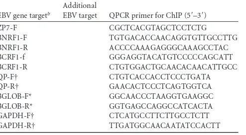
ChIP assay.ChIP experiments used a modified version of the protocol as described by Bark-Jones et al. (1). Briefly, the lysates were sonicated on ice (10 times with 10-s pulses each time, with a 30% amplitude output on a Branson model 250 Microtip at setting 5 [Sonics Vibracell]) to obtain 200- to 600-bp DNA fragments. For immunoprecipitation, 10g of an-TABLE 1Primer sequencesa
EBV gene targetb
Additional
EBV target QPCR primer for ChIP (5=–3=)
BALF5-F GATCGTGATAGCGTCTTCTGC BALF5-R GCAACATGCCTCTGGTGA BMRF1-1F GATTGGACTTCACACCAGGAA BMRF1-1R TCACAATGCGTTCAGAGAGG BMRF1-2F CACTGCGGTGGAGGTAGAG BMRF1-2R GGTGGTGTGCCATACAAGG BMRF1-3F CACCATGCTGGTGGTAGATG BMRF1-3R GCATGGTCATAGCACTTGGA BMRF1-4F CTGAGGAACGAGCAGATGATT BMRF1-4R CGTAGAGATCCGGATTGAGTG
OL1-F (Flank Orilyt L) GCGCAACAGTGCCACCAACC
OL1-R CAGGACCTGGCGGTAGTGCAG
OL2-F GCTCCACTGCACCTGGAAT
OL2-R CCAGAGGAGCCCCAGAAC
OL3-F OR3-F CTCTTTTTGGGGTCTCTGTG
OL3-R OR3-R CCTCCTCCTCTCGTTATCC
OL4-F CTAGAGGTCCGCGAGATTTG
OL4-R ACCTCTAGGCTCCACCCACT
OL5-F OR4-F CAGCTGACCGATGCTCGCCA
OL5-R OR4-R ATGGTGAGGCAGGCAAGGCG
[image:2.585.42.283.72.718.2] [image:2.585.297.544.78.214.2]OL6-F GGCCTGAAGAGGTTGACAAG OL6-R GAGGTAAGCCGTTCCAGATG OL7-F GGTACCCTGCATCCTGTGTT OL7-R GCGGAGAGGTGTTTCTCTTG OL8-F CATGACAGCCAATCCAACAC OL8-R CTGCGCCTACAGATCATCAA OR1-F (Flank Orilyt R) CCGCATGTCCAACCACCACG OR1-R ATGCTACCTAGGCCTGCGTCC OR2-F TTCCATTATCCTGGAGGTATCC OR2-R GCTGAATCCTACCTAGCTCCAC OR5-F GCTGGTTAAGCTGACGACCT OR5-R AGACATGCAGGAACACATGG OR6-F AGCCGAGCAGATTCTAATGG OR6-R CAACAGGTGTGCAGGTGTG OR7-F GGACGCAGCTACTTGACCTT OR7-R AGTGGACGCAGCACTTATCA RP1-F AGGTCCTCCTCTGGACTGTG RP1-R TTGATCTCGAGGTGCAGAAG RP2-F CCTGTTGTTTCGGAGAATGG RP2-R AATTTACAGCCGGGAGTGTG RP3-F GGCTGACATGGATTACTGGTC RP3-R TGATGCAGAGTCGCCTAATG RP4-F TCGCGATGCTATAAACCAGA RP4-R AGGGCATTCCATAAAGCAAA RP5-F CGGATGTCCAGAGTGCCTA RP5-R TGGACCAACATGTTCAGGAG RP6-F GTGATGAGGACGAGGATGGT RP6-R CCTCGTCAGACATGATTCACA ZP1-F CACGGCCATGCTATCTTGTA ZP1-R TGCCACTGGTCTCATCCTC ZP2-F GGAGGAATGCGATTCTGAAC ZP2-R CTGACCTCACGGTAGTGCTG ZP3-F AAGGCCAGCTAACTGCCTATC ZP3-R ACAGCTGAGGTGCTGCATAA ZP4-F GAGCCACAGGCATTGCTAA ZP4-R ACCAGCCTCCTCTGTGATGT ZP5-F CATGCAGCAGACATTCATCA ZP5-R GACGAACTGACCACAACACTAGA ZP6-F TGTCCACATATGGCTGCTTC ZP6-R GCAAGTCATCTGTTGGAGGAC
TABLE 1(Continued)
EBV gene targetb
Additional
EBV target QPCR primer for ChIP (5=–3=)
ZP7-F CGCTCACGTAGCTCCTCTG BNRF1-F TGTGACACCAACAGGTGTTGCCTTG BNRF1-R ACCCCAAAGAGGGCAAAGCCTAC BCRF1-f GGGAGGTACATGTCCCCCAGCATT BCRF1-R CTGTGGACTGCAACACAACATTGCC QP-F† CTGTCACCACCTCCCTGATA QP-R† GAACACTCCCTCAGTGGTCA BGLOB-F* GGCAACCCTAAGGTGAAGGC BGLOB-R* GGTGAGCCAGGCCATCACTA GAPDH-F† CTCATGCCTTCTTGCCTCTT GAPDH-R† TTGATGGCAACAATATCCACTT
aForward (F) and reverse (R) primer orientations areindicated in the primer
designations.
b*, Gallagher et al. (20); †,Palermo et al. (34).
on November 7, 2019 by guest
http://jvi.asm.org/
tibodies were used with chromatin from 5⫻106cells. The immune com-plexes were collected with preblocked 50% protein A/G-Sepharose bead slurry. After stringent washes and protein digestion of the immune com-plexes, the eluted DNA was purified using gel extraction kit (Qiagen) and eluted in 100l of double-distilled H2O or Tris-EDTA (TE) buffer to be analyzed further by real-time quantitative PCR (QPCR).
The ability of the anti-Zta antibody to precipitate Zta in the ChIP conditions was verified by resuspending the beads after immune pre-cipitation in 25l of 2⫻SDS sample buffer and analyzed by Western blotting.
ChIP-reChIP.Sequential ChIP experiments (ChIP-reChIP) (21) were undertaken using chromatin from 3⫻107cells and 60g of antibodies in
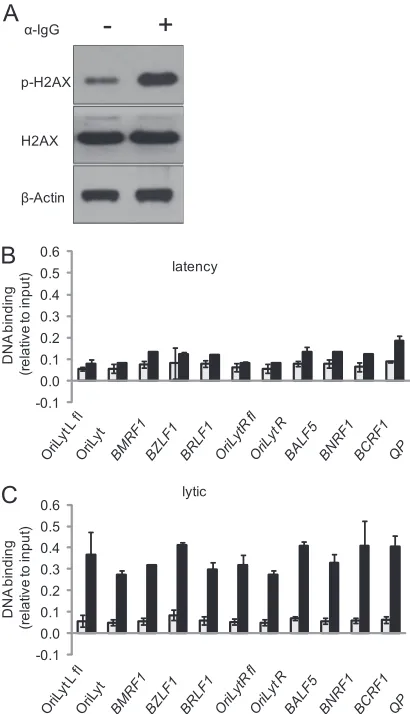
FIG 1Histone modifications associated with key lytic regulatory regions during latency. Total protein extracts and chromatin were prepared from Akata BL cells. (A) Proteins were fractionated on SDS-polyacrylamide gels and subjected to Western blotting with the indicated antibodies. (B to G) Chromatin was precipitated with the antibodies indicated above each panel, and associated DNA was purified and amplified with primers specific for each of the regulatory regions. The data are from experiments carried out on two independent batches of chromatin. In each case, the gray bars represent the signal from the control antibody, and the black bars represent the signal from the histone modification antibodies. DNA binding is expressed as the percentage of input chromatin. The primers used to amplify each region were OriLytL flank (OL1), OriLyt (OL5),BMRF1promoter (BMRF1-2),BZLF1promoter (ZP4),BRLF1promoter (RP3),BALF5promoter (BALF5), andBNRF1andBCRF1.
on November 7, 2019 by guest
http://jvi.asm.org/
the first round. The eluted complexes were diluted and subjected to a second round with 10g of each antibody. The DNA was then purified using a gel extraction column (Qiagen) and analyzed by PCR.
Real-time PCR analysis.QPCR analyses were undertaken using a Sensimix SYBR kit (Bioline) on an ABI 7500 real-time QPCR system (Applied Biosystems). The absolute quantitation method was used with dilutions of input DNA to generate the standard curve. Primer sets to detect EBV genome regions were designed using Primer3 (40) and are shown in Table 1.
Quantification of EBV genome copy number.A total of 2⫻106 Akata cells were harvested, and DNA was extracted using a Wizard genomic DNA purification kit (Promega). DNA was subjected to QPCR using primers specific to the EBV and human genomes (EBV DNA poly-merase gene and the human-globin gene). The relative amount of EBV genome copies to human genome was analyzed as described previously (20).
RESULTS
Histone modifications associated with key lytic regulatory
re-gions during latency.
The use of ChIP to map the chromatin
associated with early lytic cycle regulatory regions of the EBV
ge-nome during latency in Akata cells revealed that both of the
ori-gins of lytic replication (OriLytL and OriLytR) and the key early
lytic cycle promoters (
BZLF1
,
BRLF1
,
BMRF1
, and
BALF5
) show
evidence of being associated with a repressive histone
modifica-tion (H3K9me3) (Fig. 1). The data presented here identify a
com-mon occurrence of H3K9me3 association at several key lytic
reg-ulatory regions. In addition, a lower level of the repressive
polycomb-associated histone modification (H3K27me3) was
de-tected at these regions. Therefore, association with markers of
repressive chromatin is a common feature of these early lytic
reg-ulatory regions. Surprisingly, these experiments also revealed that
acetylated H3 was associated with these key regulatory regions,
together with lower levels of acetylated H4.
We also questioned whether H3K9me3 and acetylated H3 are
associated with late lytic genes, using BCRF1 and BNRF1 as
exam-ples. Both promoter regions have a similar high association of
H3K9me3 and lower association with acetylated H3 as the early
lytic promoters (Fig. 1F and G).
The interaction of both activation- and repression-associated
chromatin with the early lytic regulatory regions could represent
partially activated chromatin in latency, or it could result from a
mixed cell population, with a few cells undergoing lytic cycle. To
distinguish between these possibilities, we conducted sequential
ChIP to determine whether the lytic regulatory regions in the EBV
genome are associated with H3K9me3 and acetylated H3
simulta-neously. The first round of precipitation with the H3K9me3
anti-body showed substantial enrichment compared to the control
an-tibody (Fig. 2A and B). Each batch of chromatin was then eluted
and reprecipitated with either the H3K9me3 antibody or the
acetylated H3 antibody. We investigated the association at the
BRLF1
promoter and a region adjacent to OriLytL. At both loci,
only a small proportion of the H3K9me3 associated chromatin
was found to coassociate with acetylated H3. In contrast, the
con-FIG 2Coassociation of repressive marker of chromatin. Chromatin waspre-pared from Akata cells. The initial round of ChIP was undertaken with a H3K9me3 or a control antibody as shown below the histogram. The sample was then divided, and some DNA was purified directly and analyzed by PCR, while the remainder of the DNA complexes were eluted and subjected to a second round of precipitation with different antibodies. The data are from experiments carried out on two independent batches of chromatin. (A and B) Chromatin precipitated in the first round was processed to purify DNA, and this was amplified with primers sets specific for regions flanking OriLytL (OL1) (A) or at theBRLF1promoter (RP3) (B). The amount of chromatin bound in each case is expressed relative to the binding of H3K9me3 to the flank region of OriLytL (OL1). (C and D) Chromatin precipitated in the second round was processed to purify DNA, and this was amplified with primers sets specific for regions flanking OriLytL (OL1) (C) or at theBRLF1promoter (RP3) (D). The data are expressed relative to the binding of H3K9me3 to the flank of OriLytL (OL1).
FIG 3Coassociation of activation-dependent marker of chromatin. Chro-matin was prepared from Akata cells. The initial round of ChIP was under-taken with an acetylated H3 or a control antibody. The sample was then divided and some DNA was purified, while for the remainder were eluted and subjected to a second-round of precipitation with different antibodies as indicated below each data set. The data from experiments carried out on two independent batches of chromatin. (A and B) Chromatin precipitated in the first round was processed to purify DNA, and this was amplified with primers sets specific for regions flanking OriLytL (OL1) (A) or at theBRLF1 promoter (RP3) (B). The amount of chromatin bound in each case is expressed relative to the binding of acetylated H3 to OL1. (C and D) Chro-matin precipitated in the second round was processed to purify DNA, and this was amplified with primers sets specific for flanking OriLytL (OL1) (C) or at theBRLF1promoter (RP3) (D). The data are expressed relative to the binding of acetylated H3 to OL1.
on November 7, 2019 by guest
http://jvi.asm.org/
[image:4.585.44.279.67.274.2] [image:4.585.303.537.68.265.2]trol chromatin gave no signal with either antibody in the second
round.
In the reverse experiment, the chromatin was first precipitated
with the acetylated H3 antibody, or control, and subsequently with
either the H3K9me3 antibody or the acetylated H3 antibody. In this
case, the acetylated H3-associated chromatin was quantitatively
re-precipitated with the H3K9me3 antibody (Fig. 3), suggesting that all
of the acetylated H3 is associated with both histone marks. However,
since only a minority of the DNA is associated with both histone
marks and the presence of the activation-associated modification is
known to be associated with lytic cycle (6, 7, 22–24, 32, 51), this
probably represents the subpopulation of cells that spontaneously
enters into the lytic cycle during normal growth of the Akata cell line.
These data suggest that for the majority of cells in the latent
popula-tion the lytic cycle regulatory regions are marked by the repressive
histone modification H3K9me3.
Activation of H2AX associated with key lytic regulatory
re-gions.
Phosphorylation of H2AX occurs during lytic EBV
replica-tion in an inducible Zta expression system (29). We show here that
there is little phosphorylation of H2AX during latency but that a
substantial increase occurs in BL cells following entry into lytic
cycle by stimulation of the B-cell receptor (Fig. 4A). Furthermore,
it has been suggested that H2AX phosphorylation is important for
the lytic cycle of the related gammaherpesvirus MHV68 (48, 50).
We therefore sought to determine whether phosphorylated H2AX
is associated with key early- and late-lytic-cycle regulatory regions
and the latency control region Qp. This ChIP assessment of
phos-phorylated H2AX revealed negligible association of
phosphory-lated H2AX with the EBV genome during latency but a clear
in-teraction with each of the key regulatory regions specifically
during the lytic cycle (Fig. 4).
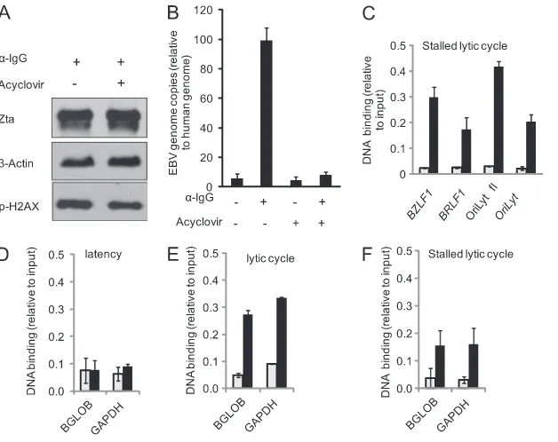
Phosphorylation of H2AX is associated with the DNA damage
response pathway and phosphorylated H2AX form foci for the
recruitment of repair components at sites of damaged DNA (30).
We speculated that H2AX might become activated in response to
the unusual DNA structures formed on the EBV genome during
lytic replication. To question this, we stalled viral DNA replication
using the drug acyclovir (stalled lytic cycle) and investigated
whether association of the EBV genome with activated H2AX
oc-curs. QPCR assays showed that acyclovir efficiently inhibited EBV
genome replication (Fig. 5B) without altering the abundance of
Zta or phosphorylated H2AX proteins (Fig. 5A). ChIP
experi-ments revealed that even during stalled lytic cycle the
phosphory-lated H2AX associates with the EBV genome (Fig. 5C).
Since replication of the EBV genome is not required to stimulate
phosphorylation of H2AX, this points to the action of a soluble
en-zyme rather than an enen-zyme that is tethered to DNA such as ATM.
Further analysis of the location of phosphorylated H2AX revealed
that it associates with the host genome during the lytic cycle (Fig. 5D
and E). Blocking EBV genome replication with acyclovir does not
prevent this increased association (Fig. 5F). Thus, the association of
phosphorylated H2AX occurs equivalently with the host and EBV
genomes. This global effect on H2AX points to the action of a soluble
enzyme such as the EBV protein kinase (BGLF4), which is known to
be able to promote phosphorylation of H2AX (48).
Association of Zta with key lytic regulatory regions on the
EBV genome.
We next explored the interaction of Zta with the
lytic cycle regulatory regions during the early stages of the lytic
cycle. We analyzed Zta binding
in vivo
during lytic replication
using ChIP with a panel of amplicons to assess the interaction of
Zta with both origins of lytic replication. This revealed a tight
footprint of Zta binding over each of OriLytL and OriLytR (Fig.
6). It is expected that Zta would interact with OriLytL, since this
has been seen in other cell lines (2). However, interaction of Zta
with OriLytR has not been documented previously (Fig. 6). These
data show that both OriLyts are recognized in an equivalent
man-ner
in vivo
during lytic replication.
Analysis of the interaction between Zta and key early lytic cycle
promoters revealed that Zta associates specifically with the
pro-moters for
BZLF1
(Zp),
BRLF1
(Rp), and
BMRF1
(Fig. 7).
Inter-estingly, the maximum binding to the Zp region (detected by ZP4
FIG 4Association of phosphorylated H2AX with EBV genome. Akata cells were induced, or not, with anti-IgG. After 48 h, total protein extracts and chromatin were prepared. (A) Proteins were fractionated on SDS-polyacrylamide gels and subjected to Western blotting with the indicated an-tibodies. Lanes “⫹” and “–” refer to stimulation with IgG. (B and C) Chroma-tin was precipitated with a control antibody or an antibody that recognizes the phosphorylated form of H2AX.The associated DNA was purified and ampli-fied with primers specific for each of the regulatory regions as indicated. The data from two independent batches of chromatin are expressed relative to the input chromatin with the standard error shown. The results for noninduced cells are shown in panel B, and the results for induced cells are shown in panel C. In each case, the gray bars represent the signal from the control antibody, and the black bars represent the signal from the phosphorylated H2AX anti-body. The primers used to amplify each region were OriLytL flank (OL1), OriLyt (OL5),BMRF1promoter (BMRF1-2),BZLF1promoter (ZP4),BRLF1 promoter (RP3),BALF5promoter (BALF5), andBNRF1,BCRF1, and the latency Qp promoter (QP).on November 7, 2019 by guest
http://jvi.asm.org/
[image:5.585.318.523.66.423.2]primers) encompasses the ZIIIA and B sites, which can mediate
autoactivation by Zta (17). It is intriguing that association with
these promoters occurs to a lesser degree than the association of
Zta with either lytic origin of replication. Indeed, the maximal
binding at the
BZLF1
promoter (Zp) is only 17% of that are
ob-served at OriLyt, and maximal binding at the
BRLF1
promoter
(Rp) is only 7% of that at OriLyt (Fig. 7E).
Since the differential association between promoters and
ori-gins of replication could be connected to stabilization of Zta
bind-ing to the origin of lytic replication, as observed by El-Guindy et al.
(10), we questioned whether there is any difference between the
kinetics of the association of Zta with key early lytic promoters
compared to the origins of lytic replication. Assessing binding
across a time course during the first 48 h after the induction of lytic
cycle revealed that the interaction between Zta and the key early
lytic cycle regulatory regions occurs in an equivalent temporal
manner detectable by 12 h postinduction (Fig. 8).
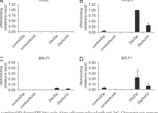
Zta coassociation with histone modifications.
We
hypothe-size that several key early lytic cycle control regions of the EBV
genome are associated with heterochromatin-like repressive
chro-matin during latency in Akata BL cells. Furthermore, once Zta is
expressed, it associates with these control regions and promotes
changes to the local chromatin environment. We tested this model
by asking whether Zta coassociates with H3K9me3 during lytic
cycle using sequential ChIP (Fig. 9). The first round of
precipita-tion isolated Zta-bound chromatin and the second round
inter-FIG 5H2AX association with the EBV and host genomes during lytic cycle. Akata cells were induced or not with anti-IgG either in the presence or in the absence of acyclovir. After 48 h, total protein extracts, genomic DNA, and chromatin were prepared. Latency is represented by no IgG and no acyclovir, a stalled lytic cycle is represented by IgG and acyclovir, and the lytic cycle is represented by IgG with no acyclovir. (A) Proteins were fractionated on SDS-polyacrylamide gels and subjected to Western blotting with the indicated antibodies. Lanes “⫹” and “⫺” refer to stimulation with IgG and the presence of acyclovir. (B) Genomic DNA was subjected to QPCR for specific regions from the EBV (BALF5) and human (beta-globin) genes. The amount of EBV genome present is expressed relative to the amount of human gene, with the standard error shown. (C to F) Chromatin was precipitated with a control antibody or an antibody that recognizes the phosphorylated form of H2AX. The associated DNA was purified and amplified with primers specific for each of the EBV or human genome regulatory regions-globin (BGLOB) or glyceraldehyde 3-phosphate dehydrogenase (GAPDH) as indicated. The data from two independent batches of chromatin are expressed relative to the input chromatin, with the standard error shown. In each case, the gray bars represent the signal from the control antibody, and the black bars represent the signal from the phosphorylated H2AX antibody. The primer sets used to amplify each region were OriLytR flank (OR1), OriLyt (OL5),BZLF1 promoter (ZP4), andBRLF1promoter (RP3).
FIG 6Zta association with the EBV origins of replication during lytic cycle. Akata cells were induced with anti-IgG where indicated. After 48 h, the chro-matin was prepared. (A and B) Chrochro-matin was precipitated with an antibody that recognizes Zta. The associated DNA was purified and amplified with primers sets specific for regions within OriLytL (A) or OriLytR (B). The data from two independent batches of chromatin are expressed relative to the max-imal Zta signal at OriLyt, with the standard error shown. (C and D) The genomic region surrounding OriLytL and OriLytR are shown with the genome coordinates and the location of characterized ZREs (identified as diamonds). The amplicons generated by the primers sets are shown below.
on November 7, 2019 by guest
http://jvi.asm.org/
[image:6.585.136.450.64.313.2] [image:6.585.52.278.433.629.2]rogated coassociation with H3K9me3. Parallel experiments with
nonspecific antibodies established the validity of binding at each
stage. Zta clearly associated with chromatin at the control regions
in OriLyt, the
BZLF1
and
BRLF1
promoters in these experiments,
whereas it did not associate with a region neighboring OrilytR
(OR1) (Fig. 9). It is clear from these experiments that Zta is able to
coassociate with H3K9me3 on DNA. Importantly, these
experi-ments demonstrate that at three early EBV lytic cycle regulatory
regions Zta is able to interact with the repressive chromatin
envi-ronment that is characteristic of EBV lytic cycle regulatory regions
during EBV latency.
We also investigated the coassociation of Zta with acetylated
histone H3 48 h postinduction of lytic replication. Sequential
ChIP revealed that approximately one-third of Zta is associated
with acetylated histone 3 at this point (Fig. 10).
DISCUSSION
We propose a model in which key early lytic promoters and the
origins of lytic replication are associated with a repressive
heterochromatin-like environment during latency, which is
over-come to allow expression of lytic cycle genes and origin function.
This model is based on our analysis of the chromatin structure at
key early lytic promoters and the origins of lytic replication and
is also supported by a low-resolution genome-wide map of
H3K9me3 association with the EBV genome in a different cell line
(group I Mutu BL cells), recently published by Tempera et al. (49).
The association of key lytic cycle regulatory regions with
H3K9me3 in two BL cell lines derived from different lymphomas
indicates that this association may be a common feature of EBV
type I latency in BL cells.
FIG 7Zta association with key lytic EBV promoters during the lytic cycle. Akata cells were induced with anti-IgG where indicated. After 48 h, the chromatin was prepared. (A and B) Chromatin was precipitated with an antibody that recognizes Zta. The associated DNA was purified and amplified with primers sets specific for regions within theBZLF1/BRLF1promoter region (A) or theBMRF1promoter region (B). The data from two independent batches of chromatin are expressed relative to the maximal Zta signal at OriLyt, with the standard error shown. (C and D) The genomic regions surrounding the promoters are shown with the genome coordinates and the location of characterized ZREs (identified as diamonds). The amplicons generated by the primers sets are shown below. Transcription start sites and the direction of transcription are indicated by arrows. (E) The maximal signal for each region is expressed relative to maximal binding to the lytic origin (OR4).
on November 7, 2019 by guest
http://jvi.asm.org/
[image:7.585.136.451.65.476.2]Several groups have shown that the
BZLF1
promoter is not
associated with acetylated histones during latency but that the
association increases significantly during lytic cycle (6, 23, 51).
This is in agreement with the negligible level of histone H3
acety-lation that we observed in the majority of cells during latency in
Akata cells, which is followed by a strong coassociation of Zta with
acetylated histone H3 during lytic cycle (Fig. 10).
In addition, we identified a further change to the chromatin
landscape at these early lytic cycle regulatory regions: association
with the activated form of the variant histone core protein H2AX.
Activation of H2AX is associated with induction of double-strand
breaks (38) and replication stress (12) and is seen during DNA
fragmentation when cells undergo apoptosis (37). Activated
H2AX plays a key role in the recruitment of DNA repair and
sig-naling factors (35). H2AX activation and the subsequent partial
activation of the DNA damage response during EBV lytic
replica-tion were documented by Kudoh et al. (29); we demonstrate here
that the H2AX modification occurs at the key regulatory regions
on the EBV genome during the lytic cycle, prior to EBV genome
replication. There is a requirement for H2AX for the lytic cycle of
the related virus MHV68 in some cell types, suggesting that it may
contribute to gamma herpesvirus replication. Since it has not
proved possible to knock down the expression of H2Ax
com-pletely, the impact that phosphorylation of H2AX has on EBV lytic
cycle remains to be determined.
Zta is required for EBV lytic replication (13). It is a
sequence-specific DNA-binding protein, with a promiscuous range of
re-sponse elements; it can interact with at least 32 sequence variants
of a 7-mer sequence (ZREs) (19). Zta has the unusual ability to
recognize methylated DNA (2–4, 9, 26); indeed, some ZREs are
entirely dependent on methylation for binding. This theoretically
provides Zta with the ability to access DNA within the EBV
ge-nome even when it has been epigenetically silenced by DNA
meth-ylation. We show here that Zta interacts with both origins of lytic
replication and with the promoters of
BZLF1
,
BRLF1
, and
BMRF1
in Akata cells at regions containing predicted ZREs (19). This
supports and extends previous reports that Zta interacts with the
EBV genome
in vivo
in other cell types (2, 9, 26, 52).
The presence of H3K9me3 correlates with repressive
chroma-tin, which is considered to prevent transcription factors from
in-FIG 8Zta association with EBV genome during the first 48 h of the lytic cycle. Akata cells were induced with anti-IgG where indicated. Total protein extracts and chromatin were prepared at 0, 4, 12, 24, and 48 h postinduction. (A) Proteins were fractionated on SDS-polyacrylamide gels and subjected to Western blotting with the indicated antibodies. Lanes “⫹” and “⫺” refer to stimulation with IgG, and the times (in hours) postinduction are indicated. (B to D) Chromatin was precipitated with an antibody that recognizes Zta. The associated DNA was purified and amplified with primers sets specific for regions at OriLyt (OL5) (B), theBZLF1promoter (ZP4) (C), theBRLF1promoter (RP3) (D), or theBMRF1promoter (BMRF1-2) (E). The data are expressed relative to the maximal Zta signal at OriLyt. In each case, the white bars represent the signal from chromatin from uninduced cells, and the black bars represent the signal from the chromatin from induced cells.on November 7, 2019 by guest
http://jvi.asm.org/
[image:8.585.135.451.63.417.2]teracting with DNA. Indeed, it was recently shown that the
pres-ence of repressive chromatin containing the H3K9me3 mark is
sufficient to prevent the interaction of the p53 transcription factor
with otherwise responsive promoters (44). It was not known
pre-viously what impact the association of Zta-responsive regulatory
elements with H3K9me3 would have on the ability of Zta to bind
to DNA. Using sequential ChIP we sought to determine whether
Zta and H3K9me3 coexist on chromatin during the EBV lytic
FIG 9Zta coassociation with H3K9me3 during EBV lytic cycle. Akata cells were induced with anti-IgG. Chromatin was prepared at 48 h postinduction. The initial round of ChIP was undertaken with a Zta or a control antibody. The sample was then split, and either DNA was purified or the protein DNA complexes were eluted and subjected to a second-round of precipitation with either Zta or H3K9me3 antibodies. DNA was prepared from chromatin precipitated in the second round and was amplified with primers sets specific for regions at OriLyt (OL5), the region flanking OriLytR (OR1), theBZLF1(ZP4), or theBRLF1(RP3) promoters. The data from experiments carried out on two independent batches of chromatin are expressed relative to the binding of Zta to OR4.FIG 10Zta coassociation with acetylated H3 during EBV lytic cycle. Akata cells were induced with anti-IgG. Chromatin was prepared at 48 h postinduction. The initial round of chromatin precipitation was undertaken with a Zta or a control antibody. The sample was then split, and either DNA was purified or the protein DNA complexes were eluted and subjected to a second round of precipitation with either Zta or acetylated H3 antibodies. DNA was prepared from chromatin precipitated in the second round and amplified with primers sets specific for regions at OriLyt (OL5), the region flanking OriLytR (OR1), theBZLF1(ZP4), or theBRLF1(RP3) promoters. The data from experiments carried out on two independent batches of chromatin are expressed relative to the binding of Zta to OR4.
on November 7, 2019 by guest
http://jvi.asm.org/
[image:9.585.135.455.63.306.2] [image:9.585.135.449.450.674.2]cycle. This revealed that Zta clearly associated with chromatin
containing the H3K9me3 modification providing proof that Zta is
able to interact with lytic cycle regulatory regions even when they
are encased in a repressive chromatin environment. Interestingly,
methylated DNA is often associated with facultative
heterochro-matin and associated with H3K9me3. Therefore, Zta may have
two routes to overcome epigenetic silencing of the EBV genome:
(i) by its ability to coassociate with H3K9me3-bound chromatin
and (ii) by its ability to interact with methylated ZREs.
ACKNOWLEDGMENT
This study was supported by a grant from the Wellcome Trust.
REFERENCES
1.Bark-Jones SJ, Webb HM, West MJ.2006. EBV EBNA 2 stimulates CDK9-dependent transcription and RNA polymerase II phosphorylation on serine 5. Oncogene25:1775–1785.
2.Bergbauer M, et al.2010. CpG-methylation regulates a class of Epstein-Barr virus promoters. PLoS Pathog.6:e1001114.
3.Bhende PM, Seaman WT, Delecluse HJ, Kenney SC. 2005. BZLF1 activation of the methylated form of the BRLF1 immediate-early pro-moter is regulated by BZLF1 residue 186. J. Virol.79:7338 –7348. 4.Bhende PM, Seaman WT, Delecluse HJ, Kenney SC.2004. The EBV lytic
switch protein, Z, preferentially binds to and activates the methylated viral genome. Nat. Genet.36:1099 –1104.
5.Bryant H, Farrell PJ.2002. Signal transduction and transcription factor modification during reactivation of Epstein-Barr virus from latency. J. Virol.76:10290 –10298.
6.Chang LK, Liu ST.2000. Activation of the BRLF1 promoter and lytic cycle of Epstein-Barr virus by histone acetylation. Nucleic Acids Res.28: 3918 –3925.
7.Countryman JK, Gradoville L, Miller G.2008. Histone hyperacetylation occurs on promoters of lytic cycle regulatory genes in Epstein-Barr virus-infected cell lines which are refractory to disruption of latency by histone deacetylase inhibitors. J. Virol.82:4706 – 4719.
8.Daigle D, et al.2011. Valproic acid antagonizes the capacity of other histone deacetylase inhibitors to activate the Epstein-Barr virus lytic cycle. J. Virol.85:5628 –5643.
9.Dickerson SJ, et al.2009. Methylation-dependent binding of the Epstein-Barr virus BZLF1 protein to viral promoters. PLoS Pathog. 5:e1000356. 10. El-Guindy A, Heston L, Miller G.2010. A subset of replication proteins
enhances origin recognition and lytic replication by the Epstein-Barr virus ZEBRA protein. PLoS Pathog.6:e1001054.
11. Ellis AL, Wang Z, Yu X, Mertz JE.2010. Either ZEB1 or ZEB2/SIP1 can play a central role in regulating the Epstein-Barr virus latent-lytic switch in a cell-type-specific manner. J. Virol.84:6139 – 6152.
12. Ewald B, Sampath D, Plunkett W.2007. H2AX phosphorylation marks gemcitabine-induced stalled replication forks and their collapse upon S-phase checkpoint abrogation. Mol. Cancer Ther.6:1239 –1248. 13. Feederle R, et al.2000. The Epstein-Barr virus lytic program is controlled
by the co-operative functions of two transactivators. EMBO J. 19: 3080 –3089.
14. Fernandez AF, et al.2009. The dynamic DNA methylomes of double-stranded DNA viruses associated with human cancer. Genome Res.19: 438 – 451.
15. Fixman ED, Hayward GS, Hayward SD.1995. Replication of Epstein-Barr virus oriLyt: lack of a dedicated virally encoded origin-binding pro-tein and dependence on Zta in cotransfection assays. J. Virol.69: 2998 –3006.
16. Fixman ED, Hayward GS, Hayward SD.1992.trans-acting requirements for replication of Epstein-Barr virus ori-Lyt. J. Virol.66:5030 –5039. 17. Flemington E, Speck SH. 1990. Autoregulation of Epstein-Barr virus
putative lytic switch gene BZLF1. J. Virol.64:1227–1232.
18. Flemington EK, Goldfeld AE, Speck SH.1991. Efficient transcription of the Epstein-Barr virus immediate-early BZLF1 and BRLF1 genes requires protein synthesis. J. Virol.65:7073–7077.
19. Flower K, et al.2011. Epigenetic control of viral life-cycle by a DNA-methylation dependent transcription factor. PLoS One6:e25922. 20. Gallagher A, et al.1999. Detection of Epstein-Barr virus (EBV) genomes
in the serum of patients with EBV-associated Hodgkin’s disease. Int. J. Cancer84:442– 448.
21. Geisberg JV, Struhl K.2004. Quantitative sequential chromatin immu-noprecipitation, a method for analyzing co-occupancy of proteins at genomic regions in vivo. Nucleic Acids Res.32:e151.
22. Gruffat H, Manet E, Sergeant A.2002. MEF2-mediated recruitment of class II HDAC at the EBV immediate-early gene BZLF1 links latency and chromatin remodeling. EMBO Rep.3:141–146.
23. Hui KF, Chiang AK.2010. Suberoylanilide hydroxamic acid induces viral lytic cycle in Epstein-Barr virus-positive epithelial malignancies and me-diates enhanced cell death. Int. J. Cancer126:2479 –2489.
24. Jenkins PJ, Binne UK, Farrell PJ.2000. Histone acetylation and reacti-vation of Epstein-Barr virus from latency. J. Virol.74:710 –720. 25. Kalla M, Schmeinck A, Bergbauer M, Pich D, Hammerschmidt W.
2010. AP-1 homolog BZLF1 of Epstein-Barr virus has two essential func-tions dependent on the epigenetic state of the viral genome. Proc. Natl. Acad. Sci. U. S. A.107:850 – 855.
26. Karlsson QH, Schelcher C, Verrall E, Petosa C, Sinclair AJ. 2008. Methylated DNA recognition during the reversal of epigenetic silencing is regulated by cysteine and serine residues in the Epstein-Barr virus lytic switch protein. PLoS Pathog.4:e1000005.
27. Kenney SC.2007. Reactivation and lytic replication of EBV, p 403– 433.In Arvin A, et al (ed), Human herpesviruses: biology, therapy, and immunopro-phylaxis. Cambridge University Press, Cambridge, United Kingdom. 28. Kraus RJ, Perrigoue JG, Mertz JE.2003. ZEB negatively regulates the
lytic-switch BZLF1 gene promoter of Epstein-Barr virus. J. Virol.77: 199 –207.
29. Kudoh A, et al.2005. Epstein-Barr virus lytic replication elicits ATM checkpoint signal transduction while providing an S-phase-like cellular environment. J. Biol. Chem.280:8156 – 8163.
30. Lukas J, Bohr VA, Halazonetis TD.2006. Cellular responses to DNA damage: current state of the field and review of the 52nd Benzon Sympo-sium. DNA Repair5:591– 601.
31. Miller G.1989. The switch between EBV latency and replication. Yale J. Biol. Med.62:205–213.
32. Murata T, et al.2011. Involvement of Jun dimerization protein 2 (JDP2) in the maintenance of Epstein-Barr virus latency. J. Biol. Chem.286: 22007–22016.
33. Packham G, Brimmell M, Cook D, Sinclair AJ, Farrell PJ.1993. Strain variation in Epstein-Barr virus immediate-early genes. Virology192: 541–550.
34. Palermo RD, Webb HM, West MJ.2011. RNA polymerase II stalling promotes nucleosome occlusion and pTEFb recruitment to drive immor-talization by Epstein-Barr virus. PLoS Pathog.7:e1002334.
35. Paull TT, et al.2000. A critical role for histone H2AX in recruitment of repair factors to nuclear foci after DNA damage. Curr. Biol.10:886 – 895. 36. Rickinson A, Kieff E (ed).2007.Epstein-Barr virus, 5th ed.Lippincott/
Williams and Wilkins, Philadelphia, PA.
37. Rogakou EP, Nieves-Neira W, Boon C, Pommier Y, Bonner WM.2000. Initiation of DNA fragmentation during apoptosis induces phosphoryla-tion of H2AX histone at serine 139. J. Biol. Chem.275:9390 –9395. 38. Rogakou EP, Pilch DR, Orr AH, Ivanova VS, Bonner WM.1998. DNA
double-stranded breaks induce histone H2AX phosphorylation on serine 139. J. Biol. Chem.273:5858 –5868.
39. Rowe M, Kelly GL, Bell AI, Rickinson AB.2009. Burkitt’s lymphoma: the Rosetta Stone deciphering Epstein-Barr virus biology. Semin. Cancer Biol. 19:377–388.
40. Rozen S, Skaletsky H.2000. Primer3 on the WWW for general users and for biologist programmers. Methods Mol. Biol.132:365–386.
41. Sinclair AJ.2003. bZIP proteins of human gammaherpesviruses. J. Gen. Virol.84:1941–1949.
42. Sinclair AJ, Brimmell M, Shanahan F, Farrell PJ. 1991. Pathways of activation of the Epstein-Barr virus productive cycle. J. Virol. 65: 2237–2244.
43. Sinclair AJ, Farrell PJ.1992. Epstein-Barr virus transcription factors. Cell Growth Differ.3:557–563.
44. Soria C, Estermann FE, Espantman KC, O’Shea CC.2010. Heterochro-matin silencing of p53 target genes by a small viral protein. Nature466: 1076 –1081.
45. Speck SH, Chatila T, Flemington E.1997. Reactivation of Epstein-Barr virus: regulation and function of the BZLF1 gene. Trends Microbiol.5:399 – 405. 46. Takada K.1984. Cross-linking of surface immunoglobulins induces
Epstein-Barr virus in Burkitt’s lymphoma cell lines. Int. J. Cancer33:27–32.
on November 7, 2019 by guest
http://jvi.asm.org/
47. Takada K, Ono Y.1989. Synchronous and sequential activation of la-tently infected Epstein-Barr virus genomes. J. Virol.63:445– 449. 48. Tarakanova VL, et al.2007. Gammaherpesvirus kinase actively initiates a
DNA damage response by inducing phosphorylation of H2AX to foster viral replication. Cell Host Microbe1:275–286.
49. Tempera I, Wiedmer A, Dheekollu J, Lieberman PM. 2010. CTCF prevents the epigenetic drift of EBV latency promoter Qp. PLoS Pathog. 6. 50. Xie A, Scully R.2007. Hijacking the DNA damage response to enhance viral replication: gammaherpesvirus 68 orf36 phosphorylates histone H2AX. Mol. Cell27:178 –179.
51. Ye J, Gradoville L, Daigle D, Miller G. 2007. De novo protein
synthesis is required for lytic cycle reactivation of Epstein-Barr virus, but not Kaposi’s sarcoma-associated herpesvirus, in response to his-tone deacetylase inhibitors and protein kinase C agonists. J. Virol. 81:9279 –9291.
52. Yin Q, Jupiter K, Flemington EK.2004. The Epstein-Barr virus transac-tivator Zta binds to its own promoter and is required for full promoter activity during anti-Ig and TGF-1 mediated reactivation. Virology327: 134 –143.
53. Young LS, et al.1991. Differentiation-associated expression of the Epstein-Barr virus BZLF1 transactivator protein in oral hairy leukoplakia. J. Virol.65:2868 –2874.
on November 7, 2019 by guest
http://jvi.asm.org/