Ruthenium Complexes
Thesis by Hang Song
In Partial Fulfillment of the Requirements for the Degree of
Doctor of Philosophy in Chemistry
California Institute of Technology Pasadena, California
2012
c
2012
Acknowledgements
My foremost acknowledgement goes to my graduate advisor, Professor Jacqueline K. Barton, for your enthusiasm, wisdom and vision in the complex worlds of science and life. Thank you for your support, guidance, and confidence in me throughout my time here. It has been a truly delightful and humbling journey along the rocky path of scientific discovery, and I am grateful that you have been there with me to help me see the gems in the piles of gravel. I would like to thank my thesis committee, Professors Harry Gray, Doug Rees, and Shu-ou Shan. Your guidance has made me a better scientist, and your generous support throughout the years has opened many doors for me.
I would also like to acknowledge Maureen Renta for her sage advice and for keeping us all out of trouble.
My mentors in the laboratory, Brian Zeglis and Mi Hee Lim, deserve special thanks for taking me under their wings. Thank you for your patience and help; your humor, creativity and scientific intuition will always be an inspiration.
Pam Sontz and Natalie Muren, you have been there with me every step of the way, and I am grateful for your love and friendship. We will always be the invincible trio!
for keeping us safe at all times; Mike Grodick, for your determination and positive energy; Anna McConnell, for your enthusiasm and advice; Mike Smeaton, for showing me tricks in cell work; Ariel Furst, for your courage and faith; Alyson Weidmann, for cheering up the corner; Helen Segal, for letting me use my initials; and Elizabeth Dervan, for a memorable summer of research and fun.
I would especially like to express my gratitude for Jens Kaiser, whose guidance and expertise in crystallography led me to solve the beautiful crystal structures presented in this work. I will always remember the day we found the crystal that diffracted to 0.92 ˚
A—my twenty-seventh birthday.
I would also like to thank members of the CCE, Biology divisions, and the Safety Office, without whom my research would have been impossible. Scott Virgil, Julie Hoy, Shelley Diamond, Pavle Nikolovski, Bill Dempsey, Tom Dunn, Agnes Tong, Steve Baldwin, Mona Shahgholi, Dave VanderVelde, Steve Gould, Joe Drew, Leah Santat, Ted Weintrob, Nathan Dalleska, Nicole Ford, Alec Durrell, Justin Chartron, and Haick Issaian: thank you for always being there to help.
I must also express my appreciation for my mentors outside of the department, Jean Huang, Elizabeth Tricomi, and Xiaolan Zhang, for your nuggets of wisdom and for being wonderful role models.
My life away from the laboratory has been colorful, thanks to my dear friends. Caglar Tanrikulu, Ozgun Konca, Muruvvet Buyukboyaci, Selim Hanay, Omer Durak, Ercan Gurses, Aycan Yurtsever, Bahar Bingol, Necmiye Ozay, Cevat Ustun, Murat Acar, and Samet Oy-mak, thank you for good company and good times.
The transformative power of graduate school does not stop at the door of the laboratory— I have also picked up a brown belt in karate. Thank you, Sensei Ian Ferguson and the Caltech Karate Club, for bringing the art of karate-do into my life.
Abstract
Deficiencies in DNA mismatch repair (MMR) have been implicated in the development of several forms of cancers, and MMR-deficient cells tend to be resistant to commonly employed cancer therapeutics such as cisplatin. Mismatch-targeting metalloinsertors developed in our laboratory have shown great promise as therapeutic and diagnostic agents for MMR-deficient cancers. In this work, we examine fundamental aspects of binding interactions of octahedral rhodium and ruthenium complexes to DNA mismatches, and strive to develop a luminescent sensor for mismatches inside cells.
We first demonstrate that the mismatch binding affinity of rhodium metalloinsertors directly correlates with their antiproliferative effect against MMR-deficient colorectal carci-noma cells. Smaller ancillary ligands on the rhodium center facilitate binding to mismatches via metalloinsertion from the narrow minor groove of DNA. Complexes with higher mis-match binding affinity in turn selectively inhibit the growth of MMR-deficient cells compared to MMR-proficient ones. This correlation suggests that DNA mismatches are indeed the biological target of rhodium metalloinsertors inside cells.
Besides rhodium metalloinsertors, luminescent ruthenium complexes are found to bind DNA mismatches as well. Mismatch binding is accompanied by enhanced luminescence intensity. We determined two crystal structures of ∆-Ru(bpy)2dppz2+ bound to oligonu-cleotide duplexes. For an oligonuoligonu-cleotide containing AA mismatches, the atomic-resolution structure revealed that the ruthenium complex binds to DNA mismatches also through metalloinsertion: the complex inserts a planar ligand into the mismatched site from the minor groove, ejecting the mismatched bases out of the helix. Several binding geometries of the complex intercalated between well-matched DNA were also observed.
Contents
Acknowledgements iii
Abstract v
1 Role of Ancillary Ligands in DNA Mismatch Recognition and Cellular Antiproliferation by Rhodium Metalloinsertors 1
1.1 Introduction . . . 2
1.2 Experimental protocols . . . 6
1.2.1 Materials . . . 6
1.2.2 DNA synthesis, purification, and quantification . . . 7
1.2.3 Rhodium complex synthesis . . . 8
1.2.4 Photocleavage titrations . . . 10
1.2.5 Binding constant determination . . . 10
1.2.6 MALDI-TOF mass spectrometry . . . 11
1.2.7 Fluorescence spectroscopy . . . 11
1.2.8 Structural modeling . . . 11
1.2.9 Cell culture . . . 11
1.2.10 Cellular proliferation ELISA . . . 12
1.3 Results . . . 12
1.3.1 Binding affinities of metal complexes at single base mismatches . . . 12
1.3.2 Inhibition of cellular proliferation by enzyme-linked immunosorbent assay (ELISA) . . . 18
1.4 Discussion . . . 25
1.5 Conclusions . . . 29
2.2.1 Materials . . . 39
2.2.2 Steady state fluorescence . . . 39
2.2.3 Time-resolved fluorescence . . . 39
2.3 Results and discussion . . . 40
2.3.1 Steady state luminescence ofrac-, ∆-, and Λ-Ru(bpy)2dppz2+ bound to oligonucleotides . . . 40
2.3.2 Comparison with other DNA-binding fluorophores . . . 41
2.3.3 Luminescence behavior of Ru with different base mismatches . . . . 41
2.3.4 Excited state lifetimes of Ru . . . 45
2.3.5 Luminescence response of Ru toDNA in the presence of Cu(phen)2+2 46 2.3.6 Selective quenching of Ru luminescence with matched DNA by NaI . 48 2.4 Conclusions . . . 49
Bibliography . . . 52
3 Crystal Structures of∆-Ru(bpy)2dppz2+ Bound to Mismatched and Well-matched DNA 57 3.1 Introduction . . . 58
3.2 Experimental procedures . . . 59
3.2.1 Materials . . . 59
3.2.2 Crystallization and data collection . . . 59
3.2.3 Structure determination and refinement . . . 60
3.2.4 Steady state fluorescence . . . 61
3.3 Results and discussion . . . 61
3.3.1 Cocrystallization of ∆-Ru(bpy)2dppz2+ with DNA . . . 61
3.3.2 Structure1 . . . 62
3.3.3 Structure2 . . . 70
3.3.4 Differences and similarities between the two structures . . . 73
3.3.5 Comparisons to other structures . . . 77
3.4 Conclusions . . . 80
Bibliography . . . 81
4 Ruthenium-Dye Conjugates as Luminescent Reporters of DNA Mismatches 85 4.1 Introduction . . . 86
4.2 Experimental protocols . . . 87
4.2.1 Materials . . . 87
4.2.2 Synthesis of dye, metal complex, and conjugates . . . 87
4.2.3 Steady state fluorescence . . . 91
4.3 Results and discussion . . . 91
4.3.1 Ru-TO-3 conjugates . . . 91
4.3.2 Ru-Eth conjugate . . . 99
4.4 Conclusions . . . 101
Bibliography . . . 103
5 Ru Complexes of DPPZ Derivatives and Chrysi Analogues 105 5.1 Introduction . . . 106
5.2 Experimental protocols . . . 106
5.2.1 Materials . . . 106
5.2.2 Synthesis of metal complexes . . . 107
5.2.3 Steady state fluorescence . . . 110
5.2.4 Fluorescence lifetimes . . . 110
5.3 Results and discussion . . . 112
5.3.1 Ru complexes bearing DPPZ derivatives . . . 112
5.3.2 Chrysi analogues of ruthenium . . . 118
5.4 Conclusions . . . 122
Bibliography . . . 123
6 RNA Mismatch Recognition by Ru(bpy)2dppz2+ 125 6.1 Introduction . . . 126
6.2 Experimental protocols . . . 126
6.2.1 Materials . . . 126
6.2.6 Confocal microscopy . . . 128 6.3 Results and discussion . . . 128 6.3.1 Ru luminescence with a mismatched RNA hairpin . . . 128 6.3.2 Confocal microscopy of Tet-on HeLa cells expressing mismatch-containing
mRNA . . . 131 6.4 Conclusions . . . 137 Bibliography . . . 138
Chapter 1
Role of Ancillary Ligands in DNA Mismatch Recognition
and Cellular Antiproliferation by Rhodium Metalloinsertors
Adapted from Russell J. Ernst, Hang Song, and Jacqueline K. Barton,Journal of the American Chemical
Society, 2009, 131 (6), 2359–2366. R. J. E. synthesized Rh(DIP)2chrysi
3+
strands of DNA: adenine (A) always pairs with thymine (T), and guanine (G) with cytosine (C) (Figure 1.1). This strict base-pairing scheme allows the sequence of a complementary strand to be determined solely from that of the template strand, which forms the basis for DNA replication. Errors in DNA replication will cause DNA base mismatches, which, left unrepaired, will become mutations in subsequent rounds of replication.1,2 Healthy cells rely on their mismatch repair (MMR) pathway to maintain a genome that is virtually free of mismatches (1 in a billion base pairs per cell division).3,4The MMR pathway corrects single base errors and insertion/deletion loops that arise during DNA synthesis, increasing the fidelity of DNA replication by a factor of 50–1000.1If uncorrected, mismatches are converted to mutations in subsequent cycles of DNA replication, and cells with MMR deficiencies, not surprisingly, exhibit elevated mutation rates.5–7 Germline mutations in hMLH1 or hMSH2, essential genes for MMR in humans, dramatically increase the risk of developing hereditary nonpolyposis colon cancer (HNPCC), the most common type of inherited colon cancer.8,9 HNPCC is marked by early onset and the presence of cancers in several other tissue types.9 Roughly 15% of sporadic colorectal cancer cases have also been linked to MMR deficiency.10 Epigenetic silencing of the MMR genes has been identified as the cause of MMR deficiency in these cases.11 In addition to colorectal cancer, mismatch repair deficiencies have been found in approximately 16% of solid tumors of all tissue types.6,12
N N N N N N N N O N N O O N N N N O N H H H R R R R H H H H H major groove minor groove minor groove major groove
Adenine Thymine Guanine Cytosine
Figure 1.1. Base pairing in DNA.
at the O6 position of guanine nucleotides triggers an apoptotic response after recognition of O6-meG:C and O6-meG:T base pairs by the MMR pathway, while MMR-deficient cells tolerate this DNA methylation.13–15 Failure to recognize DNA adducts is also involved in the resistance of MMR-deficient cells to the platinum compounds cisplatin and carbo-platin.13–16 The incorporation of antimetabolites such as 5-fluorouracil and 6-thioguanine into DNA triggers cell cycle arrest and apoptosis through the MMR pathway, and conse-quently MMR-deficient cells are resistant to these agents as well.17,18 Other studies have shown low-level resistance to the type I topoisomerase poisons camptothecin and topotecan in hMLH1-deficient lines and to the type II topoisomerase poisons doxorubicin, epirubicin, and mitoxantrone in hMLH1- or hMSH2-deficient lines.19It has also been hypothesized that treatment regimens with agents such as cisplatin might enrich tumors for MMR-deficient cells,20 and it has been shown that a substantial portion of secondary, or therapy-related, leukemias show signs of MMR deficiency.20,21 Collectively, these results show the broad involvement of MMR in mediating drug response, the effects of MMR deficiency on this re-sponse, and the need to develop therapeutic agents that specifically target MMR-deficient cells, perhaps through broad recognition of DNA mismatches.
DNA mismatch recognition is commonly achieved through oligonucleotide- or enzyme-based methods. Oligonucleotide-enzyme-based detection schemes rely on differential hybridization between fully complementary and mismatched duplexes.22–27 Enzyme-based methods typ-ically employ mismatch recognition enzymes such asE. coli MutS.28Small molecule-based detection schemes are relatively less common. Saito and Nakatani groups have demon-strated the use of naphthyridine dimers to recognize several mismatches by hydrogen bond-ing to the bases.29–31Teulade-Fichou and co-workers reported a luminescent bisanthracene derivative that discriminates between well-matched and mismatched DNA.32 In the field of inorganic small molecules, our laboratory has developed metal complexes that recognize over 80% of all base mismatches, and the therapeutic effect of these metal complexes are being investigated.33
NMR.38,40–42 These characteristics of mismatched bases led our laboratory to devise recog-nition strategies that enabled the synthesis of metal complexes which bind mismatches with high affinity and specificity.
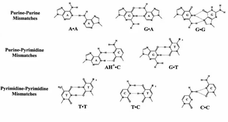
Figure 1.2. Chemical structures of base mismatches.
HN NH
N N
N N
HN NH
N N
HN NH
[image:14.612.131.519.205.414.2]phi dppz chrysi phzi
Figure 1.3. Chemical structures of intercalating and inserting ligands.
resultant metal complexes to intercalate between almost any two neighboring base pairs. Thus, although incapable of intercalation between well-matched base pairs, chrysi or phzi is able to insert into theπ-stack at destabilized mismatch sites via the minor groove, ejecting the pair of mismatched bases out of the helix. This insertion mode of binding via the mi-nor groove was fully elucidated by the crystal structure determination of Rh(bpy)2chrysi3+ bound to AC mismatches in a 12-mer duplex (Figure 1.4).43 Later crystal structures re-vealed identical structural features of Rh(bpy)2chrysi3+ insertion at an AA mismatch.44 Further, NMR studies also support the minor-groove insertion of the chrysi ligand and ejec-tion of mismatched bases in soluejec-tion.45 Interestingly, this insertion mode of binding was first proposed almost half a century ago by Lerman,46but had never been confirmed exper-imentally with small organic or inorganic molecules until the discovery of metalloinsertors. In total, Rh(bpy)2chrysi3+ has been shown to bind specifically to a single mismatch in a 2725bp linearized plasmid heteroduplex, and recognize over 80% of mismatch sites in all the possible single base pair flanking sequence contexts.47Similar to Rh(L)2(phi)3+ complexes, Rh(L)2chrysi3+ or Rh(L)2phzi3+photocleaves the DNA backbone when irradiated between 300 and 400 nm. They also show extremely enantiospecific binding, the ∆-enantiomer (Figure 1.5) being much more effective at binding to B-form DNA.48
The in vivo effects of Rh(bpy)2chrysi3+and Rh(bpy)2phzi3+ have been characterized in the isogenic cell lines HCT116N and HCT116O.49 The HCT116 cell line is a colorectal carcinoma line deficient in the hMLH1 gene. Two derivative cell lines, HCT116N and HCT116O, have been made through transfection of human chromosome 3 (ch3) and human chromosome 2 (ch2), respectively. The presence of a functional copy of ch3 restores MMR proficiency in the HCT116N line, while the HCT116O line transfected with ch2 remains MMR deficient.50 The mismatch recognition compounds were shown to selectively inhibit the proliferation of the repair-deficient HCT116O line.49
Recent work within our laboratory on luminescent ruthenium complexes has also shown that these tris(chelate) complexes are taken up inside cells through passive diffusion facili-tated by the membrane potential.51,52 Variations in ancillary ligands have dramatic effects on cellular uptake, with increased lipophilicity facilitating uptake. Uptake can also be increased through functionalization with a nuclear localizing peptide.53
Figure 1.4. Crystal structure showing the insertion of Rh(bpy)2chrysi3+ (red) via the minor groove at two AC mismatch sites in a 12-mer duplex. The mismatched bases are ejected (adenosine is colored green and cytosine colored blue).
Rh N
N
N
N H N
N H Rh
N
N
N
N H N
N H
[image:16.612.254.395.55.325.2]3+ 3+
Figure 1.5. Λ- (left) and ∆- (right) isomers of Rh(bpy)2chrysi3+.
in vivo. Importantly, we establish that the differential inhibition of cellular proliferation in MMR-deficient cells is correlated with mismatch binding affinity.
1.2
Experimental protocols
1.2.1 Materials
Rh NH3 NH3 NH3 NH3 H N N H
Rh(NH3)4chrysi3+
Rh N N N N Rh N N N N H N N H Rh H N N H N NH N N NH N Rh N N H N N H Rh N N N N H N N H H N N H
Rh(bpy)2chrysi3+ Rh(HDPA)
2chrysi3+
Rh(phen)2chrysi3+ Rh(DAP)2chrysi3+ Rh(DIP)2chrysi3+ N
N
3+ 3+ 3+
[image:17.612.131.516.53.358.2]3+ 3+ 3+
Figure 1.6. Chemical structures of [Rh(L2)(chrysi)]3+ complexes surveyed in this study.
purchased from Waters Chemical Co. (Milford, MA). Phosphoramidites were purchased from Glen Research (Sterling, VA). Media and supplements were purchased from Invitro-gen (Carlsbad, CA). BrdU, antibodies, buffers, and peroxidase substrate were purchased in kit format from Roche Molecular Biochemicals (Mannheim, Germany). All commercial materials were used as received.
1.2.2 DNA synthesis, purification, and quantification
con-1.2.3 Rhodium complex synthesis
The compound 1,12-diazaperylene (DAP) was synthesized according to literature proce-dures.54Chrysene-5,6-dione, Rh(bpy)2chrysi3+and Rh(phen)2chrysi3+were laboratory stocks synthesized according to established protocols.55
[Rh(NH3)4(chrysi)]OTf3. Rh(NH3)6OTf3was prepared as described by Sargeson.56,57 Rh(NH3)6OTf3 (200 mg, 0.3 mmol) was reacted with chrysene-5,6-dione (78 mg, 0.3 mmol) in 150 mL of acetonitrile and 50 mL of water with 0.6 mL of 1N NaOH as a catalyst. The reaction mixture (an orange suspension) was left stirring for 16 h at room temperature and turned into a dark orange-red suspension. Hydrochloric acid was added until the pH reached neutral. Acetonitrile was removed under vacuum and the resulting suspension fil-tered to remove unreacted chrysene quinone. The product was separated from unreacted Rh(NH3)6OTf3 on a Sep-Pak C18 cartridge eluting with 1:1:0.1% H2O:CH3CN:TFA. The eluant was lyophilized to recover the solid product (50 mg, 38%). 1H-NMR (300 MHz, d6-DMSO):δ 13.5 (s, 1H), 12.9 (s, 1H), 8.7–8.5 (m, 3H), 8.45 (dd, 2H), 8.2 (d, 1H), 8.0–7.6 (m, 4H), 4.4 (s, 3H), 4.35 (s, 3H), 3.7 (s, 6H). ESI-MS: calc. 427.11, obs. 427.0 (M-2H+), 410.0 (M-NH3-2H+), 391.1 (M-2NH3-2H+), 374.0 (M-3NH3-2H+), 356.9 (M-4NH3-2H+).
rac-[Rh(DAP)2(chrysi)]OTf3. Rh(NH3)4chrysi3+ (20 mg, 0.02 mmol) was reacted with 1,12-diazaperylene (DAP) (20 mg, 0.08 mmol, excess) in 45 mL of ethanol and 45 mL of water. The bright orange solution was heated under reflux for 16 h. The reaction mixture turned a darker orange color upon heating. Ethanol was removed under vacuum and the resulting solution filtered to remove any residue. The filtrate was concentrated on a Sep-Pak C18 cartridge eluting with 1:1:0.1% H2O:CH3CN:TFA, lyophilized and purified on an alumina column eluting with 5% MeOH in CH2Cl2. The fractions were collected and dried in vacuum to give an orange-brown solid (6.3 mg, 30%). ESI-MS: calc. 865.2 (M-2H+), obs. 865.3 (M-2H+), 611.1 (M-DAP-2H+), 433.2 (M-H2+). UV/vis (H2O, pH 5): 267 nm (53100 M−1 cm−1), 479 nm (16200 M−1 cm−1), 509 nm (16000 M−1 cm−1).
solution was heated under reflux for 16 h. The reaction mixture turned reddish brown upon heating. Ethanol was removed under vacuum and the resulting solution filtered to remove any residue. The filtrate was concentrated on a Sep-Pak C18 cartridge eluting with 1:1:0.1% H2O:CH3CN:TFA, lyophilized and purified on an alumina column eluting with 2% MeOH in CH2Cl2. The fractions were collected and dried under vacuum to give an orange-brown solid (8mg, 39%). 1H-NMR (300 MHz,d6-DMSO):δ 12.84 (s, 1H), 12.34 (s, 1H), 11.78 (s, 1H), 10.32 (d, 1H, 8.7 Hz), 8.63 (d, 1H, 6.9 Hz), 8.40 (d, 1H, 8.4 Hz), 8.31 (d, 1H, 9.3 Hz), 8.14 (m, 2H), 8.07 (d, 1H, 8.7 Hz), 8.04 (d, 1H, 5.4 Hz), 7.94 (m, 4H), 7.77 (m, 5H), 7.58 (m, 2H), 7.48 (d, 1H, 8.1 Hz), 7.41 (d, 1H, 8.4 Hz), 7.32 (s, 1H), 7.14 (m, 2H), 7.04 (t, 1H, 6.8 Hz), 6.98 (t, 1H, 6.9 Hz), 6.81 (t, 1H, 6.5 Hz) ppm. ESI-MS: calc. 699.2 (M-2H+), obs. 699.2 (M-2H+), 350.1 (M-H2+). UV/vis (H2O, pH 5): 287 nm (42200 M−1 cm−1), 321 nm (23000 M−1 cm−1), 442 nm (8800 M−1 cm−1).
rac-[Rh(DIP)2(NH3)2]OTf3. RhCl3 and 2 equiv. of DIP were combined in 1:1 ethanol:water and refluxed overnight. The solvent was removed in vacuo, and the product was recrystallized by dissolving in acetonitrile at 60◦C and cooling to −20◦C. The precip-itate was collected by filtration, washed in diethyl ether, and dissolved in neat triflic acid. The solution was again cooled and added dropwise to NH4OH at −20◦C. The pale white precipitate was collected by filtration and washed with a small amount of water to give [Rh(DIP)2(NH3)2]OTf3.
sis.55 A small amount of the labeled DNA (less than 1% of the total amount of DNA) was added to 2µM DNA in 100 mM NaCl, 20 mM NaPi, pH 7.1 buffer. The DNA was annealed by heating at 90◦C for 10 min and cooling down slowly to room temperature over a period of 2 h. Rhodium complex solutions ranging from nanomolar to micromolar concentration were made in Milli-Q water. 10 µL of annealed 2 µM DNA and 10 µL of Rh solution at each concentration was mixed in an eppendorf tube and incubated at 37◦C for 10 min. A light control (LC), in which the DNA was mixed with 10 µL of water and irradiated, and a dark control (DC), in which the DNA was mixed with the highest concentration of Rh complex without irradiation, were also prepared. The samples were left in the heat block and irradiated on an 1000-W Hg/Xe arc lamp (320–440 nm, Oriel) for 5 min. The irradiated samples were dried and electrophoresed in a 20% denaturing polyacrylamide gel. The gel was then exposed to a phosphor screen, and the relative amounts of DNA in each band were quantitated by phosphorimagery (ImageQuant).
1.2.5 Binding constant determination
The fraction of DNA cleaved in each lane on the gel was normalized and plotted against the log of the concentration of rhodium complex. At least three photocleavage titrations were carried out for each racemic metal complex. The pooled data were fit to a sigmoidal curve using OriginPro 6.1. The resulting midpoint value (i.e., the log of [rhodium complex] at the inflection point of the curve) was converted to units of concentration ([Rh50%]). The dissociation constant was calculated according to KD = [Rh50%] – 0.5[DNA], and
the binding constant was defined as KB = 1/KD. The errors were derived from the
1.2.6 MALDI-TOF mass spectrometry
Irradiation samples were prepared similarly to those used in photocleavage titrations, ex-cept that 2 µM of unlabeled DNA and 2 µM of Rh complex were used instead of 1 µM. The samples were heated at 90◦C for 30 min after irradiation to convert any metastable photocleavage products to stable one(s). Water (30 µL) was added to each sample, and the entire volume was passed through a Bio-Rad Microbio-spin chromatography column. The samples were dried and desalted using the ZipTip procedure (Millipore). The desalted oligonucleotides were then dried and redissolved in 1µL of water. MALDI-TOF mass spec-tra were taken on a PerSeptive Biosystem Voyager-DE Pro instrument. The samples were prepared by the dry droplet method, using 3-hydroxypicolinic acid and diammonium citrate as matrix. Several scans of 100 shots each were taken and averaged.
1.2.7 Fluorescence spectroscopy
Steady state fluorescence spectra were recorded on an ISS-K2 fluorimeter at ambient tem-peratures in aerated solution (5mM Tris, 50 mM NaCl, pH 7.5).
1.2.8 Structural modeling
Geometries of metal complexes were optimized in ChemDraw3D (CambridgeSoft) using MM2 minimization. All modeling sessions were performed in PyMol using the DNA crystal structure (PDB entry 2O1L).43 Metal complexes were combined with the DNA structure by superimposing the Rh atom with that in Rh(bpy)2chrysi3+ in the crystal structure and maximally overlapping the chrysi ligands. For clarity only one Rh complex is shown in each model.
1.2.9 Cell culture
Figure 1.7. Sequence of the DNA hairpin used in Rh(chrysi) complDNA binding ex-periments.
1.2.10 Cellular proliferation ELISA
HCT116N and HCT116O cells were plated in 96-well plates at 2000 cells/well and allowed 24 h to adhere. The cells were then incubated with rhodium complexes for the durations specified. For incubation less than 72 h, the Rh-containing medium was replaced with fresh medium, and the cells were grown for the remainder of the 72 h period. Cells were labeled with BrdU 24 h before analysis. The BrdU incorporation was quantified by antibody assay according to established procedures.58,59 Cellular proliferation was expressed as the ratio of the amount of BrdU incorporated by the treated cells to that of the untreated cells.
1.3
Results
1.3.1 Binding affinities of metal complexes at single base mismatches
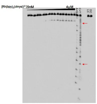
The binding constants of the family of Rh(L)2chrysi3+ complexes at a CC, AC or TT mis-match in a 29-mer DNA hairpin (Figure 1.7) were measured. The hairpin sequence allows cleavage site determination on either strand around the DNA mismatch site. By irradiating samples of DNA titrated with varying concentrations of a rhodium complex, a photocleavage titration curve is obtained, from which the binding constant of the Rh complex is deter-mined. A typical autoradiogram of electrophoresced samples in a photocleavage titration with Rh(bpy)2chrysi3+ and CC-containing DNA is shown in Figure 1.8. The position of the photocleavage band indicates that Rh(bpy)2chrysi3+ cleaves one base away from the mismatch site near the 3’-end.
Figure 1.8. Autoradiogram of a denaturing 20% polyacrylamide electrophoresis gel show-ing a representative photocleavage titration. Conditions are DNA (CC mismatch, 1 µM), Rh(bpy)2chrysi3+ (lanes 1 to 17: 5 nM, 10 nM, 20 nM, 50 nM, 0.1 µM, 0.2 µM, 0.3 µM, 0.4 µM, 0.5µM, 0.6 µM, 0.7 µM, 0.8 µM, 0.9 µM, 1 µM, 2 µM, 3 µM, 4 µM, lane 20: no Rh complex, lane 21: 4 µM), NaCl 50 mM, NaPi 10 mM, pH 7.1 at room temperature. Samples were irradiated at 365 nm on a 1000W Hg/Xe lamp for 5 min. The dark control in lane 21 was not irradiated. Lanes 18 and 19 are Maxam-Gilbert sequencing reactions. The mismatched sites are marked with red arrows.
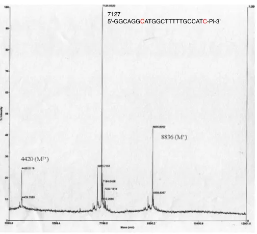
mass spectrometry.53 This cleavage pattern is found to be true of Rh(phen)2chrysi3+ and Rh(DIP)2chrysi3+as well. No other photocleavage bands are visible, demonstrating the high specificity of Rh(chrysi) complexes binding to the mismatch. The photocleavage titration curve is generated from the autoradiogram by quantitating the amount of photocleavage relative to the total amount of DNA at each Rh concentration. Pooled data from at least three repeats were fitted to a sigmoidal curve (Figure 1.10). The dissociation constant for Rh(bpy)2chrysi3+ at a CC mismatch is found to be (30 ±3) nM, which translates into a binding constant of (3.4 ± 0.3) × 107 M−1. At an AC and TT mismatch, the respective binding constants are (2.2 ± 0.2) × 106 M−1 and (6.3 ± 2.0) × 106 M−1. The dissoci-ation constant for Rh(phen)2chrysi3+ is found to be (320 ± 20) nM at a CC mismatch, which corresponds to a binding constant of (3.2 ±0.3)×106 M−1. The binding affinity of Rh(phen)2chrysi3+ to AC is (1.4±0.2) ×106 M−1. At a TT mismatch, the photocleavage efficiency of Rh(phen)2chrysi3+ is too low to allow meaningful quantification.
Figure 1.9. MALDI-TOF mass spectrum of CC-containing DNA after irradiation in the presence of Rh(bpy)2chrysi3+.
0.5 1.0 1.5 2.0 2.5 3.0 3.5 -0.2
0.0 0.2 0.4 0.6 0.8 1.0
Normalized fraction cleaved
Log[Rh(bpy) 2(chrysi)]
1.5 2.0 2.5 3.0 3.5 4.0 0.0
0.2 0.4 0.6 0.8 1.0
Normalized fraction cleaved
Log[Rh(NH3)4(chrysi) 3+
[image:24.612.199.451.104.334.2]]
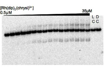
Figure 1.11. Autoradiogram of a denaturing 20% polyacrylamide electrophoresis gel showing photocleavage titration using Rh(DIP)2chrysi3+. Conditions are DNA (1.5 µM), Rh(DIP)2chrysi3+ (lanes 1 to 16: 0.5µM, 1µM, 2µM, 3µM, 4µM, 5µM, 6µM, 7µM, 8
µM, 9µM, 10µM, 15µM, 20µM, 25µM, 30µM, 35µM, lane 17: no Rh complex, lane 18: 40µM), NaCl 50 mM, NaPi 10 mM, pH 7.1 at 37◦C. Samples were irradiated on a 1000-W Hg/Xe lamp (320–440 nm) for 5 min except for the dark control in Lane 9. In addition to the photocleavage band, a slower-running band above the parent band is visible.
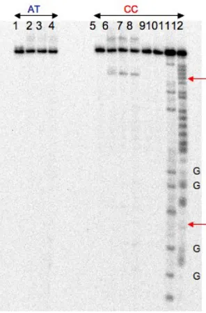
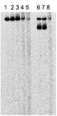
band (Figure 1.11). The intensity of this band is proportional to that of the photocleavage band, which suggests that its origin is somewhat mismatch related. This is confirmed by irradiating samples of Rh(DIP)2chrysi3+ and a DNA hairpin with a matched AT base pair in place of CC (Figure 1.12). Without a mismatch, the slower-running band is not observed. Piperidine treatment after irradiation shows that a small fraction of the slower-running band is due to the formation of a lesion prone to cleavage by piperidine, perhaps 8-oxo-guanine (Figure 1.12). MALDI-TOF mass spectrometry detects an additional cluster of peaks around m/z ≈ 9601 (m/z for parent DNA = 8836). These peaks are tentatively assigned to covalent Rh-DNA adducts with both DIP ligands but without the chrysi ligand (calculated mass for DNA+Rh+2DIP = 9603). Thus, the quantitation of the fraction of DNA photocleaved is carried out by dividing the photocleavage band intensity over the total DNA in a given lane including the slower-running band. The dissociation constant for Rh(DIP)2chrysi3+ is found to be (11 ± 2) µM and the binding constant is (9 ± 1)
× 104 M−1 (Table 1.1). It should be noted that if we treat the slower-running band as a mismatch-specific photoreaction product and combine it with the normal photocleavage band, the measured binding constant will not differ much, since the two bands are pro-portional to each other and will give similar normalized fractions regardless of whether the slower-running band is included. With an AC mismatch, Rh(DIP)2chrysi3+ does not yield any photocleavage up to 100 µM; thus, its KD value is estimated to be greater than that.
Figure 1.12. Autoradiogram of a denaturing 20% polyacrylamide electrophoresis gel com-paring Rh(DIP)2chrysi3+ reaction with matched (AT) and mismatched (CC) DNA. Condi-tions are DNA (1 µM), Rh(DIP)2chrysi3+ (lanes 1 and 5: 3µM, lanes 2–4, 6–8 and 10: 40
slower-running band increases as the concentration of Rh(DIP)2chrysi3+ increases, until the entire parent band disappears.
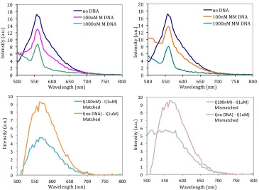
Rh(DAP)2chrysi3+ photocleaves DNA; however, instead of producing a single photo-cleavage band, a smear is seen in the autoradiogram, implying extremely nonspecific bind-ing and cleavage (Figure 1.13). The same smear is seen from irradiations at 313, 320–440, or 495 nm. We attribute this nonspecific interaction with DNA to the shape of the DAP ligand: the DAP ligand resembles a phi ligand in terms of its size and distance from the Rh center. Thus, it is not surprising that it can intercalate and cleave at multiple positions in the duplex. More interestingly, Rh(DAP)2chrysi3+ is found to be luminescent in water, emitting at 560 nm. The luminescence intensity is not proportional to its concentration, with the intensity at 1 µM being much less than 100-fold of the intensity at 10 nM, which implies that the complex is self-quenching. When DNA is added to a buffered solution of Rh(DAP)2chrysi3+, its luminesence decreases as the amount of DNA added increases. The extent of the decrease is similar in the presence of matched or mismatched DNA (Figure 1.14). Consistent with the results from irradiation experiments, these observations again point to nonspecific interactions with DNA and the quenching of the DAP ligand in a hy-drophobic environment (in this case propbably stacked between base pairs). Thus, due to its nonspecific interactions with DNA, it was not feasible to extract mismatch-specific binding constant for Rh(DAP)2chrysi3+.
Figure 1.13. Autoradiogram of a denaturing 20% polyacrylamide electrophoresis gel show-ing Rh(DAP)2chrysi3+ photoreaction. Conditions are DNA (1 µM), Rh(DAP)2chrysi3+ (lanes 1–5: 0.25 µM, 1.25 µM, 2.5 µM, 6 µM, 12.5 µM, lane 7: 0.6 µM, lane 8: 6 µM,), Rh(bpy)2chrysi3+ (lanes 6–8: 1µM), NaCl 50 mM, NaPi 10 mM, pH 7.1 at 37◦C. Samples were irradiated on a 1000-W Hg/Xe lamp (320–440 nm) for 10 min.
complexes are summarized in Table 1.1.
As Rh(NH3)4chrysi3+ exhibits very tight binding to CC and AC mismatches, its ability to recognize guanine-containing mismatches was also investigated. The metallointercalator Rh(bpy)2phi3+ was used to photocleave a DNA hairpin containing either an AG or a GG mismatch in a nonspecific manner. Rh(NH3)4chrysi3+ was added to compete for binding with Rh(bpy)2phi3+. The binding of Rh(bpy)2phi3+ at the mismatch site decreased sig-nificantly in the case of AG mismatch and to a lesser extent in the case of GG mismatch, as evident in the reduction in photocleavage around the mismatched sites (Figure 1.16). Photocleavage by Rh(bpy)2phi3+ at other sites in DNA was not as significantly affected.
1.3.2 Inhibition of cellular proliferation by enzyme-linked
immunosor-bent assay (ELISA)
!" #" $" %" &" '!" '#" '$" '%" '&" #!"
(!!" ((!" %!!" %(!" )!!" )(!" &!!" *+",-." '!!*/"/",-." '!!!*/"/",-." !" #" $" %" &" '!" '#" '$" '%" '&" #!"
(!!" ((!" %!!" %(!" )!!" )(!" &!!" *+",-." '!!*/"//",-." '!!!*/"//",-." !" #" $" %" &" '" (" )" *" +" #!"
'!!" ''!" (!!" ('!" )!!" )'!" *!!" ,-#!!./0"1",-#2/0" /345678" ,-.9":;<0"1",-#2/0" /345678" !" #" $" %" &" '" (" )" *" +" #!"
'!!" ''!" (!!" ('!" )!!" )'!" *!!" ,-#!!./0"1",-#2/0" /3456789:;" ,-.<"=>?0"1",-#2/0" /3456789:;"
Wavelength (nm) Wavelength (nm)
Wavelength (nm) Wavelength (nm)
Intens
ity (
a.u
.)
Intens
ity (
a.u
.)
Intens
ity (
a.u
.)
Intens
ity (
a.u
[image:29.612.133.498.78.345.2].)
Figure 1.14. Emission spectra of Rh(DAP)2chrysi3+ in the absence and presence of matched (top left) or mismatched (top right) DNA (excitation wavelength = 470 nm). The sharp peak at 560 nm is due to Raman scattering of solvent water. The difference spectra (i.e., the difference in intensity, I, between the spectra recorded in the presence of 100 nM and 1 µM DNA, as well as the difference inI between the spectra in the absence and presence of 1 µM DNA) are shown in the bottom row. Qualitatively there is no dif-ference between the fluorescence response in the presence of matched versus mismatched DNA. The fluorescence intensity (I) decreases with increasing amounts of either matched or mismatched DNA.
Figure 1.15. Autoradiogram of a denaturing 20% polyacrylamide electrophoresis gel showing a representative binding competition titration. Conditions are DNA (1 µM), Rh(bpy)2chrysi3+ (1 µM except for the light control), Rh(NH3)4chrysi3+ (lanes 3 to 20: 50nM, 0.1µM, 0.2µM, 0.25µM, 0.3µM, 0.4µM, 0.5µM, 0.6µM, 0.7µM, 0.8µM, 0.9µM, 1µM, 1.5µM, 2µM, 2.5µM, 5µM, 7.5µM, 10µM, lane 21: no Rh complexes, lane 22: no Rh(NH3)4chrysi3+), NaCl 50 mM, NaPi 10 mM, pH 7.1 at 37◦C. Samples were irradiated on a 1000-W Hg/Xe arc lamp (320–440 nm) for 5 min. The dark control in lane 21 was not irradiated. Lanes 1 and 2 are Maxam-Gilbert sequencing reactions. The mismatched sites are marked with red arrows.
exerting little to no effect on the HCT116N line (7±6% inhibition).
Table 1.1. Binding constantsa,b of Rh(L
2)chrysi3+ complexescto a single mismatch
Complex Structure KB(CC)/M−1 KB(AC)/M−1 KB(TT)/M−1
Rh NH3 NH3 NH3 NH3 H N N H 3+
Rh(NH3)4chrysi3+ 1.0×10⁸ 3.4× 10⁶ 8.8 ×10⁶
Rh N N N N H N N H 3+
Rh(bpy)2chrysi3+ 3.4×10⁷ 2.2× 10⁶ 6.3 ×10⁶
Rh H N N H N NH N N NH N 3+
Rh(HDPA)2chrysi3+ 2.0×10⁷ 2.6× 10⁶ n.d.d
Rh N N N N H N N H 3+
Rh(phen)2chrysi3+ 3.2×10⁶ 1.4× 10⁶ n.d.d
Rh N N H N N H N N
3+
Rh(DIP)2chrysi3+ 9 ×10⁴ <104 <104
a
Uncertainties are estimated to be 10%–15%. bBinding constants are determined from pho-tocleavage or binding competition titrations using a DNA hairpin with the sequence 5’-GGCAGGXATGGCTTTTTGCCATYCCTGCC-3’ (XY = CC, AC, or TT). Samples were irradiated with a solar simulator (320–440 nm) at 37◦C in 50 mM NaCl, 10 mM NaPi, pH 7.1, as described in the Experi-mental Protocols. cFor the polypyridyl complexes, values are given for racemic mixtures. dNot determined.
HeLa cells by Ru(bpy)2dppz2+, Ru(phen)2dppz2+, and Ru(DIP)2dppz2+, where the most rapid uptake is apparent with the lipophilic DIP complex.51
G T C C
Figure 1.16. Autoradiogram of a denaturing 20% polyacrylamide electrophoresis gel show-ing bindshow-ing competition between Rh(NH3)4chrysi3+ and Rh(bpy)2phi3+ to a DNA hairpin containing an AG or GG mismatch. Conditions are DNA (500 nM), Rh(bpy)2phi3+ (10µM except for the light control), Rh(NH3)4chrysi3+(0 nM, 100 nM, 500 nM, 1µM, 5µM), NaCl 50 mM, NaPi 10 mM, pH 7.1. Samples were irradiated on a 1000W Hg/Xe arc lamp (320 -440 nm) for 30 min and heated at 90◦C for 25 min after irradiation. The light controls were irradiated without Rh complexes. The dark controls were not irradiated. Lanes “A+G” and “C+T” are Maxam-Gilbert sequencing reactions. The mismatched sites are marked with red arrows.
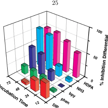
time for the family of complexes. Clear correlations with the binding constants for these complexes are evident (Table 1.1). Significantly, the differential effect in inhibiting cell proliferation in MMR-deficient cells is directly correlated to the binding affinity of the compound for DNA mismatches. Rh(NH3)4chrysi3+(KB= 1×10⁸M−1at a CC mismatch),
for example, shows the largest differential effect in inhibiting proliferation of MMR-deficient versus MMR-proficient HCT116 cells after 72 h (79 ± 5%), while Rh(phen)2chrysi3+(KB
Figure 1.18. Inhibitory effects of rhodium metalloinsertors as a function of incubation time. Shown are plots of BrdU incorporation normalized to the BrdU incorporation of untreated cells as a function of rhodium concentration. The inhibition differential is the difference of the normalized percentages of cellular proliferation for each cell line, with standard error bars (SN−O=
q s2
N +s2O). ELISA were performed as in Figure 1.17. Cells
were incubated with no rhodium, 2 µM rac-Rh(DIP)2chrysi3+, 10 µM Rh(NH3)4chrysi3+, or 20 µMrac-Rh(L)2chrysi3+ (L = HDPA, bpy, or phen).
Figure 1.20. Inhibitory effects of rhodium metalloinsertors as a function of metal complex identity. Shown are bar graphs of BrdU incorporation normalized to the BrdU incorporation of untreated cells as a function of rhodium concentration. The inhibition differential is the difference of the normalized percentages of cellular proliferation for the two cell lines, HCT116O versus HCT116N. ELISA were performed as in Figure 1.17. Cells were incubated with no rhodium, 2 µM rac-Rh(DIP)2chrysi3+, 10 µM Rh(NH3)4chrysi3+, or 20 µM rac -Rh(L)2chrysi3+(L = HDPA, bpy, or phen). A correlation between mismatch binding affinity and differential inhibition of MMR-deficient cells is evident.
1.4
Discussion
A clear trend emerges when comparing the binding constants of the series of rhodium com-plexes to mismatched sites: the DNA mismatch binding affinity increases as the size of the ancillary ligand decreases. This trend is consistent with what we have learned from the structural studies, specifically that mismatch binding by insertion via the minor groove is subject to stringent space constraints. With major groove intercalation, the base rise is increased, and the major groove offers space to accommodate the ancillary ligands. In con-trast, with insertion, there is no increase in base pair rise; the mismatched bases are instead ejected and replaced by the deeply inserted chrysi ligand. Moreover, the minor groove, small even for hydrophobic groove binding molecules, offers little space for the ancillary ligands. While little enantioselectivity is apparent for intercalation of bpy complexes into B-form DNA, ∆-Rh(bpy)2chrysi3+ binds enantiospecifically to single base mismatches.43,61 Thus, steric interactions of the ancillary ligands are seen as an extremely important factor governing the binding affinity of a metal complex at the mismatch site.
ther-ically more stable mismatch and, in this case, the melting temperature of the AC hairpin is 5◦C higher than that of the CC hairpin (Table 2.1). We assume, then, that the general trend holds for all members of this metalloinsertor family. This stabilization is translated into a higher dissociation constant (smaller binding affinity) for the entire series of rhodium complexes. This decrease in binding affinity depends upon the greater energy required to eject the mismatched bases from the base pair stack, as evident crystallographically and by NMR.43,45 Nonetheless, for the family of chrysi complexes, the inverse relationship be-tween the size of the ancillary ligand and the binding affinity still holds, with the smallest complex Rh(NH3)4chrysi3+ showing the highest affinity and that of the largest complex, Rh(DIP)2chrysi3+, more than 2 orders of magnitude lower.
bind at all to a mismatch site with some specificity, corroborate our understanding of the driving force and the dynamics of mismatch recognition: the π-stacking between the in-serted chrysi ligand and the adjacent bases provides the major stabilizing force for binding, and both the metal complex and DNA distort their conformations to accommodate each other in the bound state.
Importantly, the DNA mismatch binding affinities of the Rh(L)2chrysi3+ family corre-late well with the differential biological effects seen between the repair-proficient HCT116N and repair-deficient HCT116O cell lines. This correlation supports the hypothesis that DNA mismatches are the target of rhodium metalloinsertorsin vivo. Because of this correlation, we may attribute the preferential inhibitory effect on MMR-deficient cells to binding of the complexes to DNA mismatches. Since the MMR-deficient cells contain more mismatches, the tighter binding complexes would be expected to display a greater inhibitory effect. It should be noted that finding any inhibitory effect with these complexes was at first sur-prising, since they bind DNA noncovalently and might be expected to be readily displaced. Although the mechanism of inhibition is not yet fully understood, it is likely that protein recognition of the metal-mismatch complex, perhaps by RNA polymerase or topoisomerase, may generate a covalent protein-DNA lesion and contribute to the cellular response.
The differential inhibitory effect seen with Rh(HDPA)2chrysi3+ cannot be understood simply on the basis of binding affinities. Despite having essentially the same mismatch binding affinity as Rh(bpy)2chrysi3+, the HDPA complex preferentially inhibits the MMR-deficient cell line almost as well as Rh(NH3)4chrysi3+with long incubation times; with short times of incubation, the differential inhibitory effect by Rh(HDPA)2chrysi3+ is greatest. Both the HDPA ligand and the amine group have the potential to form hydrogen bonds. This hydrogen bonding capability and flexibility of the ligands might serve to make them more effective inhibitors of any protein-DNA interactions. Indeed, ruthenium complexes bearing HDPA ligands have been shown to exhibit DNA binding and cytotoxicity.63
much shorter incubation times and lower concentrations, which also matches the acceler-ated uptake observed for Ru(DIP)2dppz2+. Cellular uptake is surely a rate-limiting factor in biological activity of the rhodium metalloinsertors, yet cellular uptake is not the only challenge: proper subcellular localization must also be achieved in order for any drug to act on its target. It has been well established that lipophilic cations preferentially target the mitochondria, whereas hydrophilic cations do not.64,65It may be that Rh(HDPA)2chrysi3+ and Rh(NH3)4chrysi3+ lack the lipophilicity required for mitochondrial accumulation, al-lowing a greater proportion of these compounds to reach the nucleus once inside the cell. This difference in intracellular partitioning could then account for the differential effects of Rh(HDPA)2chrysi3+.
1.5
Conclusions
Bibliography
[1] R. R. Iyer, A. Pluciennik, V. Burdett, and P. L. Modrich. DNA mismatch repair: Functions and
mechanisms. Chemical Reviews, 106(2):302–323, 2006.
[2] Paul Modrich. Mechanisms in eukaryotic mismatch repair. Journal of Biological Chemistry,
281(41):30305–30309, 2006.
[3] John W. Drake. A constant rate of spontaneous mutation in DNA-based microbes. Proceedings of the
National Academy of Sciences, 88(16):7160–7164, 1991.
[4] John W. Drake. The distribution of rates of spontaneous mutation over viruses, prokaryotes, and
eukaryotes. Annals of the New York Academy of Sciences, 870(1):100–107, 1999.
[5] Lawrence A. Loeb. A mutator phenotype in cancer. Cancer Research, 61(8):3230–3239, 2001.
[6] Nitai P. Bhattacharyya, Adonis Skandalis, Anil Ganesh, Joanna Groden, and Mark Meuth. Mutator
phenotypes in human colorectal carcinoma cell lines. Proceedings of the National Academy of Sciences,
91(14):6319–6323, 1994.
[7] Bernard S. Strauss. Frameshift mutation, microsatellites and mismatch repair. Mutation
Re-search/Reviews in Mutation Research, 437(3):195–203, 1999.
[8] Nickolas Papadopoulos and Annika Lindblom. Molecular basis of HNPCC: Mutations of MMR genes.
Human Mutation, 10(2):89–99, 1997.
[9] P¨aivi Peltom¨aki. Deficient DNA mismatch repair: A common etiologic factor for colon cancer. Human
Molecular Genetics, 10(7):735–740, 2001.
[10] D. A. Lawes, S. SenGupta, and P. B. Boulos. The clinical importance and prognostic implications of
microsatellite instability in sporadic cancer.European Journal of Surgical Oncology (EJSO), 29(3):201–
212, 2003.
[11] James G. Herman, Asad Umar, Kornelia Polyak, Jeremy R. Graff, Nita Ahuja, Jean-Pierre J. Issa,
Sanford Markowitz, James K. V. Willson, Stanley R. Hamilton, Kenneth W. Kinzler, Michael F. Kane,
Richard D. Kolodner, Bert Vogelstein, Thomas A. Kunkel, and Stephen B. Baylin. Incidence and
functional consequences of hMLH1 promoter hypermethylation in colorectal carcinoma.Proceedings of
[12] Iordanis I. Arzimanoglou, Fred Gilbert, and Hugh R. K. Barber. Microsatellite instability in human
solid tumors.Cancer, 82(10):1808–1820, 1998.
[13] Klaus Pors and Laurence H. Patterson. DNA mismatch repair deficiency, resistance to cancer
chemother-apy and the development of hypersensitive agents.Current Topics in Medicinal Chemistry, 5(12):1133–
1149, 2005.
[14] Anna Maria Valentini, Raffaele Armentano, Michele Pirrelli, and Maria L. Caruso. Chemotherapeutic
agents for colorectal cancer with a defective mismatch repair system: The state of the art. Cancer
Treatment Reviews, 32(8):607–618, 2006.
[15] J. M. Carethers, M. T. Hawn, D. P. Chauhan, M. C. Luce, G. Marra, M. Koi, and C. R. Boland.
Competency in mismatch repair prohibits clonal expansion of cancer cells treated with
N-methyl-N’-nitro-N-nitrosoguanidine.The Journal of Clinical Investigation, 98(1):199–206, 1996.
[16] Daniel Fink, Sibylle Nebel, Stefan Aebi, Hua Zheng, Bruno Cenni, Alissar Nehm´e, Randolph D.
Chris-ten, and Stephen B. Howell. The role of DNA mismatch repair in platinum drug resistance. Cancer
Research, 56(21):4881–4886, 1996.
[17] John M. Carethers, Dharam P. Chauhan, Daniel Fink, Sibylle Nebel, Robert S. Bresalier, Stephen B.
Howell, and C. Richard Boland. Mismatch repair proficiency and in vitro response to 5-fluorouracil.
Gastroenterology, 117(1):123–131, 1999.
[18] S. Aebi, D. Fink, R. Gordon, H. K. Kim, H. Zheng, J. L. Fink, and S. B. Howell. Resistance to cytotoxic
drugs in DNA mismatch repair-deficient cells.Clinical Cancer Research, 3(10):1763–1767, 1997.
[19] Andr´e Fedier, Viola A. Schwarz, Heinrich Walt, Renato Delli Carpini, Urs Haller, and Daniel Fink.
Resistance to topoisomerase poisons due to loss of DNA mismatch repair. International Journal of
Cancer, 93(4):571–576, 2001.
[20] D. Fink, S. Aebi, and S. B. Howell. The role of DNA mismatch repair in drug resistance. Clinical
Cancer Research, 4(1):1–6, 1998.
[21] Peter Karran, Judith Offman, and Margherita Bignami. Human mismatch repair, drug-induced DNA
damage, and secondary cancer. Biochimie, 85(11):1149–1160, 2003.
[22] Irina V. Nesterova, S. Sibel Erdem, Serhii Pakhomov, Robert P. Hammer, and Steven A. Soper.
Ph-thalocyanine dimerization-based molecular beacons using near-ir fluorescence.Journal of the American
Chemical Society, 131(7):2432–2433, 2009.
[23] Daniela Baumstark and Hans-Achim Wagenknecht. Perylene bisimide dimers as fluorescent “glue” for
DNA and for base-mismatch detection. Angewandte Chemie International Edition, 47(14):2612–2614,
[25] Kazushige Yamana, Yoshikazu Ohshita, Yudai Fukunaga, Mitsunobu Nakamura, and Atsushi
Maruyama. Bis-pyrene-labeled molecular beacon: A monomer–excimer switching probe for the
de-tection of DNA base alteration. Bioorganic & Medicinal Chemistry, 16(1):78–83, 2008.
[26] T. Santhosh Kumar, Jesper Wengel, and Patrick J. Hrdlicka. 2-N-(pyren-1-yl)acetyl-2-amino-α-L-LNA:
Synthesis and detection of single nucleotide mismatches in DNA and RNA targets. ChemBioChem,
8(10):1122–1125, 2007.
[27] Anthony G. Frutos, Santona Pal, Mark Quesada, and Joydeep Lahiri. Method for detection of
single-base mismatches using bimolecular beacons. Journal of the American Chemical Society, 124(11):2396–
2397, 2002.
[28] Minseon Cho, Suhman Chung, Seong-Dal Heo, Jakang Ku, and Changill Ban. A simple fluorescent
method for detecting mismatched DNAs using a MutS–fluorophore conjugate. Biosensors and
Bioelec-tronics, 22(7):1376–1381, 2007.
[29] Kazuhiko Nakatani, Shinsuke Sando, and Isao Saito. Scanning of guanine-guanine mismatches in DNA
by synthetic ligands using surface plasmon resonance. Nat Biotech, 19(1):51–55, 2001.
[30] Kazuhiko Nakatani, Shinya Hagihara, Yuki Goto, Akio Kobori, Masaki Hagihara, Gosuke Hayashi,
Motoki Kyo, Makoto Nomura, Masaki Mishima, and Chojiro Kojima. Small-molecule ligand induces
nucleotide flipping in (CAG)ntrinucleotide repeats. Nat Chem Biol, 1(1):39–43, 2005.
[31] Akio Kobori, Souta Horie, Hitoshi Suda, Isao Saito, and Kazuhiko Nakatani. The SPR sensor detecting
cytosinecytosine mismatches. Journal of the American Chemical Society, 126(2):557–562, 2004.
[32] Anton Granzhan and Marie-Paule Teulade-Fichou. A fluorescent bisanthracene macrocycle
discrimi-nates between matched and mismatch-containing DNA. Chemistry–A European Journal, 15(6):1314–
1318, 2009.
[33] Brian M. Zeglis, Valerie C. Pierre, and Jacqueline K. Barton. Metallo-intercalators and
metallo-insertors.Chem Commun (Camb), (44):4565–4579, 2007.
[34] T. Brown, W. N. Hunter, G. Kneale, and O. Kennard. Molecular structure of the G.A base pair in
DNA and its implications for the mechanism of transversion mutations. Proceedings of the National
Academy of Sciences, 83(8):2402–2406, 1986.
[35] J. V. Skelly, K. J. Edwards, T. C. Jenkins, and S. Neidle. Crystal structure of an oligonucleotide duplex
containing G.G base pairs: Influence of mispairing on DNA backbone conformation.Proceedings of the
[36] William N. Hunter, Tom Brown, and Olga Kennard. Structural features and hydration of a dodecamer
duplex containing two C.A mispairs. Nucleic Acids Research, 15(16):6589–6605, 1987.
[37] W. N. Hunter, T. Brown, G. Kneale, N. N. Anand, D. Rabinovich, and O. Kennard. The structure
of guanosine-thymidine mismatches in B-DNA at 2.5-a resolution. Journal of Biological Chemistry,
262(21):9962–9970, 1987.
[38] Nicolas Peyret, P. Ananda Seneviratne, Hatim T. Allawi, and John SantaLucia. Nearest-neighbor
thermodynamics and NMR of DNA sequences with internal AA, CC, GG, and TT mismatches.
Bio-chemistry, 38(12):3468–3477, 1999.
[39] John SantaLucia and Donald Hicks. The thermodynamics of DNA structural motifs. Annual Review
of Biophysics and Biomolecular Structure, 33(1):415–440, 2004.
[40] Frances H. Arnold, Steven Wolk, Phillip Cruz, and Ignacio Tinoco. Structure, dynamics, and
ther-modynamics of mismatched DNA oligonucleotide duplexes d(CCCAGGG)2 and d(CCCTGGG)2.
Bio-chemistry, 26(13):4068–4075, 1987.
[41] Richard J. Isaacs, William S. Rayens, and H. Peter Spielmann. Structural differences in the
NOE-derived structure of G–T mismatched dna relative to normal DNA are correlated with differences in
13
C relaxation-based internal dynamics. Journal of Molecular Biology, 319(1):191–207, 2002.
[42] D. J. Patel, S. A Kozlowski, and K. Ikuta, S.and Ikuta. Dynamics of DNA duplexes containing
inter-nal GT, GA, AC, and TC pairs—hydrogen-exchange at and adjacent to mismatch sites. Federation
Proceedings, 43(11):2663–2670, 1984.
[43] Valerie C. Pierre, Jens T. Kaiser, and Jacqueline K. Barton. Insights into finding a mismatch through
the structure of a mispaired DNA bound by a rhodium intercalator.Proceedings of the National Academy
of Sciences, 104(2):429–434, 2007.
[44] Brian M. Zeglis, Valerie C. Pierre, Jens T. Kaiser, and Jacqueline K. Barton. A bulky rhodium complex
bound to an adenosine-adenosine DNA mismatch: General architecture of the metalloinsertion binding
mode. Biochemistry, 48:4247–4253, 2009.
[45] Christine Cordier, Val´erie C. Pierre, and Jacqueline K. Barton. Insertion of a bulky rhodium complex
into a DNA cytosinecytosine mismatch: An NMR solution study. Journal of the American Chemical
Society, 129(40):12287–12295, 2007.
[46] L. S. Lerman. Structural considerations in the interaction of DNA and acridines. Journal of Molecular
Biology, 3(1):18–30, 1961.
[47] Brian A. Jackson, Viktor Y. Alekseyev, and Jacqueline K. Barton. A versatile mismatch recognition
agent: Specific cleavage of a plasmid DNA at a single base mispair. Biochemistry, 38(15):4655–4662,
DNA mismatch-specific targeting and hypersensitivity of mismatch-repair-deficient cells to bulky
rhodium(III) intercalators. Proceedings of the National Academy of Sciences, 103(42):15359–15363,
2006.
[50] Minoru Koi, Asad Umar, Dharam P. Chauhan, Sajeev P. Cherian, John M. Carethers, Thomas A.
Kunkei, and C. Richard Boland. Human chromosome 3 corrects mismatch repair deficiency and
mi-crosatellite instability and reduces N-Methyl-N-nitro-N-nitrosoguanidine tolerance in colon tumor cells
with homozygous hMLH1 mutation. Cancer Research, 54(16):4308–4312, 1994.
[51] Cindy A. Puckett and Jacqueline K. Barton. Methods to explore cellular uptake of ruthenium complexes.
Journal of the American Chemical Society, 129(1):46–47, 2007.
[52] Cindy A. Puckett and Jacqueline K. Barton. Mechanism of cellular uptake of a ruthenium polypyridyl
complex. Biochemistry, 47(45):11711–11716, 2008.
[53] Jens Brunner and Jacqueline K. Barton. Targeting DNA mismatches with rhodium intercalators
func-tionalized with a cell-penetrating peptide. Biochemistry, 45(40):12295–12302, 2006.
[54] Oliver Schmelz, Alf Mews, Thomas Basch´e, Andreas Herrmann, and Klaus M¨ullen. Supramolecular
complexes from CdSe nanocrystals and organic fluorophors. Langmuir, 17(9):2861–2865, 2001.
[55] Brian M. Zeglis and Jacqueline K. Barton. DNA base mismatch detection with bulky rhodium
inter-calators: synthesis and applications.Nature Protocol, 2(2):357–371, 2007.
[56] Nicholas E. Dixon, Geoffrey A. Lawrance, Peter A. Lay, and Alan M. Sargeson.
(Trifluoromethanesulfonato-O)pentaammine complexes: Versatile synthetic intermediates.
Inor-ganic Chemistry, 22(5):846–847, 1983.
[57] Nicholas E. Dixon, Geoffrey A. Lawrance, Peter A. Lay, and Alan M. Sargeson. Synthetically versatile
(trifluoromethanesulfonato)metal amine complexes.Inorganic Chemistry, 23(19):2940–2947, 1984.
[58] Armin H. Reitmair, Robert Risley, Robert G. Bristow, Teresa Wilson, Anil Ganesh, Anne Jang, James
Peacock, Samuel Benchimol, Richard P. Hill, Tak W. Mak, Richard Fishel, and Mark Meuth. Mutator
phenotype in Msh2-deficient murine embryonic fibroblasts. Cancer Research, 57(17):3765–3771, 1997.
[59] H. G. Gratzner. Monoclonal antibody to 5-bromo- and 5-iododeoxyuridine: A new reagent for detection
of DNA replication. Science, 218(4571):474–475, 1982.
[60] Achim H. Krotz, Louis Y. Kuo, Thomas P. Shields, and Jacqueline K. Barton. Dna recognition by
rhodium(III) polyamine intercalators: Considerations of hydrogen bonding and van der Waals
[61] Jacqueline K. Barton. Metals and DNA: Molecular left-handed complements. Science, 233(4765):727–
734, 1986.
[62] Brian A. Jackson and Jacqueline K. Barton. Recognition of base mismatches in DNA by
5,6-chrysenequinone diimine complexes of rhodium(III): A proposed mechanism for preferential binding
in destabilized regions of the double helix. Biochemistry, 39(20):6176–6182, 2000.
[63] Venugopal Rajendiran, Mariappan Murali, Eringathodi Suresh, Mallayan Palaniandavar,
Vaiya-puri Subbarayan Periasamy, and Mohammad Abdulkader Akbarsha. Non-covalent DNA binding and
cytotoxicity of certain mixed-ligand ruthenium(II) complexes of 2,2’-dipyridylamine and diimines.
Dal-ton Trans., pages 2157–2170, 2008.
[64] Josephine S. Modica-Napolitano and June R. Aprille. Delocalized lipophilic cations selectively target
the mitochondria of carcinoma cells. Advanced Drug Delivery Reviews, 49(1-2):63–70, 2001.
[65] Michael P. Murphy and Robin A. J. Smith. Drug delivery to mitochondria: The key to mitochondrial
Chapter 2
Sensitivity of Ruthenium Luminescence to DNA Defects
Adapted from Mi Hee Lim, Hang Song, Eric D. Olmon, Elizabeth E. Dervan, and Jacqueline K. Barton,
Inorganic Chemistry, 2009, 48, 5392–5397. M. H. L., H. S. and E. E. D. performed the steady state
2.1
Introduction
Cells have evolved intricate enzymatic pathways to screen for and repair DNA lesions such as base pair mismatches and abasic sites to maintain the integrity of the genome.1,2 Be-cause unrepaired DNA defects can lead to cancerous transformation,3–5 a means of recog-nizing DNA lesions is a crucial step in the development of early cancer diagnostics. Many approaches have been developed to target DNA lesions and particularly single base mis-matches in DNA.6–20 As discussed in Chapter 1, our laboratory has focused on the design of bulky metalloinsertors as probes of DNA mismatches.21Although these bulky complexes target mismatches with high specificity, they have not yet achieved sufficient sensitivity to serve as luminescent probes for mismatches in cellular assays.20In this chapter, we explore the use of the simple luminescent probe for DNA, Ru(bpy)2dppz2+, in targeting single base mismatches and abasic sites.
Ru(bpy)2dppz2+, which has been dubbed a “molecular light switch for DNA,” displays a remarkable increase in luminescence upon intercalation into duplex DNA (Figure 2.1).22–26 In nonaqueous solvents, the complex luminesces brightly owing to the excitation to a metal-to-ligand charge transfer state, but in water the luminescence is quenched through hydrogen bonding with the phenazine nitrogen atoms.27,28 Studies have shown that the Ru complex binds well-matched duplex DNA avidly through intercalation, where the planar dppz ligand intercalates into the helix, stacking with the DNA but not disturbing base pairing.22–24,29 Our results have indicated that the complex, like other metallointercalators,30 binds the duplex from the major groove side.29 Others have provided some evidence in support of minor groove association.31–33In any case, through this intercalative stacking, the phenazine nitrogen atoms of the dppz become somewhat protected from water, and hence, bound to DNA in aqueous solution, Ru(bpy)2dppz2+ shows luminescence.
Figure 2.1. Chemical structures of Rh(bpy)2chrysi3+ and Ru(bpy)2dppz2+ isomers.
metalloinsertion mode, the sterically expansive chrysi ligand binds deeply into the mis-match site from the minor groove with complete ejection of the mismis-matched base pair into the major groove.37,38 In short, the Rh complex behaves as aπ-stacking replacement of the mismatched pair in the DNA base stack. Upon light activation, the Rh complexes promote direct strand cleavage adjacent to the DNA lesion. DNA photocleavage studies on the full range of single base mismatches in all sequence contexts have shown a strong correlation between mismatch binding affinity by the metal complex and the thermodynamic destabi-lization associated with the mismatch.34–36 Thus, the easier it is to eject the mismatched base pairs, the tighter the binding through metalloinsertion.
results suggest binding to these defects is through metalloinsertion. These data indicate a powerful new application of Ru(bpy)2dppz2+ in probing DNA defects.
2.2
Experimental protocols
2.2.1 Materials
All reagents and solvents were purchased from commercial suppliers and used without fur-ther purification. The DNA-binding organic fluorophores, ethidium bromide (EB) and TO-PRO-3 were obtained from Sigma-Aldrich and Invitrogen, respectively. Ruthenium complexes were prepared and enantiomers separated by previously reported methods; all complexes were utilized as chloride salts.22–24,39The oligonucleotides used for measurements of steady state luminescence and excited state lifetimes were synthesized on an ABI 3400 DNA synthesizer (Applied Biosystems) and purified as previously described.40 The copper complex Cu(phen)22+ was generated in situ by reacting the phen ligand with CuCl2 in a ratio of 3:1.41,42
2.2.2 Steady state fluorescence
Luminescence spectra with emission intensities ranged from 560 to 800 nm were recorded on an ISS-K2 spectrophotometer at ambient temperature in aerated solutions and then integrated. For all titrations, the experiments were performed at least three times. UV-visible spectra were taken on a Beckman DU7400 spectrophotometer.
2.2.3 Time-resolved fluorescence
Figure 2.2. Titrations of Ru(bpy)2dppz2+ with DNAs containing defects. Top: DNA sequences of matched, mismatched and abasic 27-mer duplex DNA (R denotes a tetrahy-drofuranyl abasic site). Bottom: plots of the integrated emission intensity (λex = 440 nm)
of rac- (left), ∆- (middle) and Λ-Ru(bpy)2dppz2+ (right) (100 nM) upon increasing the concentration of DNA in 50 mM NaCl, 5 mM Tris, pH 7.5. Error bars indicate standard deviations in the measurements.
2.3
Results and discussion
2.3.1 Steady state luminescence ofrac-,∆-, andΛ-Ru(bpy)2dppz2+ bound
to oligonucleotides
We first investigated the luminescent response of Ru(bpy)2dppz2+in the presence of in-creasing concentrations of 27-mer oligonucleotide duplexes that were either fully matched or contained a single base mismatch or an abasic site (Figure 2.2). After adding the Ru complex (100 nM, rac-, ∆-, or Λ-) to a solution containing various concentrations of DNA (0-100 nM oligomer) in 50 mM NaCl, 5 mM Tris, pH 7.5, the resulting steady state lumi-nescence was measured. We find a marked increase in Ru lumilumi-nescence as the concentration of DNA increases until saturating conditions are reached. Significantly, the luminescence increase is greater with those oligonucleotides containing the single site defect.
compared to well-matched DNA. It should be noted that ∆-Ru exhibits higher integrated luminescence intensity with all three duplexes, reflecting that the ∆-enantiomer binds more tightly than the Λ-enantiomer to right-handed B-form DNA.25,26,46 Nonetheless, for rac-and ∆-Ru, 90% saturation is reached at a Ru/DNA ratio of 3:1 with the matched duplex rac-and 4:1 with the mismatched duplex. This stoichiometry reveals that an additional Ru is bound to the mismatch without affecting the loading of Ru at matched sites. Interestingly, Λ-Ru, unlike rac-Ru and ∆-Ru, presents a significant increase in luminescence with abasic DNA over well-matched DNA (2.5-fold) or mismatched DNA (1.8-fold), as shown in Figure 2.2 (bottom right). This characteristic of Λ-Ru suggests that it may be useful in the detection of abasic sites in DNA.
2.3.2 Comparison with other DNA-binding fluorophores
To compare the luminescent properties of the Ru complex to other commonly used DNA-binding fluorescent probes, we employed ethidium bromide (EB)47 and TO-PRO-348 (Fig-ure 2.3). When the intercalator EB is incubated with each DNA duplex, no distinguishable difference in luminescence intensity is observed among the three oligonucleotides (Figure 2.3, bottom left). TO-PRO-3, a known minor groove binding agent, shows a small decrease in luminescence in the presence of DNA containing a mismatch or abasic site compared with well-matched DNA. Neither of these two commonly used luminescent DNA binding agents show any evidence of luminescence enhancement with DNAs containing a defect.
The different luminescence behavior of Ru in the presence of mismatched and abasic DNA versus the well matched duplex reports on its structural characteristics at the de-fect binding site: the dede-fect sites afford the complex a higher degree of protection from solvent water molecules versus a well-matched duplex site. This result also suggests that Ru binding at mismatches and abasic sites is fundamentally different from intercalation between matched bases or groove binding, because if Ru(bpy)2dppz2+ were to bind to mis-matches or abasic sites through classical intercalation or groove binding, we might expect the luminescence response to resemble that of EB or TO-PRO-3.
2.3.3 Luminescence behavior of Ru with different base mismatches
[image:52.612.150.500.226.482.2]
Figure 2.3. Comparisons with common DNA-binding fluorophores. Structures of EB and TO-PRO-3 are shown along with plots of the integrated emission intensity of EB (100 nM) and TO-PRO-3 (100 nM) in the presence of 27-mer duplex DNAs (Figure 2.2; λex for EB
= 512 nm, λex for TO-PRO-3 = 642 nm) in 50 mM NaCl, 5 mM Tris, pH 7.5. Error bars
Table 2.1. Melting temperatures (Tm) of DNA hairpins containing a single mismatcha
Mismatch Tm (◦C)b
CT 65
CC 65
TT 66
AA 67
AC 70
GT 70
AG 71
GG 73
AT (well-matched) 75
a
Absorbance at 260 nm was monitored as a function of temperature to determine theTm. DNA hairpins
(Figure 2.4) were used. bUncertainties are±1◦ C.
As evident in Figure 2.4, we detect enhanced luminescence intensities with AA, AC, and CC mismatches, which are relatively destabilized compared to Watson-Crick base pairs (Table 2.1).49 With the thermodynamically stable G-containing mismatches (GG, GA, GT), the Ru complex acts in a manner similar to that with well matched DNA (AT). Indeed, the luminescence intensity of Ru is correlated with the relative thermodynamic stability of each mismatch.
An exception to this thermodynamic correlation that must be noted is that there is no significant increase in the luminescence of Ru with CT and TT mismatches, even though the dppz ligand may insert into pyrimidine-pyrimidine mismatches more easily than purine-purine ones (Figure 2.4). The absence of an increase in luminescence with CT and TT may be related to an intermolecular hydrogen bonding interaction between thymine and the phenazine moiety of dppz, which, like hydrogen bonding with water, yields quenching.
T T T
T
T GCCATYCCTGCC – 3'
CGGTAXGGACGG – 5'
X Y A T T T C C A C G T G G C T A A A G
Well Matched DNA
Mismatched DNA 0 100 200 300 400 500 600 700
0 0.4 0.8 1.2 1.6 2 2.4
In te gra te d Lu mi ne sce nce In te nsi ty
[DNA] / [Ru]
GG, GT, AG, TT, AT
[image:54.612.178.472.183.498.2]CT AC AA CC
Figure 2.4. Titrations of Ru(bpy)2dppz2+ with hairpin DNAs containing different mis-matches. Top: Hairpin DNA sequences. Bottom: plots of the integrated emission intensity (λex = 440 nm) of ∆-Ru (100 nM) with with increasing concentrations of hairpin DNA
groove, possibly causing the ejection of one or both bases into the major groove. Accord-ingly, because insertion into the minor groove allows for deeper binding, one could explain the greater luminescence at these mismatched sites.
It should also be noted that a higher differential luminescence intensity of Ru between mismatched and matched DNA is observed with the hairpin oligonucleotides (3-fold in-crease) compared with the longer 27-mer duplex (1.5-fold inin-crease) for the same mismatch (CC). This observation is expected; only one Ru complex can bind to the single mismatch on both duplexes, while the longer duplex can accommodate more Ru in matched duplex sites. Thus, shortening the DNA increases the probability of binding to the destabilized site which in turn enhances the differential in luminescence intensity.
2.3.4 Excited state lifetimes of Ru
To elucidate further the luminescent characteristics of the Ru complexes bound to DNA defects, we examined their excited state lifetimes in the presence of the 27-mer oligonu-cleotides. The excited state decay profiles for the Ru complexes have been fit to biexponen-tial luminescent decays (Table 2.2). We have previously seen biexponenbiexponen-tial decays for the Ru complexes with B-form DNAs and, through quenching studies and NMR experiments, have characterized this biexponential decay structurally in terms of side-on and perpendic-ular components.22–24,29 In the perpendicular binding mode, the dppz ligand intercalates such that the Ru-dppz axis lies along the DNA dyad axis; in contrast, when Ru intercalates via a side-on approach, the Rudppz axis lies along the long axis of the base pairs. As a re-sult of these intercalative differences, the perpendicular binding mode places the phenazine moiety between the base pairs in a way that is more protected from quenching by water compared to the side-on bound phenazine moiety, yielding a longer fluorescent lifetime for the perpendicular versus side-on mode.
signif-Complex DNA τ1 (ns)b τ2 (ns)b τ1:τ2c
rac-Ru Matched 72 212 83:17
Mismatched 74 213
![Figure 1.6. Chemical structures of [Rh(L 2)(chrysi)]3+complexes surveyed in this study.](https://thumb-us.123doks.com/thumbv2/123dok_us/18125.1344/17.612.131.516.53.358/figure-chemical-structures-rh-chrysi-complexes-surveyed-study.webp)