0022-538X/08/$08.00⫹0 doi:10.1128/JVI.01441-07
Copyright © 2008, American Society for Microbiology. All Rights Reserved.
Rous Sarcoma Virus (RSV) Integration In Vivo: a CA Dinucleotide Is
Not Required in U3, and RSV Linear DNA Does Not Autointegrate
䌤
Jangsuk Oh,
1Kevin W. Chang,
1Rafal Wierzchoslawski,
1W. Gregory Alvord,
2and Stephen H. Hughes
1*
HIV Drug Resistance Program1and Data Management Services,2National Cancer Institute at Frederick,
Frederick, Maryland 21702-1201
Received 2 July 2007/Accepted 15 October 2007
The sequences required for integration of retroviral DNA have been analyzed in vitro. However, the in vitro experiments do not agree on which sequences are required for integration: for example, whether or not the conserved CA dinucleotide in the 3ⴕend of the viral DNA is required for normal integration. At least a portion of the problem is due to differences in the experimental conditions used in the in vitro assays. To avoid the issue of what experimental conditions to use, we took an in vivo approach. We made mutations in the 5ⴕend of the U3 sequence of the Rous sarcoma virus (RSV)-derived vector RSVP(A)Z. We present evidence that, in RSV, the CA dinucleotide in the 5ⴕ end of U3 is not essential for appropriate integration. This result differs from the results seen with mutations in the U5 end, where the CA appears to be essential for proper integration in vivo. In addition, based on the structure of circular viral DNAs smaller than the full-length viral genome, our results suggest that there is little, if any, integrase-mediated autointegration of RSV linear DNA in vivo.
The retroviral life cycle is characterized by the conversion of the single-stranded RNA genome found in virions into a dou-ble-stranded linear DNA that is subsequently integrated into the host cell genome. Viral DNA synthesis takes place in the cytoplasm of the infected cell. The RNA genome of the virus is copied into DNA by a virally encoded enzyme, reverse tran-scriptase (RT). RT contains two enzymatic activities, a DNA polymerase that can copy either an RNA or a DNA template, and an RNase H that cleaves RNA only if the RNA is part of an RNA/DNA hybrid. Like many other DNA polymerases, RT requires both a template and a primer. First (minus)-strand DNA synthesis is initiated from a cellular tRNA primer that is base paired at the primer binding site (PBS), which is near the 5⬘end of the viral genomic RNA. During the reverse transcrip-tion process, this tRNA primer is removed from the end of the minus-strand DNA by RNase H; this defines the right (U5) end of the linear viral DNA. Second (plus)-strand DNA syn-thesis is initiated from a polypurine tract (PPT) primer adja-cent to U3. The RNase H cleavages that generate and remove PPT primer define the left (U3) end of the linear viral DNA (31).
After reverse transcription, the linear viral DNA is trans-ported to the nucleus, where the viral DNA is inserted into the host genome in a reaction mediated by the viral enzyme inte-grase (IN). Retroviral integration is an essential step in the viral life cycle. The integrated viral DNA is called the provirus. The provirus is copied into RNA by the host DNA-dependent RNA polymerase. This viral RNA then is used both as the viral genome and as viral mRNA (2, 8, 9, 17, 27, 30). In the inte-gration step, IN recognizes sequences at the ends of linear viral
DNA, termed the attachment site (att), and usually removes two nucleotides adjacent to the conserved CA dinucleotide on each of the 3⬘ends of the linear viral DNA (6, 11, 15, 28). In the next step, the processed ends of the viral DNAs are joined to the host chromosomal DNA. The reaction catalyzed by IN leaves short gaps between the viral DNA and host DNA which are repaired by host enzymes, creating a short direct repeat (4 to 6 bp) of the host sequence that flanks the integrated provi-rus (7, 13, 14, 28).
The sequences required for integration have been analyzed in vitro for both avian leukosis-sarcoma virus (ASLV) and human immunodeficiency virus type 1 (HIV-1) IN. In integra-tion reacintegra-tions carried out in vitro, the canonical CA in the viral sequence is thought to be important; when the CA of either the U5- or the U3-derivedattsite is changed, several groups have reported that 3⬘processing and strand transfer are significantly impaired (10, 16, 18, 32). However, different researchers have reported different sequence requirements for in vitro integra-tion by HIV-1 IN: for example, that the CA is required (10) or that it is not (33). Zhou et al. (33) showed that sequences other than the CA dinucleotide can be successfully integrated by ASLV IN in an in vitro reaction. It is possible that some of the differences seen in the in vitro assays come from the fact that different laboratories have used different preparations of IN and have used somewhat different conditions for the integra-tion reacintegra-tions. One soluintegra-tion to this problem is to determine the sequence requirements in vivo. However, at least for ASLV, a linear viral DNA with one defective end can be integrated in the host genome with good efficiency even though the aberrant end is not inserted by IN (24). For this reason, titer is not sufficient to determine whether IN can properly insert an ab-errant end. Colicelli and Goff (4, 5) reported that murine leukemia virus (MLV) could, with reduced efficiency, replicate if there were one or four nucleotides between the CA and the PBS but not with an end with no nucleotides between the CA and the PBS. Analysis of autointegrants suggested that MLV
* Corresponding author. Mailing address: HIV Drug Resistance Program, NCI at Frederick, P.O. Box B, Bldg. 539, Rm. 130A, Fred-erick, MD 21702-1201. Phone: (301) 846-1619. Fax: (301) 846-6966. E-mail: [email protected].
䌤Published ahead of print on 24 October 2007.
503
on November 8, 2019 by guest
http://jvi.asm.org/
ments with RSVP(A)Z, we showed that the CA in U5 was essential for efficient integration in vivo. However, the se-quences of the U5 and U3 ends of the RSVP(A)Z vector are different, and it was not clear that mutating the CA in U3 would have the same effect on the integration of viral DNA.
Here, we made mutations that changed the sequence of the 5⬘ end of the U3 sequence of RSVP(A)Z and analyzed the effects of the mutations on the viral titer, two-long terminal repeat (two-LTR) circle junctions, and integration. RSVP (U3CATG) and RSVP(U3CATC) did not measurably affect the relative virus titer. The relative titer of RSVP(U3TCTT) was about 75% of that of the wild type; the titer of RSVP (U3CAT) was about half of that of the wild type. The two-LTR circles are derived by the ligation of the ends of the linear viral DNA and can be used as surrogates that can be used to infer the sequences at the ends of the linear viral DNA, which are difficult to analyze directly. The two-LTR circle junction se-quences that we obtained suggest that RSV RNase H removes the entire PPT primer regardless of the sequence changes in U3. In the case of RSVP(U3CAT), in which there is only a single nucleotide beyond the CA dinucleotide at the U3 end, 30% of the proviruses that we isolated were from aberrant integrations in which the insertion of the U3 end into host DNA apparently did not involve viral IN. These aberrant in-tegrations were similar to the aberrant inin-tegrations that we reported in experiments involving mutant viruses with aberrant U5 ends, in which there is one nucleotide between the CA and the PBS (24). However, in the case of RSVP(U3TCTT), in which the CA of the U3 was mutated to TC, about 60% of the proviruses were properly integrated using the TC and the re-sulting proviruses were flanked by a normal 5- or 6-bp dupli-cation of host sequence. This result was different from our recent finding with RSVP(CATC), in which the CA sequence of U5 was changed to TC (24). All of the proviruses derived from infections with RSVP(CATC) were aberrant. This result suggests that the conserved CA dinucleotide is not required for appropriate integration of the U3 end in vivo, although, in U5, the conserved CA dinucleotide was essential.
In the nucleus of an infected cell, some of the unintegrated linear viral DNA is converted into several different kinds of circular DNAs; one-LTR circles are formed by homologous recombination between the LTRs of the linear viral DNA, two-LTR circles are formed by joining the ends of the linear viral DNA, and other circular forms are generated both by IN-mediated autointegration and by other, less well defined
tion in vivo.
MATERIALS AND METHODS
Plasmid construction.Mutations were introduced into the 5⬘end of the U3 sequence next to the PPT using in vitro site-directed mutagenesis with KS/RSVP (A)Z as a template and appropriate sets of primers using the QuikChange site-directed mutagenesis kit (Stratagene, La Jolla, CA), following the manufac-turer’s recommendations. KS/RSVP(A)Z has a 0.9-kb MluI-to-SacI fragment containing the PPT, both LTRs, and the PBS region of RSVP(A)Z (24). The resulting plasmids (KS/U3CATG, KS/U3CATC, KS/U3CAT, and KS/U3TCTT) were sequenced to ensure that only the desired mutations were introduced. The MluI-SacI fragments from KS/U3CATG, KS/U3CATC, KS/U3CAT, and KS/ U3TCTT were used to replace the corresponding MluI-SacI fragment in RSVP (A)Z, which generated RSVP(U3CATG), RSVP(U3CATC), RSVP(U3CAT), and RSVP(U3TCTT), respectively.
To measure virus titer, the MluI-PvuI fragment of RSVP(A)Z1-LTRgfp(3) was replaced with the corresponding MluI-PvuI fragments in RSVP(U3CATG), RSVP(U3CATC), RSVP(U3CAT), and RSVP(U3TCTT), generating RSVP/ GFP(U3CATG), RSVP/GFP(U3CATC), RSVP/GFP(U3CAT), and RSVP/ GFP(U3TCTT), respectively.
In order to express recombinant Lac repressor, the nucleotide sequence cor-responding to nucleotides 366734 to 365652 of theEscherichia coliK-12 strain (GenBank accession no. NC_000913) was PCR amplified from plasmid pLS1 (a generous gift from Kathleen S. Matthews, Rice University) with oligonucleotides Lac5 (5⬘-CGCGGATCCAAACCAGTAACGTTATACGATGTCGC-3⬘) and Lac3 (5⬘-CCGGAATTCCTGCCCGCTTTCCAGTCGGGAAACCTGTC-3⬘) carrying BamHI and EcoRI restriction sites, respectively (both sites are under-lined). The PCR product was digested with EcoRI and BamHI and ligated between the corresponding sites of the pTrcHisA expression plasmid (Invitrogen, Carlsbad, CA), in frame with the polyhistidine (six-His) region, creating the N-terminal fusion six-His-Lac (plasmid pTrc-His-Lac).
The C terminus of the Lac repressor was modified by inserting a recombinant tobacco etch virus (rTEV) protease cleavage site, followed by a 23-residue peptide (BP; amino acid sequence, MASSLRQILDSQKMEWRSNAGGS) pre-viously identified as the minimal substrate for BirA-catalyzed biotinylation in vivo. Proteins carrying this sequence are biotinylated by the endogenousE. coli
biotin synthetase BirA, which catalyzes the transfer of biotin to the epsilon amino group of a lysine residue in the BP sequence (underlined in the amino acid sequence). The rTEV-BP coding sequence was PCR amplified using a pair of self-annealing oligonucleotides, RTEVB-5 (5⬘-CCGGAATTCGAAAACCTGTA
TTTTCAGGGCATGGCTTCTTCCCTGAGGCAAATCCTGGACTCTC-3⬘) and
RTEVB-3 (5⬘-CAGCCAAGCTTCGTCATCGGGATCCGCCAGCGTTAGATC TCCACTCCATCTTCTGAGAGTCCAGGATTTGCCTCAG-3⬘) carrying EcoRI and HindIII restriction sites, respectively (both sites are underlined, the nucle-otide sequence corresponding to the BP peptide is shown in bold, and rTEV is italicized). The PCR product was cleaved with EcoRI and HindIII and ligated into the corresponding sites of the pTrc-His-Lac expression plasmid.
Cells, transfection, and infection.DF-1, a continuous line of chicken fibro-blasts, was derived from EV-O embryos (12, 29). The cells were maintained in Dulbecco’s modified Eagle’s medium (Gibco, Carlsbad, CA) supplemented with 5% fetal bovine serum, 5% newborn calf serum, 100 U of penicillin per ml, and 100g of streptomycin (Quality Biological, Inc., Gaithersburg, MD) per ml. DF-1 cells were incubated at 39°C with 5% CO2, and 293-tva cells were incubated
on November 8, 2019 by guest
at 37°C with 5% CO2. Cells were passaged 1:5 at confluence with trypsin De-Larco (pH 6.8). Ten micrograms of plasmid DNA encoding RSVP(U3CATG), RSVP(U3CATC), RSVP(U3CAT), and RSVP(U3TCTT) was introduced into DF-1 cells by using the calcium phosphate transfection kit (Invitrogen), following the manufacturer’s recommendations. DF-1 cells were incubated with medium containing 15% glycerol for 5 min at 39°C 16 h after transfection. The cells were washed twice with phosphate-buffered saline and incubated in fresh medium for 48 h. The 48-h supernatants were harvested and subjected to low-speed centrif-ugation to remove cellular debris. A portion of the infectious virions was used to infect fresh DF-1 cells. Selection for zeocin resistance was initiated 48 h postin-fection with 300g/ml of zeocin (Invitrogen).
Measurement of virus titer.Titers of viral stocks generated by transfection with the RSVP(A)Z1-LTRgfpvectors carrying the mutations were determined on 293-tva cells, and the percentage of green fluorescent protein (GFP)-positive cells was determined by flow cytometry 48 h after infection. The 293 cells expressing the tva receptor (293-tva) were kindly provided by John Young. The
titers were normalized based on the amount of p27 antigen present in the viral stocks, as measured by p27 antigen capture enzyme-linked immunosor-bent assay, giving a relative titer by normalizing the data to the values for wild-type RSVP/GFP.
Recovery of two-LTR circle junctions.Genomic DNA was isolated from in-fected DF-1 cells ca. 48 h after infection by using a DNeasy tissue kit (Qiagen). A portion of the recovered DNA was used to transform ElectroMax DH10B or DH5␣(Invitrogen) by electroporation. Electroporation was performed as de-scribed previously (23, 24).
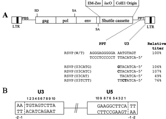
[image:3.585.119.460.71.316.2]Lac repressor protein expression.To express the recombinant Lac repressor protein, the pTrc-His-Lac-biotin plasmid was heat shock transformed into the competentE. coliStbl2 strain (Invitrogen). A single colony was used to inoculate FIG. 1. Structures of the RSVP vectors. (A) Schematic representation (not to scale) of the recovery cassette and the mutants. Mutations are indicated in bold. The virus titers, normalized to the amount of p27 (capsid), are shown relative to that of the wild-type virus. These results are the averages from three independent experiments. EM-Zeo is the bacterial promoter linked to the zeocin resistance gene;lacO is thelac
operator. (B) Schematic numbering of the nucleotide positions in the ends of the provirus. The last nucleotide on each end of a normal provirus is number 1.
[image:3.585.301.540.534.707.2]FIG. 2. Two-LTR circle junctions isolated from infected cells. A consensus circle junction is shown at the top. Different types of aber-rant two-LTR circle junctions are shown. The tRNA insertion is indicated by a white box with black horizontal bars; the PPT is a white box with black vertical bars; a deletion is indicated by a black jagged end.
TABLE 1. Recovery of full-length integrated viral DNA
Provirus U3a
U5a Size of
duplication (bp)
No. of cases
RSVP(U3CATG) 1 1 15 3
1 1 6 17
⫺62 1 6 1
RSVP(U3CATC) 1 1 5 3
1 1 6 9
1 26 191 1
RSVP(U3CAT) 1 1 5 2
1 1 6 9
Variable 1 Variable 5
RSVP(U3TCTT) 1 1 5 2
1 1 6 8
Variable 1 Variable 6
aThe last viral nucleotide at each end of the proviruses is indicated by a
number, using the numbering system in Fig. 1B.
on November 8, 2019 by guest
http://jvi.asm.org/
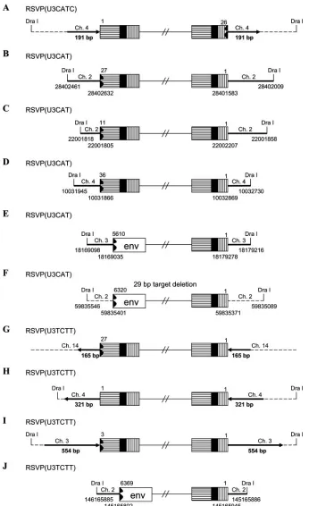
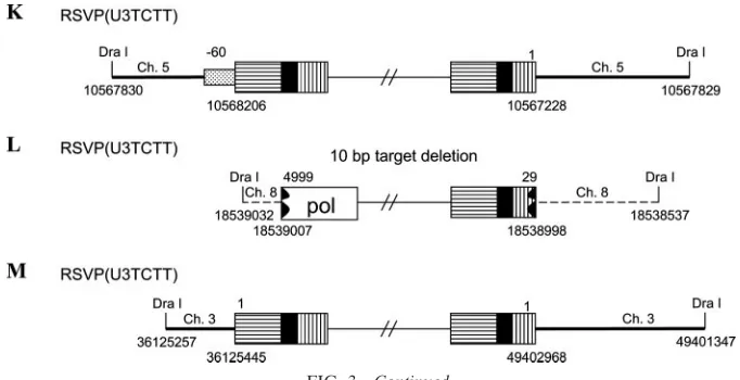
[image:3.585.43.290.556.671.2]FIG. 3. Structures of the aberrant proviruses. U3 is shown as a box with horizontal bars, R is shown as a black box, and U5 is shown as a box with vertical bars. A deletion in U5 and/or U3 is indicated by a black jagged end. Insertion of PPT and flanking viral sequence is shown by a box with black dots. Host DNA is shown as a line. Large duplications of host sequences are indicated by thick black lines/arrows (not to scale). The direction of the arrow corresponds to the numbering of the host genomic DNA. In some cases in which the nature of the origin of the viral DNA is ambiguous (see the text), the positions of DraI sites and the viral/host DNA junctions are indicated by nucleotide position numbers from a BLAT search.
on November 8, 2019 by guest
2 ml of LB medium (supplemented with 50g/ml ampicillin for selection of transformed colonies; 2 mg/ml biotin was provided as a substrate for BP bio-tinylation), and the cells were grown overnight at 37°C with shaking. The next day, 1 liter of LB containing ampicillin and biotin was inoculated with 1 ml of the overnight culture and grown at 37°C with shaking to an optical density at 600 nm of 0.6 (mid-log phase of the cells). Lac expression was induced with 1 mM isopropyl--D-thiogalactopyranoside (IPTG) and, after growth of the culture for another 5 h and removal of a 1-ml aliquot for protein analysis, the suspension was centrifuged and the resulting pellet was stored at⫺20°C for further use.
To confirm the expression of the recombinant Lac repressor protein, the pellet from the 1-ml aliquot was resuspended in 100l of 20 mM phosphate buffer at neutral pH, frozen in liquid nitrogen, and thawed at 42°C (the freeze-thaw was repeated three additional times) followed by pelleting of the insoluble proteins for 10 min in a conventional tabletop microcentrifuge at maximum speed. The supernatant was fractionated on a 4 to 12% sodium dodecyl sulfate-polyacryl-amide gel which was stained with Coomassie blue. This procedure confirmed the presence of a 43-kDa band corresponding to the recombinant Lac repressor monomer.
To confirm the presence of introduced C- and N-terminal modifications, the sample was subjected to standard Western blot analysis using two primary anti-bodies which detected the His tag (anti-His antibody from Sigma) and the biotinylated BP peptide (streptavidin-alkaline phosphatase conjugate), respec-tively (both antibodies were from Sigma). Both antibodies detected the 43-kDa protein, identifying it as the modified recombinant Lac repressor.
Lac repressor protein purification.To extract the Lac repressor protein, theE. colicell pellet was resuspended in 10 ml of lysis buffer (50 mM NaPO4, pH 8.0, 50 mM NaCl, 0.75 mg/ml lysozyme, 1 mM phenylmethylsulfonyl fluoride) and incubated on ice for 30 min, after which the concentration of NaCl in the lysate was adjusted to 300 mM. The lysate was sonicated three times for 30 s (60% power plus 70% pulse) and centrifuged at 85,000⫻gfor 1 h, and the supernatant was diluted 1:1 with 50 mM NaPO4, pH 6.8, 300 mM NaCl and passed over a Poly-Prep chromatography column (Bio-Rad) prepacked with 1 ml of BD Talon resin (BD Biosciences) that had been preequilibrated with 50 mM NaPO4, pH 7.0, 300 mM NaCl, 6 mM imidazole. The column was washed four times with 20 ml of the equilibrating buffer, and the bound protein was eluted with 20 ml of the elution buffer (50 mM NaPO4, pH 6.0, 300 mM NaCl, 600 mM imidazole, 10% glycerol).
Lac repressor-mediated recovery of integrated retroviral DNA. Genomic DNA was isolated from infected DF-1 (24, 25) cells that survived zeocin selection using a QIAamp DNA blood maxikit (Qiagen). To recover full-length proviruses, 100 to 200g of genomic DNA was digested with DraI overnight at 37°C. One milligram of Dynabeads M-280 streptavidin (Dynal Biotech) was washed three times with a wash buffer (150 mM NaCl, 10 mM EDTA) and incubated with 3 to 6g of purified biotinylated Lac repressor protein for 1 to 2 h at room temper-ature with gentle rotation of the tube. The Dynabeads-biotinylated Lac repressor mixture was washed three times with a wash buffer. The digested DNA was adjusted to 150 mM NaCl, 10 mM EDTA, 50g of bovine serum albumin per ml, and 10% (vol/vol) glycerol in a final volume of 600l and then incubated with the Dynabeads-biotinylated Lac repressor mixture for 2 h at room temperature with gentle rotation of the tube. The DNA-Dynabeads-biotinylated Lac repressor
mixture was washed three times with 1 ml of wash buffer and eluted twice with 1 ml of elution buffer (10 mM Tris-HCl [pH 7.5], 10 mM EDTA, 10 mM IPTG) for 30 min at 37°C. The enriched DNA was extracted with phenol-chloroform and precipitated with ethanol. The precipitated DNA was ligated with T4 DNA ligase (30 U/200l; Roche, Indianapolis, IN) for 18 h at 16°C. The ligated DNA was then used to transformE. colias described above. After a 3-h recovery period in SOC medium at 37°C, the transformed bacteria were plated onto low-salt Luria-Bertani plates containing 50g of zeocin per ml.
Sequencing of two-LTR circle junctions and integration sites.Recovered plasmids were directly sequenced using the following primers: the PBS primer (5⬘-ACTATCACGTCGGGGTCACCA) for the two-LTR circle junction and the U3 integration sites and the Ori-2 primer (5⬘-GCAAGCAGCAGATTACGC GCA) for the U5 integration sites. The Ori-2 primer was also used to confirm that the mutations were present in the recovered plasmids. In those cases in which the provirus was flanked by substantial duplications, the ends of the duplications were sequenced to demonstrate that the duplicated regions were present on both ends of the provirus. Chicken genomic sequences were analyzed by BLAT searches (http://genome.ucsc.edu/cgi-bin/hgBlat).
RESULTS AND DISCUSSION
The U3-end mutations affected virus titer. Starting with RSVP(A)Z, we changed the sequence that is normally found at the 5⬘end of U3 adjacent to the PPT (TT) and the CA dinu-cleotide that is normally found at the U3 end of the provirus. The behavior of the mutant viruses should help us understand how the sequence adjacent to the PPT affects the generation of the linear viral DNA and how IN inserts the linear DNAs produced by the mutants into the host genome. We also changed the CA dinucleotide in the 5⬘end of U3 to TC to ask how this substitution affects integration. The changes made in the viral mutants are shown in Fig. 1.
To measure virus titer, viruses were generated by transfect-ing DF-1 cells with plasmids containtransfect-ing the U3-end mutations and the resulting viral stock was used to infect 293-tva cells. GFP expression was measured using fluorescence-activated cell sorting; the titers were normalized relative to the amount of p27 (capsid) in the infecting viral stock. RSVP(U3CATG) and RSVP(U3CATC) had relative virus titers that were indis-tinguishable from those of the wild type. However, RSVP (U3CAT) decreased virus titer to about 49% of that of the wild type. A similar result was obtained with the corresponding U5 mutant, RSVP(HIV1), in which the two nucleotides (TT) be-tween the PBS and the CA dinucleotide of RSVAP(A)Z were
FIG. 3—Continued.
on November 8, 2019 by guest
http://jvi.asm.org/
[image:5.585.121.462.68.243.2]changed to the one nucleotide that matches the HIV-1 se-quence (G). The RSVP(U3CAT) mutation should put the canonical CA in the U3 end one nucleotide from the end of the linear DNA, just as the RSVP(HIV1) mutation puts the ca-nonical CA on the U5 end one nucleotide from the other end of the linear DNA. The RSVP(HIV1) mutation decreased virus titer to about 46% of the wild-type level (24). Surpris-ingly, the RSVP(U3TCTT) mutation had a more modest effect on viral titer, reducing the titer to 76% of the wild-type level, even though the canonical CA dinucleotide in the U3 end was changed to TC. This result differed from our previous result with the corresponding RSVP(CATC) mutant, in which the canonical CA dinucleotide in the U5 end was changed to TC; RSVP(CATC) decreased virus titer to about 36% of the wild-type level (24). Because the titer of the mutants was within twofold of that of the wild type, we did not attempt to measure the synthesis of the viral DNA using real-time PCR. In our hands, the accuracy of real-time PCR is approximately twofold, so we were unable to determine whether any of the mutations had an effect on the efficiency of viral DNA synthesis; if there is an effect, it would be small (twofold or less).
Analysis of two-LTR circle junctions.Analysis of the two-LTR circle junctions from the mutants is shown in Fig. 2. Although the two-LTR circle junctions obtained from the mu-tants included both insertions and deletions, the proportion of consensus sequence junctions that were derived from the join-ing of complete, normal copies of both ends of the linear viral DNA was not significantly different between the mutants and the wild type. Although in Table 2 the percentage of two-LTR circles in the wild type is slightly lower than the percentage of two-LTR circles seen with the mutants (62% versus approxi-mately 80%), in previous experiments we found that the per-centage of two-LTR circles produced in a wild-type infection was approximately 80% (24). We previously reported that re-placing the terminal 3⬘A of the RSV PPT with a G caused a preferential miscleavage one nucleotide into U3, following the newly created GA dinucleotide sequence. This miscleavage resulted in the deletion of the terminal adenine normally present at the 5⬘ end of the U3, making the resulting linear viral DNA 1 bp shorter on the U3 end (3). Here, we replaced the T at the 3⬘ end of U3 with a C (U3CATC) or with a G
[image:6.585.300.541.539.692.2]⬘ the host sequence ABC is captured between the two ends of a circular viral DNA.
TABLE 2. Microhomology at the host and viral DNA junctions in aberrant provirusesa
Provirus U3 U5
A Acat-5⬘ 5⬘-tacGAG
B AGAaca-5⬘ 5⬘-ttca
C ACgtta-5⬘ 5⬘-ttca
D taccat-5⬘ 5⬘-ttca
E aggctc-5⬘ 5⬘-ttca
F Cgatgg-5⬘ 5⬘-ttca
G AGAcag-5⬘ 5⬘-ttca
H CTatca-5⬘ 5⬘-ttca
I ATcaga-5⬘ 5⬘-ttca
J Taaggg-5⬘ 5⬘-ttca
K TGgtag-5⬘ 5⬘-ttca
L Ccccct-5⬘ 5⬘-gacTAC
M ctatca-5⬘ 5⬘-gcttca
aFour nucleotides from normal host/virus DNA junctions and six nucleotides
from aberrant junctions are shown. Capital letters indicate sequences showing microhomology with the host DNA. The letters A to M correspond to the proviruses described in Fig. 3.
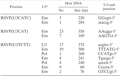
TABLE 3. Recovery of circles derived from abortive integrations
Provirus U3a Host DNA U3-end
junction No. Size (bp)
RSVP(U3CATC) Env 3 220 GGcgtt-5⬘
Env 1 284 ataccg-5⬘
RSVP(U3CAT) Env 23 358 AAcgga-5⬘
Env 7 189 AAGTct-5⬘
RSVP(U3TCTT) U3 17 374 acgtta-5⬘
Env 19 300 TTTATG-5⬘
Pol 1 260 CCATgt-5⬘
Env 4 241 Tgacga-5⬘
Pol 4 240 aacctt-5⬘
Env 9 60 Ccccta-5⬘
Env 2 76 GTCCgt-5⬘
aAt the U5 junctions, the last nucleotide was a 1 in all cases, using the
numbering system in Fig. 1B. Six nucleotides from U3-end junctions are shown, and capital letters indicate sequences showing microhomology with the host DNA.
on November 8, 2019 by guest
[image:6.585.44.284.552.692.2](U3CATG), which resulted in a linear DNA in which there was either a 5⬘-GA or a 5⬘-CA at the U3 end of the linear viral DNA. Neither of these mutations affected the specificity of the cleavage of the PPT by Rous sarcoma virus (RSV) RNase H. These results suggest that, as expected, the specific cleavage that removes the PPT primer in vivo is much more dependent on the sequence of the PPT than it is on the adjacent sequence in U3.
Recovery of full-length integrated viral DNA.Although RSV RNase H properly cleaves the PPT primer in RSVP(U3CAT) and RSVP(U3TCTT), the mutations lead to the generation of linear viral DNAs with aberrant U3 ends, which raises the question of whether the aberrant ends are substrates that can be properly integrated by RSV IN. Full-length integrated viral DNA was recovered as described in Materials and Methods (see also references 24 and 25).
Based on sequence analysis, most of the proviruses derived from infections with RSVP(U3CATG) and RSVP(U3CATC) arose from normal integrations in which two nucleotides were removed from each end of the linear viral DNA and the viral DNA was inserted into the host genome by IN, creating a 5- or 6-bp duplication of the target site sequence flanking the pro-virus (Table 1). However, in one case, an RSVP(U3CATG) provirus was isolated in which the viral DNA was integrated using an internal CA sequence in U3, giving rise to a provirus flanked by a normal host DNA repeat of 6 bp (Table 1). In another case, a provirus derived from infection with the RSVP (U3CATC) mutant virus had a deletion in U5 but had a nor-mal U3 junction (Table 1; Fig. 3A). Infections with this mutant gave rise to two-LTR circle junctions with U5 deletions; sug-gesting that some of the linear viral DNAs synthesized by this mutant had aberrant U5 ends. If we compare the aberrant U3 junctions obtained in infections with RSVP(U3CATC) (in which the mutations are in U3) with the aberrant U5 junctions of proviruses that arise from infections with a virus with mu-tations in the U5 end, the aberrant U3 and U5 junctions are similar (24).
In the case of RSVP(U3CAT), about 70% of the proviruses were integrated normally, indicating that RSV IN can remove a single nucleotide from the U3 LTR terminus of the linear viral DNA during integration (Table 1). However, 30% of the proviruses were aberrant (Fig. 3B to F). In each of the aberrant proviruses, the terminus of the U3 LTR was deleted and none of the aberrant proviruses were flanked by a 5- or 6-bp dupli-cation at the target site. In one case, there was a 29-bp deletion of host sequence at the target site. The proviruses were ob-tained by digesting host genomic DNA with DraI, which does not cleave in viral DNA. If an aberrant provirus is flanked by a large direct repeat of host sequence and the DraI sites lie outside the repeat, DraI cleavage allows us to recover host DNA that contains the complete direct repeats. However, if there is a DraI site in the repeated portion of the flanking host DNA, portions of both repeats are lost, and the recovered viral DNA is indistinguishable from an unintegrated circular DNA with host sequences between the two LTRs that could arise from an abortive integration event like the one shown in Fig. 4 (described below; see also reference 24). We suggest that most of the ambiguous DNAs that we have recovered are derived from proviruses for two reasons. (i) We selected cells that were zeocin resistant. The fact that we were able to select the cells
suggests that a significant portion of the viral DNA is inte-grated because zeocin is expressed at a high-enough level to allow the cells to survive the selection. (ii) Many of the ambig-uous DNAs have a DraI site in the host sequences. We did recover some circular DNAs in which the host DNA lacks a DraI site. These DNAs almost certainly derive from abortive integrations; these abortive integrations are discussed in a later section.
In the case of an infection with RSVP(U3TCTT), about 60% of the proviruses that we recovered were integrated nor-mally, indicating that RSV IN can successfully remove two nucleotides beyond the mutated TC sequence, which, in this mutant, replaces the canonical CA that is normally found two nucleotides from the U3 LTR terminus of the linear viral DNA. In about 40% of the RSVP(U3TCTT) proviruses, the U3 LTR terminus was deleted and, more rarely, there was an insertion of the PPT and flanking sequences. In the aberrant integrations there were either large duplications or a deletion of the flanking host sequence, instead of the normal 5- or 6-bp duplication. For some of the aberrant proviruses (Fig. 3G to I), we sequenced through the duplicated regions of host DNA at both ends of the proviruses into the nonduplicated sequences. One apparently normal provirus had what appeared to be the mutated TC sequence in the U3 LTR junction linked directly to host DNA. This would suggest that this was an IN-mediated insertion; however, there was a 321-bp duplication of the host sequence at the target site, which suggests that this provirus was not derived from a normal integration event (Fig. 3H). This interpretation was supported by the fact that the last two nucleotides of the U3 LTR terminus were identical with the first two nucleotides of the host DNA. Microhomology at the host/virus DNA junction is not a characteristic of normal IN-mediated integration events; however, microhomologies are often found at aberrant host/virus DNA junctions that are generated by host enzymes (24, 25) (Table 2). The fact that there is microhomology at the host/virus DNA junction also means we cannot define the exact U3 junction for this provirus (Table 2, H). We isolated another apparently normal provirus; however, it was flanked by an inversion of host sequences (Fig. 3M). There was no microhomology between the viral and host sequences at either of the host/virus junctions. We previously proposed a model to explain how an aberrant proviral insertion could generate an inversion of the flanking host sequence (25). Others previously showed that mutating the CA in either U3 or U5 caused a moderate reduction in the titer of HIV-1 (1, 22). However, at least for RSV, a linear viral DNA with one defective end can be integrated into the host genome with good efficiency, even though the aberrant end is not inserted by IN. For this reason, titer is not sufficient to determine whether IN can properly insert an aberrant end. In RSV, mutating the CA in the viral DNA that is joined to host sequences has different consequences in U3 and U5. We previously reported that changing the CA dinucleotide in the U5 end to TC, the mutation in RSVP(CATC), prevented the normal IN-medi-ated insertion of the mutIN-medi-ated U5 end in all the proviruses that we isolated (24). However, in the experiments that we present here, making the corresponding mutation in U3, changing the CA sequence in the U3 end to TC, allowed IN-mediated in-sertion of the mutant end in 60% of the RSVP(U3TCTT) proviruses; these proviruses were flanked by the normal 5- or
on November 8, 2019 by guest
http://jvi.asm.org/
RSVP(U3CATC) Env ⫺20 5⬘-ccTGAT U3 37 5⬘-tcccTG
Env ⫺8 5⬘-tggTGA Gag 58 5⬘-gttgAT
Env ⫺5 5⬘-atttgG Gag 69 5⬘-tgtgCA
Gag 1 5⬘-gcttca Env 130 5⬘-atattG
U3 3 5⬘-aggctt
Pol 5 5⬘-gaaGGC D64K Pol ⫺18 5⬘-gacgtG
Env 18 5⬘-cctGCA Gag ⫺2 5⬘-ttcatt
Gag 24 5⬘-cgagCA Gag 1 5⬘-gcttCA
Env 27 5⬘-ctacgA Env 2 5⬘-ggcttC
Env 29 5⬘-gacTAC Gag 4 5⬘-aaggct
Pol 38 5⬘-ttccCT Gag 5 5⬘-gaaggc
Env 134 5⬘-agaGAT Gag 5 5⬘-gaaGGC
Env 176 5⬘-gggtcT Pol 6 5⬘-aGAAGG
Gag 205 5⬘-gatcGT Pol 7 5⬘-cagaAG
Pol 10 5⬘-aagcAG
RSVP(U3TCTT) Pol ⫺15 5⬘-cccgaC Gag 15 5⬘-gcatGA
Gag ⫺13 5⬘-accCCG Env 16 5⬘-tGCATG
Pol-Env ⫺12 5⬘-gaccCC Gag 18 5⬘-cctgCA
Env ⫺8 5⬘-tggTGA Env 20 5⬘-cacCTG
Env ⫺2 5⬘-ttcatt Env 22 5⬘-agcacc
Env ⫺2 5⬘-ttcatT Pol 23 5⬘-gagcaC
Env 1 5⬘-gcttcA Gag 28 5⬘-actacG
Pol 1 5⬘-gcttCA Env 37 5⬘-tccctG
Gag 1 5⬘-gcttCA Gag 58 5⬘-gttgaT
Gag 5 5⬘-gaaggc
U3 5 5⬘-gaaggc RSVP(U3CAT) Env ⫺16 5⬘-ccgacG
Env 5 5⬘-gaagGC Env ⫺9 5⬘-ggtgAC
Pol 6 5⬘-agaaGG Gag ⫺2 5⬘-ttcatT
Gag 6 5⬘-agaAGG Env ⫺2 5⬘-ttcatt
Env 8 5⬘-gcaGAA Env 3 5⬘-aggCTT
Gag 9 5⬘-agcaGA Gag 6 5⬘-agaagG
Gag 9 5⬘-agcagA Gag 7 5⬘-cagaAG
Pol 9 5⬘-agcaGA Env 9 5⬘-agcAGA
Env 13 5⬘-atgaAG Gag 13 5⬘-atgaAG
U3 17 5⬘-ctgcat Env 19 5⬘-acctGC
Env 20 5⬘-cacctg U3 20 5⬘-cacctg
Gag 20 5⬘-cacctg U3 20 5⬘-caccTG
Gag 21 5⬘-gcacCT U3 21 5⬘-gcaccT
Pol-Env 21 5⬘-gcaccT Env 23 5⬘-gagcaC
Gag 26 5⬘-tacgAG Pol 25 5⬘-acgaGC
Pol 30 5⬘-cgaCTA Gag 25 5⬘-acGAGC
Gag 33 5⬘-tgacgA U3 27 5⬘-ctacga
Env 47 5⬘-gaccgT Gag 27 5⬘-ctaCGA
Gag 54 5⬘-aTGGCC Gag 28 5⬘-actacG
Gag 73 5⬘-ttggtG Gag 28 5⬘-actaCG
Pol 86 5⬘-accatt Gag 30 5⬘-cgacta
Pol 69 5⬘-tgtgcA
RSVP(U3CATG) Env ⫺30 5⬘-agggAA Env 299 5⬘-tagtCT
Gag ⫺13 5⬘-accccG Gag 423 5⬘-gggacT
Env ⫺9 5⬘-ggtgAC
aSix nucleotides from U5-end junctions are shown, and capital letters indicate sequences showing microhomology between the viral DNAs. The name of the viral
mutant from which the circular DNAs were derived is given in the left column for each group. D64K has a mutation in the active site of IN. W/T, wild type. For Pol-Env, the position of the junction is between thepolandenvgenes.
510
on November 8, 2019 by guest
[image:8.585.48.541.58.688.2]6-bp duplication of host sequence. Not surprisingly the titer of the U3 mutant RSVP(U3TCTT) was higher than the titer of the U5 mutant RSVP(CATC), 76% and 36% of the wild-type level, respectively. These results suggest that, in the context of the U3 end, TC, instead of the canonical CA, can be used reasonably efficiently by RSV IN, which means that the CA dinucleotide is not required at the U3 end for appropriate integration in vivo.
Recovery of circular viral DNAs derived from abortive inte-grations. When we recovered full-length proviruses, we also recovered 11 circular viral DNAs that contained host DNA sequences ranging in size from tens to hundreds of nucleotides, in which there was no DraI site (Table 3). All of these circles contained a normal U5 junction; however, portions of the U3 end of the viral DNA were deleted and the new U3 end was joined to the host DNA in a way that caused the host sequence to be captured between the ends of a circular viral DNA (Fig. 3). There were, in 8 out of 11 cases, microhomologies involving one to five nucleotides between the ends of viral sequences and host sequences at the aberrant junction (Table 3); as has al-ready been discussed, this is a characteristic of aberrant host/ virus junctions that are generated by host enzymes (24).
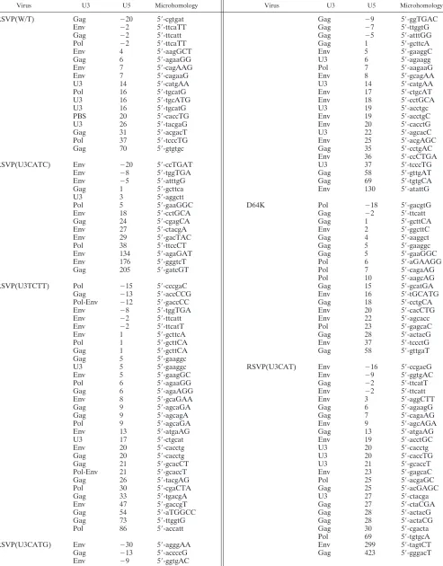
Recovery of smaller circular viral DNAs. When we recov-ered the two-LTR circle junctions, we also recovrecov-ered 130 cir-cular viral DNAs in which a portion of the viral DNA was deleted from the U3 end (Table 4). In most of these smaller circles, the U5 end of the viral DNAs was abnormal: either the U5 end was deleted or a portion of the PBS/flanking sequence was retained. The U5 end of the viral DNA was then joined to various positions of the viral DNA, creating smaller circular viral DNAs. In six cases, a canonical CA sequence was present at the U5 end of the viral DNA, which suggests that these circular forms could have come from IN-mediated autointe-gration events. However, in five out of these six cases, we cannot define the exact U5 junction, because there are micro-homologies involving one or two nucleotides between the two ends of the viral DNAs.
Olsen et al. (26) previously reported the structure of circular viral DNAs produced in RSV infections. Approximately 30% of the circular viral DNAs had aberrant U5 ends, and there were deletions in the 5⬘portion of the viral genome, similar to the circular viral DNAs reported here. Although they pointed out the common nucleotides present at each boundary of the deletion, the statistical significance of the microhomologies in their circular DNA isolates was not determined, due to limited numbers of circular viral DNAs recovered. Olsen et al. discuss the possibility that IN has a role in generating the small circles that arise during RSV infections without reaching a firm con-clusion; however, their data are consistent with our interpre-tation that IN-mediated autointegration occurs rarely, if at all, in RSV infections. We have recently done experiments using an HIV-1-based shuttle vector that is similar to the RSV-based vectors used in the experiments reported here and recovered numerous circular viral DNAs that were obviously derived from one-ended and two-ended IN-mediated autointegrations, which shows that the technology would have allowed us to recover IN-mediated autointegrated viral DNAs if they had been present (unpublished observations).
In the present study, when the sequences used to form the viral/viral DNA junctions were compared, there were
micro-homologies involving from one to five nucleotides between the ends (Table 4). Although the junctions with one homologous nucleotide were found at a frequency that was not higher than what could have occurred by chance, the frequencies of junc-tions in which there were two or more homologous nucleotides were high enough to be statistically significant. The probabil-ities (P) of obtaining the number of two, three, four, and five homologous nucleotides by chance were ⬍0.0003, ⬍0.0001, 0.0018, and 0.0022, respectively. In addition, we did not re-cover any circles arising from a concerted autointegration event in which both ends are processed correctly and inserted into the viral DNA in an orientation opposite from their own polarity, which creates a circular viral DNA that contains two unlinked LTRs with a 5- or 6-bp duplication at the target site. This type of autointegrated circular DNA is commonly found in cells infected by MLV and HIV-1. To exclude the involve-ment of IN in generating smaller circles, we recovered a set of small circular DNAs from cells infected with a virus mutated in the IN active site (D64K). When the viral/viral DNA junctions were examined, the smaller viral DNAs recovered in the ab-sence of IN enzymatic activity were similar in their structure to those that we recovered from an infection with a virus that has a normal wild-type IN (Table 4), suggesting that the generation of smaller circles is independent of integrase activity. Taken together, these results suggest that RSV linear DNA, in con-trast to MLV and HIV-1 linear DNA, does little, if any, auto-integration. This should provide RSV with an important ad-vantage. From the point of view of the virus, autointegration is a dead end that wastes a linear viral DNA that might otherwise be used to establish a provirus. Despite the fact that linear RSV DNA does not appear to autointegrate in vivo, it auto-integrates efficiently when the preintegration complexes (PICs) are isolated from infected cells (19, 20). The PICs of HIV and MLV appear to interact with a host factor, BAF (barrier to autointegration factor), whose presence reduces, but does not eliminate, autointegration. The striking contrast between the in vivo and in vitro results with RSV autointegration suggests the possibility that the RSV PIC interacts with some as-yet-unidentified host factor that efficiently blocks autointegration of linear RSV DNA. Presumably, the procedures used by Lee and Coffin to isolate RSV PICs from infected cells removed this protective factor (19, 20).
ACKNOWLEDGMENTS
We are grateful to Terri Burdette for help in preparing the manu-script.
This research was supported by the Intramural Research Program of the NIH, National Cancer Institute, Center for Cancer Research.
REFERENCES
1.Brown, H. E. V., H. Chen, and A. Engelman.1999. Structure-based mutagen-esis of the human immunodeficiency virus type 1 DNA attachment site: effects on integration and cDNA synthesis. J. Virol.73:9011–9020. 2.Cannon, P. M., W. Wilson, E. Byles, S. M. Kingsman, and A. J. Kingsman.
1994. Human immunodeficiency virus type 1 integrase: effect on viral repli-cation of mutations at highly conserved residues. J. Virol.68:4768–4775. 3.Chang, K. W., J. G. Julias, W. G. Alvord, J. Oh, and S. H. Hughes.2005.
Alternate polypurine tracts (PPTs) affect the Rous sarcoma virus RNase H cleavage specificity and reveal a preferential cleavage following a GA dinu-cleotide sequence at the PPT-U3 junction. J. Virol.79:13694–13704. 4.Colicelli, J., and S. P. Goff.1985. Mutants and pseudorevertants of Moloney
murine leukemia virus with alterations at the integration site. Cell42:573– 580.
5.Colicelli, J., and S. P. Goff.1986. Isolation of a recombinant murine leuke-mia virus utilizing a new primer tRNA. J. Virol.57:37–45.
on November 8, 2019 by guest
http://jvi.asm.org/
DF-1 chicken fibroblast cell line: transformation induced by diverse onco-genes and cell death resulting from infection by avian leucosis viruses. Vi-rology248:295–304.
13.Hughes, S. H., A. Mutschler, J. M. Bishop, and H. E. Varmus.1981. A Rous sarcoma virus provirus is flanked by short direct repeats of a cellular DNA sequence present in only one copy prior to integration. Proc. Natl. Acad. Sci. USA78:4299–4303.
14.Ju, G., and A. M. Skalka.1980. Nucleotide sequence analysis of the long terminal repeat (LTR) of avian retroviruses: structural similarities with transposable elements. Cell22:379–386.
15.Julias, J. G., M. J. McWilliams, S. G. Sarafianos, W. G. Alvord, E. Arnold, and S. H. Hughes.2004. Effects of mutations in the G tract of the human immunodeficiency virus type 1 polypurine tract on virus replication and RNase H cleavage. J. Virol.78:13315–13324.
16.LaFemina, R. L., P. L. Callahan, and M. G. Cordingley.1991. Substrate specificity of recombinant human immunodeficiency virus integrase protein. J. Virol.65:5624–5630.
17.LaFemina, R. L., C. L. Schneider, H. L. Robbins, P. L. Callahan, K. LeGrow, E. Roth, W. A. Schleif, and E. A. Emini.1992. Requirement of active human immunodeficiency virus type 1 integrase enzyme for productive infection of human T-lymphoid cells. J. Virol.66:7414–7419.
18.Leavitt, A. D., R. B. Ross, and H. E. Varmus.1992. Both substrate and target oligonucleotide sequences affect in vitro integration mediated by human immunodeficiency virus type 1 integrase protein produced inSaccharomyces cerevisiae. J. Virol.66:2359–2368.
19.Lee, Y. M., and J. M. Coffin.1990. Efficient autointegration of avian retro-virus DNA in vitro. J. Virol.64:5958–5965.
and R. Swanstrom.1990. Rearrangements in unintegrated retroviral DNA are complex and are the results of multiple genetic determinants. J. Virol. 64:5475–5484.
27.Panganiban, A. T., and H. M. Temin.1983. The terminal nucleotides of retrovirus DNA are required for integration but not virus production. Nature 306:155–160.
28.Roth, M. C., P. L. Schwartzberg, and S. P. Goff.1989. Structure of the termini of DNA intermediates in the integration of retroviral DNA: depen-dence on IN function and terminal DNA sequence. Cell58:47–54. 29.Schaefer-Klein, J., I. Givol, E. V. Barsov, J. M. Whitcomb, M. VanBrocklin,
D. N. Foster, M. J. Federspiel, and S. H. Hughes.1998. The EV-O derived cell line DF-1 supports the efficient replication of avian leucosis-sarcoma viruses and vectors. Virology248:305–311.
30.Schwartzberg, P., J. Colicelli, and S. P. Goff.1984. Construction and analysis of deletion mutations in the pol gene of Moloney murine leukemia virus: a new viral function required for productive infection. Cell37:1043–1052. 31.Telenitsky, A., and S. P. Goff.1997. Reverse transcriptase and the generation
of retroviral DNA, p. 121–160.InJ. M. Coffin, S. H. Hughes, and H. E. Varmus (ed.), Retroviruses. Cold Spring Harbor Laboratory Press, Cold Spring Harbor, NY.
32.Vink, C., D. C. van Gent, Y. Elgersma, and R. H. Plasterk.1991. Human immunodeficiency virus integrase protein requires a subterminal position of its viral DNA recognition sequence for efficient cleavage. J. Virol.65:4636– 4644.
33.Zhou, H., J. Rainey, S. K. Wong, and J. M. Coffin.2001. Substrate sequence selection by retroviral integrase. J. Virol.75:1359–1370.