REVIEW
Immunomodulatory plasticity
of mesenchymal stem cells: a potential key
to successful solid organ transplantation
Urvashi Kaundal
1,2, Upma Bagai
2and Aruna Rakha
1*Abstract
Organ transplantation remains to be a treatment of choice for patients suffering from irreversible organ failure.
Immunosuppressive (IS) drugs employed to maintain the allograft have shown excellent short-term graft survival, but,
their long-term use could contribute to immunological and non-immunological risk factors, resulting in graft
dys-functionalities. Upcoming IS regimes have highlighted the use of cell-based therapies, which can eliminate the risk of
drug-borne toxicities while maintaining efficacy of the treatment. Mesenchymal stem cells (MSCs) have been
consid-ered as an invaluable cell type, owing to their unique immunomodulatory properties, which makes them desirable
for application in transplant settings, where hyper-activation of the immune system is evident. The immunoregulatory
potential of MSCs holds true for preclinical studies while achieving it in clinical studies continues to be a challenge.
Understanding the biological factors responsible for subdued responses of MSCs in vivo would allow uninhibited use
of this therapy for countless conditions. In this review, we summarize the variations in the preclinical and clinical
stud-ies utilizing MSCs, discuss the factors which might be responsible for variability in outcome and propose the
advance-ments likely to occur in future for using this as a “boutique/personalised therapy” for patient care.
Keywords: Organ transplantation, Graft survival, Mesenchymal stem cells, Microenvironment
© The Author(s) 2018. This article is distributed under the terms of the Creative Commons Attribution 4.0 International License (http://creat iveco mmons .org/licen ses/by/4.0/), which permits unrestricted use, distribution, and reproduction in any medium, provided you give appropriate credit to the original author(s) and the source, provide a link to the Creative Commons license, and indicate if changes were made. The Creative Commons Public Domain Dedication waiver (http://creat iveco mmons .org/ publi cdoma in/zero/1.0/) applies to the data made available in this article, unless otherwise stated.
Background
Over recent years tremendous progress has been made
to understand the basic mechanisms underlying the state
of allograft rejection. Regardless of substantial
improve-ments in short-term allograft survival, long-term
out-come remains subpar [
1
–
4
]. The current maintenance
regimen to support organ transplantation and to reduce
transplant-related morbidity includes a combination of
immunosuppressive (IS) drugs including calcineurin
inhibitors, mTOR inhibitors and anti-proliferative agents
[
5
]. Application of IS drugs has a therapeutic and
sup-pressive effect on host’s immune system. Nevertheless,
non-specific immunosuppression produced by IS drugs,
also result in instances of undesired immunodeficiency,
toxicity to other non-immune cells, cardiovascular
disorders and malignancies [
6
–
11
]. In the last decade,
extensive research in the field of translational medicine
has indicated the use of cell-based therapies
comple-mentary to IS drugs for achieving the goal of ultimate IS
therapy i.e. a therapy that can induce a balance between
maximum efficacy and minimal adverse effects.
Mesenchymal stem cells (MSCs), have recently gained
the interest of clinicians and researchers. The
likeli-hood of these MSC based therapies depends upon, their
regenerative facets and modulation of the immunological
responses engendered through their secreted paracrine
mediators [
12
]. MSCs are recognized for the activation of
regulatory immune cells in conjunction with interference
in maturation and activation of antigen presenting cells
(APCs). As already known, exogenously cultured MSCs
upon administration into the patient’s body, interact with
the microenvironment in vivo which leads to their
acti-vation or licensing. Clinical studies have suggested that
this licensing process in vivo is mediated by the presence
Open Access
*Correspondence: aruna_pgi@yahoo.com
1 Department of Translational and Regenerative Medicine, Postgraduate
of soluble factors and cytokines in the circulation. MSCs
upon exposure to different concentrations of
inflamma-tory mediators either produce Th1 or Th2 cytokines,
growth factors, cell migration factors which assist in
tis-sue maintenance and repair. Along with the inflammatory
cytokines, other factors like in vitro culture conditions,
Toll-like receptor (TLR) signalling and drug interactions
in vivo, may also determine the clinical efficacy of MSCs.
This review aims to describe the influence of
micro-environment both in vitro and in vivo on MSC and their
implications on various preclinical and clinical studies.
Mesenchymal stem cells—physical and functional
profile
Mesenchymal stem cells originally reported by
Frieden-stein et al. [
13
,
14
], are multipotent progenitor cells
accomplished to differentiate into several specialized cell
types. At high density, MSCs, align with each other in a
typical spatial pattern and have spindle-shaped
fibro-blastoid morphology [
15
]. MSCs righteously referred to
as mesenchymal stromal cells, possess trans-differential
potential, triggered by, placing MSCs under specific
stimuli which advance their development into various
lineages namely mesodermal i.e. myocyte, adipocytes,
osteocytes, cardiomyocytes, endothelium; ectodermal i.e.
neuronal; and endodermal i.e. hepatic, respiratory,
pan-creatic epithelium [
16
–
18
]. Bone marrow (BM) is
con-sidered as a primary source of MSCs while other sources
include adult connective tissues such as dental pulp,
peripheral blood, adipose tissue and foetal tissues such as
Wharton’s jelly, placenta, amniotic fluid, umbilical cord
(UC) and umbilical cord blood [
19
].
Phenotypically, MSCs are recognized by expression
of surface markers CD105, CD73, CD90 (mesenchymal
lineage markers) and lack of expression of CD34, CD19,
CD45, CD11a (hematopoietic lineage markers), CD31
(endothelial lineage marker), HLA-DR (human leukocyte
antigen) [
18
].
Mesenchymal stem cells express intermediate levels of
class I major histocompatibility complex (MHC) and do
not express class II MHC [
18
,
20
] or other co-stimulatory
molecules like B7-1, B7-2, CD80, CD40, CD40L or Fas
ligand on their surface [
21
], which play a crucial role in
immune activation. Even though the expression of
MHC-II molecules on MSCs is upregulated when stimulated
with a low-dose of pro-inflammatory
cytokine—inter-feron (IFN)-γ, no modification in the expression of
co-stimulatory molecules is observed [
21
,
22
]. This peculiar
profile of MSCs, makes them immune evasive and thus
an attractive candidate for cell-based therapies for
vari-ous clinical conditions.
Immunological tolerance and mesenchymal stem
cells
During transplant rejection, graft from a genetically
dif-ferent donor elicits an allogeneic immune response inside
recipient’s body, generated against antigens present on
donor graft. The allogeneic immune response is a
conse-quence of an intricate seconse-quence of interactions involving
both innate (dendritic cells, macrophages, neutrophils,
mast cells and natural killer cells) and adaptive (T and
B cells) immune system, ultimately leading to rejection
and transplant failure [
23
–
25
]. In order to manage early
graft rejections, IS drugs are administered but their effect
on controlling long-term graft rejections is
question-able [
26
]. Keeping this in mind, the clinical emphasis
recently has shifted towards induction of a state of
toler-ance towards an organ allograft [
27
,
28
]. Transplantation
tolerance is a state marked by the absence of
donor-specific inimical immune responsiveness which can be
maintained devoid of chronic immune suppression [
29
,
30
]. Although transplant tolerance has been successfully
achieved in various animal models, its accomplishment
in humans remains incomprehensible [
31
–
33
]. Insights
into mechanisms involved in immune activation have
led scientists to evaluate cell-based therapies specifically
using MSCs for various disease conditions owing to their
powerful immunomodulatory potential, without any
long-term deleterious effects [
34
,
35
].
already active M1 macrophages into anti-inflammatory
M2 macrophages to resolve the hyper-inflammatory state
[
44
]. MSCs have also been shown to suppress the IL-2
mediated proliferation and cytotoxic activity of Natural
Killer (NK) cells [
45
]. These aforementioned
immuno-suppressive properties inflicted by MSCs possibly lead to
induction of a state of peripheral tolerance [
46
,
47
].
For contact-dependent mechanisms, MSCs express a
large number of chemokines which lead to chemotaxis
of immune cells in the close vicinity of MSCs. As soon
as immune cells begin to migrate, MSCs secrete locally
active immunosuppressive factors which act on these
migrating immune cells in a reciprocal fashion [
48
].
While paracrine mechanisms are conceded by MSCs
through direct secretion of anti-inflammatory factors
such as transforming growth factor (TGF-β), hepatocyte
growth factor (HGF), nitric oxide (NO), heme oxygenase
(HO)-1, indoleamine 2,3-dioxygenase (IDO) and
expres-sion of inhibitory co-stimulatory molecules such as
TNF-related apoptosis-inducing ligand (TRAIL), programmed
death ligand (PD-L1) that work together and influence
the effector populations [
49
]. Through indirect
mecha-nisms, MSCs can either inhibit the maturation of the
antigen presenting cells (APCs) or generate regulatory or
suppressor cell populations through paracrine signalling
[
50
]. Paracrine signalling pathways are thought to be
cru-cial as MSCs themselves are short-lived due to their
sus-ceptibility to lysis by CD8
+T cells and NK cells [
51
,
52
].
Interestingly, a recent study has indicated the “apoptotic
demise” of MSCs as a key step for activation of effector
mechanisms of immunosuppression lead by MSCs [
53
].
Although a number of factors are known that help in
MSC-mediated immunomodulation but every factor
serves a different function depending upon the MSC
source and its microenvironment. Therefore, further
understanding of how MSCs can be directed to produce
the “beneficial factors” and how these factors can regulate
immune cells, might lead to the achievement of a state of
tolerance/partial tolerance through immunomodulation.
Inconsistency amongst the preclinical and clinical
findings using MSCs
Theoretically, the idea of utilizing MSCs as an adjunct
immunosuppressive therapy for solid organ
transplanta-tion is very alluring as MSCs aid in stimulating
tolero-genic immune responses. To test this hypothesis, plenty
of preclinical studies in experimental models of organ
transplantation have been conducted. The first
preclini-cal study was performed by Bartholomew et al. [
54
] in
a baboon model of skin transplant which illustrated a
diminished lymphocyte proliferation with an enhanced
graft survival as a result of intravenously infused
allo-geneic MSCs. Further, studies performed in different
Table
1
C
linic
al trials r
ela
ted t
o use of MSCs f
or or
gan tr
ansplan
ta
tion
O rgan tr ansplan ted MSC sour ce MSC dosage IS r eg ime Rout e of administr ation Pa tien ts enr olled/ estima ted enr olmen t Trial phase Sta tus Kidne y A ut ologous Bone mar ro w-der iv ed MSCs (BM -MSCs) D oses f or diff er ent gr oups 2 × 10⁶ MSCs/k
g, 48 h bef or e T x 2 × 10
6 MSCs/
kg
+
5
×
10
6 cell
, 48 h bef or e T x 2 × 10
6 MSCs/k
g at da
y 1, da y 7 Anti-th ymoc yt e globulin (A TG), Pr ednisolone , M ycophenolat e mof etil (MMF), C alcineur in inhibit or ( CNI),
IVIG ATG Plasma ex
change , IVIG, anti-CD20 monoclonal antibody Intra venous (I.V
.) or
intra-ar ter ial (I.A.) I.V . +
I.A. I.V.
260 1 Enr olling NC T number -NC T02490020 Liv er Umbilical cor d D er iv
ed MSCs (UC-MSCs)
M
ultiple doses: 1
×
10
6
MSCs/k
g body w
eight for 12 w eeks ( once per 4 w eeks) Not mentioned I.V . 50 1 Unk no wn NC T number -NC T01690247 Kidne y Thir d par ty BM -MSCs
Four doses: 1
* 10 6/k g ev er y w eek Not mentioned I.V . 120 1/2 Ongoing NC T number -NC T02563366 Kidne y A ut ologous , BM -MSCs
Single dose 2
×
10
6
MSCs/k
g body wt g
iv en 1 da y bef or e Tx MMF , Tacr olimus ( TA C )
CsA, Ster
oids I.V . 159 2 Complet ed NC T number -NC T00658073 [ 62 ] Kidne y A ut ologous , BM -MSCs Tw
o doses: 1–2
× 10 6 MSCs/k g dur ing Tx and da
y 7 post
Tx
MMF
,
TA
C or CsA,
Pr ednisolone I.V . 15 1 Complet ed NC T number -NC T00734396 [ 63 ] Kidne y A ut ologous , BM -MSCs
Single dose 2
×
10
6 MSCs/k
g body wt g iv en 1 da y bef or e transplantation ( Tx) AT G , MMF , Cy clospor
ine A (
CsA), St er oids I.V . 4 1/2 Ter minat ed NC T number -NC T00752479 [ 69 ] Kidne y Allogeneic , BM -MSCs Tw
o doses: 1st dose of 1–2
×
10
6 cells/k
g 1 da y bef or e Tx, 2nd dose - 1–2 × 10
6 cells
30 da ys af ter Tx TA C , MMF , Pr ednisolone I.V . 15 1 A ctiv
e, not r
ecruiting NC T number -NC T02409940 Kidne y/liv er Allogeneic , BM -MSCs (T hir d par ty)
Single dose of 1.5–3
×
10
6 MSCs/k
g at 3 ( ± 2) da ys af ter Tx TA C , MMF , St er oids Not mentioned 40 1/2 Unk no wn NC T number -NC T01429038 Kidne y Allogeneic , BM -MSCs (T hir d par ty)
4 doses: 1
×
10
6 cells/k
g
dur
ing
Tx and at da
y 7,
14, 21 post
-T
x
Induc
tion with basilixi
-mab
,
Lo
w dose of
[image:4.595.86.521.93.727.2]Table
1
(c
on
tinued)
O rgan tr ansplan ted MSC sour ce MSC dosage IS r eg ime Rout e of administr ation Pa tien ts enr olled/ estima ted enr olmen t Trial phase Sta tus Kidne y A ut ologous , str omal vas -cular frac tion der iv ed MSCs4 doses: 1
×
10
6 cells/k
g
dur
ing
Tx and at da
y 7,
14, 21 post
-T x Basiliximab induc tion Not mentioned 120 1/2 Not y et r ecruiting , NC T number -NC T02492308 Liv er Allogeneic , BM -MSCs Tw
o doses: 1
× 10 6 MSCs/k g dur ing Tx and da
y 2 (
± 1) post -T x TA C , Basiliximab , St er oids
1st dose: intrapor
tal
infusion,
2nd dose: I.V
.
7
1 (Pilot study) Recruiting NC T number -NC T02957552 Kidne y Allogeneic , BM -MSCs Tw
o doses: F
irst dose
of 5
×
10
6 cells/k
g dur ing Tx, S econd dose of 2 × 10
6 cells/k
g 30 da ys af ter Tx Cyt oxan, M eth ylpr ednisolone (MEP), Lo
w dose of
TA
C,
MMF
1st dose: r
enal allog raf t ar ter y,
2nd dose: I.V
. 12 Pilot study Complet ed [ 64 ] Kidne y Allogeneic , BM -MSCs Tw
o doses: F
irst dose
of 5
×
10
6 cells/k
g
dur
ing
Tx, second dose
of 2
×
10
6 cells/k
g 30 da ys af ter Tx Cyt oxan, MEP , Lo
w dose of
TA
C,
MMF
1st dose: r
enal allog raf t ar ter y,
2nd dose: I.V
. 32 Pilot study Complet ed [ 68 ] Kidne y A dipose
-MSCs and BM
-HSCs
0.03
×
10
6 MSCs/k
g
+
8–10
×
10
8 HSCs per
[image:5.595.184.420.83.729.2]effector T cell responses [
61
,
65
]. A majority of
docu-mented studies have explored the use of MSCs in kidney
transplant patients and autologous BM aspirates have
been used as a preferable MSC source. Although studies
in experimental models have indicated that MSCs of both
autologous and allogeneic origin display the same
immu-nosuppressive potential still the decision of whether to
use donor-derived MSCs or not needs powerful
con-sideration. Allogeneic MSCs have been shown to have
enhanced immunogenicity in vivo which might lead to an
anti-donor immune response [
66
,
67
]. For the same
rea-son, very few studies (n
=
2) have employed allogeneic
MSC infusions for patient trials [
64
,
68
].
Data from clinical trials have repeatedly highlighted
safety and tolerance of MSCs in humans but given the
current scenario, it is difficult to state the long-term
ther-apeutic efficacy of MSCs. Small sample size, the primary
cause of the disease in a patient, treatment regime,
dif-ference in the immune cell and cytokine profile which
decide the effectiveness of a treatment course are some of
the factors which make the translation of preclinical
find-ings challenging in human subjects. Moreover,
follow-up for a limited time period, different efficacy endpoints
and insufficient cellular and molecular findings are some
factors which make it difficult to infer anything concrete
from these studies.
Microenvironmental cues influencing therapeutic
efficacy of MSCs
Although immunosuppression by MSCs is a
well-docu-mented notion, its mere understanding does not
guaran-tee a successful outcome in vivo. In vitro studies provide
a better control as they allow close monitoring of MSC
fate. While after in vivo administration, MSCs become
exposed to host immune cells and soluble cytokine/
chemokine mediators which modulate their phenotype,
thus indirectly controlling their fate, which can either
impact the outcome positively or negatively. During
disease progression, the role of cytokines is
domineer-ing in the acute phase of inflammation. But the
inflam-matory profile of patients is not stable and it varies from
time to time at different stages of disease pathogenesis.
These fluctuating cytokine profiles are responsible for the
incompetence of MSC therapy in preclinical and clinical
studies. Moreover, the drugs used for managing organ
transplants and immune-mediated disorders are mostly
immunosuppressive in nature which further add to the
complexity. The concept of MSC microenvironment
has gained the significant attention of the researchers
worldwide and may serve as the key element for
decid-ing the success of stem cell therapy. To adapt these cells
effectively for clinical applications, in this review we have
enlisted relevant factors/conditions which have already
been indicated to affect the potency of MSCs.
Oxygen conditions
Maintenance of appropriate oxygen concentration
in vitro is important for anticipating therapeutic efficacy
of MSCs.
Mesenchymal stem cells in vivo reside in perivascular
niches in close association with blood vessels in nearly
all the tissues. Although MSCs reside near
microvas-culature, yet, the various tissues where they are located
exhibit depleted levels of oxygen. The oxygen
concentra-tion in MSC niche is about 2–8% that is almost half of the
oxygen tension in arterial blood [
71
,
72
].
Along with this, oxygen pressures experienced by
dif-ferent tissues from where MSCs can be isolated are
vari-able, i.e. 1–7% for bone marrow, 15% for adipose tissue
and 1.5–5% for reproductive tract and birth-associated
tissues [
73
].
Mammalian cells are cultured in vitro at 21% O
2, which
is considered normoxic according to conventional
stand-ards set in cell culture practice. These non-physiological
culture conditions expose these cells to approximately
10 times the concentration of O
2which they normally
encounter in vivo. However, recently it has been
estab-lished that lower oxygen concentration is crucial for
maintaining the undifferentiated state of MSCs and can
also influence their proliferation rate and cell-fate
com-mitment [
74
,
75
]. Studies have revealed
Hypoxia-induci-ble factor (HIF) pathway as the crucial signalling pathway
which gets activated in MSCs when cultured in low
oxy-gen conditions [
76
]. HIF-1 alpha (HIF-1α) and HIF-2
alpha (HIF-2α) are the key molecules which have
protec-tive effects on MSCs and help them in promoting cellular
adaptation in response to hypoxic condition [
77
–
79
].
Table
2
P
reclinic
al studies r
ela
ted t
o pr
ec
onditioning of MSCs
Cell sour ce Pr ec onditioning Experimen tal model Fac tors aff ec ted Implica tion Ref er enc es Human BM -MSCs H ypo xia G vHD (mice) Increase in st
emness fac
tors:
Kruppel lik
e fac
tor 4 (KLF4),
O
ctamer
-binding transcr
iption fac
tor 4 (
OC T4), v-m yc a vian m yeloc yt omat
osis viral oncogene
Homolog (
C-MY
C
),
Incr
ease in chemok
ine genes- C
CL2, and CX
CL10
Enhanced chemotaxis
, viabilit
y and homing
[ 90 ] M ur ine BM -MSCs H ypo xia Cellular car diom yoplast y Incr
ease in anti-inflammat
or y c yt ok ine expr es
-sion as compar
ed t o pr o-inflammat or y cyt ok ines Impr ov ed car diac func tion [ 91 ] Human BM -MSCs H ypo xia
Limb ischemia (mur
ine)
Reduced NK cell c
yt ot oxicit y Enhanced ang iogenesis [ 92 ] Rat BM -MSCs H ypo xia Diabetic car diom yopath y (R at) Upr
egulation of Bcl-2/Bax ratio
,
Inhibit
ed expr
ession and
A
ctivation of caspase 3
Anti-apopt otic Impr ov ed car diac func tion [ 93 ] Human BM -MSCs IFN-γ
Induced colitis (mice)
Incr
ease in le
vels of IDO
, iNOS
D
ecr
ease in
T cell pr
olif
eration,
D
ecr
ease in inflammat
or y mar kers: TNF-a, IL -6, IL -17A D ecr
ease in disease scor
e and diminished the
se
ver
ity of colitis
[ 94 ] M ur ine BM -MSCs IFN- γ G vHD (mur ine) G vHD mor talit y D ecr
ease in G
vHD scor
e af
ter IFN- γ tr
eatment at
high concentrations
[
95
]
Human Whar
ton jelly MSCs IFN- γ A ut oimmune encephalom yelitis (mice) Incr
ease in immunosuppr
essiv e fac tors: TGF-β , VEGF
, HGF and IL
-10, IL -4, Incr ease in T r egulat or y cells , D ecr
ease in inflammat
or
y fac
tors- IL 17A
D
ecr
ease in disease scor
e [ 96 ] Human BM -MSCs IFN- γ G vHD (Humaniz ed mice)
T cell apopt
osis and aner
gy
Reduced G
vHD pathology and pr
olonged sur vival [ 97 ] UC-MSCs
TLR3 (poly I:C
) Tr initr obenz ene sulf onat e ( TNBS)-induced
colitis model (mur
ine) Reduced pr oduc tion of Th1/17 sig natur e cyt ok ines: IFN-γ , IL -17A, IL
-21, and IL
-23,
Incr
eased IL
-10 pr
oduc
tion in the colon,
Incr
eased localization of
Tr
eg in colon
D
ecr
eased infiltration of pathogenic
Th1/17
subsets;
Enhanced mig
ration of UC-MSCs t
o inflammat or y Sit es , PGE2 mediat
ed inhibition of mononuclear cell
Pr olif eration Enhanced immunosuppr essiv e pr ot ec tiv e eff ec t
of UC-MSCs on exper
imental colitis
[
98
]
UC-MSCs
TLR3 (poly I:C
) and
TLR4 (LPS) pr
iming
D
ex
tran sulfat
e sodium-induced colitis model
(mur
ine)
TLR3 pr
iming: inhibit
ed
T cell pr
olif
eration, H
igher
expr
ession of IDO
,
TLR4 pr
iming: H
igher expr
ession of pr
oinflamma -tor y c yt ok ines- IL
-6 and IL
-8 Poly(I:C ) pr imed UC-MSCs sig nificantly ameliorat
ed clinical and
hist
opatholog
ical se
ver
ity of DSS-induced
colitis
[
99
[image:7.595.93.512.80.728.2]Table
2
(c
on
tinued)
Cell sour
ce
Pr
ec
onditioning
Experimen
tal model
Fac
tors aff
ec
ted
Implica
tion
Ref
er
enc
es
M
ur
ine BM
-MSCs
TLR1/2 (P
am3CSK4),
TLR2 (PGN), TLR3 (polyI:C
),
TLR4 (LPS), TLR5 (flagellin), TLR2/6 (FSL
-1),
TLR7/8 (R848),
TLR9-ODN 1826
D
ex
tran sulfat
e sodium-induced colitis model
(mur
ine)
TLR3 pr
iming: incr
eased IDO expr
ession
Poly(I:C
)-tr
eat
ed MSCs att
enuat
ed the patholog
ic
se
ver
ity of DSS-induced mur
ine colitis
[
100
]
M
ur
ine BM
-
MSCs
TLR3 (poly I:C
) and
TLR4 (LPS) pr
iming
Exper
imental aut
oimmune encephalom
yeli
-tis (mur
ine)
TLR 3 pr
iming: r
educed pr
olif
eration of CD3
+
T cells
, r
educed \diff
er
entiation/ac
tivation of
pr
oinflammat
or
y
lymphoc
yt
es
, T
h1
and T
h17
TLR4 pr
iming: I
ncr
eased CD3
+
T
-cell
pr
olif
era
-tion, induced
Th1 and
Th17 cells
,
Incr
eased le
vels of pr
oinflammat
or
y c
yt
ok
ine IL
-6
Pr
e-tr
eatment of MSCs with poly(I:C
) impr
ov
ed
their therapeutic immunosuppr
essiv
e abilities
[
101
[image:8.595.198.357.109.723.2]MSC licensing and activation
Mesenchymal stem cells are not spontaneously
immu-noregulatory, but they sense their microenvironment
and perform accordingly i.e. either to induce immune
tolerance or inflammation. For exerting the
immu-nomodulatory functions, MSCs have to be primed with
proinflammatory cytokines i.e. IFN-γ alone or in
com-bination with TNF-α, IL-1α, IL-1β, or IL-17 [
102
,
103
].
Most in vitro assays have indicated the importance of
IFN-γ secreted by activated T cells for the
commence-ment of MSC-mediated inhibitory mechanisms.
How-ever, MSCs might produce different responses under
variable concentrations of IFN-γ and TNF-α. While
lower concentrations of IFN-γ drive them to act as
effi-cient antigen presenting cells (APCs) [
104
], higher
con-centrations inflict an inhibitory response [
103
]. The
significance of an inflammatory environment for MSC
immunosuppressive potential has been shown both
in vitro and in vivo. The proinflammatory cytokines
regu-late a number of immunomodulatory soluble molecules
produced by MSCs including IDO, NO, prostaglandin
E2 (PGE2), TSG-6, TGF-β [
105
]. Under normal
condi-tions, low levels of adhesion molecules are exhibited
on the surface of MSCs. Pre-treatment of MSCs with
appropriate concentration of proinflammatory cytokines,
promotes immunosuppression, by enhancing the
expres-sion of cell adheexpres-sion molecules such as galectin-1,
vas-cular cell adhesion molecule-1 (VCAM-1), chemokine
ligands of C-C chemokine receptor type (CCR)-5 and
C-X-C chemokine receptor type (CXCR)-3, that increase
the cell–cell contact [
106
,
107
]. In addition to this,
pro-inflammatory cytokines also induce MSCs to secrete
chemokine (C-X-C motif) ligand (CXCL)-9, CXCL10,
and CCL2 (monocyte chemotactic protein-1), which are
known to attract effector T cells [
48
]. Once MSCs and
effector T cells come in contact, direct
immunomodula-tion of T cells occurs via NO or FAS/FASL (FAS
Ligand)-induced apoptosis [
48
,
108
] and this response is further
elevated when apoptotic T cells stimulate macrophage s
to secrete TGF-β which in turn increase the regulatory
cells.
A recent study has demonstrated decreased
suscep-tibility of IFN-γ pre-treated cryopreserved MSCs to T
cell-mediated apoptosis [
109
]. IFN-γ priming triggers
immunosuppression by MSCs through up-regulation
of B7-H1 molecule, also known as PD-L1, which acts as
an inhibitory co-stimulatory molecule during immune
responses [
102
]. Additionally, a recent study has reported
that MSCs suppress T cell proliferation, seemingly
through a cumulative effect of IFN-γ, TNF-α, and IL-17,
leading to increased expression of inducible nitric oxide
synthase (iNOS) [
110
]. Another study has revealed that
IFN-γ promotes IDO expression in MSCs, which,
con-secutively suppresses the proliferation of effector T or
B cells through the tryptophan pathway [
111
]. These
findings indicate that pre-treatment of MSCs with
pro-inflammatory cytokines enhance their
immunoregula-tory ability, which may prove valuable while evaluating
them as a potential therapy.
MSCs and toll‑like receptors
Depending upon the type of TLR ligand involved in
acti-vation of MSCs, the MSCs become polarized en route
for anti-inflammatory and pro-inflammatory phenotype
[
112
]. This concept substantiates MSCs on one hand
augmenting cell survival and function and on the other
hand inhibiting inflammation and enhancing repair.
Toll-like receptors are a set of an evolutionarily conserved
family of receptors [
113
] with a tendency to identify
molecular patterns associated with pathogens or “danger
signals” associated with tissue injury. TLRs are present
on immune as well as non-immune cells and are
respon-sible for regulating both innate and adaptive immune
responses [
114
]. Once a ligand binds to the respective
toll-like receptor, a cascade of intracellular signalling
pathways is instigated that direct activation of immune
cells and release of cytokines and soluble mediators [
115
].
adenosine [
120
]. TLR3 activation has also been shown
to have protective effects on MSCs against NK cell
kill-ing and henceforth lead to successful and increased
sup-pression of NK cells by MSCs [
121
]. Table
2
shows the
compilation of studies attempted to monitor the effect
of other TLRs on the immuno-modulatory property of
MSCs. However, the studies highlighting the role of TLR
in the immunomodulatory function of MSCs have
pre-sented mixed results and therefore extensive studies are
required to elucidate the effects of TLR activation on
MSCs.
Drug interactions in vivo
Patients undergoing solid organ transplantation are
usu-ally medicated with a combination of IS drugs both pre
and post-transplantation, in order to facilitate the graft
outcome. IS drugs such as calcineurin inhibitors
(Tacroli-mus (TAC)), glucocorticosteroids and mTOR inhibitors,
in combination improve the graft function by repressing
the effector immune cell population thereby inhibiting
the inflammatory responses [
5
], which is similar to the
responses produced by MSCs [
122
]. Moreover, MSCs can
weaken the negative effects produced by IS drugs on the
immune system [
123
]. In view of that, various clinical
tri-als concerning the use of MSCs to improve the outcome
of graft have used MSCs as a supplemental therapy in
addition to IS drugs. Of note, most of the clinical trials
have not been able to reproduce the otherwise expected
results. Perhaps, the immunoregulatory function of
MSCs, which is dependent upon
microenvironmen-tal factors might also be influenced by the IS drugs. In
patients suffering from end stage organ failure,
hyperacti-vation of the immune system is evident [
124
,
125
] and IS
drugs resolve this by suppressing the pro-inflammatory
cytokines [
126
,
127
]. However, suppression of
inflam-matory mediators might adversely affect the licensing
of functionally naive MSCs towards MSCs with
anti-inflammatory function in solid organ transplant patients.
A study conducted in an in vivo heart transplantation
rat model examined the consequence of MSC
admin-istration in parallel to a common immunosuppressant
cyclosporine A (CsA). Experiments suggested that while
MSCs displayed immunosuppressive properties in vitro,
this effect was reversed in presence of CsA which
indi-cated a potential interaction between CsA and infused
MSCs [
58
]. Similar findings were observed in another
study in murine heart transplantation model using MSCs
in combination with conventional immunosuppressants
CsA, sirolimus and MMF [
128
]. Data from this study
sug-gested that calcineurin inhibitors (CsA) prevented the
activation of MSCs due to disrupted pro-inflammatory
cytokine milieu which led to an aggravated anti-donor
response while the combination of MSCs and MMF led
to prolonged allograft survival. On contrary to this, a
recent study in allogeneic mice model of skin
transplan-tation has suggested induction of an alternatively
“heal-ing” phenotype of macrophages capable of producing
high levels of IL-10 upon topical application of MSCs in
combination with CsA [
129
].
Another form of immunosuppressants- glucocorticoids
which act by blocking the biosynthesis of PGE2 [
130
]
might interfere with MSC functionality as MSCs
sup-port synthesis of PGE2 which in return is responsible for
inhibiting T cell proliferation [
131
]. A report on lung
res-ident MSCs derived from human lung allograft patients
demonstrated that both COX2 selective and
non-selec-tive COX inhibitor drugs block the immunosuppressive
potential of MSCs on host immune cells [
132
]. These
findings indicate that different drugs might behave
dif-ferently as a result of interaction with MSCs in vivo.
Acknowledging these findings, the IS drugs to be used
together with MSCs should be selected very carefully.
Other factors
improving the overall therapeutic efficacy and for
facili-tating the utilization of MSC therapy.
Future perspectives
The chief objective of applying MSCs as maintenance
immunosuppressive therapy is to augment allograft
acceptance and function. Ongoing research has
sug-gested a key role of the microenvironment in defining the
fate of MSC therapy. In light of these stimulating
find-ings, we hereby propose different approaches for MSC
modifications that can contribute to the success of this
therapy.
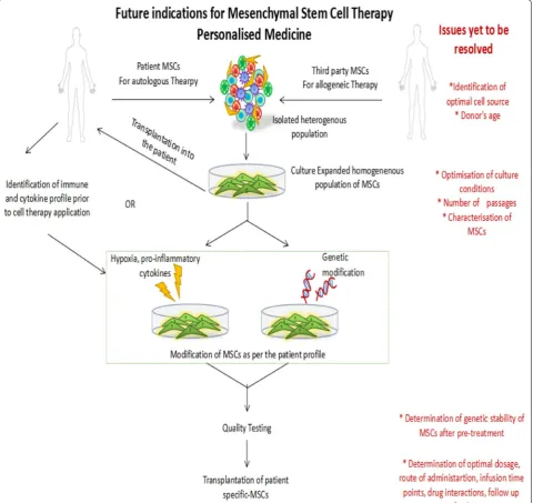
MSC modification: preconditioned or genetically modified
To overcome the current limitations, MSCs after
iso-lation can be cultured or pre-conditioned in hypoxic
conditions, so as to maintain a native healthy profile
which enhances their immunomodulatory and
regen-erative capacity. Further, preconditioning of MSCs with
proinflammatory factors could also help in abolishing
their behavioural heterogeneity [
137
] thus making them
appropriate for application in transplant patients. Some
preclinical studies have also demonstrated that genetic
modification or engineering of MSCs could also benefit
in disease management [
138
–
140
]. Targeted delivery of
MSCs with triple engineering (P-selectin glycoprotein
ligand-1 (PSGL-1)/Sialyl-Lewis(x) (SLeX)/IL-10) has
shown superior therapeutic function over the
unmodi-fied MSCs in a murine model of autoimmune
encepha-lomyelitis (EAE) [
141
]. Most recently, MSCs engineered
with Etanercept, a TNF-α blocker, were used effectively
in a mice model of collagen-induced arthritis [
142
]. On
similar lines, there exist several cytokines, growth
fac-tors, TLR agonists which can be used individually or in
combination to treat MSCs, for encouraging their
thera-peutic efficacy.
MSCs: for supplemental therapy
Mesenchymal stem cells have long been used in
combi-nation with IS drugs for immune-mediated conditions.
However, in most trials IS drugs are administered prior
to MSC infusion, which might lead to an altered cytokine
profile that MSCs experience in vivo resulting in their
poor efficacy. Therefore, optimization of timing, number
of cells per dose, number of doses and route of
adminis-tration would be of immense advantage while
consider-ing the use of MSCs for immune-mediated conditions.
MSCs: for personalized therapy
Clinical trial outcomes have emphasized the concept
of “licensing”, which is easily controllable in vitro, but,
remains to be a challenge in in vivo condition. In patients,
the inflammatory milieu is variable depending upon the
immune disorder. This variation in personal
microen-vironment is responsible for altering the behaviour of
infused MSCs. To ensure the success of MSC therapy,
it, therefore, becomes necessary to study and
under-stand the signalling molecules and cellular interactions
in the prospective microenvironment of a patient. It is
the heterogeneity in MSC profile based on isolation and
culture protocols and the patient factors which
substan-tiates the need for personalized medicine. Thus it would
be beneficial to identify the cytokine and immune status
of patients prior to MSC application. Patient population
likely to benefit may be given the MSC therapy without
any modification while for others, individualization of
MSC therapy using either genetically modified or
pre-conditioned MSCs may prove to be beneficial (Fig.
1
).
Conclusion
foundation of MSC therapy. Moreover, as a part of
per-sonalized medicine, it would be beneficial to standardize
the protocols for pre-conditioning or genetically
modify-ing MSCs as per the patient’s need to further enhance the
applicability and success of these cellular therapies which
in future may substitute the current drug therapies.
Abbreviations
IS drugs: immunosuppressive drugs; MSC: mesenchymal stem cell; APC: anti-gen presenting cells; TLR: toll-like receptor; BM: bone marrow; UC: umbilical cord; MHC: major histocompatibility complex; IFN-γ: interferon-γ; Tregs: regula-tory T cells; Bregs: regularegula-tory B cells; DC: dendritic cell; MEM: MSC-educated macrophages; IL-10: interleukin-10; NK: natural killer cells; TGF-β: transform-ing growth factor; HGF: hepatocyte growth factor; NO: nitric oxide; HO-1: heme oxygenase-1; IDO: indoleamine 2,3-dioxygenase; TRAIL: TNF-related
[image:12.595.59.540.89.542.2]apoptosis-inducing ligand; PD-L1: programmed death ligand 1; BM-MSCs: bone marrow derived MSCs; UC-MSCs: umbilical cord derived MSCs; PBMC: peripheral blood mononuclear cell; Tx: transplantation; TAC : tacrolimus; ATG : anti-thymocyte globulin; MMF: mycophenolate mofetil; CNI: calcineurin inhibi-tor; MEP: methylpremnisolone; CsA: cyclosporine A; HIF: hypoxia inducible fac-tor; HIF-1α: HIF-1 alpha; HIF-2α: HIF-2 alpha; KLF4: Kruppel like factor 4; OCT4: octamer-binding transcription factor 4; C-MYC: v-myc avian myelocytomatosis viral oncogene homolog; PGE2: prostaglandin-E2; DSS: dextran sulfate sodium; VCAM-1: vascular cell adhesion molecule-1; CCR5: chemokine ligands of C-C chemokine receptor type-5; CXCR3: C-X-C chemokine receptor type-3; CXCL9: chemokine (C-X-C motif ) ligand-9; CXCL10: chemokine (C-X-C motif ) ligand-10; CCL2: monocyte chemotactic protein-1; iNOS: inducible nitric oxide synthase; IFN-β: interferon-β; PSGL-1: P-selectin glycoprotein ligand-1; SLeX: Sialyl-Lewis(x); EAE: autoimmune encephalomyelitis.
Authors’ contributions
AR and UK conceived the idea for the paper, performed the literature search and drafted the manuscript. AR revised the manuscript for important intel-lectual content. All authors read and approved the final manuscript.
Author details
1 Department of Translational and Regenerative Medicine, Postgraduate
Institute of Medical Education and Research, Sector 12, Chandigarh, India.
2 Department of Zoology, Panjab University, Sector 14, Chandigarh, India.
Acknowledgements Not applicable.
Competing interests
The authors declare that they have no competing interests.
Availability of data and materials Not applicable.
Consent for publication Not applicable.
Ethics approval and consent to participate Not applicable.
Funding
No funding was received.
Publisher’s Note
Springer Nature remains neutral with regard to jurisdictional claims in pub-lished maps and institutional affiliations.
Received: 22 November 2017 Accepted: 7 February 2018
References
1. Silkensen JR. Long-term complications in renal transplantation. J Am Soc Nephrol JASN. 2000;11(3):582–8.
2. Chang D, Kobashigawa J, Luu M. Outpatient management and long-term complications in heart transplantation. In: Kobashigawa JC, editor. Clinical guide to heart transplantation. Cham: Springer International Publishing; 2017. p. 171–83.
3. Dharnidharka VR, Lamb KE, Zheng J, Schechtman KB, Meier-Kriesche HU. Lack of significant improvements in long-term allograft survival in pediatric solid organ transplantation: a US national registry analysis. Pediatr Transpl. 2015;19(5):477–83.
4. Neuberger JM, Bechstein WO, Kuypers DR, Burra P, Citterio F, De Geest S, Duvoux C, Jardine AG, Kamar N, Kramer BK, et al. Practical recommen-dations for long-term management of modifiable risks in kidney and liver transplant recipients: a guidance report and clinical checklist by the consensus on managing modifiable risk in transplantation (COM-MIT) group. Transplantation. 2017;101(4S Suppl 2):S1–56.
5. Halloran PF. Immunosuppressive drugs for kidney transplantation. N Engl J Med. 2004;351(26):2715–29.
6. Alangaden GJ, Thyagarajan R, Gruber SA, Morawski K, Garnick J, El-Amm JM, West MS, Sillix DH, Chandrasekar PH, Haririan A. Infectious complications after kidney transplantation: current epidemiology and associated risk factors. Clin Transpl. 2006;20(4):401–9.
7. Fernandes-Silva G, Ivani de Paula M, Rangel EB. mTOR inhibitors in pancreas transplant: adverse effects and drug-drug interactions. Expert Opin Drug Metab Toxicol. 2017;13(4):367–85.
8. Shivaswamy V, Boerner B, Larsen J. Post-transplant diabetes mel-litus: causes, treatment, and impact on outcomes. Endocr Rev. 2016;37(1):37–61.
9. Engels EA, Pfeiffer RM, Fraumeni JF, Kasiske BL, Israni AK, Snyder JJ, Wolfe RA, Goodrich NP, Bayakly AR, Clarke CA. Spectrum of cancer risk among US solid organ transplant recipients. JAMA. 2011;306(17):1891–901. 10. Stallone G, Infante B, Grandaliano G. Management and prevention of post-transplant malignancies in kidney transplant recipients. Clin Kidney J. 2015;8(5):637–44.
11. Glicklich D, Lamba R, Pawar R. Hypertension in the kidney transplant recipient: overview of pathogenesis, clinical assessment, and treatment. Cardiol Rev. 2017;25(3):102–9.
12. Shi Y, Hu G, Su J, Li W, Chen Q, Shou P, Xu C, Chen X, Huang Y, Zhu Z, et al. Mesenchymal stem cells: a new strategy for immunosuppression and tissue repair. Cell Res. 2010;20(5):510–8.
13. Friedenstein AJ. Stromal mechanisms of bone marrow: cloning in vitro and retransplantation in vivo. Haematol Blood Transfus. 1980;25:19–29. 14. Friedenstein AJ, Chailakhjan RK, Lalykina KS. The development of fibro-blast colonies in monolayer cultures of guinea-pig bone marrow and spleen cells. Cell Tissue Kinet. 1970;3(4):393–403.
15. Hoffmann M, Kuska J-P, Zscharnack M, Loeffler M, Galle J. Spatial organization of mesenchymal stem cells in vitro—results from a new individual cell-based model with podia. PLoS ONE. 2011;6(7):e21960. 16. Bianco P, Robey PG, Simmons PJ. Mesenchymal stem cells: revisiting
history, concepts, and assays. Cell Stem Cell. 2008;2(4):313–9. 17. Prockop DJ. Marrow stromal cells as stem cells for nonhematopoietic
tissues. Science (New York, NY). 1997;276(5309):71–4.
18. Pittenger MF, Mackay AM, Beck SC, Jaiswal RK, Douglas R, Mosca JD, Moorman MA, Simonetti DW, Craig S, Marshak DR. Multiline-age potential of adult human mesenchymal stem cells. Science. 1999;284(5411):143–7.
19. Hass R, Kasper C, Böhm S, Jacobs R. Different populations and sources of human mesenchymal stem cells (MSC): a comparison of adult and neonatal tissue-derived MSC. Cell Commu Signal CCS. 2011;9:12. 20. Deans RJ, Moseley AB. Mesenchymal stem cells: biology and potential
clinical uses. Exp Hematol. 2000;28(8):875–84.
21. William TT, Pendleton JD, Beyer WM, Egalka MC, Guinan EC. Suppres-sion of allogeneic T-cell proliferation by human marrow stromal cells: implications in transplantation. Transplantation. 2003;75(3):389–97. 22. Fu X, Chen Y, Xie F-N, Dong P, Liu W-B, Cao Y, Zhang W-J, Xiao R.
Com-parison of immunological characteristics of mesenchymal stem cells derived from human embryonic stem cells and bone marrow. Tissue Eng Part A. 2015;21(3–4):616–26.
23. LaRosa DF, Rahman AH, Turka LA. The innate immune system in allo-graft rejection and tolerance. J Immunol. 2007;178(12):7503–9. 24. Ingulli E. Mechanism of cellular rejection in transplantation. Pediatr
Nephrol. 2010;25(1):61–74.
25. Benichou G, Yamada Y, Aoyama A, Madsen JC. Natural killer cells in rejection and tolerance of solid organ allografts. Curr Opin Organ Transpl. 2011;16(1):47–53.
26. de Mattos AM, Olyaei AJ, Bennett WM. Nephrotoxicity of immunosup-pressive drugs: long-term consequences and challenges for the future. Am J Kidney Dis. 2000;35(2):333–46.
27. Scandling JD, Busque S, Shizuru JA, Engleman EG, Strober S. Induced immune tolerance for kidney transplantation. N Engl J Med. 2011;365(14):1359–60.
28. Ruiz P, Maldonado P, Hidalgo Y, Gleisner A, Sauma D, Silva C, Saez JJ, Nuñez S, et al. Transplant tolerance: new insights and strategies for long-term allograft acceptance. Clin Dev Immunol. 2013;2013:15. 29. Salisbury EM, Game DS, Lechler RI. Transplantation tolerance. Pediatr
30. Waldmann H. Tolerance: an overview and perspectives. Nat Rev Neph-rol. 2010;6(10):569–76.
31. Calne R. Liver transplantation tolerance in animal models for ency-clopedia of medical immunology. In: Mackay IR, Rose NR, Diamond B, Davidson A, editors. Encyclopedia of medical immunology: autoim-mune diseases. New York: Springer; 2014. p. 656–9.
32. Kingsley CI, Nadig SN, Wood KJ. Transplantation tolerance: lessons from experimental rodent models. Transpl Int. 2007;20(10):828–41. 33. Miller ML, Chong AS, Alegre M-L. Fifty shades of transplantation
toler-ance: beyond a binary tolerant/non-tolerant paradigm. Curr Transpl Rep. 2017;4(4):262–9.
34. Scalea JR, Tomita Y, Lindholm CR, Burlingham W. Transplantation toler-ance induction: cell therapies and their mechanisms. Front Immunol. 2016;7:87.
35. Zakrzewski JL, van den Brink MRM, Hubbell JA. Overcoming immuno-logical barriers in regenerative medicine. Nat Biotechnol. 2014;32:786. 36. Guo K, Ikehara S, Meng X. Mesenchymal stem cells for inducing
toler-ance in organ transplantation. Front Cell Dev Biol. 2014;2:8. 37. Deng W, Han Q, Liao L, You S, Deng H, Zhao RC. Effects of allogeneic
bone marrow-derived mesenchymal stem cells on T and B lympho-cytes from BXSB mice. DNA Cell Biol. 2005;24(7):458–63.
38. Aggarwal S, Pittenger MF. Human mesenchymal stem cells modulate allogeneic immune cell responses. Blood. 2005;105(4):1815–22. 39. Rasmusson I, Ringden O, Sundberg B, Le Blanc K. Mesenchymal stem
cells inhibit lymphocyte proliferation by mitogens and alloantigens by different mechanisms. Exp Cell Res. 2005;305(1):33–41.
40. Ma OK, Chan KH. Immunomodulation by mesenchymal stem cells: interplay between mesenchymal stem cells and regulatory lympho-cytes. World J Stem Cells. 2016;8(9):268–78.
41. Zhao Z-G, Xu W, Sun L, You Y, Li F, Li Q-B, Zou P. Immunomodulatory function of regulatory dendritic cells induced by mesenchymal stem cells. Immunol Investig. 2012;41(2):183–98.
42. Rutz S, Janke M, Kassner N, Hohnstein T, Krueger M, Scheffold A. Notch regulates IL-10 production by T helper 1 cells. Proc Natl Acad Sci USA. 2008;105(9):3497–502.
43. Cho D-I, Kim MR, Jeong H-Y, Jeong HC, Jeong MH, Yoon SH, Kim YS, Ahn Y. Mesenchymal stem cells reciprocally regulate the M1/M2 bal-ance in mouse bone marrow-derived macrophages. Exp Mol Med. 2014;46(1):e70.
44. Vasandan AB, Jahnavi S, Shashank C, Prasad P, Kumar A, Prasanna SJ. Human Mesenchymal stem cells program macrophage plasticity by altering their metabolic status via a PGE(2)-dependent mechanism. Sci Rep. 2016;6:38308.
45. Sotiropoulou PA, Perez SA, Gritzapis AD, Baxevanis CN, Papamichail M. Interactions between human mesenchymal stem cells and natural killer cells. Stem Cells. 2006;24(1):74–85.
46. Casiraghi F, Azzollini N, Cassis P, Imberti B, Morigi M, Cugini D, Cavinato RA, Todeschini M, Solini S, Sonzogni A. Pretransplant infusion of mes-enchymal stem cells prolongs the survival of a semiallogeneic heart transplant through the generation of regulatory T cells. J Immunol. 2008;181(6):3933–46.
47. Davies LC, Heldring N, Kadri N, Le Blanc K. Mesenchymal stromal cell secretion of programmed death-1 ligands regulates T cell mediated immunosuppression. Stem Cells. 2017;35(3):766–76.
48. Ren G, Zhang L, Zhao X, Xu G, Zhang Y, Roberts AI, Zhao RC, Shi Y. Mesenchymal stem cell-mediated immunosuppression occurs via concerted action of chemokines and nitric oxide. Cell Stem Cell. 2008;2(2):141–50.
49. Kyurkchiev D, Bochev I, Ivanova-Todorova E, Mourdjeva M, Oreshkova T, Belemezova K, Kyurkchiev S. Secretion of immunoregulatory cytokines by mesenchymal stem cells. World J Stem Cells. 2014;6(5):552–70. 50. Ma S, Xie N, Li W, Yuan B, Shi Y, Wang Y. Immunobiology of
mesenchy-mal stem cells. Cell Death Differ. 2014;21(2):216–25.
51. Crop MJ, Korevaar SS, de Kuiper R, Ijzermans JN, van Besouw NM, Baan CC, Weimar W, Hoogduijn MJ. Human mesenchymal stem cells are susceptible to lysis by CD8(+) T cells and NK cells. Cell Transpl. 2011;20(10):1547–59.
52. Gotherstrom C, Lundqvist A, Duprez IR, Childs R, Berg L, le Blanc K. Fetal and adult multipotent mesenchymal stromal cells are killed by different pathways. Cytotherapy. 2011;13(3):269–78.
53. Galleu A, Riffo-Vasquez Y. Apoptosis in mesenchymal stromal cells induces in vivo recipient-mediated immunomodulation. Sci Transl Med. 2017;9(416):eaam7828.
54. Bartholomew A, Sturgeon C, Siatskas M, Ferrer K, McIntosh K, Patil S, Hardy W, Devine S, Ucker D, Deans R, et al. Mesenchymal stem cells suppress lymphocyte proliferation in vitro and prolong skin graft survival in vivo. Exp Hematol. 2002;30(1):42–8.
55. Zhou HP, Yi DH, Yu SQ, Sun GC, Cui Q, Zhu HL, Liu JC, Zhang JZ, Wu TJ. Administration of donor-derived mesenchymal stem cells can prolong the survival of rat cardiac allograft. Transpl Proc. 2006;38(9):3046–51. 56. Popp FC, Eggenhofer E, Renner P, Slowik P, Lang SA, Kaspar H, Geissler
EK, Piso P, Schlitt HJ, Dahlke MH. Mesenchymal stem cells can induce long-term acceptance of solid organ allografts in synergy with low-dose mycophenolate. Transpl Immunol. 2008;20(1–2):55–60.
57. Ge W, Jiang J, Baroja ML, Arp J, Zassoko R, Liu W, Bartholomew A, Garcia B, Wang H. Infusion of mesenchymal stem cells and rapamycin syner-gize to attenuate alloimmune responses and promote cardiac allograft tolerance. Am J Transpl. 2009;9(8):1760–72.
58. Inoue S, Popp FC, Koehl GE, Piso P, Schlitt HJ, Geissler EK, Dahlke MH. Immunomodulatory effects of mesenchymal stem cells in a rat organ transplant model. Transplantation. 2006;81(11):1589–95.
59. Xu DM, Yu XF, Zhang D, Zhang MX, Zhou JF, Tan PH, Ding YC. Mesenchy-mal stem cells differentially mediate regulatory T cells and conventional effector T cells to protect fully allogeneic islet grafts in mice. Diabetolo-gia. 2012;55(4):1091–102.
60. Ge W, Jiang J, Arp J, Liu W, Garcia B, Wang H. Regulatory T-cell genera-tion and kidney allograft tolerance induced by mesenchymal stem cells associated with indoleamine 2,3-dioxygenase expression. Transplanta-tion. 2010;90(12):1312–20.
61. Perico N, Casiraghi F, Introna M, Gotti E, Todeschini M, Cavinato RA, Capelli C, Rambaldi A, Cassis P, Rizzo P, et al. Autologous mesenchymal stromal cells and kidney transplantation: a pilot study of safety and clinical feasibility. Clin J Am Soc Nephrol CJASN. 2011;6(2):412–22. 62. Tan J, Wu W, Xu X, et al. Induction therapy with autologous
mesen-chymal stem cells in living-related kidney transplants: a randomized controlled trial. JAMA. 2012;307(11):1169–77.
63. Reinders ME, de Fijter JW, Roelofs H, Bajema IM, de Vries DK,
Schaapherder AF, Claas FH, van Miert PP, Roelen DL, van Kooten C, et al. Autologous bone marrow-derived mesenchymal stromal cells for the treatment of allograft rejection after renal transplantation: results of a phase I study. Stem Cells Transl Med. 2013;2(2):107–11.
64. Peng Y, Ke M, Xu L, Liu L, Chen X, Xia W, Li X, Chen Z, Ma J, Liao D, et al. Donor-derived mesenchymal stem cells combined with low-dose tacrolimus prevent acute rejection after renal transplantation: a clinical pilot study. Transplantation. 2013;95(1):161–8.
65. Mudrabettu C, Kumar V, Rakha A, Yadav AK, Ramachandran R, Kanwar DB, Nada R, Minz M, Sakhuja V, Marwaha N, et al. Safety and efficacy of autologous mesenchymal stromal cells transplantation in patients undergoing living donor kidney transplantation: a pilot study. Nephrol-ogy. 2015;20(1):25–33.
66. Griffin MD, Ryan AE, Alagesan S, Lohan P, Treacy O, Ritter T. Anti-donor immune responses elicited by allogeneic mesenchymal stem cells: what have we learned so far? Immunol Cell Biol. 2013;91(1):40–51. 67. Oliveira RL, Chagastelles PC, Sesterheim P, Pranke P. In vivo
immuno-genic response to allogeneic mesenchymal stem cells and the role of preactivated mesenchymal stem cells cotransplanted with allogeneic islets. Stem Cells Int. 2017;2017:12.
68. Pan GH, Chen Z, Xu L, Zhu JH, Xiang P, Ma JJ, Peng YW, Li GH, Chen XY, Fang JL, et al. Low-dose tacrolimus combined with donor-derived mesenchymal stem cells after renal transplantation: a prospective, non-randomized study. Oncotarget. 2016;7(11):12089–101.
69. Perico N, Casiraghi F, Gotti E, Introna M, Todeschini M, Cavinato RA, Capelli C, Rambaldi A, Cassis P, Rizzo P, et al. Mesenchymal stromal cells and kidney transplantation: pretransplant infusion protects from graft dysfunction while fostering immunoregulation. Transpl Int. 2013;26(9):867–78.
71. Grant JL, Smith B. Bone marrow gas tensions, bone marrow blood flow, and erythropoiesis in man. Ann Intern Med. 1963;58(5):801–9. 72. Simon MC, Keith B. The role of oxygen availability in embryonic
devel-opment and stem cell function. Nat Rev Mol Cell Biol. 2008;9(4):285–96. 73. Brahimi-Horn MC, Pouysségur J. Oxygen, a source of life and stress. FEBS
Lett. 2007;581(19):3582–91.
74. Abdollahi H, Harris LJ, Zhang P, McIlhenny S, Tulenko T, DiMuzio PJ. The role of hypoxia in stem cell differentiation and therapeutics. J Surg Res. 2011;165(1):112–7.
75. Gustafsson MV, Zheng X, Pereira T, Gradin K, Jin S, Lundkvist J, Ruas JL, Poellinger L, Lendahl U, Bondesson M. Hypoxia requires notch signaling to maintain the undifferentiated cell state. Dev Cell. 2005;9(5):617–28. 76. Palomaki S, Pietila M, Laitinen S, Pesala J, Sormunen R, Lehenkari P,
Koivunen P. HIF-1alpha is upregulated in human mesenchymal stem cells. Stem Cells. 2013;31(9):1902–9.
77. Keith B, Simon MC. Hypoxia inducible factors, stem cells and cancer. Cell. 2007;129(3):465–72.
78. Lv B, Li F, Fang J, Xu L, Sun C, Han J, Hua T, Zhang Z, Feng Z, Jiang X. Hypoxia inducible factor 1α promotes survival of mesenchymal stem cells under hypoxia. Am J Transl Res. 2017;9(3):1521–9.
79. Zhu C, Yu J, Pan Q, Yang J, Hao G, Wang Y, Li L, Cao H. Hypoxia-inducible factor-2 alpha promotes the proliferation of human placenta-derived mesenchymal stem cells through the MAPK/ERK signaling pathway. Sci Rep. 2016;6:35489.
80. Lan YW, Choo KB, Chen CM, Hung TH, Chen YB, Hsieh CH, Kuo HP, Chong KY. Hypoxia-preconditioned mesenchymal stem cells attenuate bleomycin-induced pulmonary fibrosis. Stem Cell Res Ther. 2015;6:97. 81. Jiang C, Liu J, Zhao J, Xiao L, An S, Gou Y, Quan H, Cheng Q, Zhang
Y, He W. Effects of hypoxia on the immunomodulatory properties of human Gingiva—derived mesenchymal stem cells. J Dent Res. 2015;94(1):69–77.
82. Cicione C, Muiños-López E, Hermida-Gómez T, Fuentes-Boquete I, et al. Effects of severe hypoxia on bone marrow mesenchymal stem cells differentiation potential. Stem Cells Int. 2013;2013:11.
83. Basciano L, Nemos C, Foliguet B, de Isla N, de Carvalho M, Tran N, Dalloul A. Long term culture of mesenchymal stem cells in hypoxia promotes a genetic program maintaining their undifferentiated and multipotent status. BMC Cell Biol. 2011;12(1):12.
84. Estrada J, Albo C, Benguria A, Dopazo A, Lopez-Romero P, Carrera-Quintanar L, Roche E, Clemente E, Enriquez J, Bernad A. Culture of human mesenchymal stem cells at low oxygen tension improves growth and genetic stability by activating glycolysis. Cell Death Differ. 2012;19(5):743.
85. Chacko SM, Ahmed S, Selvendiran K, Kuppusamy ML, Khan M, Kuppusamy P. Hypoxic preconditioning induces the expression of prosurvival and proangiogenic markers in mesenchymal stem cells. Am J Physiol Cell Physiol. 2010;299(6):C1562–70.
86. Brandl A, Meyer M, Bechmann V, Nerlich M, Angele P. Oxidative stress induces senescence in human mesenchymal stem cells. Exp Cell Res. 2011;317(11):1541–7.
87. Rosová I, Dao M, Capoccia B, Link D, Nolta JA. Hypoxic precondition-ing results in increased motility and improved therapeutic potential of human mesenchymal stem cells. Stem Cells. 2008;26(8):2173–82. 88. Beegle J, Lakatos K, Kalomoiris S, Stewart H, Isseroff RR, Nolta JA, Fierro
FA. Hypoxic preconditioning of mesenchymal stromal cells induces metabolic changes, enhances survival, and promotes cell retention in vivo. Stem Cells. 2015;33(6):1818–28.
89. Chang C-P, Chio C-C, Cheong C-U, Chao C-M, Cheng B-C, Lin M-T. Hypoxic preconditioning enhances the therapeutic potential of the secretome from cultured human mesenchymal stem cells in experi-mental traumatic brain injury. Clin Sci. 2013;124(3):165.
90. Kim DS, Lee MW, Ko YJ, Park HJ, Park YJ, Kim DI, Jung HL, Sung KW, Koo HH, Yoo KH. Application of human mesenchymal stem cells cultured in different oxygen concentrations for treatment of graft-versus-host disease in mice. Biomed Res. 2016;37(5):311–7.
91. Chen G, Nayan M, Duong M, Asenjo J-F, Ge Y, Chiu RCJ, Shum-Tim D. Marrow stromal cells for cell-based therapy: the role of antiinflam-matory cytokines in cellular cardiomyoplasty. Ann Thorac Surg. 2010;90(1):190–7.
92. Huang WH, Chen HL, Huang PH, Yew TL, Lin MW, Lin SJ, Hung SC. Hypoxic mesenchymal stem cells engraft and ameliorate limb ischae-mia in allogeneic recipients. Cardiovasc Res. 2014;101(2):266–76. 93. Li JH, Zhang N, Wang JA. Improved anti-apoptotic and
anti-remod-eling potency of bone marrow mesenchymal stem cells by anoxic pre-conditioning in diabetic cardiomyopathy. J Endocrinol Investig. 2008;31(2):103–10.
94. Duijvestein M, Wildenberg ME, Welling MM, Hennink S, Molendijk I, van Zuylen VL, Bosse T, Vos ACW, de Jonge-Muller ESM, Roelofs H, et al. Pretreatment with interferon-γ enhances the therapeutic activity of mesenchymal stromal cells in animal models of colitis. Stem Cells. 2011;29(10):1549–58.
95. Polchert D, Sobinsky J, Douglas GW, Kidd M, Moadsiri A, Reina E, Genrich K, Mehrotra S, Setty S, Smith B, et al. IFN-γ activation of mes-enchymal stem cells for treatment and prevention of graft versus host disease. Eur J Immunol. 2008;38(6):1745–55.
96. Torkaman M, Ghollasi M, Mohammadnia-Afrouzi M, Salimi A, Amari A. The effect of transplanted human Wharton’s jelly mesenchymal stem cells treated with IFN-γ on experimental autoimmune encephalomyeli-tis mice. Cell Immunol. 2017;311:1–12.
97. Tobin LM, Healy ME, English K, Mahon BP. Human mesenchymal stem cells suppress donor CD4(+) T cell proliferation and reduce pathology in a humanized mouse model of acute graft-versus-host disease. Clin Exp Immunol. 2013;172(2):333–48.
98. Qiu Y, Guo J, Mao R, Chao K, Chen BL, He Y, Zeng ZR, Zhang SH, Chen MH. TLR3 preconditioning enhances the therapeutic efficacy of umbili-cal cord mesenchymal stem cells in TNBS-induced colitis via the TLR3-Jagged-1-Notch-1 pathway. Mucosal Immunol. 2016;10:727. 99. Fuenzalida P, Kurte M, Fernández-O’ryan C, Ibañez C, Gauthier-Abeliuk
M, Vega-Letter AM, Gonzalez P, Irarrázabal C, Quezada N, Figueroa F, et al. Toll-like receptor 3 pre-conditioning increases the therapeutic effi-cacy of umbilical cord mesenchymal stromal cells in a dextran sulfate sodium—induced colitis model. Cytotherapy. 2016;18(5):630–41. 100. Ryu D-B, Lim J-Y, Lee S-E, Park G, Min C-K. Induction of indoleamine
2,3-dioxygenase by pre-treatment with poly(I:C) may enhance the efficacy of MSC treatment in DSS-induced colitis. Immune Netw. 2016;16(6):358–65.
101. Vega-Letter AM, Kurte M, Fernández-O’Ryan C, Gauthier-Abeliuk M, Fuenzalida P, Moya-Uribe I, Altamirano C, Figueroa F, Irarrázabal C, Carrión F. Differential TLR activation of murine mesenchymal stem cells generates distinct immunomodulatory effects in EAE. Stem Cell Res Ther. 2016;7(1):150.
102. Sheng H, Wang Y, Jin Y, Zhang Q, Zhang Y, Wang L, Shen B, Yin S, Liu W, Cui L, et al. A critical role of IFNgamma in priming MSC-mediated sup-pression of T cell proliferation through up-regulation of B7-H1. Cell Res. 2008;18(8):846–57.
103. Krampera M, Cosmi L, Angeli R, Pasini A, Liotta F, Adreini A, Santarlasci V, Mazzinghi B. Role for interferon-gamma in the immunomodulatory activity of human bone marrow mesenchymal stem cells. Stem Cells. 2006;24:386–98.
104. Chan JL, Tang KC, Patel AP, Bonilla LM, Pierobon N, Ponzio NM, Ramesh-war P. Antigen-presenting property of mesenchymal stem cells occurs during a narrow window at low levels of interferon-gamma. Blood. 2006;107(12):4817–24.
105. English K. Mechanisms of mesenchymal stromal cell immunomodula-tion. Immunol Cell Biol. 2013;91(1):19–26.
106. Ren G, Zhao X, Zhang L, Zhang J, L’Huillier A, Ling W, Roberts AI, Le AD, Shi S, Shao C, et al. Inflammatory cytokine-induced intercellular adhesion molecule-1 and vascular cell adhesion molecule-1 in mes-enchymal stem cells are critical for immunosuppression. J Immunol. 2010;184(5):2321–8.
107. Ren G, Roberts AI, Shi Y. Adhesion molecules: key players in mesen-chymal stem cell-mediated immunosuppression. Cell Adhes Migr. 2011;5(1):20–2.
108. Akiyama K, Chen C, Wang D, Xu X, Qu C, Yamaza T, Cai T, Chen W, Sun L, Shi S. Mesenchymal-stem-cell-induced immunoregulation involves FAS-ligand-/FAS-mediated T cell apoptosis. Cell Stem Cell. 2012;10(5):544–55.