INV ASCULAR SMOOTH MUSCLE
Penelope-Jane Linnett BSc(Hons) ANU BVSc Syd
A thesis submitted for the degree of
Master of Philosophy
at the
Australian National University
STATEMENT
The contents of this thesis are original and all experiments were planned and
carried out by the author under the supervision of Dr. Caryl Hill and Dr. Klaus
Matthaei, except where otherwise stated or referenced.
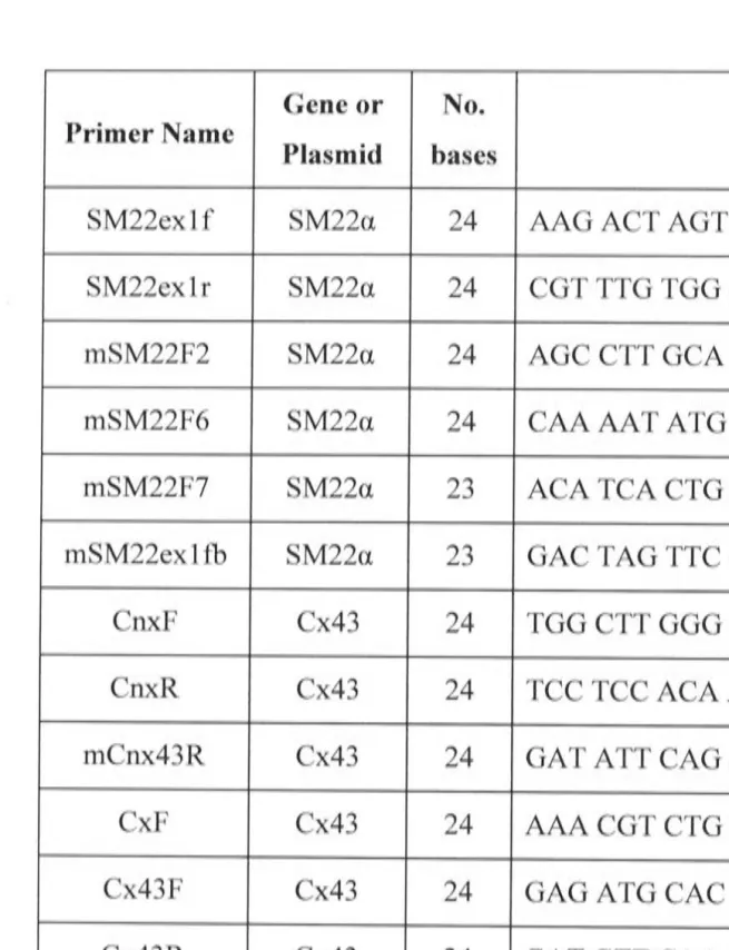
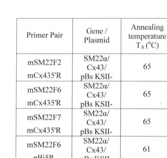
The oligonucleotide primers (Cx43F, Cx43R, Cx43R2, Cx43R3, CMVF,
pEGFPR) were designed by Dr. Caryl Hill and Dr. Klaus Matthaei, whilst the HOTT3
and HOTT7 primers were designed by Mr. David Mann. Preparation and testing of
the smooth muscle cells was carried out by Ms. Haruyo Hickey. The set up and
culture of the embryonic stem cells was done by Dr Klaus Matthaei, and the
blastocyst injections were performed by Ms Helen Taylor. The blood pressure
measurements in Chapter 4 were done by Mrs Kate MacKenzie.
I wish to acknowledge the following: Dr Caryl Hill, Dr Klaus Matthaei, Wayne
Damcevski, David Mann and Matthew Newhouse. Special thanks must go to Sue
Henderson, Professor Ian Hendry, Haruyo Hickey, Dr Nesrin Ozarak and Dr Shaun
Sandow.
Finally, I wish to acknowledge 'the very special support and guidance provided to
ABSTRACT
Transgenic mice overexpressing Cx43 in specific regions of the vasculature were
generated to examine the hypothesis that vascular function ( specifically blood pres$ure)
is influenced by increased connexin (Cx) 43 expression. The creation of a DNA
construct containing a restricted portion of the SM22a promoter (SM22a I Cx43) to
drive Cx43 gene expression, met the tissue specific requirement of the mice.
Unfortunately, the establishment of a temporal and tissue specific transgenic mouse line
to ensure that the mouse would not succumb to embryonic or neo-natal mortality,
exceeded the time limitations of the current project.
When tested in vitro in murine aortic smooth muscle cells (prior to
transformation into BALB/c embryonic stem cells), this DNA construct (SM22a / Cx43
I pBs KSII-) produced inconclusive results. This construct was, however, used to
generate a transgenic mouse line with a truncated-promoter in the germ-line. It was
found that only 204 to 103 bp of the SM22a promoter was present and so the functional
capability of the truncated-promoter was unknown. Subsequently, this mouse line was
interbred to increase the gene copy number and hence, the Cx43 expression level. A
second attempt produced mice with the full-promoter in the germ-line.
Preliminary analyses on both mouse lines revealed the following: the aortic and
tail artery tissues of all transgenic mice were more fragile than the control mouse; and
the blood pressures of all mice, as determined by tail-cuff plethysmography, were within
the normal range. Western blotting was used to determine the Cx43 protein levels in the
aortae and tail arteries of the mice. In the single transgenic-promoter mice, there was no
significant increase in Cx43 expression in either the aorta or tail artery. The double
transgenic truncated-promoter mice had significantly increased Cx43 protein levels in
was minimally functional. In the single transgenic full-promoter mice, aortic Cx43
protein levels were significantly increased with a trend towards increased Cx43 in the
tail arteries, which demonstrated that the SM22a / Cx43 construct was functional in
VIVO.
Together, the Cx43 expression data and the blood pressure measurements.
suggest that increased Cx43 expression does not cause hypertension or hypotension in
the mouse. The results obtained from this thesis provide the groundwork for further
experimentation into the physiological effects of Cx43 overexpression in the vasculature
PUBLICATIONS AND PRESENTATIONS
Some of the work emanating from this thesis was presented at scientific
meetings. The following abstract was published in conjunction with one of these
presentations.
Linnert, P-J., Matthaei, K.I., Hickey, H. and Hill, C.E. (1999) Tissue and temporal
specific expression of connexin 43 in mice. Proceedings of the Australian Neuroscience
Society 10: 225.
Other presentations:
Neuroscience Colloquium, Kioloa, NSW. (1999)
Statement
Acknowledgements
Abstract
Publications and Presentations
Table of Contents
Abbreviations
CHAPTER 1 - INTRODUCTION
1.1 The Circulatory System
1.1.1 General overview
1.1.2 Types of arteries
1.1.3 Arterial morphology
1.1.3 .1 Vascular smooth muscle cells
1.1.4 Arterial function
1.1.5 Gap junctions
1.1.5 .1 Gap junction structure
..
II...
Ill.
IV.
VI..
vu...
XIII 1 2 2 2 3 3 5 6 71.1.5.2 Gap junctions: homomeric, heteromeric, homotypic & heterotypic forms 10
1.1.5.3 Gap junction expression
1.1.5.4 Regulation of gap junction function
1.1.6 Gap junctions and arterial function
1.2 Vascular Control Mechanisms
1.2.1 Endothelium derived factors
1.2.2 Endothelial influences in vascular disease
1.3 Vascular Disease and Gap Junctions
1.3 .2 Hypertension
1.3.2.1 Blood pressure control mechanisms
1.3 .2.2 Types and causes of hypertension
1.3 .2.3 Vascular morphology in hypertension
1.4.1 General overview
1. 4 .2 Thesis aims
CHAPTER 2 - MATERIALS AND METHODS
2.1 Recombinant DNA Techniques
2.1.1 Oligonucleotide design and preparation
2.1.2 The polymerase chain reaction (PCR)
2.1.3 Restriction endonuclease digests
2.1.4 Analytical and preparative separation of DNA fragments
2.1.5 Gel purification of restricted DNA fragments
2.1.6 DNA concentration by ethanol precipitation
2.1. 7 Dephosphorylation of vector DNA
2.1.8 Blunt ending of DNA fragments with Klenow
2.1.9 Purification of products
2.1.10 Ligation of restriction fragments into vector DNA
2.1.11 Transformation and selection of bacterial clones
(a) Preparation of electrocompetent cells
(b) Preparation of L-ampicillin agar plates
( c) Electroporation and plating
2.1.12 Screening cloned inserts
( a) a-complementation (blue / white colony screening)
(b) Colony cracking
(c) PCR
2.1.13 Plasmid preparation
2.1.14 DNA sequencing
2.2 Generation of the DNA Construct
2.3 Evaluation of the DNA Construct in Cell Culture
2.3 .1 Dissection and culture of aortic smooth muscle cells
2.3 .2 Culture of fibroblasts
2.3.3 Transfection of cultured cells
2.4 Generation of the Transgenic Mice 58
2.4.1 General outline 58
2.4.2 The chimaeras and progeny 60
2.4.3 Preparation of tail DNA 60
2.5 Analysis of the Transgenic Mice 61
2.5 .1 Immunohistochemistry 61
2.5.2 Western blots 62
2.5.3 Blood pressure recordings 64
CHAPTER 3 - GENERATION OF THE SM22oc/ CX43 CONSTRUCT AND MOUSE 66
3.1 Introduction 66
PART I - Cloning of the SM22a promoter and Cx43 cDNA 68
3.2 Additional Methods 68
3 .2.1 Cloning of the SM22a promoter 68
3 .2.1.1 Isolation of the SM22a promoter 68
3.2.1.2 Construction of the SM22a / pBs KSII- plasmid 68
3 .2.2 Cloning of the Cx43 cDNA 69
3.2.2.1 Isolation of the Cx43 cDNA 69
3.2.2.2 Construction of the Cx43 I pBs KSII- plasmid 69
3.3 Results 70
3.3.1 SM22a I pBs KSII- 70
3.3.2 Cx43 I pBs KSII- 71
PART II - Cloning of the Cx43 / pBi5 construct 72
3.4 Additional Methods 72
3.4.1 Preparation and verification of the pBi5 plasmid 72
PART III - Cloning of the SM22a I Cx43 / pBs KSII- construct 7 4
3.6 Additional Methods 7 4
3.6.1 Cloning of the SM22a I Cx43 construct 74 3.6.1.1 Preparation of the SM22a / pBs KSII- plasmid 74
3.6.1.2 Preparation of the Cx43 / pBi5 plasmid 75
3.6.1.3 Cloning of the insert into the vector 75
3.6.1.4 Preparation and electroporation of the SM22a I Cx43 I pBs KSII- 75
3.6.1.5 Screening of the progeny 76
3.7 Results 76
3.7.1 Cloning of SM22a / Cx43 76
3.7.2 Testing in cell culture 77
3.7.3 Preparation and electroporation of the SM22a / Cx43 fragment into ES cells 78 3. 7 .4 Identification of positive clones
3.7.5 PCR screening of the progeny
3.8 Generation of a Second SM22a I Cx43 Transgenic Mouse
3.9 Discussion
CHAPTER 4 -ANALYSIS OF THE SM2~/ CX43 MOUSE
4.1 Introduction
4.2 Results
4.2.1 General observations
4.2.2 Litter sizes and pup gender
4.2.3 Body weights
4.2.4 Organ weights
4.2.5 Blood pressure measurements
4.2.6 Determination of Cx43 expression
4 .2. 7 Immunohistochemistry
4.3 Discussion
REFERENCE LIST
APPENDIX I - METHODS
AI. I Restriction endonuclease digests
AI.2 Analytical and preparative separation of DNA fragments
AI.3 Smooth muscle cell specificity
AI.4 Protocol for fresh tissue immunohistochemistry
AI.5 Western blots
113
156
156 157 158 159 160APPENDIX II - GENERATION OF THE TETRACYCLINE RESPONSE CONSTRUCTS
161
All.I Introduction
AII.2 Additional Methods
AII.2.1 Cloning of SM22a / pTetOn
AII.2.1.1 Preparation and verification of the pTetOn plasmid
AII.2.1.2 Preparation of the SM22a / pBs KSII- plasmid
AII.2.1.3 Cloning of SM22a / pTetOn
AII.2.2 Cloning of Cx43 / pBi5
AII.2.3 Cloning of EGFP / Cx43 / pBi5
AII.2.3.1 Preparation of the pEGFP-NI plasmid
AII.2.3 .2 Preparation of the Cx43 / pBi5 plasmid
AII.2.3.3 Preparation of the Cx43 / pBi5 vector
AII.2.3.4 Preparation of the EGFP gene insert
AII.2.3 .5 Cloning of the insert into the vector
AII.2.4 Imrnunohistochemistry
AII.3.1 Cloning of SM22a / pTetOn. 167
AII.3.2 Testing of the constructs in cell culture 168
AII.3 .2.1 Electroporation experiments 168
AII.3 .2.2 Single and double transfections of the fibroblasts 168
(a) Single transfections 168
(b) Double transf ections 169
AII.3 .2.3 Single and double transfections of the smooth muscle cells 170
AII.3 .2.4 Cloning of EGFP / Cx43 / pBi5 170
AII.3.3 Cloning ofEGFP I Cx43 I pBi5 171
AII.3.4 Testing of the construct in cell culture 171
AII.3.4.1 Single and double transfections of the fibroblasts 172
(a) Single transfections 172
(b) Double transfections 1 73
AII.3 .4.2 Single and double transfections of the smooth muscle cells 173
Abbreviations AIS ATP 4-AP bp bpm BRF BSA cAMP cGMP cDNA cm
Ca2+
CAT CIAP CMV promoter COOH-CT Cx Cx43 dNTP dox Da DFS DMEM DNA DTT EDTA ES cell EtOH FCS g G418
Analytical Imaging System
adenosine triphosphate
4-aminopyridine
base pair( s)
beats per minute
Biomolecular Resource Facility
bovine serum albumin
cyclic adenosine monophosphate
cyclic guanosine 3',5'-monophosphate
complimentary DNA
centimetre
calcium
choramphenicol acetyltransferase
calf intestinal alkaline phosphatase
cytomegalovirus promoter
carboxy-carboxy-tail or carboxy terminal tail
.
connexm
connexin 4 3 gene
deoxynucleotidetri phosphate
doxycycline
dalton
doxycycline free serum
Dulbecco modified Eagles medium
deoxyribonucleic acid
dithiothreitol
ethylenediaminetetraacetic acid
embryonic stem cell
ethanol
foetal calf serum
gram(s)
GA GFA hr HCl I/P IGF kb kDa kV KCl L L-amp plate LB LIF mg min(s) ml mM
mRNA
M MCS MgCh ng nm NaCl Na2HP04 NaOAc NaOH NEB NH2_ oligo OD pBsKSII-glycyrrhetinic acid ( 18a- and 18 ~-)
glutamine:fructose-6-phosphate-amidotransf erase
hour(s)
hydrochloric acid
intraperitoneal
interleuken growth factor
kilo base( s)
kilodalton( s)
kilovolt( s)
potassium
potassium chloride
litre(s)
, L-ampicillin agar plate
Luria-Bertani broth
leukaemia inhibitory factor
milligram(s) minute(s) millilitre( s) millimolar messenger RNA molar
multiple cloning site
magnesium chloride
nano gram( s)
nanometre( s)
sodium chloride
sodium hydrogen phosphate
sodium acetate
sodium hydroxide
New England Biolabs
.
ammo-oligonucleotide
optical density
per os pmol PBS PBS-T PCR rpm rtTA
s / sec(s)
SDS SM22a
TA
Tm
Taq TEA TRE Tris Tris-HCl µg µl µmu
UTR UV light v/v V VDCC w/v X-gal by mouth picomolephosphate buffered saline
phosphate buffered saline with Tween-20
polymerase chain reaction
revolutions per minute
reverse transacti vator
second(s)
sodium dodecyl sulphate
smooth muscle 22a gene
annealing temperature
melting temperature
Thermus aquaticus
tetraethylammonium
tetracycline response element
tris(hydroxymethyl)aminomethane
tris-hydrochloride
microgram( s)
micro litre( s)
micron(s)
unit(s)
untranslated region
ultraviolet light
volume per volume
volt(s)
voltage-dependent calcium channel
weight per volume
This thesis examines the hypothesis that vascular function is influenced by
connexin expression by specifically determining the effects of increased connexin (Cx)
4 3 expression on blood pressure. To date, over fourteen individual mammalian
connexins have been described (Beyer et al. 1990; Goodenough et al. 1996; Kumar and
Gilula, 1992, 1996; Lo, 1999), with three having been identified in vascular smooth
muscle: Cx43, Cx40 (Beyer et al. 1992; Little et al. 1995a; Christ et al. 1996; Severs et
al. 1996; Xie et al. 1997; Hill et al. 2002) and Cx37 (Nakamura et al. 1999; Hill et al.
2002).
The characteristics of gap junctions, as determined by the properties of their
component Cx properties, permit the selective intercellular passage of various ions,
metabolites and small molecules ( < 1 kD) between adjacent coupled cells (Loewenstein,
1981; Beyer et al. 1990; Bennett et al. 1991; Beny and Pacicca, 1994; Bruzzone et al.
1996; Goodenough et al. 1996; Veenstra, 1996). Such coupling can vary both within,
and between, vascular beds. Changes in the distribution and expression of the vascular
connexins have been correlated with a number of disease states such as hypertension,
atherosclerosis, coronary heart disease and renal failure (Brink, 1998; Severs, 1999;
Kwak et al. 2002).
In order to investigate aspects of some of these disease states, and in particular
hypertension, the primary aim of this thesis was to produce a transgenic mouse model
that would selectively overexpress Cx43 in the arterial system. This would enable an
examination of the physiological effects of such overexpression on a number of aspects
1.1 The Circulatory System
1.1.1 General overview
The circulatory system consists of the heart and an extensive system of arteries,
veins and lymph vessels that are responsible for the supply of essential substances, such
as oxygen and nutrients, and the removal of metabolic by-products. In this regard, the
circulatory system is essential for the maintenance of homeostasis, including
temperature regulation, systemic humoral communication, and adjustments to oxygen
and nutrient supply in different physiological states (Berne and Levy, 1988; Tortora and
Grabowski, 1993).
The vascular system consists of three general types of vessels that can be
subdivided on a functional basis: the aorta and arteries (a distributing system), the
microcirculation (a diffusion and filtration system), and the veins (a collecting system).
1.1.2 Types of arteries
Arteries have traditionally been classified into three types: large or elastic
arteries ( eg. the aorta), medium or muscular arteries ( eg. the coronary, mesenteric and
middle cerebral arteries), and small arteries and arterioles. In general, large diameter
vessels with a media consisting of smooth muscle cells and elastic laminae, are referred
to as elastic arteries, whilst vessels with a much reduced or absent medial elastic lamina
are referred to as muscular arteries (Duling et al. 1991; Luff, 1991 ). The latter generally
decrease in diameter as they approach the periphery at which point, the media consists
of only 1 to 2 smooth muscle cell layers, and where the vessel is commonly referred to
as an arteriole (Mulvany and Aalkjaer, 1990). Elastic arteries may give rise to elastic or
muscular branches, although the reverse does not occur (Bunce, 1970). Functionally,
elastic arteries serve as conduction tubes that facilitate the passage of blood (Berne and
1.1.3 Arterial morphology
In all arteries, the vessel wall is divided into three distinct layers: the intima,
media and adventitia (Gutterman, 1999). Although variations in the characteristics of
each layer have been observed in different vascular beds, the general arrangement of the
endothelial and smooth muscle cells is similar in all arteries. A description of the
morphology based upon several publications is presented in Figure 1.1 and Section
1.1.3.1 (Rhodin, 1967, 1980; Gabella, 1981; Ross and Reith, 1985; Deitweiler, 1989;
Mulvany and Aalkjaer, 1990; Duling et al. 1991; Luff, 1991), with the latter
concentrating on the vascular smooth muscle cells, which are of particular relevance to
the current study.
1.1.3 .1 Vascular smooth muscle cells
Vascular smooth muscle cells, the predominant cell type in the media, are
mononucleate and are arranged in helical or circular layers around the larger blood
vessels with a generally decreased pitch ( defined as the degree of angle in relation to the
parallel arrangement of the vessel) in more peripheral vessels (Rhodin, 1967; Luff,
1991 ). The structural arrangement of vascular smooth muscle cells reflects both
functional and regulatory variations between different blood vessels, both in different
parts of the vasculature and within the same vascular region (Jacobsen et al. 1966;
Bevan and Torok, 1970; Shiraishi et al. 1986; Frid et al. 1997).
The size of vascular smooth muscle cells varies considerably in different
vascular beds ranging from 30 to approximately 2000 µm in length (Bumstock, 1970;
Gabella, 1981) and from 3 to 20 µmin width (Komuro et al. 1982; Todd et al. 1983;
Shiraishi et al. 1986). The overall shape of vascular smooth muscle cells has been
shown to vary along the one vascular bed, between different vascular beds and between
media
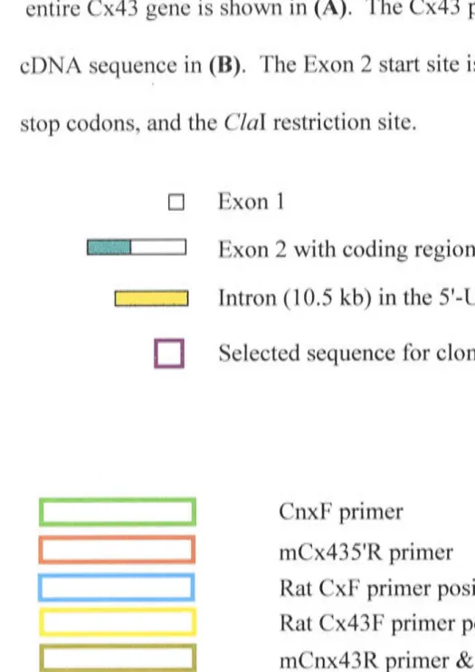
[image:19.1183.322.657.70.306.2]intima
Figure 1.1 Schematic representation of the X-sectional appearance of an artery
The intima consists of a tubular lining of endothelial cells which are joined by tight ( occluding) junctions and gap ( communicating) junctions, with the specific arrangement of junctions varying in different blood vessels, tissues and animal species. In elastic arteries, the
intima is usually relatively thick, consisting of the lining endothelium with its basal lamina, a subendothelial layer of connective tissue with both collagenous and elastic fibres, and the concentrically arranged internal elastic lamina. In muscular arteries, the subendothelial
connective tissue is sparse and in some arteries, the basal lamina of the endothelium contacts the internal elastic membrane.
The medial layer consists of either single or multiple layers of smooth muscle cells that vary in size, shape and arrangement in different blood vessels. These cells are connected by tight and gap junctions, with collagen fibres often being present in the region between smooth muscle cells. In both elastic and muscular arteries, the smooth muscle cells produce
extracellular collagen and elastic fibres. Arterioles have one layer of smooth muscle cells and a small artery may have up to about eight layers of smooth muscle cells. Muscular arteries often have a distinct external elastic lamina.
The adventitia confers structural support to the vessel, as well as connecting it to the surrounding tissues. It is composed of connective tissue and extracellular collagen fibres which contain cellular components such as fibroblasts, macrophages, and sympathetic,
Kohler et al. 1999), as well as during development (Hill and Sandow, unpublished
results) and maturation (Cliff, 1967; Miller et al. 1987).
A variety off orms of vascular smooth muscle cells have been found to coexist
within the vascular wall (Giuriato et al. 1995; Frid et al. 1997). Phenotypically distinct
vascular smooth muscle cells, characterized by different patterns of ultrastructural
morphology, cytoskeletal protein and ion channel expression, electrical responses,
capacity for extracellular matrix synthesis, migration and growth, have been identified
in the adult arterial media and in culture (Ko et al. 1999; Kohler et al. 1999; Halayko
and Solway, 2001). Thus, specific types of vascular smooth muscle cells may be
differentially involved in the processes associated with vascular disease (Frid et al.
1997; Seidel, 1997). A characteristic property of vascular smooth muscle cells is their
ability to modulate between the contractile phenotype and the synthetic phenotype,
which is characterized by a reduction in myofilament content and an increase in
synthetic organelles (Campbell et al. 1981 ), for the synthesis of extracellular matrix
molecules (Blackbum et al. 1997). In the adult mammalian vascular system, vascular
smooth muscle cells are found predominantly in the contractile state, but during vascular
disease (in hypertension, for example) or following injury or during atherosclerotic
plaque formation, the secretory synthetic type is expressed (Rennick et al. 1993; Lindop
et al. 1995; Ross, 1996; Dzau et al. 1993; Severs, 1999).
Abnormal growth of vascular smooth muscle cells plays an important role in the
pathogenesis of many vascular diseases including atherosclerosis and hypertension
(Jackson and Schwartz, 1992; Christ et al. 1996; Haefliger et al. 1997b; Shimokawa,
1999). In hypertension, the vascular pathology is characterized by hypertrophy of the
media, which in tum results in elevated blood pressure (Weissberg et al. 1995), whilst
development of the intimal cells occurs in atherosclerosis (Blackbum et al. 1995;
related to the position of the particular vessel in the vascular tree (Nakamura et al.
1998).
In hypertension, hypertrophy of the media is greater in the smaller arteries or
arterioles than in the larger arteries, such as the coronary artery (Isoyama et al. 1992).
Thus, in this state, the main pathological change in the vascular smooth muscle cells
varies according to the size and position in the vasculature of the specific blood vessel.
In the small arteries or arterioles, the smooth muscle cells undergo hyperplasia rather
than hypertrophy (Mulvany et al. 1985; Berk et al. 1989) whilst in the larger arteries,
they undergo hypertrophy rather than hyperplasia (Owens et al. 1981 ; Berk et al. 1989).
Morphologically and biochemically there is little to distinguish these cells from normal
vascular smooth muscle cells other than the hypertrophy / hyperplasia and a tendency to
form multiple branches (Lindop et al. 1995; Weissberg et al. 1995).
1.1.4 Arterial function
As a functional whole, vascular smooth muscle cells contribute to the control of
total peripheral resistance, and arterial and venous tone (Christ et al. 1996). These cells
are in close contact with the endothelial cells which maintain the homeostatic state of
the blood vessel by producing factors that regulate vessel tone, coagulation state,
cellular responses ( eg. proliferation), and leukocyte movements (Kunsch and Medford,
1999).
Contraction of the smooth muscle cells assists in the maintenance of blood
pressure (Sherwood, 1993; Tortora and Grabowski, 1993). Contraction is initiated by
an increase in the cytoplasmic concentration of calcium resulting from the release of
calcium from the sarcoplasmic reticulum and/or from the influx of calcium from the
extracellular space though voltage-dependent calcium channels (VDCC: L-type and
T-type, of which the L-type VDCC is a major contributor to vascular tone) or receptor
1998). The specific mechanisms involved in the entry and activity of calcium vary
amongst vascular beds (van Breemen et al. 1995; Hirst and Edwards, 1989; Jagger and
Ashmore, 1999). Calcium increase in the cytosol activates myosin light chain kinase, an
enzyme that uses ATP to phosphorylate the myosin so that it can bind to actin for
contraction to occur. Smooth muscle tone, which is integral for the maintenance of
vascular tone and tension, is maintained by the continual entry and removal of calcium
from the cytosol of smooth muscle cells, resulting in the preservation of a state of
continual partial contraction (Sherwood, 1993; Tortora and Grabowski, 1993).
1.1.5 Gap junctions
The coupling of vascular smooth muscle cells via gap junctions is essential for
the co-ordination, modulation and propagation of the contractile activity of vascular
smooth muscle cells (Segal and Duling, 1986; Hirst and Edwards, 1989; Duling et al.
1991; Christ et al. 1993a,b; Segal, 1994; Brink et al. 2000). The absence or reduction in
gap junctions in tumours or transformed cells (Loewenstein, 1979; Ruch et al. 1998)
and their corrective effects on cell proliferation when connexins, their constituent
proteins, are transfected into these cells (Mehta et al. 1991; Rose et al. 1993 ), suggests
that gap junctions also play an important role in the control of cell growth and
development (Loewenstein, 1987; Bennett et al. 1991; Loewenstein and Rose, 1992;
Warner, 1992; Goodenough et al. 1996; Lo, 1996).
Analysis of connexin gene knockout animals and naturally occurring connexin
mutations, has indicated that gap junctional communication has diverse functions in the
development and physiological activity of not only the vascular system, but also of
several organ systems (Lo, 1996; Martyn et al. 1997; Chanson et al. 1999; Waldo et al.
1999).
Gap junctions are dynamic structures, continually undergoing the process of
other membrane proteins, this appears to be rapid in normal adult vascular tissues with,
for example, Cx43 half-life between 1.5 and 3 hours in primary cultures of neonatal
myocytes (Beardslee et al. 1998) as well as in other cultured cells (Fallon and
Goodenough, 1981; Yancey et al. 1981 ; Laird et al. 1991; Darrow et al. 1995; Laing
and Beyer, 1995; Darrow et al. 1996; Laing et al. 1997). Regulation of gap junction
function and structure by various means, including via the activity of growth and
differentiation factors, has been extensively reported (Meda et al. 1993; Brissette et al.
1994) and will be discussed further in Section 1.1.5.4.
1.1.5 .1 Gap junction structure
Dye-tracer studies, high resolution electron microscopy and nuclear resonance
imaging (NMR) electron microscopy have enabled the morphological structure of gap
junctions to be well characterized (Brightman and Reese, 1969; Makowski et al. 1977,
1984; Lampe et al. 1991; Yeager and Gilula, 1992; Yeager, 1995). Cell-to-cell contact
through gap junctions occurs at sites of close apposition (2 to 3 nm apart) of plasma
membranes of adjacent cells (Goodenough and Revel, 1970; Gabella, 1973; Caspar et
al. 1977; Little, 1995a,b; Unger et al. 1999). These sites appear as electron dense
pentalaminar structures (Luff, 1991) approximately 10 nm in width (Brink, 1998).
At the molecular level, gap junctions have been described as consisting of a
highly conserved group of membrane protein subunits termed connexins; different types
and combinations of which allow the selective intercellular passage of various ions,
metabolites and small molecules(< 1 kD) in different vascular beds and tissues
(Loewenstein, 1981 ; Beyer et al. 1990; Bennett et al. 1991; Beny and Pacicca, 1994;
Bruzzone et al. 1996; Goodenough et al. 1996; Kumar and Gilula, 1996; Veenstra,
1996; Simon, 1999; Falk, 2000). Each gap junction channel is composed of 12
connexin protein subunits farmed by the association of two hemichannels ( connexons ),
which together surround a central pore 1.5 to 2.0 run in diameter (Loewenstein, 1980,
1987; Figure 1.2).
The characteristic pentalaminar appearance of the gap junctions at the
ultrastructural level, together with the arrangement of transmembrane connexins, is
illustrated in Figure 1.3. Electron micrographs have shown that gap junctions have
variable numbers of closely packed membrane channels, ranging from a few to many
thousands (also called gap junct.ion plaques) (Goodenough and Revel, 1970), with the
packing morphology varying between different tissues (Shivers and Mc Vicar, 1995).
Since the first connexin cloning studies of Kumar and Gilula ( 1986) and Paul
(1986) that demonstrated the full length amino acid sequences for specific connexins,
over fourteen distinct mammalian connexins have been cloned and designated according
to their molecular mass (in the range 26 to 60 kD) (Beyer et al. 1990; Willecke et al.
1991; Kumar and Gilula, 1992; Goodenough et al. 1996) or into three major subclasses,
a or type II,~ or type I and y or type III (Gimlich et al. 1990; Kumar and Gilula, 1996;
O'Brien et al. 1998). Their presence has been demonstrated in a large variety of
organisms (Bennett et al. 1995; Waltzman and Spray, 1995; Morley et al. 1997), as well
as in various mammalian cell types and tissues (Paul, 1986; Waltzman and Spray, 1995;
Coppen et al. 1999; Lo, 1999). Although connexin expression is conserved among
species, it has been shown to be both tissue- and cell-specific, with some cell types
expressing multiple connexins (Donaldson et al. 1997). Multiple connexins may also be
present in the same gap junctional plaque (Nicholson et al. 1987).
In vascular smooth muscle, two connexins have been identified - connexin (Cx)
43 and Cx40 (Christ et al. 1996), whilst Cx43 , Cx40 and Cx37 have been demonstrated
in vascular endothelial cells (Beyer et al. 1992; Christ et al. 1996; Delorme et al. 1997).
Localization of Cx3 7 has recently been demonstrated in the medial layer of both the
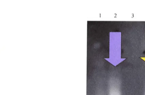
Figure 1.2 Gap junction structure between smooth muscle cells
High power electron micrograph from a 14 day rat iris
arteriole showing a typical gap junction between smooth muscle cells
and the appearance of connexins at the electron microscope level.
The characteristic pentalaminar structure can be seen clearly. The
lighter regions (arrows; 1-3) denote individual connexin channel
pores. The distance between the connexins is 9 nm. The scale bar
represents 5 nm.
(Electron micrograph courtesy of Dr. Shaun Sandow, Division of
Neuroscience, John Curtin School of Medical Research, Australian
Schematic diagram of the structure of gap junction channels between two cells
A and B, demonstrating the position of the connexons in the cell membranes and their
constituent connexins. Also shown are the lipid bilayers, the intercellular space and
some of the molecules which are known to pass through the central pore of each gap
junction.
~ "zSiill'I~ - Lipid
Bilayer
pers com). Connexins may also have overlapping or complementary expression patterns
(Delorme et al. 1997).
High resolution ultrastructural studies and sequencing data have shown that the
connexins comprise a family of homologous proteins, all of which have four a-helical
transmembrane domains that form the channel, two extracellular loops, a cytoplasmic
loop, and cytoplasmic NH2_ and COOH- termini (Figure 1.4) (Bennett et al. 1991;
Locke, 1998; Unger et al. 1999) which can influence or regulate the physical properties
of the channel (Makowski et al. 1977; Kumar and Gilula, 1992; Paul, 1995; Sosinsky,
1996; Yeager and Nicholson, 1996; Nicholson and Bruzzone, 1997; Yeager, 1998;
Pumick et al. 2000). The extracellular loops of all connexins contain highly conserved
cysteine residues, which are important in intercellular recognition and the docking of
two hemichannels ( connexons) (Yeager and Nicholson, 1996). Studies of the carboxy
terminal ( CT) tail have shown that it is an important site for channel gating in response
to intracellular signalling (Moreno et al. 1994; Ek-Vitorin et al. 1996; Castro et al.
1999) with each domain interacting with the other domains (Verselis et al. 1994). The
CT tail also has a role in regulating connexin-mediated growth (Omori and Yamasaki,
1999).
Sequence similarity amongst the connexins is concentrated in the transmembrane
domains and extracellular loops, whilst most of the sequence and length variation is in
the cytoplasmic loop and CT tail (Beyer et al. 1990; Haefliger et al. 1992; Donaldson et
al. 1997). Domain swapping experiments have determined that compatibility between
the connexins (defined as the ability of adjacent cells expressing different connexins to
communicate) is attributable to the second extracellular domain (White et al. 1994,
Two typical connexins, Cx43 and Cx32, are illustrated, showing the four
membrane spanning domains - two extracellular loops, a cytoplasmic loop, and
cytoplasmic amino (N) and carboxy (C) termini.
1.1.5.2 Gap junctions: homomeric, heteromeric, homotypic and heterotypic forms
As alluded to above, each connexon (hemichannel) may be composed of either
one type of connexin (homomeric) or multiple connexins (heteromeric). Theoretically,
the heteromeric connexon can result in a variety of connexin combinations (Brink,
1998; White and Paul, 1999) which would be physiologically advantageous, in terms of
diversity, for intercellular communication. Adjacent cells can contribute different types
of connexon to the intercellular channel giving rise to homotypic ( when the same
connexin is present in both connexons ), heterotypic ( when each connexon is composed
of a different connexin) or heteromeric (when different connexins mix in the
connexons) intercellular channels (Figure 1.5) (Bukauskas et al. 1995; White and
Bruzzone, 1996; Brink et al. 1997; Yeh et al. 1998; White and Paul, 1999).
Some combinations of connexins, however, will not interact and consequently,
are unable to form functional channels. Cx43 and Cx40, for example, are coexpressed
in vascular smooth muscle and together, they form homomeric / homotypic channels
with distinct permeability and gating properties but not functional homomeric /
heterotypic channels (Kanter et al. 1993; He et al. 1999; Li and Simard, 1999).
Conversely, He et al. (1999) demonstrated that in vascular smooth muscle cells in vitro,
Cx43 and Cx40 can form functional channels with unique gating and conductance
properties in the form of heteromeric channels, whilst Valiunas et al. (2000) have
demonstrated that these connexins can form functional heterotypic channels in human
HeLa cells.
Electrophysiological studies of heterotypic channels have shown that some
exhibit unique properties compared to homotypic channels (Barrio et al. 1991; Bruzzone
et al. 1994; White et al. 1994; Darrow et al. 1996; Brink et al. 1997). Studies of
homotypic Cx43, Cx40 and Cx37 gap junction channels (Brink et al. 1996; Veenstra,
1996) have shown that they all display symmetric voltage dependence but different
The arrangement of connexins into connexons, intercellular channels and gap
junctions is shown. Adjacent cells contribute different types of connexon to the
intercellular channel giving rise to heteromeric, homomeric, heterotypic and homotypic
channels.
a _·:_
?
.
connextns
cytoplasm
of cell one
heteromeric
heterotypic
homomeric
hom.otypic
connexons
intercellular channels
intercellular gap
JJ
gap junction
cytoplasm
though at different levels, to fluorescent dye molecules in the range 350 to 450 Da such
as Lucifer Yellow or carboxyfluorescein (Robinson et al. 1993; Moreno et al. 1994;
Little et al. 1995b; Brink, 1996, 1997; Darrow et al. 1996), with Cx37-Cx40 having
reduced permeability to 6-carboxyfluroscein compared to Cx43 (Veenstra et al. 1994).
1.1.5.3 Gap junction expression
The previous sections have alluded to the fact that different connexins and
differences in the characteristics of connexins can confer specific physiological
properties on gap junctions (Bruzzone et al. 1996; Beyer et al. 1990; Willecke et al.
1991; Goodenough et al. 1996). Since most, if not all cells and tissues express at least
two connexins (Nicholson et al. 1987; Kanter et al. 1992,1993; Darrow et al, 1995;
Stauffer, 1995; Van Rijen et al. 1997; Coppen et al. 1999; Lo, 1999; Mehta et al. 1999),
it follows that the properties of the gap junctions of any cell or tissue are complex.
Physiological analysis of channel formation and gating has revealed unique
patterns of connexin-connexin interaction, as well as unique functional characteristics of
channels containing more than one type of connexin protein (White et al. 1995; White
and Bruzzone, 1996). Each connexin has tissue-specific distributions, differential
temporal expression and specific channel permeability properties, and is subject to
unique types of biochemical regulation (Veenstra, 1992; Bruzzone et al. 1996; Kumar
and Gilula, 1996; Bevans et al. 1998; Goodenough et al. 1996). Data from animal
model studies and human disease states have shown that genetic defects in each
connexin produce distinct developmental and/or physiological defects
(Britz-Cunningham et al. 1995; Paul, 1995; Anzini et al. 1997; Donaldson et al. 1997; Ewart
et al. 1997; Huang et al. 1998; Chanson et al. 1999; Sullivan et al. 1999; ·White and
Paul, 1999; Willecke et al. 1999). Specific mutations in Cx32 result in X-linked
Charcot-Marie-Tooth disease, a demyelinating peripheral neuropathy (Bergoffen et al.
1999) whilst congenital hereditary bilateral cataracts have been associated with a
missense mutation in Cx50 (White and Paul, 1999; Xu and Ebihara, 1999).
Mutations in Cx43 (also referred to al connexin, and is the predominant gap
junction gene in developing and adult heart tissue) have been shown to be associated
with atrial defects ( eg. visceroatrial heterotaxia; Britz-Cunningham et al. 1995) and
other congenital cardiac abnormalities (Reaume et al. 1995). In the transgenic
( overexpressing Cx 4 3) and knockout Cx 4 3 mice, there are major right ventricular
cardiac defects (Reaume et al. 1995; Ewart et al. 1997; Martyn et al. 1997; Ya et al.
1998) leading to failure of pulmonary gas exchange and neonatal lethality at birth.
Consequently, Cx43 appears to play an important role in cardiac morphogenesis with
the precise level of Cx43-mediated gap junctional communication also being potentially
critically important (Reaume et al. 1995; Ewart et al. 1997).
Coexpression of multiple connexins enables cells to achieve levels of
intercellular communication that could not be achieved by expression of a single
connexin (Koval et al. 1995). Coexpression also enables functional compensation to
occur (Minkoff et al. 1999) as has been demonstrated in Cx43 knockout mice. In this
mouse, the absence of gross abnormalities, other than the ( critical) heart defect, has been
taken as evidence that the loss of Cx43 can be compensated for by other connexins
whose distributions overlap (De Sousa et al. 1997; Houghton et al. 1999). Furthermore,
in preimplantation embryos lacking Cx43, a low level of intercellular coupling is
maintained by gap junction channels having characteristics of Cx45 ( a6 connexin; De
Sousa et al. 1997). Cx32 has also been suggested to compensate for the absence of
Cx43 in some instances (Houghton et al. 1999). The ability of one type of connexin to
either induce or suppress the expression of other connexins during development (van
Kempen et al. 1995) has been demonstrated with respect to Cx45 in the Cx43 knockout
Depending on the surrounding microenvironment to which cells are exposed, the
extent of intercellular communication may also vary during development and aging
(Mombouli and Vanhoutte, 1999). Modification of the Cx43 gene in mice has
highlighted the importance of this connexin in heart morphogenesis (Reaume et al.
1995; Ewart et al. 1997; Huang et al. 1998; Sullivan et al. 1999). In the Cx43 knockout
mouse, abnormal cardiac tissue growth has been observed during development (Sullivan
et al. 1999). Responses to other stimuli (pharmacological, hormonal or oncogenic) may
also be distinct to a particular connexin or group of connexins (Mehta et al. 1999).
1.1.5.4 Regulation of gap junction function
Many aspects of the regulatory processes of gap junctions have been elucidated
as a result of physical, biochemical and cloning studies. Regulation of gap junction
function, and hence gap junction-mediated communication, can occur both acutely
(gating) and over a longer time period. In the latter, overall connexin expression levels
are altered, with or without switching to functionally different connexin isoforms
(Bennett et al. 1991; Beyer, 1991).
Dye coupling experiments and dual whole-cell voltage-clamp experiments
(Flagg-Newton et al. 1979; Schwarzmann et al. 1981; Little et al. 1995b; White et al.
1995; Darrow et al. 1996; Kwak and Jongsma, 1996; Bevans et al. 1998; Ebihara et al.
1999; Manthey et al. l 999; Srinivas et al. 1999) have been used extensively to
determine the functional state of gap junctions. Dye coupling studies have provided
information about the extent of diffusional coupling and permeability of the channels,
whilst dual whole-cell voltage-clamp experiments have allowed the gap junctional
conductance (gj), which depends on the number of channels present, the conductance of
each channel (yj) and their probability of being ' open' , to be determined (Kwak and
Jongsma, 1996). As the majority of these studies have been done in the adult animal,
Gap junctions can 'open' and 'close' ( or be in an 'intermediate' state) rapidly in
response to various stimuli (Moreno et al. 1994; Morley et al. 1997); an ability which
may enable cells to adapt quickly to different physiological conditions. In human
corporal cells, for example, the mean open time for Cx 4 3 channels ranged from O. 43 to
5.25 seconds while the mean closed time was in the range of 0.21 to 1.49 seconds
(Brink et al. 1997). At any one time, the proportion of channels open depends on the
characteristics of the constituent connexin/s in conjunction with such variables as the
calcium concentration, transjunctional voltage, phosphorylation state, cytoplasmic pH
and levels of cAMP (Laing and Beyer, 1995). These are briefly discussed below.
In prolonged transjunctional voltage, where the voltage differs between two
cellular interiors (Revilla et al. 1999), vascular smooth muscle cell Cx43 gap junction
channels are prone to prolonged closures (Brink et al. 1996). Phosphorylation (Kwak et
al. 1995a,b; Kwak and Jongsma, 1996) and changes in intracellular pH (Bennett et al.
1991; Morley et al. 1997; Francis et al. 1999) are known to be important regulators of
gap junctional communication. Phosphorylation has been implicated in connexin
trafficking, assembly, insertion into membranes, degradation and retrieval from the
plasma membrane (Laird, 1996; White and Bruzzone, 1996; Stahl and Sies, 1998). It
also influences the gating of gap junction channels, as do changes in intracellular pH
and the concentration of calcium (Obaid et al. 1983). The effect phosphorylation exerts
on junctional coupling is dependent on two factors: the type of connexin and the type of
kinase involved (Kwak et al. 1995a,b; Kwak and Jongsma, 1996). Phosphorylation is
associated with both increases and decreases in junctional coupling (Musil and
Goodenough, 1991; Kwak and Jongsma, 1996; Hertlein et al. 1998), probably in
consequence to conformational changes in the connexin/s that make up the channel.
Changes in the phosphorylation state of Cx43, for example, appear to correlate with
changes in the functional state of the gap junctions formed by this protein (Saez et al.
(1997) showed reduced functional coupling of neonatal rat cardiac myocytes after
treatment with staurosporine, a protein kinase inhibitor. In contrast, Crow et al. (1990)
demonstrated that in transformed mammalian fibroblasts, phosphorylation of Cx43 on
tyrosine in the presence of v-src is correlated with inhibited communication.
With respect to the regulation of gap junctions and changes in pH, Morley et al.
(1997) showed that the closure of Cx43 channels is significantly impaired by the
truncation of the CT domain at amino acid 257, with a loss of pH sensitivity. This
implies that the pH gating of Cx43 results from an interaction between the CT domain
and the rest of the channel. Trexler et al. (1999) demonstrated that the CT domain of
Cx43 enhanced the pH sensitivity of truncated Cx40, and that the CT domain of Cx40
restored the pH sensitivity of truncated Cx43. Similar results have also been obtained
by Gu et al. (2000). Thus, the regulatory domain of one connexin can specifically
interact with a channel formed by another connexin, which may be an important factor
in the regulation of heteromeric channels (Section 1.1.5.2).
Functional differences between the various connexins are likely to be
physiologically relevant (Peters, 1997) with respect to the selective permeabilities to
small signalling and regulatory molecules, such as cAMP and cGMP (Spray and
Bennett, 1985; Spray and Burt, 1990; Moreno et al. 1994; Veenstra et al. 1995;
Nicholson et al. 2000). cAMP and cGMP play important roles in the mediation of the
contractile process and can alter the functional status of gap junctions (Burt and Spray,
1988; Burt, 1990; Bevans and Harris, 1999). Peters (1997) and Brink (1998) have
suggested that cAMP and cGMP are of a size, charge and conformation that may cause
them to diffuse selectively and thus exert their effects differentially between cells with
different connexin expression. Other studies however, have shown that as a whole, gap
junction channels appear to be poorly selective (Tsien and Weingart, 1976; Beblo and
Veenstra, 1997; Wang and Veenstra, 1997) allowing the diffusion of small solutes,
Other regulatory factors influencing gap junction function include the number,
size and spatial distribution of gap junctions; these being important determinants of
passive electrophysiological properties (Beblo et al. 1995). In cultured smooth muscle
cells, gap junctions are numerous between cells with a synthetic phenotype but few are
observed between contractile cells (Rennick et al. 1993). This may be important for
vessel development, following injury, or in various vascular disease states (Christ et al.
1996). Increased expression of Cx43 has been observed in some forms of hypertension
(Bastide et al. 1993; Gabriels et al. 1998) and atherosclerosis (Blackbum et al. 1995).
Recent advances in the cloning of connexins have made it possible to manipulate
the regulation of cellular growth and neoplastic transformation associated with
intercellular communication (Ruch et al. 1998). It has been proposed that the loss of
gap junctional communication leads to uncontrolled cell growth (Pitts et al. 1988;
Loewenstein and Rose, 1992). Support for this contention comes from a number of
studies (Mehta et al. 1991; Zhu et al. 1991; Rose et al. 1993; Hirschi et al. 1996;
Temme et al. 1997; Naus, 1998; Rae et al. 1998; Mehta et al. 1999). In general, an
inverse relationship exists between cell growth and connexin expression and
intercellular coupling (Bennett et al. 1991; Beyer, 1991; Loewenstein and Rose, 1992).
In Cx32 (~1) knockout mice, susceptibility to spontaneous and chemically induced
hepatocarcinogenesis is increased (Temme et al. 1997). In mouse and human lung
carcinoma cell lines, Ruch et al. (1998) have shown that expression of Cx43 is reduced
compared to control. However, conflicting evidence is provided by Sia et al. (1999) in
their study of gap junctional communication in quiescent mammary epithelial cells,
where Cx43 expression was reduced but a high level of intercellular communication was
maintained.
A number of other factors have been shown to influence gap junctional activity.
anaesthetics (halothane, isoflurane ), calmodulin binding, cadherins (the
calcium-dependent cell-cell adhesion molecules), neurotransmitters (dopamine, acetylcholine
and y-aminobutyric acid), and selective pharmacological agents (heptanol, Gap 27
mimetic peptide, 18a- and 18~-glycyrrhetinic acid (GA) and tetraethylammonium)
(Davidson et al. 1986; Peracchia, 1991; Schiller et al. 1992; Hertig et al. 1996; Proulx
et al. 1997; Wang and Rose, 1997; Chaytor et al. 1998; Yamamoto et al. 1999; He and
Burt, 2000).
1.1.6 Gap junctions and arterial function
Gap junctions enable coupling between smooth muscle cells, between
endothelial cells and in some cases, between adjacent smooth muscle and endothelial
cells (Rhodin, 1967; Somlyo and Somlyo, 1968; Gabella, 1981; Duling et al. 1991;
Luff, 1991; Beny, 1999). Dye tracer studies performed in isolated arterioles indicate
that in some vessels, gap junctional coupling between endothelial cells is more
extensive than that between the underlying medial smooth muscle cells (Little et al.
1995b ). Connections between endothelial cells and the innermost smooth muscle cells
have been suggested to occur through discontinuities in the internal elastic lamina of
smaller coronary arteries and arterioles, with heteromeric gap junctions (Section 1.1.5 .2)
sometimes being present at these sites (Severs, 1999). In such cases, communication
occurs mainly in a unidirectional fashion from the endothelium to the smooth muscle
layer (Little et al. 1995b; Beny, 1999), although bi-directional coupling is possible (Hill
et al. 2001 ).
Whilst the full functional gamut of gap junctional coupling remains to be
elucidated, it has been established that this coupling is important for the regulation and
co-ordination of blood flow both within and into tissue beds. The localized application
of various vasoconstrictor and vasodilator substances ( acetylcholine and norepinephrine,
generalized response (vasoconstriction or vasodilation) which is propagated proximally
and distally along the vessel (Duling and Berne, 1970; Segal and Duling, 1986; Segal et
al. 1989; Delashaw and Duling, 1991; Gustafsson and Holstein-Rathlou, 1999). This
response is referred to as the conducted vasomotor response. However, not all
substances that are capable of inducing local vasoconstriction or vasodilation initiate a
propagated vascular response - sodium nitroprusside, for example (Segal et al. 1989).
The ability of local vasomotor responses ( constriction or dilation) to be
conducted relates to the stimulus (Delashaw and Duling, 1991 ), whilst the actual
conduction of the response has been shown to be due to electrotonic transmission of the
signal along the vascular wall through gap junctions between adjacent cells in some
vessels (Segal and Duling, 1989; Xia and Duling, 1995; de Wit et al. 2000; Emerson
and Segal, 2000). Basically, vasomotor responses may originate in either the
endothelial or smooth muscle cell layer, with the expression of the final mechanical
response depending on the specific signal transduction pathway involved (Hill et al.
2001).
The connexin knockout mice models have demonstrated the important role
played by gap junctions in the vasomotor response, particularly in cardiac conduction
and morphogenesis. Cx40-deficient mice have impaired conduction of
endothelium-dependent vasodilator responses along intact skeletal muscle arterioles ( de Wit et al.
2000), and cardiac conduction abnormalities (Kirchhoff et al. 1998; Simon et al. 1998).
Cx40 has been proposed to be important in the heteromeric coupling of the endothelium
and vascular smooth muscle (de Wit et al. 2000). Cardiac conduction abnormalities
have also been observed in the Cx40 and Cx43 double-deficient knockout mice
(Kirchhoff et al. 2000). In Cx45-deficient mice, vascular development is defective
(Kruger et al. 2000; Kumai et al. 2000), whilst in the Cx37 knockout mice, females are
results support the contention that connexins play a vital role in intercellular
communication in the vasculature, and hence the conducted vasomotor response.
Antagonism of gap junction activity by pharmacological agents provides a useful
tool for examining gap junction function although, to date, no agent has been shown to
be universally effective. The specificity of action of these agents has been questioned
and is indeed variable at best. The long chain alcohols, heptanol and octanol, for
example, have been shown to block gap junction activity in a variety of tissues and cell
cultures (Burt and Spray, 1989; Takens-Kwak et al. 1992; Bastiaanse et al. 1993; Christ
et al. 1993b; Deutsch et al. 1995; Zhang et al. 1996; Largo et al. 1997; Proulx et al.
1997). Christ et al. (1993b, 1999), for example, have shown that alpha-adrenoceptor
agonist stimulated contraction is inhibited by heptanol in rat aortic smooth muscle cells,
whilst in the rabbit superior mesenteric artery, heptanol abolishes rhythmic contractile
activity and inhibits contractions evoked by phenylephrine (Chaytor et al. 1997). In
myoblasts, 1-octanol and P-glycyrrhetinic acid inhibit gap junctional intercellular
communication (Proulx et al. 1997). Inhibition of gap junction activities by
P-glycyrrhetinic acid has also been observed in rat hepatocytes (Eugenin et al. 1998) and
guinea-pig mesenteric arterioles (Yamamoto et al. 1999). In rabbit jugular veins,
18a-glycyrrhetinic acid blocks heterocellular endothelium-vascular smooth muscle coupling
(Griffith and Taylor, 1999) and in immature rat irideal arterioles, it blocks spontaneous
depolarisations and contractions (Hill et al. 1999).
Another putative gap junction antagonist, Gap 27, a synthetic peptide possessing
sequence homology with the extracellular loop 2 of Cx43, has been shown to block the
acetylcholine induced endothelium-dependent relaxation in rabbit mesenteric arteries
(Chaytor et al. 1998; Dora et al. 1999; Hutcheson et al. 1999) and jugular veins (Griffith
and Taylor, 1999). Other synthetic peptides consisting of regions of the connexin
smooth muscle cells in mesenteric arteries (Chaytor et al. 1997). Calero et al. (1998),
for example, showed that a synthetic l 7mer peptide formed by amino acids 271 to 287
of Cx43 interfered, in a sequence specific manner, with the acidification-induced
uncoupling of Cx43. Similarly, dye coupling and dual whole-cell voltage clamp have
been used to demonstrate, via the synthetic oligopeptide P 180-195 which corresponds to
a segment of the second extracellular loop of Cx43 , selective inhibition of Cx43 gap
junction channels (Kwak and Jongsma, 1999). The selective block of gap junction
channel expression with connexin-specific antisense oligodeoxynucleotides has also
been documented (Moore and Burt, 1994; Minkoff et al. 1999).
A variety of pharmacological agents ( agonists) have been used to study the
activation or promotion of gap junction activity. Two examples are the potassium
channel inhibitors tetraethylammonium (TEA) and 4-aminopyridine ( 4-AP) (Jones et al.
1978; Kannan and Daniel, 1978; Peracchia, 1991). These agents are nonselective
inhibitors of K+ channels. In vascular smooth muscle, it has been suggested that
tetraethylammonium induces rhythmic contractions by promoting gap junction activity
(Takens-Kwak et al. 1992; Christ et al. 1996).
Thus, both in vivo and in vitro , diverse mechanisms of gap junction regulation
exist that can vary between vascular beds and within vascular beds and for different
connexins, that are susceptible to manipulation via a number of mechanisms.
1.2 Vascular Control Mechanisms
The control of blood flow in the vasculature is mediated by a variety of
mechanisms, which can result in either arterial constriction or dilation. Such
mechanisms include neural, humoral, myogenic, metabolic, endothelial-derived and
Differences in the activity of one or more of these mechanisms can result in
specific control of different regions of the vasculature (Bylund, 1995), which can result
in both systemic and localized control of blood flow and vascular resistance (tone)
(Bevan and Purdy, 1973; Johnson, 1978; Bumstock and Ralevic, 1994). Control of
vascular tone is important in the small arteries and arterioles, as they are the major
resistance vessels, containing more than 50% of the total systemic vascular resistance
(Berne and Levy, 1989; Sherwood, 1993). The integration of the vascular response
occurs through electrical and mechanical coupling of vascular smooth muscle cells
(Hirst and Edwards, 1989); factors that are moderated by the autonomic nervous system
(Janssen and Smits, 2002).
Neurotransmitters released from the autonomic nervous system act on specific
receptors located on the smooth muscle cells and/or endothelial cells to modify vascular
tone (Todd, 1980; Bevan, 1988). Contraction or relaxation of many blood vessels
occurs in response to either an increase or decrease in intracellular calcium (Walsh et al.
1995), initiated via various pathways and signalling mechanisms. However,
nerve-mediated vascular responses vary between vascular beds and between different regions
along the one blood vessel (Bylund, 1995), as well as within the same vascular bed
during development (Sandow and Hill, 1999). Not only can signalling pathways vary,
but the type of neurotransmitter released, their associated pre- and post-synaptic
receptors and pathways associated with such receptors, and mechanisms of intracellular
calcium, may also vary.
Thus, in order to understand the implications of variable gap junctional activity
in the vasculature, an understanding and recognition of other factors associated with
vascular control is required. One such entity that is relevant to this thesis, in terms of its
influence on the vascular smooth muscle cells, is the neurovascular control provided by
1.2.1 Endothelium derived factors
The endothelium plays an important role in maintaining vascular resistance and
blood flow in many vascular beds (Griffith and Henderson, 1989; Mombouli and
Vanhoutte, 1999), by releasing a variety of vasodilator mediators, including nitric oxide,
endothelium-derived hyperpolarizing factor (EDHF) and prostaglandins (Moncada et al.
1991; Fleming and Busse, 1999; Shimokawa, 1999). In some cases, this function may
be hampered by the release of vasoconstrictors. Nitric oxide and EDHF activity
(release) have also been linked to the activity of various neurotransmitters (Griffith et al.
1988; Toda and Okamura, 1998).
Nitric oxide is synthesized from L-arginine via the enzyme nitric oxide synthase
(Palmer and Moncada, 1989; Vanhoutte and Feletou, 1998). In smooth muscle cells,
nitric oxide mediates vasodilation via the activation of guanylate cyclase and cyclic 3',
5'-guanosine monophosphate ( cGMP) signaling pathways (McDonald and Murad,
1995). In endothelial cells, activation of nitric oxide synthase, which is calcium /
calmodulin dependent (Bredt and Snyder, 1992), may occur via receptor activation, by
shear stress and/or aggregating platelets (Fleming and Busse, 1995; Amal et al. 1999;
Takamura et al. 1999). Nitric oxide synthase has been shown to co-localize with
acetylcholine and vasoactive intestinal peptide (VIP) in parasympathetic nerves (Lumme
et al. 1996; Yoshida and Toda, 1997), and to be present in sensory nerves (Lumme et al.
1996).
The labile substance EDHF (Feletou and Vanhoutte, 1999) induces
hyperpolarization of smooth muscle cells due to an increase in potassium conductance
(Komori and Suzuki, 1987), which in turn leads to reduced intracellular calcium and
relaxation (Walsh et al. 1995). The exact nature and mechanisms of action of EDHF are
unknown to date (Mombouli and Vanhoutte, 1997, 1999; Feletou and Vanhoutte, 1999).
dilatory response is often tissue specific. In most vascular diseases, the vasodilator
function of the endothelium is attenuated (Mombouli and Vanhoutte, 1999).
1.2.2 Endothelial influences in vascular disease
Furchgott and Zawadzki (1980) first demonstrated the role of the endothelium in
the acetylcholine induced relaxation of isolated rabbit aorta where a factor, subsequently
shown to be nitric oxide, caused relaxation of vascular smooth muscle (Ignarro et al.
1987; Palmer et al. 1987; Moncada and Palmer, 1991 ; Feletou and Vanhoutte, 1999).
Subsequently, it has been shown that the endothelium can control vascular
smooth muscle tone primarily by the release of mediators of vasodilation (prostacyclin
and EDHF, for example) in response to factors such as increases in the level of shear
stress, neurotransmitters ( such as acetylcholine and noradrenaline), endothelial produced
autacoids, platelet products and coagulation derivatives (including serotonin and
thrombin) and hormones (angiotensin II) (Vanhoutte and Miller, 1989; Vanhoutte and
Boulanger, 1995; Shimokawa, 1998; Boulanger, 1999; Burnstock, 1999; Feletou and
Vanhoutte, 1999; Fleming and Busse, 1999; Mombouli and Vanhoutte, 1999; Takamura
et al. 1999).
In a number of blood vessels, selective a2-adrenergic agonists can cause
endothelium-dependent relaxations and stimulate post-junctional a2-adrenoceptors on
vascular smooth muscle cells to result in a contraction (Vanhoutte and Miller, 1989;
Aburto et al. 1993). Although these receptors are more abundant in hypertensive blood
vessels, the responses they evoke are very sensitive to functional agonists such as nitric
oxide (Vanhoutte and Miller, 1989).
A summary of the effects of the main endothelium-derived vasodilators (nitric
oxide, derived hyperpolarizing factor and prostacyclin) and
endothelium-derived contracting factors (vasoconstrictor prostaglandins, endothelin and reactive
Summary of the effects of endothelial-derived vasoconstrictors and vasodilators
on the contraction and proliferation of the underlying vascular smooth muscle cells.
The figure also demonstrates coupling between adjacent endothelial cells (1), between
adjacent smooth muscle cells (2) and between endothelial and smooth muscle cells (3).
Shear stress
BK Ach
l l
Cytokines Thrombin
Hypoxia SHT
r'
l
Achi
Al:23187
:J
Endothelin · 02· TxA2 PGH2 .
Under normal conditions, an accurate and balanced release of relaxing and
contracting factors contribute to vascular perfusion (Tortora and Grabowski, 1993). In
disease states however, such as hypertension, atherosclerosis, coronary artery disease,
diabetes and heart failure, this balance is shifted (Sherwood, 1993). Arteries obtained
from hypertensive animal models have demonstrated, for example, that
endothelium-dependent vasodilation is reduced (Vanhoutte and Miller, 1989; Vanhoutte and
Boulanger, 1995; Shimokawa, 1998), with the mechanisms underlying this dysfunction
being variable between different types of hypertension.
Endothelial dysfunction in most arterial diseases is characterized by either
decreased secretion of vasodilator mediators, increased production of
derived vasoconstrictors, and/or resistance of vascular smooth muscle to
endothelium-derived vasodilators (Vanhoutte and Boulanger, 1995; Shimokawa, 1998; Mombouli
and Vanhoutte, 1999). Endothelial dysfunction differentially affects shear stress- or
hormone-induced endothelium-dependent vasodilation in vascular beds for two reasons:
firstly, the distribution of endothelial receptors for acetylcholine, ADP, UTP,
bradykinin, serotonin, vasopressin and catecholamines ( stimulators of endothelial
vasodilators) may vary, as well as the signal transduction pathway/s to which they are
coupled, within an artery or be modified by the physiological conditions present in the
artery (Furchgott, 1983; Vanhoutte and Miller, 1989; London and Safar, 1996;
Mombouli and Vanhoutte, 1999).
1.3 Vascular Disease and Gap Junctions
1.3 .1 Gap junctions in injury and vascular disease
Remodelling of vascular structures in response to haemodynamic signals
associated with changes in blood flow readily occurs as a result of changes in a variety