Data Article
Histological data of axons, astrocytes, and myelin
in deep subcortical white matter populations
Santiago Coelho
a,b,*, Jose M. Pozo
a,b, Marina Costantini
b,
J. Robin Highley
c, Meghdoot Mozumder
b, Julie E. Simpson
c,
Paul G. Ince
c, Alejandro F. Frangi
a,baCentre for Computational Imaging&Simulation Technologies in Biomedicine (CISTIB) and Leeds Institute
for Cardiac and Metabolic Medicine (LICAMM), School of Computing&School of Medicine, University of Leeds, Leeds, UK
bCISTIB, Electronic and Electrical Engineering Department, The University of Sheffield, Sheffield, UK cSheffield Institute for Translational Neuroscience (SITraN), The University of Sheffield, Sheffield, UK
a r t i c l e i n f o
Article history:
Received 29 November 2018
Received in revised form 7 February 2019 Accepted 8 February 2019
Available online 6 March 2019
a b s t r a c t
This immunohistochemistry dataset contains the main structures in deep subcortical white matter (axons, astrocytes, and myelin-ated axons) in a representative cohort of an ageing population. A set of samples from 90 subjects of the Cognitive Function and Ageing Study (CFAS) were analysed, stratified into three groups of 30 subjects each, in relation to the presence of age-associated deep subcortical lesions. High-resolution microscopy data enables the extraction of valuable information, such as volume fractions, for the construction and validation of diffusion MRI (dMRI) models. The dataset provided here was used in Coelho et al. [1].
©2019 The Author(s). Published by Elsevier Inc. This is an open access article under the CC BY license (http://creativecommons. org/licenses/by/4.0/).
*Corresponding author. Centre for Computational Imaging&Simulation Technologies in Biomedicine (CISTIB) and Leeds Institute for Cardiac and Metabolic Medicine (LICAMM), School of Computing&School of Medicine, University of Leeds, Leeds, UK.
E-mail address:S.Coelho@leeds.ac.uk(S. Coelho).
Contents lists available atScienceDirect
Data in brief
j o u r n a l h o m e p a g e : w w w . e l s e v i e r . c o m / l o c a t e / d i b
https://doi.org/10.1016/j.dib.2019.103762
2352-3409/©2019 The Author(s). Published by Elsevier Inc. This is an open access article under the CC BY license (http://
1. Data
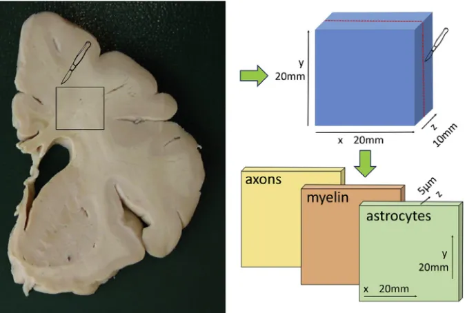
The dataset contains histopathology images of brain deep white matter samples from 90 subjects of the Cognitive Function and Ageing Study (CFAS) cohort. For each sample digital images of axons,
as-trocytes and myelin markers at 40X magnification are provided (see Fig. 1). The resolution of the
images is 0.23
m
m/pix. These are available in NanoZoomer Digital Pathology Image (ndpi) format andwere allocated in three groups: Control (no DSCLs were present in the subject), Lesion (the sample presented DSCLs), and Normal Appearing White Matter (NAWM, the subject presented DSCLs but not in the sampled tissue).
2. Experimental design, materials and methods
Age-associated cerebral white matter lesions can be classified into those within deep white matter
of thecentrum semiovale(deep subcortical lesion, DSCL) and those close to the angles of the lateral
ventricles (periventricular lesion, PVL). These are thought to be originated from small vessel-related vascular pathology such as vascular dementia. This work focus on DSCLs, which are associated with
loss of myelin components[2]and astrogliosis[3,4]. To this purpose, several subjects belonging to
groups representing healthy and diseased conditions were imaged. Immunohistochemically stained sections of three populations of deep white matter samples were analysed: Control, Lesion, and NAWM.
Samples used in this work came from the Cognitive Function and Ageing Study (CFAS)
neuropa-thology cohort[5,6]. Brains were removed with the consent of the next of kin and with multicentre
research ethics committee approval, according to standard CFAS protocols[7]. Brains were removed
within 60 hours of death, one cerebral hemisphere wasfixed in buffered formaldehyde and sliced into
10 mm thick coronal slices. These slices were: 1) immediately anterior to the temporal stem (anterior), 2) at the level of the pulvinar (middle), and 3) at the posterior most limit of the occipital horn of the
lateral ventricle (posterior). Slices were scanned using T1and T2weighted MRI (details available in[7]).
The MR images were rated by three experienced observers (blind to clinical status) and given a score Specifications table
Subject area NeuroImaging
More specific subject area Computational modelling in neuroimaging
Type of data Images in NanoZoomer Digital Pathology Image [.ndpi] format of histologically stained brain sections. Excel sheet with metadata.
How data was acquired Digital whole slide scanner, Nanozoomer XR (Hamamatsu, Photonics Ltd., Hertfordshire, UK).
Data format Raw colour images
Experimental factors Deep White Matter samples from 90 human subjects divided into Control (N¼30), Normal Appearing White Matter (N¼30), and Lesion (N¼30).
Experimental features High resolution microscopy data was acquired for axons, astrocytes, and myelin from three populations of Deep White Matter samples.
Data source location Sheffield, United Kingdom
Data accessibility Full dataset is available upon registration in MULTI-X repository:https://multi-x.org/(`Data’–>
`OCEAN histology’folder)
Related research article Coelho, S., Pozo, J. M., Costantini, M., Highley, J. R., Mozumder, M., Simpson, J. E., Ince, P. G., Frangi, A. F., 2018. Local volume fraction distributions of axons, astrocytes, and myelin in deep subcortical white matter, NeuroImage 179, 275e287[1].
Value of the data
Elderly populations with various degrees of Deep Subcortical Lesions (DSCL) were scanned.
Population representative histology data let us extract specific information useful to develop and validate sensitive and specific dMRI models.
for DSCLs using a modified Scheltens' scale[8]. Following this scoring, the coronal slices were stored in formalin until required for this study (at least four weeks). One block of approximately
20 mm20 mm x 10 mm was sampled from one of the slices of every subject. Blocks were allocated in
three groups: Control, NAWM, and Lesion. Control blocks were taken from cases where all three levels were scored as 0 on this scale or where only one slice had a score of a maximum of 1. Lesion blocks were taken from regions with a Scheltens' score of 4 or greater. NAWM blocks were taken from lesion free regions of deep white matter in which a DSCL of score 3 or greater was present elsewhere.
The formalin-fixed blocks of tissue were processed to paraffin and embedded in paraffin wax using
conventional protocols[7]. Three sections of 5
m
m thickness were cut from each block forimmuno-histochemistry (in-plane dimensions of the samples were around 20 mm20 mm, seeFig. 1). Sections
were collected onto charged slides and underwent Ag retrieval with Access Revelation RTU (A. Menarini Diagnostics Ltd, Winnersh, UK) in a pressure cooker. Sections were immunostained for
phosphorylated neurofilament (SMI31, an axonal marker), glial fibrillary acidic protein (GFAP, an
astrocyte marker), and proteolipid protein (PLP, a myelin marker) using an intelliPATH FLX system (A. Menarini Diagnostics Ltd, Winnersh, UK). These immunostaining markers were chosen due to being
the best option for analysingex vivosamples of these structures[9e11]. Immunohistochemistry was
performed using a standard ABC method, visualised with diaminobenzidine tetrachloride (DAB), and the sections counterstained with haematoxylin. Prepared sections were scanned and digitised at 40X
magnification using a Nanozoomer XR (Hamamatsu, Photonics Ltd., Hertfordshire, UK).
3. Dataset structure
Three main folders separate all the samples according to different degrees of DSCLs (Control, NAWM, and Lesion groups). Inside each of these, there is a subfolder for each subject, named by the
subject id. Inside each subject-specific folder there are three files with the histological images for
[image:3.468.71.408.64.291.2]https://www.hamamatsu.com/eu/en/product/type/U12388-01/index.html. The metadata with the age and
sex of the subjects and position of the samples is provided as a table in an Excelfile named‘
Meta-data_sorted’located in the main folder of the dataset.
4. Dataset license
This dataset is distributed with the Creative Commons License CC BY-NC-ND 4.0 (Creative Commons Attribution-NonCommercial-NoDerivatives 4.0). Free download and any non-commercial applications
are permitted provided that the source is given full credit. Redistribution of any modification or
de-rivative of the dataset is prohibited. Please cite present article if you use the dataset on your research.
Acknowledgements
This work has been supported by the OCEAN project (EP/M006328/1) and MedIAN Network (EP/ N026993/1) both funded by the Engineering and Physical Sciences Research Council (EPSRC) and the European Commission FP7 Project VPH-DARE@IT (FP7-ICT-2011-9-601055). The CFAS study is sup-ported by the Department of Health and the Medical Research Council (grants MRC/G9901400 and MRC U.1052.00.0013); the UKNIHR Biomedical Research Centre for Ageing and Age-related Disease Award to the Newcastle upon Tyne Hospitals Foundation Trust; the Cambridge Brain Bank is supported by the NIHR Cambridge Biomedical Research Centre; The Cambridgeshire and Peterborough NIHR
CLAHRC; Nottingham University Hospitals NHS Trust; University of Sheffield and the Sheffield Teaching
Hospitals NHS Foundation Trust; The Thomas Willis Oxford Brain Collection, supported by the Oxford Biomedical Research Centre; The Walton Centre NHS Foundation Trust, Liverpool. The authors would
like to acknowledge the essential contribution of the liaison officers, the general practitioners, their
staff, and nursing and residential home staff. They are also grateful to CFAS respondents and their families for their generous gift to medical research, which has made this study possible. The Nano-zoomer XR scanner was funded by the bet365/Denise Coates Foundation. The authors thank Dan Fil-lingham, Lynne Baxter and Rachel Waller from SITraN, and Leandro Beltrachini and Manu Raghavan-Sareena from CISTIB for their help in the acquisition and processing of the digitised histology im-ages and in early discussions of this work.
Transparency document
Transparency document associated with this article can be found in the online version athttps://
doi.org/10.1016/j.dib.2019.103762.
References
[1] S. Coelho, J.M. Pozo, M. Costantini, J.R. Highley, M. Mozumder, J.E. Simpson, P.G. Ince, A.F. Frangi, Local volume fraction
distributions of axons, astrocytes, and myelin in deep subcortical white matter, Neuroimage. 179 (2018) 275e287.
[2] S.B. Wharton, J.E. Simpson, C. Brayne, P.G. Ince, Age associated white matter lesions: the MRC cognitive function and
ageing study, Brain Pathol. 25 (1) (2015) 35e43.
[3] J.E. Simpson, M.S. Fernando, L. Clark, P.G. Ince, G.F.M. Forster, R.J.T.O. Barber, R.N. Kalaria, C. Brayne, P.J. Shaw, C.E. Lewis, S.
B. Wharton, M.C. Function, A.N.S. Group, White matter lesions in an unselected cohort of the elderly: astrocytic, microglial
and oligodendrocyte precursor cell responses, Neuropathol. Appl. Neurobiol. 33 (4) (2007) 410e419.
[4] J.E. Simpson, S.B. Wharton, J. Cooper, C. Gelsthorpe, L. Baxter, G. Forster, P.J. Shaw, G. Savva, F.E. Matthews, C. Brayne, P.G.
Ince, Alterations of the blood brain barrier in cerebral white matter lesions in the ageing brain, Neurosci. Lett. 486 (3)
(2010) 246e251.
[5] C. Brayne, C. McCracken, F.E. Matthews, Cohort profile: the medical research council cognitive function and ageing study
(CFAS), Int. J. Epidemiol. 35 (2006) 1140e1145.
[6] Cognitive Function and Ageing Studies (CFAS) Collaboration, Cognitive Function&Ageing Study, 2017.http://www.cfas.ac. uk/(Accessed 10 October 2017).
[7] M.S. Fernando, J.T. O'Brien, R.H. Perry, P. English, G. Forster, W. McMeekin, J.Y. Slade, A. Golkhar, F.E. Matthews, R. Barber, R.
N. Kalaria, Ince, P.G., of MRC CFAS, N.G., Comparison of the pathology of cerebral white matter with postmortem magnetic
resonance imaging (MRI) in the elderly brain, Neuropathol. Appl. Neurobiol. 30 (4) (2004) 385e395.
[8] P. Scheltens, F. Barkhof, D. Leys, J.P. Pruvo, J.J. Nauta, P. Vermersch, M. Steinling, J. Valk, A semiquantative rating scale for the
[9] R. Barker, D. Wellington, M.M. Esiri, S. Love, Assessing white matter ischemic damage in dementia patients by
mea-surement of myelin proteins, J. Cereb. Blood Flow Metab. 33 (7) (2013) 1050e1057.
[10] C. Garwood, L. Ratcliffe, J. Simpson, P. Heath, P. Ince, S. Wharton, Review: astrocytes in Alzheimer's disease and other
age-associated dementias: a supporting player with a central role, Neuropathol. Appl. Neurobiol. 43 (4) (2016) 281e298.
[11] L.A. Sternberger, N.H. Sternberger, Monoclonal antibodies distinguish phosphorylated and nonphosphorylated forms of