University of Tasmania
Barriers of the Developing Brain:
In Vivo
and
In Vitro
Studies
by
Graham
KnottSubmitted in accordance with the requirements of the
University of Tasmania
for the degree of Doctor of Philosophy
Department of Physiology Medical Sciences University of Tasmania
Declaration:
This thesis contains no material used for the award of any other degree or diploma in any university and, to the best of my knowledge contains no material previously published by another person, unless due reference is made.
Graham Knott
Authority of Access:
This thesis may be made available for loan and limited copying in accordance with the Copyright Act 1968.
February 1995
Acknowledgements
The work I have written about in this thesis first began in the United Kingdom. I am deeply grateful to my supervisors Professor Norman Saunders and Doctor Kate Dziegielewska, firstly for their excellent supervision, and secondly for giving me the opportunity to continue these studies with them in Tasmania. Their endless patience and encouragement has made this work not only possible but extremely enjoyable.
I owe special thanks to Professor John Nicholls FRS, Doctor Mark Habgood and Ms. Yael Balsev with whom I have also been fortunate enough to work and learn from during the course of my studies.
UNIVERSITY OF TASMANIA FACULTY OF MEDICAL SCIENCES
PHYSIOLOGY
BARRIERS IN THE DEVELOPING BRAIN; IN VIVO AND IN VITRO STUDIES by
Graham Knott ABSTRACT
The work presented in this thesis addresses certain aspects of brain development using two models; one in vivo and the other in vitro. The main theme of the work presented centres around the importance of plasma proteins contained in the cerebrospinal fluid and found in high concentration early in development, and the barriers which separate this fluid from the blood and the brain.
The in vivo model was based on the marsupial species Monodelphis domestica, or grey short tailed opossum, which was used to study the transfer mechanism for the plasma protein albumin, transported from the blood to the CSF during the very early period of brain development at the time when the cortical plate of the neocortex first starts to appear. Previous studies have not examined this transfer at such an early stage of brain development. This thesis shows that during the earliest ages studied the movement of albumin from the blood into the CSF appeared to be via a selective transport mechanism which was able to distinguish between different species of albumin. The barrier between the CSF and the brain was shown to be well developed.
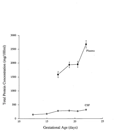
Measurements of total protein concentration in fetal rat lateral ventricular CSF showed that the peak in concentration occurred at E15, coincident with the appearance of the cortical plate, as has been described in other species.
The in vitro model used fetal rats (gestational day 15). The entire central nervous system was isolated and maintained in culture for up to 40 hours. This second model showed that the commonly used culture medium supplement, fetal calf serum (FCS), causes rapid growth and proliferation of cells of the neocortex. Cell division occurred at the ventricular zone as in vivo. Evidence provided by the light and electron microscope showed that the barrier between the CSF and the brain was maintained throughout the culture period. Immunocytochemical staining demonstrated that the fetal protein fetuin is specifically taken up by certain cells of the neocortex both in vivo and in vitro. After a 24 hour period in culture, fetuin positive cells could be seen in a region of the cortical plate similar to those found in development in vivo.
PUBLICATIONS
Refereed Papers
Saunders, N. R., Balkwill, P., Knott, G. W., Habgood, M. D., Mollgard, K., Treherne, J. M., and Nicholls, J. G. Growth of axons through a lesion in the intact CNS of fetal rat maintained in long-term culture. Proc. R. Soc. Lond. B. 250, 171-180. (1992).
Habgood, M. D., Knott, G. W., Dziegielewska, K. M., and Saunders, N. R. The nature of the decrease in blood-cerebrospinal fluid barrier exchange during postnatal brain development in the rat. J. Physiol. 468, 73-83. (1993).
Woodward, S. K. A., Treherne, J. M., Knott, G. W., Fernandez, J., Varga, Z. M., and Nicholls, J. G. Development of connections by axons growing through injured spinal cord of neonatal opossum in culture. J Exp. Biol. 176, 77- 88. (1993).
Saunders, N.R., Deal, A., Knott, G.W., Varga, Z.M., and Nicholls, J.G. Repair and recovery, following spinal cord injury in a neonatal marsupial (Monodelphis domestica). Clinical Experimental Pharmacology and Physiology. (In Press).
Reviews/Book Chapters
Abstracts
Knott, G. W., Dziegielewska, K. M., Habgood, and Saunders, N. R. Species specific blood-CSF transfer of albumins in the grey short-tailed opossum (Monodelphis domestica). European Journal of Neuroscience supplement. 4, 4158. (1991).
Balkwill, P., Habgood, M.D., Knott, G.W., Mollgard, K., Saunders, N.R., and Treherne, J.M. Maintenance of fetal rat CNS and recovery from injury in vitro. J. Physiol. 452, 34P. (1992).
Habgood, M.D., Knott, G.W., and Saunders, N.R. The nature of the barrier permeability decrease during postnatal brain development in the rat. J. Physiol. 446, 502P. (1992).
Knott, G.W., and Habgood, M.D. 1- 4C-Theophylline permeability in the developing rat brain. J. Physiol. 446, 34P. (1992).
Knott, G. W., Brodbeck, D., Deal, A., Dziegielewska, K. M., and Saunders, N. R. Growth and development of the neocortex in vitro. Proc. Aust. Physiological and Pharmacological Soc. 24, 212P. (1993).
Knott, G., Dziegielewska, K. M., Habgood, M. D., and Saunders, N. R. Transfer of albumin from milk across the immature gut of postnatal Monodelphis domestica. Proc. Aust. Physiological and Pharmacological Soc. 24, 43P. (1993).
Knott, G. W., and Smith, T. J. Continued growth, division and survival of cells within the fetal rodent central nervous system maintained in culture. Proc. Aust. Neuroscience Soc. 5, P3. (1994).
Knott, G. W., Deal, A., Dziegielewska, K. M., Saunders, N. R. Survival of fetal central nervous system maintained in culture: specific cellular uptake of proteins and preservation of the CSF-brain barrier. Proc. Aust. Physiological and Pharmacological Soc. 25, 106P. (1994).
Saunders, N. R., and Knott, G. W. High resolution 3-D light microscopy using an Edge scientific instruments microscope. Proc. Aust. Physiological and Pharmacological Soc. 25, 216P. (1994).
Saunders, N. R., Deal, A., and Knott, G. W. Recovery from complete spinal cord crush and subsequent development of locomotor function in neonatal opossum (Monodelphis domestica). Eur. J. Neurosci. Suppl. 7. (1994).
Saunders, N. R., Deal, A., and Knott, G. W. Regeneration and recovery following spinal cord injury in a neonatal marsupial (Monodelphis domestica). Proc. Aust. Physiological and Pharmacological Soc. 25, 222P. (1994).
Saunders, N. R., Deal, A., Nicholls, J. G., Varga, Z., and Knott, G. W. Recovery from spinal cord injury in immature rats and opossums. Proc. Aust. Neuroscience Soc. 5, 92. (1994).
Saunders, N.R., Weller, L., Deal, A., and Knott, G.W. Recovery from spinal cord injury in neonatal S. American opossums. Society for Neuroscience Abstracts. 20, 520.9. (1994).
Kitchener, P. D., Dziegielewska, K. M., and Knott, G. W., and Saunders, N. R. SNA lectin binding in the spinal cord and DRG of perinatal rats. Proc. Aust. Neuroscience Soc. 6, 127. (1995).
Knott, G. W., Dziegielewska, K. M., Deal, A., and Saunders, N. R. Specific uptake of fetuin by early cortical plate and migrating cells in the neocortex of the fetal rat. Proc. Aust. Neuroscience Soc. 6, 60. (1995).
Tab
le
of
Con
ten
ts
Declaration and Authority of Access
Acknowledgments II
Abstract iii
Publications iv
Chapter One 1
Introduction and Background 2
Outline 5
Development of the Neocortex 6
Ventricular Zone 7
Marginal Zone 10
Cortical Plate 13
Intermediate Zone 13
Subyentricular Zone 14 Migration of Cells during Neurogenesis 14
Barriers of the Developing Brain 17 The Blood-Brain Barrier 17
CSF-Brain Barrier 18
Blood-CSF Barrier 21
General Methods 22
In Vivo Methods 23
Handling and Injection ofMonodelphis domestica 23
Injected Albumins 24
Sampling of CSF and Blood fromthe Young
Monodelphis 25
Measurements of Total Protein Concentrations. 28 Measurement of Individual Proteinsin Plasma and
CSF Samples 29
Identification of Individual Proteins 29 Counting of 125! Labelled Samples 30
In Vitro Methods . 31
Dissection ofthe CNS . 31 Culturingthe Preparation . 32 Survival ofthe Preparation 33 Sampling of Fluid withinthe Ventricles. . 33 .
Morphological Methods 34
Tissue Fixation for Light Microscopy 34 Bouin's fixative 34 Immunogold Silver Staining 34
Procedure 35
Incorporation of BrdU . 36
Antibodies 36
Primary Antibodies 37 Fixation and Preparation for Electron Microscopy 38
Post Fixation 39
Basal Medium Eagle (BME) Formula 40
Chapter Two 41
CSF Proteins during Development 41
Introduction .42
Backgroundto Plasma Proteinsin CSF and Relation
to Early Brain Development . 42 Origin of Proteinsinthe CSF 47 Transfer of Proteins fromthe Bloodintothe CSF 48 Synthesis of Plasma Proteinsinthe CNS 53
CSF Sink Effect 55
Present Work 55
CSF Protein Concentration withinthe Fetal Rat. . 56 Transfer of Protein Between Blood and CSF Before
Results 59 Total Protein Concentrationin CSF 59 Concentrations of Total Proteinin Cistemal and
Ventricular CSF of Fetal Rats 62
Permeability Experiments in Monodelphis
domestica 63
Estimations of Steady State 63 CSF and Plasma Ratio forMonodelphis Albumin 64 Steady State CSF/plasma Ratio for Exogenous
Albumins 64
Steady State CSF/plasma Ratio for Iodinated
Albumin 65
Steady State CSF/plasma Ratio for Modified
Exogenous Albumin 66
Discussion 67
CSF Protein Concentrations withinthe Ventricles of
the Fetal Rat 67
Albumininthe Developing Choroid Plexuses 72 Blood-CSF Barrier Protein Permeabilityin
Monodelphis doniestica 73 Permeability of Iodinated Albumin 78 Functional Significance 80
Chapter Three 82
InVitro Studies of Neocortical Development 82
Introduction 83
Backgroundto Culturing Nerve Tissue/Cells 83 Isolated Entire CNS InVitro 89
The Present Work 91
Results 92
Labelling of Dividing Cellsinthe CNS 101 Labelling of Cells with BrdU during Culture 101 Exogenous CSF Protein Staining withinthe
Neocortex 107
In Vivo Staining for Exogenous and Endogenous
Proteins 108
Electron Microscopical Appearance ofthe Ventricular
and Pial Surfaces .111 Analysis of CSF andthe Culture Medium 113
Discussion 117
Proliferation of Cells In Vitro 120 CSF-Brain Barrier 123 Staining for Fetuin and Albumin 124
General Discussion •.129
Albumin Transferintothe CSF 130 In Vitro Studies 132
Chapter One; General Introduction
Chapter One
Chapter One; General Introduction
Introduction and Background
From the very earliest stages of development the brain is surrounded by a fluid, cerebrospinal fluid (CS F), which bathes and protects the central nervous system (CNS). The blood has access to both the CSF and the brain and in more recent years definite barriers have been recognised between the blood, brain and the CSF (see Davson et al, 1987). The term barrier is used to describe a restriction against the diffusion of lipid insoluble molecules between the different compartments. The barriers are:
1) A blood-brain barrier created by the cerebral vessel walls.
2) A blood-CSF barrier created by choroid plexus epithelial cells, between the extracellular fluid of the choroid plexus and the CSF secreted by these cells.
3) A pia-arachnoid-brain barrier separating the CSF in the sub-arachnoid space from the extracellular fluid of the brain.
A CSF-brain barrier present only in developing animals which separates the CSF in the ventricular system from the extracellular fluid of the immature brain.
Chapter One; General Introduction
The CSF of fetal brains is characterised by a high concentration of protein compared to the adult (Dziegielewska and Saunders, 1988). The nature, origin and possible functions of the increased protein concentration have been the subjects of numerous studies in the last few decades. At present it is known that the majority of the proteins in the CSF are immunologically identical to those in the plasma. The origin of these proteins in the CSF seems to be from blood plasma, at least in the later part of brain development and especially in the adult (Davson, 1967; Felgenhauer, 1974; Davson et al, 1987). The possible functions of such an increased concentration of protein in brain development are still unknown, although several possible suggestions have been made (Saunders et al, 1992b):
i) A contribution to CSF formation by exerting colloid osmotic pressure between ventricular spaces and the brain extracellular fluid.
ii) Providing binding proteins with which a number of ligands essential for brain development are brought to the CNS.
Providing an environment essential for the high levels of cell division occurring within the ventricular zone regions of the brain that are in contact with the CSF within the ventricles.
Chapter One; General Introduction
extremely small size and fragility of the available animal models (eg. sheep fetus of gestational age of 30 days, E30).
In the present thesis a new model, the marsupial Monodelphis domestica (grey short-tailed opossum), will be introduced for a study of the development of the peak of total protein concentration in the CSF.
The peak of protein concentration in CSF does not seem to be related to any particular stage of general animal development. Table 2.1 (page 44) illustrates the available data on the timing of the peak of protein concentration in the CSF, in days after conception. This table shows that there is no relationship between the timing of the peak and the time of birth. The only obvious similarity is the fact that the peak occurs early in gestation.
Analysis of the available data on the studies of the brain development in different species and on the CSF composition in these species suggests one possible link; the peak protein concentration in the CSF seems to be occurring at the time when cortical plate formation in the neocortex first occurs (see Page 6). Table 2.2 summarises available data on neocortical development on different species and the time of peak CSF protein concentration in the same species early in brain development. These studies show a correlation between the peak in CSF protein concentration and the initial formation of the cortical plate. The exception appears to be the rat. In previous studies it was established that the peak of CSF protein concentration in the rat occurs at around the time of birth (E20-E22) but the cortical plate begins to form earlier, at about E15-E16. Several possible explanations for this discrepancy are discussed below. In the present work this problem is re-examined in the light of available information on differences in protein concentration in different regions of the ventricular system.
Chapter One; General Introduction
of externally applied (or exogenous) proteins on brain growth and differentiation. This model also provides a convenient way of studying the CSF-brain barrier at both the pial and ventricular surfaces.
The analysis of previous work shows that early in development, when the concentration of protein is high in the CSF, these proteins are prevented from diffusing into the brain, due to the CSF-brain barrier. Additionally, at least in the sheep fetus, during this period neurogenesis is also occurring (Astrom, 1967). It is these observations from previously published work that provide the main focus for the work presented in this thesis.
Outline
This thesis is presented in two main parts: in vivo studies using Monodelphis and fetal rat and in vitro studies using the entire central nervous system of the fetal rat isolated and maintained in culture. Each part has its own introduction followed by sections on results and discussion.
The first part is concerned with both the protein composition of the CSF and its changes during development, as well as regional differences in the CSF compartments (lateral, IIIrd and IVth ventricles). In this part experiments are presented that also investigate the mechanism which transfers proteins from the blood to the CSF at a developmental stage earlier than has been studied before, by using a marsupial species.
Chapter One: Development of the Neocortex
Development of the Neocortex
During the development of the CNS an enormous diversity is created between the different cell types. Cells formed during development undergo maturation and during this process show altered characteristics which define the particular cell phenotype. The timing of phenotype appearance is specific for particular cells. Progenitor cells form two daughter cells, these cells may then migrate out to a particular region of the CNS (Sauer, 1935a, b). In the case of the neocortex the migration continues until the formation of the different layers within the cortex. During the course of development maturing cells within the central nervous system will express specific molecules: receptors, neurotransmitters and structural proteins which give the cell its specific molecular identity closely related to the function it will serve. As development continues axonal projections interconnect with other cells either from the same region or elsewhere in the nervous system forming synaptic contacts and communication between cells. During this period of axiogenesis the formation of shorter processes, dendrites, form dendritic arbors which add to the overall cytoarchitecture within the brain. Throughout the modelling of the central and peripheral nervous system certain cells or groups of cells will die, having already differentiated and established contacts with other regions (Jacobson, 1991).
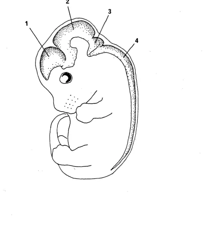
Marginal Zone Cortical Plate
Intermediate Zone
• Subventricular zone
Ventricular Zone Chapter One: Development of the Neocortex
recommended terminology.
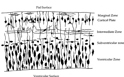
Pial Surface
[image:19.562.105.524.108.370.2]Ventricular Surface
Figure 1.1: Diagrammatic representation of a transverse section through a region of vertebrate neocortex during development showing the five layers. As described by the Boulder Committee (1970).
Ventricular Zone
Chapter One: Development of the Neocortex
ventricular system of the adult brain. The ventricular zone has been the source of much interest to the early anatomists (Vignal, 1888; Ramon Y Cajal, 1891, reviewed by Jacobsen, 1991), especially the mechanism of cell division in that region. Cells of the ventricular zone appear to be morphologically identical and undergo a process of interkinetic nuclear migration (Sauer, 1935b; Sidman
et al,
1959; Sauer and Walker, 1959) during their cell generation cycle. This cell division occurs at the ventricular surface and the DNA replication (S phase) occurs on the outer border of the ventricular zone (see Figure 1.2). The nuclei in this region are continually moving perpendicular to the ventricular surface, backwards and forwards, across the ventricular zone. More recently this movement and mechanism of division has been studied extensively using a variety of techniques.Chapter One: Development of the Neocortex
cycling time is 11 hours, at E18 this cycling time is 19 hours (Waechter and Jaensch, 1972). This intermitotic nucleic cycling has also been confirmed using other methods such as cytophoto metric measurement of DNA content of nuclei obtained from different depths of the ventricular zone (Sauer and Chittenden,
1959).
As cortical development progresses, dividing cells are later seen (E15) outside the ventricular zone, the proportion of these more superficial cells to those at the ventricular surface increases during development (Lewis, 1968).
Chapter One: Development of the Neocortex
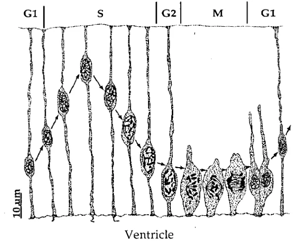
[image:22.562.136.432.70.314.2]Ventricle
Figure 1.2: The movement of the nuclei to different levels of the ventricular zone during the mitotic cycle. From Jacobson (1991).
Marginal Zone
At E14 in the rat there are only two distinguishable layers of the neocortex which have been classically described as: the ventricular zone and the marginal zone (Boulder Committee, 1970). However, this superficial, cell sparse layer, the marginal zone, was later termed the primordial plexiform layer (Marin-Padilla, 1978) and contains as well as cells at various stages of differentiation, the terminally differentiated Cajal-Retzius cells. Marin-Padilla (1978, 1983) proposed that the term "marginal zone" should refer to the outermost and most superficial layer of the neocortex after the appearance of the cortical plate. He suggested the term primordial plexiform layer for the outer zone prior to the appearance of the cortical plate. The primordial plexiform layer therefore has a distinctive structure and only a short duration, immediately before the cortical plate appears.
Chapter One: Development of the Neocortex
The first cells to move out of the ventricular zone settle furthest away from the ventricular surface with the later, young neurons of the subplate staying in the less superficial region closer to this germinal layer. Therefore the oldest neurons in the cortex are the large Cajal Retzius cells which reside in the outer layer I. However, subsequent neurogenesis forming the cortical plate has a neurogenetic gradient which is "inside out". In other words, neurons generated in the ventricular and subventricular zones migrate in a radial fashion through the intermediate zone to occupy a region just below layer I. This means that younger cells will migrate through layers of older neurons before reaching their final position in the cortex.
Conical Plate
Cells of the ventricular zone extend their processes out to the pial surface, the main part of the cell body and nuclei remaining in the well defined, densely stained band, next to the ventricles. This primordial plexiform layer contains cytoplasmic filaments from cells of the ventricular zone. As these cells elongate, the primordial plexiform layer increases its thickness until the formation of other layers. As well as the Cajal-Retzius neurons, which have a horizontal orientation, there are other, younger neurons deeper in the layer which will become subplate neurons forming a subplate zone (Kostovic and Molliver, 1974; Marin-Padilla, 1983). These two regions of the primordial plexiform layer will become split into the outer marginal zone (or layer I) and the inner subplate with the formation of the cortical plate between these two layers; this occurs at E16 in the rat (Marin-Padilla, 1983) and E31 in the cat (Luskin and Shatz, 1985).
Chapter One: Development of the Neocortex
subplate neurogenesis, appeared in the cortical plate at E16, however the subplate itself did not become distinct until E18 (Bayer and Altman, 1991). This population of neurons residing in the cortical plate has been described in other species. Kostovic and Rakic (1980, 1990), also using the 3H- thymidine technique, showed that interstitial cells of white matter in neocortex of humans and monkeys originated from early subplate cells. Luskin and Shatz (1985), working on the cat neocortex, referred to these cells not as a cortical plate but the "upper subplate". Saunders et at (1992b) showed that the neurons in the region of the cortical plate, which contribute to the formation of the primordial plexiform layer, the initial subplate and early cortical plate, are positive for the fetal protein fetuin in the sheep. This was shown using immunocytochemical methods combined with 3H-thymidine autoradiography. Therefore it seems that fetuin, in certain species, is a suitable marker for the early neurons in the cortical plate and subplate (eg., sheep, MoIlgard et al, 1984; cow, Reynolds eta!, 1987; wallaby, Jones et al, 1991).
McConnell et al (1989) showed in the cat that it is cells of the subplate which send out the initial projections to subcortical regions, such as the thalamus. These findings have been supported by more recent studies (Shatz et al, 1990; Erzurumlu and Jhaveri, 1992). These projections are however only temporary and disappear soon after permanent thalamocortical projections are established from layer 5 and 6 (Shatz et al, 1988). Kim et at (1991) provided evidence based on morphology of growth cones growing out of the subplate region that suggested that these projections are the pioneering fibres for the later forming corticothalamic pathway.
Chapter One: Development of the Neocortex
do not grow tangentially into the cortical plate as soon as they arrive at their appropriate position below the subplate but appear to wait for varying periods of time, depending on the species, in the subplate (Molliver and Van der Loos, 1970; Lund and Mustari, 1977; Rakic, 1977; Shatz and Luskin, 1986; Ghosh and Shatz, 1992). Using "DiI" tracing methods Ghosh and Shatz (1992) found that afferent fibres arrived in the subplate of the cat at E36 and invaded the cortical plate at E55. These dates were much earlier than originally thought (Shatz and Luskin, 1986), from studies using a less sensitive technique. The emergence of better fibre tracing methods (such as DiI) has meant that the waiting periods in different species has been better defined. In rats the rate of development is comparatively fast making it difficult to establish whether a waiting period exists or not (Catalano et al, 1991).
The region of the subplate is therefore known as the "waiting zone" and is suggested to occur because the cells in the cortical plate are too immature to attract or permit the ingrowth of thalamocortical fibres until the end of the waiting period (O'Leary and Koester, 1993). Ghosh and Shatz (1993), using fetal cats, removed the subplate neurons at the onset of the waiting period and found that the number of cells (ie. in the lateral geniculate nucleus) sending incoming fibres into the cortical regions was markedly reduced. This work suggests the importance of subplate neurons in formation of the thalamocortical pathway before the appearance of cells in the cortical plate. Synapses between thalamocortical fibres and subplate zone cells in the rat have recently been described (Kageyatria and Robertson, 1993).
Intermediate zone
Chapter One: Development of the Neocortex
cells will pass through the intermediate zone before settling in more superficial regions (Rakic, 1972). Fibres growing to or from the cortical layers will pass through the intermediate zone before projecting up to cells in the cortical layers.
Subventricular zone
The subventricular zone appears first at EIS in the rat in the more ventrolateral portion of the neocortex above the ventricular zone. This region is sometimes referred to as the secondary mitotic region, although cells here remain in a fixed position throughout their cell cycle. They continue to divide long after the disappearance of the ventricular zone and have a smaller, more rounded appearance compared to the cells of the ventricular zone (Boulder Committee, 1970). This region is thought to give rise to certain specific classes of neurons as well as most of the macroglia in the CNS (Boulder Committee, 1970).
Migration of Cells during Neurogenesis
Chapter One: Development of the Neocortex
(eg. Valverde et al, 1989). The difficulty of this technique is that labelling all cells undergoing DNA replication during a short period of time, using a pulse label, does not show from which region of the ventricular zone labelled nuclei in the cortex originated. In other words, the position of labelled nuclei sometime after the exposure to radioactive thymidine does not give any information as to the original position of the nuclei at the time of labelling.
Another difficulty with this method is due to marker dilution, caused by further cell divisions after labelling. One cell division halves the quantity of the radioactive thymidine within the nuclei of the daughter cell.
In recent years more sophisticated techniques, using inheritable markers, have been used to trace the movements of dividing cells as they leave the ventricular zone. The use of retroviruses to introduce "reporter genes" into cells eliminates the problem of marker dilution such as 3 H-thymidine (Price et al,
1987). When a retrovirus infects a dividing cell its genome integrates into a chromosome of the infected cell and is inherited by progeny of that cell (Sanes, 1989). Clonal analysis and lineage studies in the cerebral cortex appear to show that radial migration is not the only mechanism through which the cortex is built (Walsh and Cepko, 1992, 1993; Price and Thurlow, 1989). Cells migrating tangentially after leaving the ventricular zone have been shown to disperse widely across functional areas of the cortex (Walsh and Cepko, 1993). The relationship between the number of cells migrating tangentially and the stage of development of the neocortex is unclear. Walsh and'Cepko (1993) showed a large amount of clonal dispersion (43%) occurring between E14 and E20 in the rat neocortex whereas Nakatsuji et al (1991) suggested, on the basis of experiments using mouse neocortex, that the majority of the dispersion and mixing of cells between radial columns was occurring later in development.
Chapter One: Development of the Ncocortex
framework appears well understood. However, certain in vitro studies have shown that cells destined for the cortical plate exhibit both perpendicular and parallel contact guidance on parallel aligned neurite bundles (Nakatsuji and Nagata, 1989; Nagata and Nakatsuji, 1990).
Chapter One; GENERAL INTRODUCTION; Barriers of the Developing Brain
Barriers of the Developing Brain
The term blood-brain barrier is used widely in the literature and encompasses a number of barriers which separate and protect the brain from the very earliest stages of development. These are; the blood-brain barrier, the blood-CSF barrier and the CSF-brain barrier.
The Blood-Brain Barrier
Saunders (1992) describes the barrier which prevents the movement of proteins within the plasma from entering the brain and the CSF as being the most fundamental. Blood vessels which first invade the developing brain (blood-brain interface) and form within the choroid plexus (blood-CSF interface) provide tight and well developed barriers to protein from the outset (Saunders, 1992).
The exact nature of these barriers, between the blood and the brain, is the tight junctions between cerebral endothelial cells (cells lining the blood vessels) making the blood-brain barrier and between choroid epithelial cells making the blood-CSF barrier. These restrict diffusion of molecules from the blood into the brain or its environment therefore protecting it from rapid changes which may occur in the rest of the body.
Chapter One; GENERAL INTRODUCTION; Barriers of the Developing Brain
Dziegielewska and Saunders (1988) describe a number of shortcomings in these early studies which have led to incorrect interpretations of results. Studying the morphology of the endothelial and epithelial cell tight junctions in the very young brain require extreme care in the tissue fixation. Poor fixation may lead to an unnecessary amount of tissue shrinkage and disruption of the junctions between cells. Studying the permeability of this barrier to marker proteins introduced into the adult as well as the young has been used in a number of studies to examine the nature of the barrier. However, before the results from these type of experiments can be interpreted the physiological state of the animal and degree of disruption to which it is exposed must be considered, ie. the conditions during the experiment must be kept within physiological limits.
In spite of some claims to the contrary, the tight junctions between endothelial and epithelial cells have been shown to be well formed in the earliest brains using freeze fracture techniques (Mollgard and Saunders, 1975, 1986; Saunders and Mollgard, 1984).
The first part of this thesis uses the marsupial species Monodelphis domestica to examine the nature of the barrier (to albumin) between the blood and the CSF at a very early stage of brain development not previously investigated in physiological experiments.
CSF-B rain Barrier
Chapter One; GENERAL INTRODUCTION; Barriers of the Developing Brain
but its ultrastructure has not been adequately studied. Some preliminary information is included in this thesis.
In the past, chemical markers such as horseradish peroxidase (HRP), injected into the ventricles of adult brain have been shown to penetrate into the extracellular space of the brain between the ependymal cells lining the ventricles (Brightman and Reese 1969). However, Fossan et al (1985) looked at this barrier in the fetal sheep at 2 different gestational ages, E60 and E125 (term is 150 days). This experiment involved the perfusion of the same marker protein, HRP, into the ventricles, as well as 1251 labelled human albumin, 3H sucrose and
14C inulin. The total protein concentration of the perfusate was adjusted to be around that which would normally be found in the CSF at these two different ages, thus avoiding disturbance of the surrounding tissue due to osmotic effects. The entry of the horseradish peroxidase into the brain from the CSF, at E125, was time dependent, ie. the longer the perfusion time the greater the penetration of protein from the CSF and into the brain. This was a similar result to that of Brightman and Reese (1969) in the adult mouse although the levels of CSF protein concentration in Fossan's experiment closely matched the endogenous levels, whereas those in Brightman and Reese's experiments were much higher than found in adult mouse CSF. However in the younger fetus, E60, the penetration of the HRP into the brain was very much restricted. This barrier, present only during these early stages, was shown to be coincident with the occurrence of morphological structures ("strap junctions") not present in the older fetuses. There appeared to be specialised junctions between cells of the ventricular zone not normally present between ependymal cells that line the ventricles in the adult. These junctions and the appearance of this barrier has been subsequently studied at various stages of development in a number of different species (human, Mollgard and Saunders, 1986; sheep, Mollgard et al,
Chapter One; GENERAL INTRODUCTION; Barriers of the Developing Brain
adherens) and desmosomes (macular adherens), (Farquhar and Palade, 1963). These and the junctions between early ependymal cells were studied at different stages of development by Mollgard et at (1987). Apposed normal ependymal cells at the ventricular zone of the E125 sheep were linked close to the luminal surface by intermediate junctions, with 20 nm intercellular gaps and gap junctions were also seen (M011gard et al, 1987). This same study examined closely the morphology of the junctions present in the much younger fetuses (E19-E40). These were described as "complex" with the two adjacent membranes folded and exhibiting a "tortuous configuration" with a narrow intercellular gap. Intermediate-like junctions were apparent although there was no evidence of any desmosomes or definite gap junctions. At the apical surface, adjacent cell membranes were fused with a tight junction-like appearance. These junctions however did not show a typical belt-like structure but were shown to
spiral around the cells membrane forming an average of 4 "kissing points" with the adjacent cell. These junctions were not characteristic of any other type of junction in an early developmental stage and the authors describe this new type of junction as a "strap junction" (Mollg&rd et al, 1987).
Chapter One; GENERAL INTRODUCTION; Barriers of the Developing Brain
Blood-CSF Barrier
GENERAL METHODS
GENERAL METHODS
In
V
ivo
Methods
Monodelphis domestica, a marsupial species, has an averagelitter size of between 6 and 10 young which are born at an extremely early stage of brain development. Thus eachlitter provides a number of young which are an accessible modelin whichto studythe barriersinthe CNS.
(i) Handling andInjection ofMonodelphis domestica
Animals supplied forthis work came fromthelocal colony (Animal House, University of Tasmania, Hobart). Details of breeding and care ofthese animals have been published previously (VandeBerg, 1983; Adamet al,1988).
Animals from between 3 days old and adulthood were usedinthese experiments. From birth (PO) until about 15 days old (P15)the young are permanently attachedtothe mother andthen detach periodically until nearerthe time of weaning, at around 48 days. The younger animals hadto beinjected whilst still attachedtothe mother asthey would not return back ontothe mother'steat and would be eaten bythe mother within minutes of removal. Once the young wereinjected IPthey werethenleft onthe mother for a period oftime (until steady state was reached), and werethen carefully removedtaking carethat no damage was doneto eithertheteat orthe mouth ofthe young. Forthese barrier permeability experiments (see below)individual animals wereinjected with standardised volumes (8% of estimatedtotal blood volume).
Sincethe circulating volume of bloodinthese young animalsis small
GENERAL METHODS
the amountsinjected did notincreasethe circulated blood volume enoughto cause any disruptionto blood vessels. This was achieved bylimitingtheinjected volume and by givingtheinjectionintothe peritoneal cavity from where it entered only slowlyintothe circulation. It was alsoimportantthatthe amount of foreign proteininjected did not markedly changethe existing concentration of protein already circulatinginthe plasma. Alitter based model was used for determiningthetimeto reach steady state (Habgood, 1990; see also section (iv) below),ie.individual animals or pairs of animals represent a singletime point. Withthis methoditis essentialthat each animal hadthe same concentration of foreign marker circulatinginthe plasmain-espective ofits size. The volume injected was 8m.1/g of body weight which was equivalentto 8% ofitstotal blood volume, assumingthetotal amount of bloodin each animal was equivalentto 10% ofits body weight. The different exogenous albumin markers (see below), which were injected IP, were made up as 20% (w/v) solutions in sodium chloride (0.9% w/v). Inthe youngest animals a 30 gauge needle was used for theinjection,taking carethatthere was noleak of solution backthroughthe entry hole. This was done by displacingthe skinto one sideimmediately before insertingthe needleintothe peritoneal cavity. After removingthe needlethe skin relaxes backintoits original position moving acrossthe entry holeintothe peritoneum. If a substantialleak of solution occurred backthroughthe entry hole (greaterthan 40%)thenthe animal was notincludedinthe experiment.
(ii) Injected Albumins
Bovine serum albumin(BSA);Sigma; code No. A 2153.
Human serum albumin(HSA); Sigma; code No. A 1653.
GENERAL METHODS
1251 radiolabelled human serum albumin(125IHSA), Amersham; code No.IM
17P.
(iii) Sampling of CSF and Bloodfromthe YoungMonodelphis
After careful removal fromthe mother,individual animals were killed by an overdose of halothane anaesthetic priortothe sampling of CSF and blood. Blood was collected bytwo methods, either by direct cardiac punctureinthe older animals or by cuttingtheleft subclavian artery asit emerged fromthe chest cavityinthe very young. This ensuredthat pleural fluids ofthe chest cavity were not removed withthe blood (which was foundto be a problem with cardiac punctureinthe youngest animals). Samples were removed using gentle suction into fine, glass, heparinised micropipettes. CSF wasthen removed by carefully cutting awaythe muscles atthe back ofthe neck betweenthe base ofthe skull andthe first vertebra. This exposedthedura matercoveringthe CSF space of thecisterna magna.Thedura materwas pierced with a fine glass rrdcropipette and by gentle suction a sample of CSF was removed,taking care notto disturb any ofthe blood vesselsinthe region. CSF and blood samples were also checked under a stereo microscope for blood and any showing detectable signs of contamination were discarded. Levels aslow as 0.05% could be detectedin this manner by comparison with reference samples with knownlevels of contamination. CSF and blood samples were storedin microglass sealed capillarytubes at -20°C until used for further analysis.
(vi) Determination of Steady State
GENERAL METHODS
plasma concentration at regularintervals (or estimatingthe concentrationinthe litter based model)itis possibleto obtain atime weighted mean ofthe plasma concentration over a period priorto obtaining a CSF sample. To ensurethatthe concentrations of marker (exogenous albumin, BSA)inthe plasma and CSF of the youngMonodelphis were at a constantlevel atthetime of samplingthelitter based model was used (Habgoodet al,1992). Each pup from a litterof Monodelphis was given a standardised IPinjection (see above) sothat each littermate was estimatedto havethe same amount of markerinjected per unit weight. This would resultin each animal havingthe same marker plasma concentration. Individual animals werethen subsequently removed fromthe mother after different time intervals and their CSF and plasma marker concentration measured. Individuals were sampled after 6, 11, 15 and 24 hours. The concentration of proteinin CSF was estimated and results expressed as a ratio, CSF/plasma. Once CSF/plasma ratios were nolonger rising significantly, then steady stateis approached.
The following sections describe methods usingthe fetal rattoinvestigate certain aspects ofthe changesin CSF protein concentration during development. This animal was chosen as previous studies have examined similar changes (Dziegielewskaeta!,1981) and also becausethe CSFinthe ventricular system of the fetal rat was easily removed using a micropipette. The midbrain vesicle and thelateral ventricles arelargein relationtothe rest ofthe brain and easily seen underthe dissecting microscope.
(v) Handling and Anaesthesia of Pregnant and Fetal Rats
GENERAL METHODS
(vi) Sampling of CS Ffrom Fetal Rats.
Between 13 and 20 days gestation CSF from within ventricles ofthe fetal rat was removed as described forMonodelphis(see section (iii)). The fetus was separated fromthe mother andimmediately exsanguinated bythe removal ofthe heart. Fine, drawn-out glass micropipettes weretheninsertedthroughthe skin and skull and through the thin layer of neural tissue. Once the tip of the micropipette waslocatedinside eitherthe IIIrd ventricle orthe region ofthe
cisterna magna,by gentle suctionthrough plastictubing attachedtothe endit was possible to withdraw exact amounts of fluid. These samples were then centrifuged and checked for contamination. Any samples showing any signs of blood contamination were discarded.
Injection of Solutionsinto Ventricles
The pregnant rat was anaesthetised (see section, xii) andthe uterus exposed with a medialincisioninthelower abdomen. After exposurethe fetuses were kept dampthroughoutthe experiment using gauze swabs soakedin sodium chloride solution (0.9% w/v),to ensurethatthey remained viablethroughout. Fetal rats (E15-E16) wereinjected with different protein solutions (200mg/100m1 bovine albumin + 200mg/100m1 bovine fetuin)into one ofthetwolateral ventricles. Care wastaken nottoinjecttoolarge a volume (<51A which may cause damagetothetissues. Forthese experimentsthe fetus remained alive and
in uterofor a period of 30 minutes after foreign proteins wereinjectedintothe ventricles.
Using fine glass micropipettes (see above) a small volume of CSF was removed from either ofthetwolateral ventricles. This wasimmediately replaced bythe same volume of protein solution using a similar micropipette.
GENERAL METHODS
Analysis of Plasma and CSF Samples
(viii) Measurements of Total Protein Concentrations.
The total protein concentration in the culture medium, CSF and plasma was measured using the method of Bradford (1976). One ml of protein reagent (100mg. Coomassie Brilliant Blue G-250 was dissolved in 50 ml of 95% ethanol. 100m1 of 85% w/v phosphoric acid was added and the solution was then made up to llitre) was added to tubes containing 1 to 10 1.1.g of protein standard (Sigma, protein standard, code No. 540-10) in 100W volume. The protein standard contains 5.0g/100m1 of albumin and 3.0g/100m1 of globulin, the major protein components present in concentrations similar to those found in the adult human plasma. Although the protein composition of the CSF and plasma changes with, age and the dye used in this method may react differently with different proteins, this protein standard was found most satisfactory, as has been discussed by Dziegielewska et al (1991) who used this method for estimating the total protein levels in fetal sheep and recognised differences in values obtained from previous studies where only one protein (ie. albumin) was used as the standard. In the present study the same protein standard was used throughout.
GENERAL METHODS
(ix) Measurement of Individual Proteins in Plasma and CSF Samples
The concentrations of individual proteins, either endogenous or exogenous were measured using radial immunodiffusion assays (Mancini et al,
1965).
The different anti-albumin antibodies used are known to have some cross-reactivity with albumins of different species. Therefore all the antibodies used were preabsorbed against all other possible cross-reactants. For example, if human serum albumin was the marker injected into the young Monodelphis, and-HSA antibody was firstly preabsorbed overnight with an excess of antigen (Monodelphis control plasma). Finally, lack of cross-reactivity was checked using immunodiffusion assays in the case of the Mancini method and on the appropriate histological sections in the case of immtmocytochemistry (see section (xvii). If cross-reactivity had not been eliminated this would have shown on these controls. Only samples of absorbed antibodies that showed no cross-reactivity were used.
Significant differences between Monodelphis and exogenous albumins were calculated using unpaired Student's t-tests.
(x) Identification of Individual Pmteins
Different proteins within CSF samples, culture media and plasma samples were identified in crossed immunoelectrophoresis using the method of Laurell (1965) which separates different proteins in an electric field (20volts/cm for 1 hour in one dimension, then at 4 volts/cm overnight at right angle in the 2nd dimension which contains appropriate antibodies). The individual proteins can be identified using monospecific antibodies (see section (xix))
Tris-GENERAL METHODS
the staining ofthe proteins, acetic acid and ethanol were used as a destainer (eg. Dziegielewskaet al,1979).
(xi) Counting of 125I Labelled Samples
GENERAL METHODS
In Vitro
Methods
(xii) Dissection of the CNS
Female rats were time mated to ensure that the fetuses were of the correct age. Males and females (Hooded Wistar) were put together for a 24 hour period only. The day they were separated was designated as EC). The age of the fetus was additionally checked against the crown rump length (Butler and Juurlink,
1987).
Pregnant rats were anaesthetised intraperitoneally (I.P.)with 0.6m1/100g body weight of 25% urethane (ethyl carbatnate, Sigma, lot. 69F0633) and maintained on a heated pad at 35°C. Fetuses, at day 15 of gestation (E15),were removed by caesarean section immediately prior to the dissection and killed by rapid exsanguination. The whole fetuses were then completely immersed in culture medium in a small Petri dish lined with a clear silicone polymer (SYLGARD, Seneffe, Belgium). The silicone polymer allows for a more overall illumination during the operation as well as being able to accept dissecting pins, keeping the specimen anchored throughout. Initially the fetus was pinned out so that its dorsal surface was uppermost and kept for the duration of the dissection in basal medium Eagle's (BME) and bubbled continuously with 5% carbon ' dioxide in oxygen.
GENERAL METHODS
kept to a minimum by carefully cutting away the brain in the area of the optic nerves as well as the trigeminal ganglia. Any serious damage to the brain caused a collapse of the ventricle due, to the loss of CSF. Preparations with such distortions were discarded as the culturing process led to further damage of these fragile preparations.
The dissection procedure was performed in clean but asterile conditions, using a dissecting microscope (Wild M8) and media bubbled continuously with 5% CO2 in oxygen that was between 5°C and 12°C. Once removed, the preparations were then transferred to fresh media under sterile conditions in a laminar flow cabinet ready for culture.
(xiii) CuRuling the Preparation
Under sterile conditions of a laminar flow cabinet, preparations were sequentially washed, with sterile medium (BME, Gibco, Life Technologies, Scotland, UK, see below for medium composition) 10-15 times, using sterile plastic pipettes (Falcon, 1 m1). They were then transferred individually to culture flasks containing 15 ml of medium with 0.1% gentamycin with or without 10% fetal calf serum (FCS).
These flasks were stoppered using sterile rubber bungs and bubbled with 5% carbon dioxide in oxygen through sterile, surgical needles (21G, 1 1/4"). Sterile filters (Sigma, 0.21.tm) were attached at both the gas inlet and outlet of the flask reducing the chances of infection during culture.
GENERAL METHODS
(xiv) Survival of the Preparation
Electrophysiological recordings from individual cells within the cortex were not attempted. The preservation of cell structure and the overall state of the preparation were determined histologically. Preparations which did not survive the culture, for example when the gas supply to the medium stopped, quickly deteriorated and soon broke up into smaller fragments. The tissue under the dissecting microscope had a granular appearance and was more fragile than the original and those which survived.
(xv) Sampling of Fluid within the Venhicles.
At various times during the culture of the whole central nervous system the protein concentration and composition of the fluid within the ventricles of the brain was measured. The preparation was first removed from the culture vial and washed in fresh, protein free medium; this was to remove from its surface any protein which were present during culture. A fine glass micropipette (inside tip diameter, approximately 801_tm; outside diameter, approximately 1004m), drawn out over a bunsen flame, was then inserted in turn into each of the ventricular spaces. By gentle suction through a tubing (Portex, ref;800/010/125/800), connected to the micropipette, fluid from within the ventricles of the brain was withdrawn. Samples from different ventricular spaces within the same brain were collected together into Eppendorf tubes and stored at -20°C until further analysis.
GENERAL METHODS
Morphological Methods
(xvi) Tissue Fixation for Light 1VEcroseopy
Bouin's fixative.
Composition of Bouin's fixative: 75 ml of saturated aqueous solution of picric acid, 25m1 of 40% formaldehyde and 5m1 of glacial acetic acid (Culling,
1963).
The preparations were immersed in Bouin's fixative overnight. After several washes in tap water they were then dehydrated through 70%, 95% and then 100% ethanol (minimum 6 hours in each with 2 changes) followed by two changes of chloroform, one overnight. This was followed by 3 changes of paraffin wax at 56°C for at least 4 hours in each. Once embedded and cooled to room temperature the blocks were then cut serially in 3pm sections and placed on gelatin coated slides (Culling, 1963).
(xvii) Immunogold Silver Staining
GENERAL METHODS
Procedure;
a) Dewax Sections
i) Xylene 2 changes.
ii) Absolute alcohol (2 changes, 10 minutes each).
iii) 2 ml 100% hydrogen peroxidein 400m1 absolute alcohol (20-30 minutes).
iv) Graded alcohols, 95% and 70, 5 minutesin each.
v) Tap water (5 minutes)
b). Staining of Sections
i) Phosphate buffer solution, (PBS, phosphate buffered saline, 3 changes, 20 minutes each) pH 7.2 with 0.2% Tween detergent (polyoxyethylene sorbitan monolaurate, Sigma; code No. P1379).
ii) Blocking solution for 1 hour (solution used; 10% gelatinin PBS with 0.2% Tween.
iii) Incubatedin primary antibody at 4°C overnight. Antibody was diluted appropriately and appliedin blocking solution.
iv) Washed 3timesin PBS/Tween.
v) Gold conjugated secondary antibody applied for 2 hours at room temperature. Antibody was diluted (X50) and appliedin blocking solution.
vi) Washed 3timesin PBS with Tween for atotal of 15 minutes.
GENERAL METHODS
ix) Running distilled water for 15 minutes.
x) Counterstained in toluidine blue for 30 seconds.
xi) Washed in distilled water until appropriate level of staining remains.
xii) Dehydrated in graded alcohols, cleared in xylene and mounted in DPX.
(xviii) Incorporation of B nclU
Dividing cells in vitro and in vivo were detected using immunocytochemistry. The method involves introducing a small quantity of the thymidine analogue, bromodeoxyuridine (BrdU, Sigma code No. B 5002), into the culture medium for a short period of time, immediately after the CNS has been isolated from the fetus. The nucleotide analogue will be incorporated into newly made DNA and can be detected using the immunocytochemical technique and antibodies specific to BrdU (Schutte et al, 1987).
Preparations were exposed to 30uM BrdU (MW 307.11) for 1 hour, 29°C or 33°C, in fresh medium. After labelling they were thoroughly washed in sterile medium before being separated into sterile culture vials as described above.
Localisation of the BrdU incorporated into DNA was done on fixed and paraffin embedded preparations as described above. Acid or enzyme digestion procedures were found to be unnecessary in the present work. Significant staining of the labelled cells was achieved using a X20 dilution of the BrdU antibody (Sigma).
(xix) Antibodies
GENERAL METHODS
adjuvant (Sigma Code No. F-5506) (Dziegielewska et al, 1989). Complete adjuvant was used initially followed by incomplete adjuvant for all the subsequent injections (Harboe and Ingild, 1973).
Antisera to other species of albumin (human serum albumin, bovine serum albumin), with the exception of Monodelphis albumin, were obtained from Dako (see below).
(xx) Primary Antibodies
Rabbit Anti-Monodelphis Albumin (Supplied by Dr. K. M. Dziegielewska)
This antibody was preabsorbed With against BSA and HSA. Each albumin (10mg) was mixed with lml of the antibody, left overnight at 4°C, centrifuged for 10 minutes and separated from the supernatant. The antibody was used at concentrations between 1:20 and 1:100.
Rabbit Anti-Calf Fetuin (DA K 0, Denmark, Code No. Z 249).
This antibody has been shown not to cross-react with rat fetuin (Dr. K. M. Dziegielewska, personal communication) and therefore making preabsorption unnecessary. The concentrations used as the primary antibody were between 1:50 and 1:200 with a 10% gelatin blocker.
Rabbit Anti-Rat Serum (DAK 0, Denmark, Code No. Z 179).
GENERAL METHODS
This solution was used as the primary antibody (diluted 100 times) and blocker combined.
Rabbit Anti-Cow Albumin (DAKO, Denmark, Code No. Z229)
This antibody was found not to cross react with rat albumin; however as a precaution it was preabsorbed with rat plasma (1001.0 plasma to 1 ml of antibody). The antibody was used at concentrations between 1:200 and 1:300 in a 10% gelatin blocker solution.
Rabbit Anti-Rat Albuinin (NORDIC, Netherlands, Code No. RARalAlb).
This antibody cross reacts with albumin in fetal calf serum. Antibody (100111) was mixed with 10 ml of 10% FCS in PBS, left at 4°C overnight, centrifuged for 10 minutes and separated from the supernatant. This solution was used as the primary antibody and blocker combined (dilution X100).
Monoclonal Mouse Anti-BrdU (DAKO, Denmark, Code No. M744)
This monoclonal mouse antibody shows no cross reactivity with any fetal rat proteins and was diluted 1:20 in PBS and blocker.
(xxi) Fixation and Preparation for Electron Nlicroscopy
GENERAL METHODS
the pieces of fixed tissue were cut, using scalpel blades, into small blocks (not more than 5mm in any dimension).
Post Fixation
These fixed pieces of tissue then underwent post fixation in an osmium-potassium feiTocyanide mixture (De Bruijn and Den Breejen, 1975; De Bruijn and Den Breejen, 1976; Goldfischer et al, 1981). The solution was made up as follows; 0.21g fen-ocyanide (0.5M) in 10m1 of 0.2M cacodylate buffer with 10111 of NaOH. This solution was mixed with an equal volume of 4% reduced osmium (0s04). Therefore the final solution is 2% reduced osmium, 0.025M K4Fe(CN)6 and 0.1M cacodylate buffer. The pieces of tissue were postfixed in this solution for 2 hours at 4°C, agitating continuously.
At room temperature the tissue was then added to 1% uranyl acetate (in water) for 1 hour before being dehydrated in alcohols (4°C); 70% for 15 minutes, 96% for 15 minutes, 2X 100% for 15 minutes. Once dehydrated the tissue was then prepared for embedding;
i) Propylene oxide (2X 15 minutes at 4°C)
ii) 1: 1 Propylene oxide: Epon. 30 minutes at 4°C.
1: 2 Propylene oxide: Epon. 2 hours at room temperature.
iv) Overnight in 100% Epon.
v) Change of fresh Epon and in oven for at least 12 hours at 65°C.
GENERAL METHODS
(xxii) Basal Medium Eagle (BME) Formula
COMPONENT CONCENTRATION mg/ml
Inorganic Salts Calcium Chloride .2H20 Magnesium Sulphate(anhydrous) Potassium Chloride
Potassium Phosphate Monobasic (anhydrous) Sodium Bicarbonate
Sodium Chloride
Sodium Phosphate Dibasic (anhydrous) Amino Acids
L-Arginine.HCL L-Cystine.2HCL L-Histidine (free base) L-Isoleucine L-Leucine L-Lysine.HCL L-Methionine L-Phenylalanine L-Threonine L-Tryptophan L-Tyrosine.2Na.2H20 L-Valine Vitamins D-Biotin Choline Chloride Folic Acid myo-Inositol Niacinamide
Chapter Two; CSF Proteins during Development
Chapter Two
Chapter Two CSF Proteins during Development
Introduction
Background to Plasma Proteins in CSF and Relation to Early Brain Development
From the very earliest stages of development the inner surface and later also the outer surface of the central nervous system is bathed in a fluid (cerebrospinal fluid, CSF) which is constantly being secreted and reabsorbed. Its composition, protein and electrolyte content is highly specific for particular stages of brain development and has been investigated extensively in various species (for reviews see S'aunders and Bradbury, 1973; Davson eta! , 1987; Dziegielewska and Saunders, 1988; Saunders, 1992).
CSF Protein Concentration
(mg/100m1) Sheep Pig Rat Rabbit Chicken Wallaby Opossum*
Peak during 1143 ± 83 961 + 95 317 ± 15 529 + 18 523 ± 16 405 ± 21 499 ± 25 Development
(31 days) (31 days) (22 days) (20 days) (11 days) (42 days) (23 days)
Adult 26 ± 2 31 + 3 24 ± 8 29 + 3 141± 15 66 ± 3 23 ± 3.8 CSF
Age (in days) post- conception at
birth
150 115 22 32 21 28 14
Chapter Two; CSF Proteins during Development
The protein concentrations within the CSF have been measured in many
different species and in all those studied so far the levels during the early stages
of brain development are much higher than in the adult. The variety of animal
species studied so far is wide-ranging in both size and evolutionary terms.
However in all species studied, a peak of protein concentration in the CSF occurs
early in brain development (see Table 2.1) and the timing of this peak's
appearance appears to have no correlation to the time of birth.
In the eutherian species studied (see Table 2.1) the peak of CSF protein
concentration appears in the fetal stages of development. This is compared with
the marsupial species (wallaby and opossum), born at an extremely early stage of
development, in which the peak occurs postnatally.
In all these species studied, it appears that the timing of the peak of
protein concentration in the CSF coincides with the first appearance of the cortical
plate in the neocortex (see section, 1.2; Development of the Neocortex and Table
2.2). The exception seems to be the rat. The rat, which shows very rapid brain
development, has a gestational period of 22 days and the cortical plate first
appears on day E16 (Bayer and Altman, 1991). From the studies of
Dziegielewska
eta!(1981), the peak of protein concentration in the CSF in the
ratoccurs between E20-E22. The human has a gestational period of 40 weeks and
the cortical plate appears during the 7th week (Marin-Padilla, 1983). The
concentration of the CSF early in development is also high but it is not known
when the peak of total protein occurs. In the marsupial species the appearance of
the cortical plate and the CSF protein peak occurs
postpartum.Chapter Two; CSF Proteins during Development
Figure 2.1
ZA
d3
ZA
N
V
r
'
4.
.. . -• 7
7
.
.
454
AO At4' a 7 --,4
•
-
ea, Aih-
4.
. •d-. is.
.
.
.
„-AV
•
l
•
!SAW, --_,,.• .ft,...-#•2. ..."P
e
r
b
.
.
.
•
4
1
1".. V ... 4 4if l■At 1 & 41 Ily "e 41
:4
•
• dd. ,igoOrmir, 411147Pkt -..ft ;A'-,-b/a - ii 'I% •Am,,,
•-A • 9 ... A s, 11.,,,, 94 Ir & 74 • 8—
.
, 'r-7 , _ 4 • 4 ‘ 111‘ 41111 p ia 7 • . 4•IK
0 A tg •'' ft i
O.
Chapter Two: CSF Proteins during Development
In general, the cortical plate, which is the beginnings of the cortical gray matter, forms within the primordial plexiform layer splitting this region into the outer marginal zone and the inner subplate region (Marin-Padilla, 1983) (see General Introduction). This primordial plexifonn layer, which is present before the formation of the cortical plate lies superficially to the ventricular zone, immediately below the pial surface (see section, 1.2, Development of the Neocortex).
Only in the rat does the timing of the peak of total protein concentration appear to be well after the first cells of the cortical plate begin to differentiate. However there are two points which need to be considered:
(1) From which region was the CSF sampled and how does this region compare with other regions during this critical time of development?
(2) If there is indeed a difference in the rat, then what could be the reasons for it?
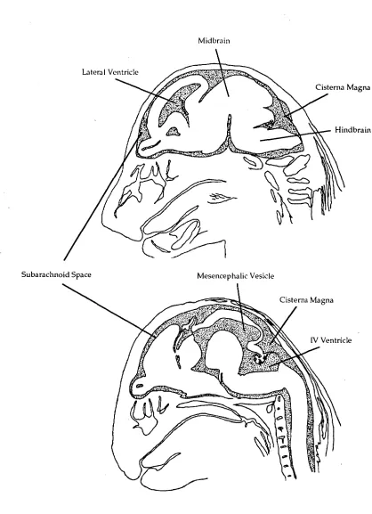
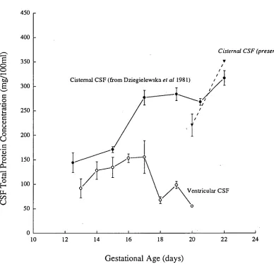
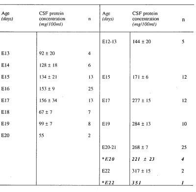
The study of Dziegielewska eta! (1981) sampled CSF mostly from the cisterna magna, a region close to the IV ventricle (see Figure 2.6), and they found the peak to coincide with the time of birth. A more extensive investigation of the nature of the raised peak of protein concentration within the CSF in different regions of the brain may give a different developmental pattern and a clearer picture as to the changes occurring throughout the CSF.
Chapter Two; CSF Proteins during Development
ventricle fell sharply whilethatinthe IVth ventricle remained similar (lateral, 373
± 72 mg/100m1; IVth, 1184 ± 133 mg/100m1). Atlater stages,the CSF protein
concentrations withinthetwo different regions have both dropped considerably,
thelateral ventricular CSF concentration was alwayslowerthanthe IVth, evenin
the adult, as has been known for many years for adult humans (see Davsonet al,
1987). Therefore,thereis a much sharper drop oftotal protein concentration
withinthelateral ventriclesthaninthe IVth ventricle. Regional differenceslike
these may accountinthe rat forthe apparent differenceinthetiming of a protein
concentration peak afterthe appearance ofthe cortical plate.
Sheepl pig2 Rat3 Wallaby4 Opossum5
Timing ofthe Protein Peakin CSF(in days after Conception) and
total Protein Concentration (mg/100m1)in CSF
31 days (1143 ± 83)
31 days (961 ± 95)
22 days (317 ± 15)
41 days (405 ± 21)
23 days (499 ± 25)
Time(days after conception) of cortical
plate appearance 33-34 26-31 15-16 34-43 17-21
Table 2.2: Thetiming ofthe CSF peak oftotal protein concentrationin five
animal species (in days after conception) comparedtothetimein
days after conception when cells ofthe cortical plate first startto
differentiate. 1, Reynolds and Mollgard (1985); 2,Cavanagh and
Mollgard (1985); 3,Bayer and Altman (1991); 4,Reynoldset al
(1985); 5,Saunderset al (1989).
The nature and origin ofthe peak of protein concentration withinthe CSF
could be more accurately describedif more was known aboutthe characteristics
Chapter Two; CSF Proteins during Development
Previous permeability experiments have been successful in determining the origins of proteins in CSF at developmental stages after the concentration of protein in CSF has declined from its peak values.
Results so far suggest that the peak of protein concentration is more related to a specific stage in brain development than to the time of birth. To date, no studies have investigated the mechanism(s) which brings about the peak at this early stage.
Origin of Proteins in the CSF
Felgenhauer (1974) compared the molecular radius of individual proteins within adult CSF and compared these with their CSF/plasma ratios. These studies in the adult showed that many proteins in the CSF, which are present in much lower concentration than in the fetus, could be accounted for by simple diffusion through the barrier which separates the blood from the CSF.
The idea of a barrier providing a protection against non-specific leak of molecules from the rest of the body is not a new one. Reviews by Dobbing (1969), and Davson (1967) describe in detail the various underlying mechanisms which collectively are known as the "blood-brain barrier" and more recently reviews have been published by Bradbury (1979), Davson eta! (1987), M011gard and Saunders (1986) and Saunders (1992).
Chapter Two; CSF Proteins duiing Development
circulating blood from very early in its development, but developmentally regulated mechanisms result in a high concentration of protein in fetal CSF.
Proteins within the CSF during the early stages of development are thought to have contributions from four sources:
(1) A component from the plasma itself by passive transfer.
(2) By a specific transport mechanism from the plasma.
(3) From synthesis by epithelial cells of the choroid plexus.
(4) From synthesis within cells of the brain itself.
The quantitative contribution from these sources may be different at different stages of development and also for different proteins.
Transfer of Pinteins from the Blood into the CSF
Over the years the nature of the "blood-brain barrier" and the permeability of different kinds of molecules have been investigated. These experiments have usually involved the introduction of a foreign "marker molecule" into the blood of an animal followed by the use of various techniques to detect the marker on the other side of the barrier, within the CSF or brain. In their review of the development of the blood brain barrier, Dziegielewska and Saunders (1988) describe in detail the criteria which must be met in such permeability experiments so that the results are valid. Important points concerning experimental detail are described, the omission of which in some of the previous work has led to much confusion concerning the development of the blood brain barrier in the fetus and newborn.