Fundamental Contribution and Host Range Determination of
ANP32A and ANP32B in Influenza A Virus Polymerase Activity
Haili Zhang,
aZhenyu Zhang,
aYujie Wang,
aMeiyue Wang,
aXuefeng Wang,
aXiang Zhang,
aShuang Ji,
aCheng Du,
aHualan Chen,
aXiaojun Wang
aaState Key Laboratory of Veterinary Biotechnology, Harbin Veterinary Research Institute, The Chinese Academy of Agricultural Sciences, Harbin, China
ABSTRACT
The polymerase of the influenza virus is part of the key machinery
neces-sary for viral replication. However, the avian influenza virus polymerase is restricted in
mammalian cells. The cellular protein ANP32A has been recently found to interact with
viral polymerase and to influence both polymerase activity and interspecies restriction.
We report here that either human ANP32A or ANP32B is indispensable for human
influ-enza A virus RNA replication. The contribution of huANP32B is equal to that of
huANP32A, and together they play a fundamental role in the activity of human
influ-enza A virus polymerase, while neither human ANP32A nor ANP32B supports the
activ-ity of avian viral polymerase. Interestingly, we found that avian ANP32B was naturally
in-active, leaving avian ANP32A alone to support viral replication. Two amino acid
mutations at sites 129 to 130 in chicken ANP32B lead to the loss of support of viral
rep-lication and weak interaction with the viral polymerase complex, and these amino acids
are also crucial in the maintenance of viral polymerase activity in other ANP32 proteins.
Our findings strongly support ANP32A and ANP32B as key factors for both virus
replica-tion and adaptareplica-tion.
IMPORTANCE
The key host factors involved in the influenza A viral polymerase
ac-tivity and RNA replication remain largely unknown. We provide evidence here that
ANP32A and ANP32B from different species are powerful factors in the maintenance
of viral polymerase activity. Human ANP32A and ANP32B contribute equally to
sup-port human influenza viral RNA replication. However, unlike avian ANP32A, the avian
ANP32B is evolutionarily nonfunctional in supporting viral replication because of a
mutation at sites 129 and 130. These sites play an important role in ANP32A/
ANP32B and viral polymerase interaction and therefore determine viral replication,
suggesting a novel interface as a potential target for the development of
anti-influenza strategies.
KEYWORDS
ANP32A, ANP32B, RNA replication, influenza A virus, interspecies
transmission, polymerase activity
V
irus transmission from its natural host species to a different species reflects
mechanisms of molecular restriction, evolution, and adaptation. Birds harbor most
of the influenza A viruses, which are typically replication-restricted in human hosts
because of receptor incompatibility and limited viral polymerase activity in human cells.
Although many host factors have been reported to be involved in viral replication (1–8),
the key mechanisms that determine viral polymerase activity and host range are poorly
understood.
Influenza A viral ribonucleoprotein (vRNP), the viral minimum replicon, comprises
the viral genome, the heterotrimeric RNA polymerase PB1, PB2, PA, and the
nucleo-protein (NP), and carries out viral RNA transcription and replication in infected cells.
Avian influenza A polymerases have very limited activity in mammalian cells, indicating
an unknown host-specific restriction mechanism that directly affects viral RNA
replica-CitationZhang H, Zhang Z, Wang Y, Wang M,
Wang X, Zhang X, Ji S, Du C, Chen H, Wang X. 2019. Fundamental contribution and host range determination of ANP32A and ANP32B in influenza A virus polymerase activity. J Virol 93:e00174-19.https://doi.org/10.1128/JVI .00174-19.
EditorAdolfo García-Sastre, Icahn School of
Medicine at Mount Sinai
Copyright© 2019 Zhang et al. This is an
open-access article distributed under the terms of theCreative Commons Attribution 4.0 International license.
Address correspondence to Xiaojun Wang, wangxiaojun@caas.cn.
H.Z. and Z.Z. contributed equally to this work.
Received4 February 2019
Accepted9 April 2019
Accepted manuscript posted online17
April 2019 Published
crossm
14 June 2019
on November 6, 2019 by guest
http://jvi.asm.org/
Downloaded from
on November 6, 2019 by guest
http://jvi.asm.org/
Downloaded from
on November 6, 2019 by guest
http://jvi.asm.org/
Downloaded from
on November 6, 2019 by guest
http://jvi.asm.org/
Downloaded from
on November 6, 2019 by guest
http://jvi.asm.org/
tion. The adaptation of avian viruses to mammals, such as occurred with the H5N1 and
H7N9 avian viruses, occurs along with substitutions on the viral polymerase, mainly on
the PB2 subunit (E627K and other signatures), which enhance viral replication (9–28).
Many host factors have been reported to interact with the vRNP complex to help viral
replication (7, 29–40). Of these proteins, the acidic nuclear phosphoprotein 32 family,
members A and B (ANP32A and ANP32B), have been found to regulate viral RNA
synthesis (7). Interestingly, chicken ANP32A (chANP32A) has been reported to
specifi-cally promote avian influenza replication due to the 33-amino-acid insert (8). This
33-amino-acid insert includes a hydrophobic SUMO interaction motif, which connects
to host SUMOylation to partially contribute to the promotion of avian viral polymerase
activity (41). Furthermore, ANP32A from birds has splicing variants without SUMO
interaction motif also support viral replication and polymerase adaptation (42). These
studies suggest that ANP32A plays an important role in viral replication.
The acidic (leucine-rich) nuclear phosphoprotein 32-kDa (ANP32) family comprises
several members, including ANP32A, ANP32B, and ANP32E, which have various
func-tions in the regulation of gene expression, intracellular transportation, and cell death
(43). Although ANP32A is considered to be an important cofactor of the influenza virus
polymerase and to influence the viral host range, the roles of different ANP32 members
in viral replication, and the extent to which the proteins are involved in the activity of
the polymerase remain unclear. In this study, by using CRISPR/Cas9 knockout screening,
we found that huANP32A and huANP32B play fundamental roles in the facilitation of
human influenza A viral RNA synthesis, and that both huANP32A and huANP32B
contribute equally. The mammalian ANP32 proteins give no or only limited support to
the avian virus polymerase. Human ANP32A&B do not support the replication of the
avian influenza virus, but this restriction can be overcome by E627K substitution in the
viral PB2 protein. Furthermore, we found that chicken ANP32B has no effect on
polymerase activity, which can be ascribed to mutations in two key amino acid residues
of ANP32A&B that determine their activity in supporting viral RNA replication. Together,
these data reveal fundamental roles for ANP32A and ANP32B in supporting influenza
virus A polymerase activity as well as a site key for their function, and show that both
ANP32A and B determine viral polymerase adaptation and host range.
(This article was submitted to an online preprint archive [44].)
RESULTS
Human ANP32A&B are critical host factors that determine viral polymerase
activity and virus replication.
Previous studies have reported that several host factors,
including BUB3, CLTC, CYC1, NIBP, ZC3H15, C14orf173, CTNNB1, ANP32A, ANP32B,
SUPT5H, HTATSF1, and DDX17, interact with influenza viral polymerase and that some
of these factors have an effect on viral polymerase activity (3, 4, 29). All of these
observations were based on the gene transient knockdown technique, meaning that
the results may vary because of the different knockdown efficiency of target genes.
Using a CRISPR/Cas9 system, however, allows rapid knockout of certain genes and
accurate evaluation of target proteins. To identify the critical roles of the
above-mentioned host factors in influenza viral replication, we used a CRISPR/Cas9 system to
establish a series of knockout 293T cell lines, and a model virus-like luciferase RNA was
expressed, together with the viral polymerases PB1, PB2, PA, and NP, to determine the
polymerase activity in these cell lines. First, we tested the polymerase activity of 2009
pandemic H1N1 virus A/Sichuan/01/2009 (H1N1
SC09) (45) on these knockout 293T cells
and found that individual knockout of NIBP, ZC3H15, or DDX17 results in a 2- to 4-fold
decrease in viral polymerase activity, which is consistent with previous results (3, 29).
However, in none of the other single-gene-knockout cells was viral polymerase activity
blocked (Fig. 1A). We did not observe any reduction of viral polymerase activity in
ANP32A or ANP32B knockout cells, which was a surprising result since ANP32A and
ANP32B (ANP32A&B) were reported to be important host factors supporting viral
polymerase activity (7, 8, 41, 42). Since ANP32A and ANP32B have high similarity in both
structure and known functions, we predicted that in single-knockout cell lines of
on November 6, 2019 by guest
http://jvi.asm.org/
A
B
F
ir
ef
ly
pol
y
m
er
as
e ac
tiv
ity
nor
m
al
iz
ed t
o
R
e
ni
lla
ns ns ns nsns ns nsns ns
*
* **
C
293T AKO BKO DK
O
0.001 0.01 0.1 1 10 100 1000
H7N9AH13
**** ns ns
WSN
0.01 0.1 1 10 100 1000
**** ns ns
293T AKO BKO DK
O
F
ir
e
fly
p
o
ly
m
e
ra
se
a
c
tiv
ity
n
o
rm
a
liz
ed
t
o
R
e
n
ill
a
F
ir
e
fly
p
o
ly
m
e
ra
se
a
c
tiv
ity
nor
m
a
liz
e
d
t
o R
e
ni
lla
293T AKO BKO DK
O
H1N1SC09
ns ns
**** 0.01
0.1 1 10 100 1000 0.01
0.1 1 10 100 1000
D
293T BU
B3
CL
T
C
CY
C1
NI
B
P
Z
C
3H
15
C
1
4or
f1
73
CT
NNB
1
AN
P32A
AN
P32B
SU
PT
5H
H
T
AT
SF
1
DDX
1
7
PB1
NP PA
β-Actin
E
V
iru
s
t
it
e
r (l
g
T
C
ID
5
0
/0
.1
m
l)
0 12 24 36 48 0
1 2 3 4 5 6
7 293T AKO BKO DKO
Hours post infection
* ***
** β-Actin
293T AKO BKO DK
O
huANP32A
huANP32B
G
F
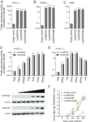
FIG 1huANP32A&B are indispensable for influenza A virus polymerase activity and viral replication. (A) Wild-type 293T cells and single-gene-knockout 293T cell lines, including BUB3, CLTC, CYC1, NIBP, ZC3H15, C14orf173, CTNNB1, ANP32A, ANP32B, SUPT5H, HTATSF1, and DDX17, were transfected with firefly
minige-nome reporter, Renilla expression control, and the polymerase of H1N1SC09. The luciferase activity was
measured 24 h after transfection, and data indicate the firefly luciferase gene activity normalized to theRenilla
luciferase gene activity. Statistical differences between samples are indicated, according to a one-way ANOVA,
followed by a Dunnett’s test (NS, not significant;*,P⬍0.05;**,P⬍0.01;****,P⬍0.0001). Error bars represent
the SEM within one representative experiment. (B) Wild-type 293T cells were transfected with pMJ920 vector (a plasmid expressing eGFP and Cas9) and gRNAs targeting huANP32A and huANP32B to generate huANP32A&B double-knockout cells (DKO). The endogenous proteins of different cells (293T cells, huANP32A knockout cells [AKO], huANP32B knockout cells [BKO], and DKO cells) were identified by Western blotting with
antibodies against -actin, huANP32A, and huANP32B as described in Materials and Methods. (C) Design
scheme of sgRNAs for huANP32A and huANP32B. (D to F) Wild-type 293T cells, huANP32A knockout cells (AKO), huANP32B knockout cells (BKO), and huANP32A&B double-knockout cells (DKO) were transfected with
firefly minigenome reporter, Renilla expression control, and either H1N1SC09 polymerase (D), H7N9AH13
polymerase (E), or WSN polymerase (F). The luciferase activity was measured at 24 h posttransfection. For
panels D to F, the data are the firefly luciferase gene activity normalized to that ofRenillaluciferase activity.
Statistical differences between cells are indicated, following a one-way ANOVA and subsequent Dunnett’s test
(NS, not significant;**,P⬍0.01;***,P⬍0.001;****,P⬍0.0001). Error bars represent the SEM of replicates
within one representative experiment. The expression levels of H1N1SC09polymerase proteins were assessed
by Western blotting in panel D. (G) Wild-type 293T, AKO, BKO, and DKO cells were infected with WSN virus at an MOI of 0.01. The supernatants were sampled at 0, 12, 24, 36, and 48 h postinfection, and the virus titers were determined by endpoint titration in MDCK cells. Error bars indicate the SD from three independent
experiments.*,P⬍0.05;**,P⬍0.01;***,P⬍0.001.
on November 6, 2019 by guest
http://jvi.asm.org/
[image:3.585.40.400.71.472.2]ANP32A or ANP32B, the presence of either one of these two proteins could support
viral replication in the absence of the other. We then developed an ANP32A and
ANP32B double-knockout cell line to confirm this hypothesis. Human ANP32A
(huANP32A) knockout 293T cells (AKO cells), human ANP32B (huANP32B) knockout
293T cells (BKO cells), and ANP32A and ANP32B double-knockout cells (DKO) were
confirmed by Western blotting with ANP32A- and ANP32B-specific antibodies (Fig. 1B).
The target sequences of sgRNAs for huANP32A and huANP32B are shown in Fig. 1C. In
AKO and BKO cells, the viral polymerases have similar activities to wild-type (WT) 293T
cells, but when both ANP32A and ANP32B were knocked out (DKO), the polymerase
activity was abolished (
⬃
10,000-fold reduction) (Fig. 1D), although the polymerase was
expressed equally in the 293T, AKO, BKO, and DKO cells (Fig. 1D). We then tested the
polymerase activities of 2013 China H7N9 human isolate A/Anhui/01/2013 (H7N9
AH13)
(46), and H1N1 virus A/WSN/1933 (WSN) on AKO, BKO, and DKO cell lines and found
that the viral polymerase complex lost all activity in the DKO cells but not in the AKO
or BKO cells (Fig. 1E and F). We further confirmed the effect of huANP32A&B on viral
infectivity by infecting 293T cells and KO cells with WSN virus. In DKO cells, but not AKO
or BKO cells, the infectivity of WSN decreased by
⬎
10,000-fold (Fig. 1G). These results
indicate a crucial role for both ANP32A&B in viral RNA replication.
The knockout of both ANP32A and ANP32B led to dramatic loss of viral polymerase
activity (
⬃
10,000-fold), which is distinct from a previous report that used a gene
knockdown method and resulted in an approximately 3- to 5-fold reduction in viral
polymerase activity (8). We found that reconstitution of either huANP32A or huANP32B,
or both of them, in the DKO cells restored the viral polymerase activities of H1N1
SC09,
H7N9
AH13, and WSN viruses (Fig. 2A to C). Expression of huANP32A or huANP32B in
DKO cells supported the H1N1
SC09viral polymerase activity in a dose-dependent
manner (Fig. 2D). We confirmed that the required level of expression of huANP32A or
B is very low, and overdose expression has a negative effect (Fig. 2E), suggesting an
explanation of the confusing phenotype previously observed, that in normal 293T cells
overexpression of ANP32 protein decreased viral polymerase activity (8). Reconstitution
of huANP32A or huANP32B or both of them in DKO cells restored full viral infectivity
(Fig. 2F), indicating that huANP32A and huANP32B are of fundamental importance in
human influenza viral replication. These data proved that huANP32A and huANP32B
are key factors required for polymerase activity, that they have similar functions in the
support of viral replication, and that they can function independently and contribute
equally to influenza virus polymerase activity.
Influenza A virus replication starts from the transcription and replication of the
negative single-stranded viral RNA (vRNA) by the vRNP complex. vRNA is transcribed
into positive cRNA (cRNA) and mRNA; the cRNA is then used as a template to amplify
into new vRNA (47, 48). We found that vRNA, cRNA, and mRNA synthesis was
dramat-ically reduced in DKO cells. However, when huANP32A was reconstituted in the DKO
cells, vRNA, cRNA, and mRNA synthesis was fully recovered (Fig. 3A to C). We observed
similar results from the reconstitution of ANP32B or both ANP32A and ANP32B (Fig. 3A
to C), indicating that huANP32A&B are key factors in triggering the replication of the
human influenza viral genome.
We next used an eight-plasmid reverse genetics system in H1N1
SC09and tested the
virus production in the supernatant of the transfected cells using an antigen capture
enzyme-linked immunosorbent assay (ELISA) (49). The result showed that the DKO cells
had very low NP production in the cell supernatant compared to the 293T, AKO, or BKO
cells (Fig. 3D). The NP production was recovered when the huANP32A and/or
huANP32B was expressed in DKO cells (Fig. 3E). Taken together, these results suggest
that huANP32A and huANP32B determine viral RNA replication efficiency and
subse-quent viral production in 293T cells.
Support of influenza A viral replication by ANP32A or ANP32B from different
species.
ANP32A&B are members of the evolutionarily conserved ANP32 family, which
has various functions in the regulation of gene expression, intracellular transport, and
cell death (43). ANP32A&B exist in almost all eukaryotic cells, with the exception of early
on November 6, 2019 by guest
http://jvi.asm.org/
eukaryotic life (yeast and other fungi) (50). We then investigated the support of ANP32A
or ANP32B from different species for viral polymerase activity in DKO cells. ANP32A
from human, chicken, duck, turkey, zebra finch, mouse, pig, and horse sources and
ANP32B from human and chicken sources were expressed individually with
minig-enomes of either H1N1
SC09, human isolate H7N9
AH13, H3N2 canine influenza virus
A/canine/Guangdong/1/2011(H3N2
GD11), H3N8 equine influenza virus
A/equine/Xinji-ang/1/2007(H3N8
XJ07), A/equine/Jilin/1/1989(H3N8
JL89), or H9N2 chicken virus
A/chick-A
C
F
D
B
H7N9AH13 H1N1SC09****
**** ****
100
10
1
0.1
0.01
100
10
1
0.1
0.01 WSN
V
ir
u
s
t
it
e
r (
lg
T
C
ID50/0.1ml)
******** ****
**** **** ****
0.01 0.1 1 10 100 1000
F
ir
e
fly
p
o
ly
m
e
ra
se
a
c
tiv
ity
nor
m
a
liz
e
d
t
o
R
e
ni
lla
F
ire
fly
p
o
ly
m
e
ra
se
a
c
tiv
ity
nor
m
a
liz
e
d
t
o R
e
ni
lla
**** ****
**** **** ****
**
0pg 10pg
100pg 1ng 5ng 10ng 20ng
0 12 24 36 48
0 1 2 3 4 5 6 7
Empty vector huANP32A huANP32B huANP32A&B
Hours post infection huANP32A
huANP32B β-Actin
β-Actin **ns
** * ***
** **** **** ****
0pg
100pg
1ng
20ng
100ng 500ng
1ug
0.01 0.1 1 10 100
**** ****
**** ****
**** ****
H1N1SC09
E
H1N1SC09
huANP32A huANP32B huANP32A
huANP32B
0.01 0.1 1 10 100
0.001
Emp
ty ve
ct
o
r
h
u
AN
P3
2
A
huA
N
P
32B
h
u
AN
P3
2
A
&B
Emp
ty ve
ct
o
r
h
u
AN
P3
2
A
huA
N
P
32B
h
u
AN
P3
2
A
&B
Emp
ty ve
ct
o
r
h
u
AN
P3
2
A
huA
N
P
32B
h
u
AN
P3
2
A
&B
FIG 2Reconstitutions of huANP32A&B rescue polymerase activity and replication of influenza A virus. We cotransfected 20 ng of huANP32A or huANP32B, 10 ng of huANP32A and 10 ng of huANP32B, or 20 ng
of empty vector with either H1N1SC09polymerase (A), H7N9AH13polymerase (B), or WSN polymerase (C)
into DKO cells and then assayed the luciferase activity at 24 h after transfection. (D and E) Increasing
doses of huANP32A or huANP32B were cotransfected with H1N1SC09polymerase into DKO cells. The
luciferase activity was measured at 24 h posttransfection. For panels A to E, the data are the firefly
luciferase gene activities normalized to that of the Renillaluciferase activity. Statistical differences
between cells are indicated, following a one-way ANOVA and subsequent Dunnett’s test (NS, not
significant;**,P⬍0.01;***,P⬍0.001;****,P⬍0.0001). Error bars represent the SEM of the replicates
within one representative experiment. (F) DKO cells were transfected with 1g of huANP32A and/or
huANP32B, or empty vector. After 24 h, the cells were infected with WSN virus at an MOI of 0.01. The supernatants were sampled at 0, 12, 24, 36, and 48 h postinfection, and the virus titers in these supernatants were determined as described above. Error bars indicate the SD from three independent
experiments (NS, not significant;*,P⬍0.05;**,P⬍0.01;***,P⬍0.001;****,P⬍0.0001).
on November 6, 2019 by guest
http://jvi.asm.org/
[image:5.585.52.362.73.501.2]en/Zhejiang/B2013/2012(H9N2
ZJ12) in DKO cells (Fig. 4A to G). Interestingly, we found
that chicken and other avian ANP32A proteins, which contain an additional 33 amino
acids compared to the ANP32 proteins of mammals (8), supported viral polymerase
activities in all cases, whereas the ANP32A proteins from humans, pigs, and horses, as
well as human ANP32B, supported mammalian influenza virus polymerase activities,
but not that of chicken H9N2
ZJ12or the H3N8
JL89(an avian-like virus). This result is
consistent with previous reports that avian ANP32A can promote avian viral polymerase
activity in human cells (8, 42). PB2 is the most important polymerase subunit and affects
host range (31, 51, 52). Almost all the avian viruses had a glutamic acid (E) at PB2
residue 627, while it could be rapidly selected as a lysine (K) when the virus adapted to
mammals, accompanied with increased pathogenicity and transmission abilities. The
PB2 E627K mutation has long been regarded as a key signature of the avian influenza
virus in overcoming the block to replicate in mammalian cells (9, 20, 22, 27, 28, 45, 53,
54). The H7N9 virus strain A/Anhui/01/2013 is an avian original virus with E627K (human
viral signature) on the PB2 subunit; however, other key residues of PB2, including 588A,
591Q, 598 V, and 701D, are all avian viral signatures. We observed that the K627E
mutation in H7N9
AH13lost support from mammalian ANP32 proteins (Fig. 4H), while
B
C
E
D
huANP32A Empty vector huANP32B huANP32A&B
Hours post transfection Hours post transfection
293T AKO BKO DKO
0 12 24 36 48 60 0 12 24 36 48 60
1000
0 2000 3000
vRNA cRNA
A
mRNA***
ns ns ns
ns ns
ns ns ns ns
R
el
at
e
d R
N
A
ex
pr
es
s
ion
*
293T DK
O
D
K
O
+
huAN
P
32A
DK
O
+
h
u
A
NP
3
2
B
D
K
O
+
huAN
P32A&B
0.01 0.1 1 10
293T DK
O
D
K
O
+
huAN
P
32A
D
K
O
+
huAN
P32B
D
K
O
+
huAN
P32A&B
0.001 0.01 0.1 1 10
293T DK
O
D
KO
+
huAN
P
32A
D
K
O
+
huAN
P32B
D
KO
+
huAN
P32A&B
0.001 0.01 0.1 1 10
**
4000
1000
0 2000 3000 4000
Virus production (N
P
n
g
/ml)
** **** *
*
**** ****
**** **** **** *** **** ****
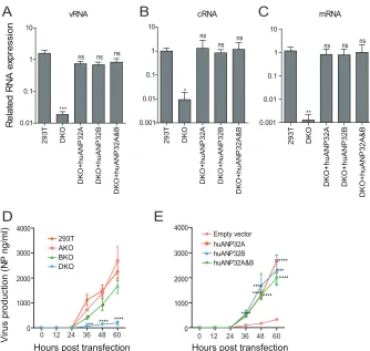
FIG 3huANP32A&B determine viral RNA replication efficiency and viral production. (A to C) Wild-type 293T
or DKO cells were transfected with the H1N1SC09minigenome reporting system, together with 20 ng of
empty vector, huANP32A, huANP32B, or huANP32A plus huANP32B. The cells were incubated at 37°C for 24 h before reverse transcription, followed by quantitative PCR (qRT-PCR) for vRNA, mRNA, and cRNA of the
luciferase gene. Error bars represent the SD from three independent experiments (NS, not significant;*,
P⬍0.05;**,P⬍0.01;***,P⬍0.001;****,P⬍0.0001). (D and E) Replication kinetics of H1N1SC09in ANP32
knockout cells. 293T, AKO, BKO, and DKO cells in 6-well plates were transfected with 0.5g each of the
eight plasmids of H1N1SC09(D), or, in DKO cells, 40 ng of huANP32A, 40 ng of huANP32B, 20 ng each of
huANP32A and huANP32B, or 40 ng of empty vector was cotransfected with the eight plasmids of H1N1SC09
(E). The cells were cultured at 37°C, and supernatants were collected at 0, 12, 24, 48, and 60 h posttrans-fection and subjected to virus production assay by ELISA as described in Materials and Methods. Error bars
indicate the SD from three independent experiments (*, P⬍0.05; **, P⬍0.01; ***, P⬍0.001; ****,
P⬍0.0001).
on November 6, 2019 by guest
http://jvi.asm.org/
[image:6.585.42.376.69.386.2]H1N1SC09 Emp ty ve ct o r ch AN P3 2 A ch AN P3 2 B huA N P 32A huA N P 3 2B dk A N P 32A ty A N P 3 2 A zf AN P3 2 A mu AN P3 2 A pgA N P 32A eqA N P 32A 0.01 0.1 1 10 100 H7N9AH13 Emp ty ve ct o r ch AN P3 2 A ch AN P3 2 B huA N P 32A huA N P 32B dk A N P 32A ty A N P 32A z fA N P 32A m uA N P 32A pgA N P 32A eqA N P 32A 0.001 0.01 0.1 1 10 100 H3N2GD11 Emp ty ve ct o r ch AN P3 2 A ch AN P3 2 B huA N P 32A huA N P 32B dk A N P 3 2A tyAN P3 2 A zf AN P3 2 A mu AN P3 2 A pgA N P 32A eqA N P 32A 0.001 0.01 0.1 1 10 100 H3N8XJ07 Emp ty ve ct o r ch AN P3 2 A ch AN P3 2 B huA N P 32A huA N P 32B dk A N P 32A ty A N P 32A z fA N P 32A mu AN P3 2 A pgA N P 32A eqA N P 32A 0.0001 0.001 0.01 0.1 1 H3N8JL89 Emp ty ve ct o r c hA N P 32A ch AN P3 2 B huA N P 32A h u AN P3 2 B dk A N P 32A ty A N P 3 2 A zf AN P3 2 A mu AN P3 2 A pgA N P 32A eqA N P 32A 0.001 0.01 0.1 1 10 H9N2ZJ12 Emp ty ve ct o r ch AN P3 2 A ch AN P3 2 B h u AN P3 2 A huA N P 32B d k AN P3 2 A ty A N P 32A z fA N P 32A mu AN P3 2 A p g AN P3 2 A e q AN P3 2 A 0.001 0.01 0.1 1 10 A B C D F E F ir e fly p o ly m e ra s e a c tiv ity nor m al iz ed t o R e ni lla F ire fly p o ly m e ra s e a c tiv ity nor m al iz e d t o R e ni lla F ir e fly p o ly m e ra s e a c tiv ity nor m al iz ed t o R e ni lla F ir e fly pol y m er as e ac tiv ity nor m al iz ed t o R eni lla F ir e fly pol y m e ra s e ac tiv ity nor m al iz ed t o R e ni lla ns ** **** **** **** ns *** **** **** **** **** **** **** **** **** **** ns **** ** ns **** **** **** **** **** **** ns **** *** ns **** **** ns ******** ************ ******** ns **** ****
ns ns ns ****
***
ns * ns
**** **** **************** **** **** **** E m p ty ve ct o r c h AN P3 2 A c hA N P 32B h u AN P3 2 A huA N P 32B d k AN P3 2 A ty AN P3 2 A z fAN P3 2 A m u AN P3 2 A p g AN P3 2 A e q AN P3 2 A 0.001 0.01 0.1 1 10 100 **** ******** **** **
ns ns ns ns
ns E m pt y v ec tor ch A N P 3 2 A ch A N P 3 2 B huA N P 32A huA N P 32B dk A N P 3 2A ty A N P 3 2 A zf A N P 3 2 A mu A N P 3 2 A pgA N P 32A eqA N P 32A 0.001 0.01 0.1 1 10 E m pt y v ec tor ch A N P 3 2 A ch A N P 3 2 B huA N P 32A huA N P 32B dk A N P 3 2A ty A N P 3 2 A zf A N P 3 2 A mu A N P 3 2 A pgA N P 32A eqA N P 32A 0.001 0.01 0.1 1 10 *** **** *** ************ **** *** ** ns ns **** **** **** **** **** **** **** **** ns
H7N9AH13 (PB2 K627E)
H3N8JL89 (PB2 E627K) H9N2ZJ12 (PB2 E627K)
G
H
J
I
β-Actin Flag
Empty vector chAN
P3 2 A ch AN P3 2 B huA N P 32A huA N P 32B d kAN P3 2 A zf AN P3 2 A ty AN P3 2 A pgA N P 32A eqA N P 32A m uAN P3 2A
FIG 4Support of influenza A viral replication by ANP32A or ANP32B from different species. (A) One microgram of each ANP32 plasmid was transfected into 293T cells using Lipofectamine 2000. At 48 h posttransfection, the cell lysates were analyzed using SDS-PAGE and Western blotting with antibodies
(Continued on next page)
on November 6, 2019 by guest
http://jvi.asm.org/
[image:7.585.60.360.69.704.2]E627K mutations in isolates of avian origin (horse H3N8
JL89or chicken H9N2
ZJ12)
dramatically changed viral fitness to mammalian ANP32 proteins (Fig. 4I and J). These
results revealed that ANP32A&B play important roles in influenza A viral replication
across different species and that mammalian ANP32 proteins provide poor support for
avian influenza viral RNA replication, which may be the major determinant for the
adaptation of influenza A virus to different host.
A novel 129-130 site vital for influenza polymerase activity in ANP32A&B.
Surprisingly, chicken ANP32B did not support any viral RNA replication, and mouse
ANP32A showed limited support to H1N1
SC09and H3N8
XJ07, indicating a functional loss
in these molecules (Fig. 4). The ANP32 protein family has a conserved structure
containing an N-terminal leucine-rich repeat (LRR) and a C-terminal low-complexity
acidic region (LCAR) domain (43). ANP32A&B have been reported to interact directly
with the polymerase complex (7, 42). The key functional domains of ANP32 proteins
remain largely unknown. We noticed that the chicken ANP32B (chANP32B) was
func-tionally inactive and hypothesized that certain amino acids may be responsible for this
phenotype. Alignment of huANP32B, pgANP32B, and chANP32B revealed scattered
substitutions on the proteins (Fig. 5A). Chimeric clones between chicken and human
ANP32B were constructed and evaluated (Fig. 5B). We found that the replacement of
amino acids 111 to 160 of huANP32B with those of chANP32B aborted the activity of
this protein, while chANP32B with the human fragment from amino acids 111 to 161
(fragment 111-161) gained the ability to boost viral polymerase activity (Fig. 5C and D).
Further comparing of ANP32B fragments 111-161 for chickens, humans, and pigs
showed eight amino acid substitutions (Fig. 6A). Of these, a combined mutation
N129I/D130N, but not others, on huANP32B impaired H1N1 polymerase activity (Fig.
6B), and the N129I showed stronger impairment than did the D130N mutation (Fig. 6C).
We confirmed this phenotype on H7N9 polymerase activity (Fig. 6D). The 129-130
mutations also aborted the support of huANP32A of H1N1 and H7N9 polymerases (Fig.
6E and F). Furthermore, substitutions at amino acids 129 and 130 in chANP32A and
chANP32B reversed the support of these proteins of the H7N9 virus human isolate (Fig.
6G). Interestingly, in the context of a chicken H7N9 viral minigenome, although the
mutation of amino acids 129 and 130 of chANP32A impaired the polymerase activity,
the chANP32B reverse mutation did not restore its support to chicken-like H7N9 viral
RNA replication (Fig. 6H), indicating that chicken virus replication may require both the
129-130 site and an extra 33-amino-acid peptide as in chANP32A. Most terrestrial
mammalian ANP32A&B proteins have a 129-ND-130 signature, but most of the avian
ANP32Bs have IN-130 residues (Table 1). Murine ANP32A (muANP32A) has
129-NA-130, and this may explain the impaired support of H1N1 polymerase by muANP32A
(Fig. 4 and 6I). To further confirm the function of the 129-130 site, we made inactivating
mutations of huANP32A and huANP32B at the sites 129 and 130 (N129I, D130N) on the
293T genome using CRISPR-Cas9 mediated recombination (Fig. 7A to D); the double
mutated proteins expressed well and fully impaired the replication of both H1N1
SC09and H7N9
AH13(Fig. 7E and F). Interestingly, the single ANP32A or ANP32B mutations led
to slightly decreased polymerase activities compared to wild-type 293T cells,
suggest-ing competition between the mutated proteins and the native proteins for viral
polymerase binding.
FIG 4Legend (Continued)
against Flag peptide and-actin. (B to J) Twenty nanograms of either empty vector or ANP32A or
ANP32B from one of several different species was cotransfected with a minigenome reporter,Renilla
expression control, and human influenza virus polymerase from H1N1SC09 (B), H7N9AH13(C), canine
influenza virus H3N2GD11(D), equine influenza virus H3N8XJ07(E) and an avian origin equine influenza
virus H3N8JL89(F), avian influenza virus H9N2ZJ12(G), H7N9AH13with PB2 K627E (H), H3N8XJ07with PB2
E627K (I), or H9N2ZJ12with PB2 E627K (J) into DKO cells. The luciferase activity was measured 24 h later.
Data indicate the firefly luciferase gene activity normalized to that ofRenillaluciferase activity. Statistical
differences between cells are indicated, following a one-way ANOVA and subsequent Dunnett’s test (NS,
not significant;*,P⬍0.05;**,P⬍0.01;***,P⬍0.001;****,P⬍0.0001). Error bars represent the SEM of
the replicates within one representative experiment. dk, duck; ty, turkey; zf, zebra finch; mu, mouse; pg, pig; eq, equine.
on November 6, 2019 by guest
http://jvi.asm.org/
The 129-130 site in ANP32B affects its interaction with viral polymerase.
It has
been reported that ANP32A variants interact with the polymerase subunit PB2 and that
the avian ANP32A can enhance avian viral RNP assembly in human cells (42). ANP32A
and ANP32B can interact with polymerase only when three subunits of the polymerase
are present (7). ANP32B proteins from different species have a conserved structure that
comprises a N-terminal LRR region and a C-terminal LCAR (Fig. 8A). To investigate the
function of the 129-130 site in ANP32B in the interaction of this protein with viral
polymerase, we used ANP32B and its variants to cotransfect with the WSN minireplicon
into DKO cells. Coimmunoprecipitations detected strong interactions between
huANP32A and the polymerase subunits PB1 and PA (Fig. 8B, lane 1) but weak
interactions when chANP32B was present (Fig. 8B, lane 2). No interactions were
detected when PB1 was absent (Fig. 8B, lane 3), indicating that this interaction occurred
between ANP32B and the viral trimeric polymerase complex, which is consistent with
previous findings (7, 41). As controls, we also observed that the LCAR truncations of
huANP32B were unable to interact with the viral polymerase (Fig. 8B, lanes 4 to 6), in
agreement with previous observations (41). When we mutated the functional human
huANP32B
chANP32B
chANP32B(1-161)
chANP32B(162-262)
chANP32B(1-41)
chANP32B(42-110)
chANP32B(111-161)
chANP32B(hu111-161)
41 110 166 251 262
LRR(Leucine-rich repeat) 1 LRR2
LRR3 LRR4
LRRT LCAR
LCAR
huA
N
P
32B
c
h
AN
P3
2
B
c
hA
N
P
32B
(1-161)
c
hA
N
P
32B
(162-262)
0.01 0.1 1 10 100
h
u
AN
P3
2
B
ch
AN
P3
2
B
ch
AN
P3
2
B
(1
-4
1
)
ch
AN
P3
2
B(4
2
-1
1
0
)
chA
N
P
32B
(111-1
61)
ch
A
N
P
32B
(hu111-1
61)
0.01 0.1 1 10 100 ns
**** ****
**** ****
ns ns ns
F
ir
ef
ly
pol
y
m
er
as
e ac
ti
vi
ty
nor
m
a
liz
e
d
t
o R
e
ni
lla
A
B
C
D
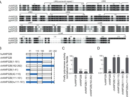
FIG 5Mapping of crucial sites in ANP32 proteins that influence viral polymerase activity. (A) ANP32B of chicken, human, and pig sequences were aligned using Geneious R6 software. The notches are marked with dashes. The similarity of amino acid identity is highlighted in different colors. (B) Schematic diagram of chimeric clones between chicken and human ANP32B, constructed according to the known domains (separated by dotted lines: 1 to 41 amino acids, LRR1 region; 42 to 110 amino acids, LRR2,3&4 region; 111 to 161 amino acids, LRRCT region; 162 to 251 amino acids, LCAR region [as showed in panel A]). The bars indicate the origins of the genes by color as follows: white, huANP32B; and blue, chANP32B. (C and D) Chimeric clones were cotransfected with minigenome
reporter,Renillaexpression control, and H1N1SC09polymerase into DKO cells. The luciferase activity was measured 24 h later. Data indicate the polymerase
activity normalized to that ofRenilla. The statistical differences between samples are indicated, following one-way ANOVA and subsequent Dunnett’s test (NS,
not significant;****,P⬍0.0001). Error bars represent the SEM of replicates within a representative experiment.
on November 6, 2019 by guest
http://jvi.asm.org/
[image:9.585.45.542.72.441.2]ANP32B at the 129-130 site to the chicken ANP32B signature, the huANP32B lost the
ability to interact with viral polymerase. Conversely, chANP32B gained the ability to
coimmunoprecipitate with viral polymerase when its 129-130 sites were mutated to the
human ANP32B signature (Fig. 8C). Together, these results revealed a fundamental
function of ANP32A and ANP32B in supporting influenza A viral polymerase activity and
Chicken
Human
Pig
F ire fl y p o ly m e ra s e a c ti vi ty nor m a liz ed t o R e ni lla F ir ef ly pol y m er as e ac ti vi ty nor m a liz ed t o R eni llaH1N1
SC09H1N1
SC09H7N9
AH13
H7N9
AH13
H7N9
AH13H7N9
AH13(K627E)
****H
G
F
E
D
C
A
B
huAN P32B huAN P32B E113P huAN P32B K116H huAN P32B N 127M huAN P32B N 129I huAN P 32B D 1 30N huAN P 32B I 137T huAN P 32B R 150A huANP32B A160P Em
p ty ve ct o r 0.01 0.1 1 10 100 1000 0.01 0.1 1 10 100 1000 **** ns **** **** ns *** *** * ns ns
ns ns ns
huAN P32B c hAN P32B huAN P32B E113P huAN P32B K116H huAN P32B N 1 27M huAN P32B N 1 29I /D 1 30N huAN P32B I 137T huAN P32B R 150A huAN
P32B A160P Em
p ty ve ct o r 0.01 0.1 1 10 100 1000 **** **** **** *
ns ns ns * ns
huA N P 3 2B h u ANP3 2 B N1 2 9 I huA N P 32B D 130N E m pt y vect or **** **** ** huAN P32B N 1 29I /D 1 30N huAN P32B N 1 29I /D 1 30N 0.01 0.1 1 10 100 1000
I
*** **** **** * m u AN P32A m u AN P32A A 1 3 0 D huAN P32A huAN P32B E m p ty ve ct o r c h AN P32A ch A N P 3 2 A N 1 2 9 I c h AN P32A D 1 30N ch A N P 3 2 A N 129I /D 130N c h AN P32B c h AN P32B I 129N c h AN P32B N 1 30D c h AN P32B I 129N /N 130D E m p ty ve ct o r 0.01 0.1 1 10 100 **** **** *** **** c hAN P32A c h AN P32A N 129I c hAN P32A D 130N c hAN P32A N1 2 9 I/ D1 3 0 N c h AN P32B c h A N P 3 2 B I1 2 9 N c hAN P32B N 130D E m p ty ve ct o r 0.01 0.1 1 10 100 huAN P32A huAN P32A N 129I huAN P32A D 130N E m p ty ve ct o r 0.01 0.1 1 10 100 **** **** **** huAN P32A N 1 29I /D 1 30N c hAN P32B I 129N /N 130D **** **** **** **** ns **H1N1
SC09 huAN P32A huAN P32A N 129I huAN P32A D 1 30N huAN P32A N 129I /D 1 30N E m p ty ve ct o r 0.01 0.1 1 10 100 1000H1N1
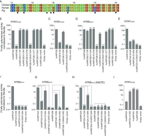
SC09 ******** **** ****FIG 6Key amino acids in ANP32A&B determine the activity of influenza viral polymerases. (A) Amino acid sequence comparison for chicken, human, and pig ANP32B proteins (amino acids 110 to 161). The amino acid sequences are available in GenBank. The black dots indicate the different amino
acid within these proteins. (B to D) huANP32B mutants were cotransfected with polymerase plasmids from H1N1SC09(B and C) or H7N9AH13(D), plus
a minigenome reporter and a Renillaluciferase control, into DKO cells. The luciferase activity was then assayed 24 h after transfection. (E and F)
huANP32A mutants were cotransfected with polymerase plasmids from H1N1SC09(E) or H7N9AH13(F), plus a minigenome reporter and aRenillaluciferase
control, into DKO cells. (G and H) chANP32A or chANP32B mutants were cotransfected with polymerase plasmids from H7N9AH13with either PB2 627K
(G) or 627E (H), respectively. (I) Mutated muANP32A or human ANP32 proteins were cotransfected with minigenome reporter,Renillaexpression control,
and H1N1SC09polymerase into DKO cells. Luciferase activity was analyzed as mentioned above. The data indicate the firefly luciferase gene activity
normalized to that of theRenillaluciferase activity. Statistical differences between cells are indicated, following one-way ANOVA and subsequent
Dunnett’s test (NS, not significant;*,P⬍0.05;**,P⬍0.01;***,P⬍0.001;****,P⬍0.0001). Error bars represent the SEM of the replicates within a
representative experiment.
on November 6, 2019 by guest
http://jvi.asm.org/
[image:10.585.44.542.73.544.2]a novel 129-130 site of ANP32A&B in different species that may influence influenza virus
replication.
DISCUSSION
Although the host factors that are involved in influenza A virus replication have
been long investigated, and genome-wide screening has shown that many host
proteins interact with viral polymerase, the key mechanisms that determine viral
polymerase activity in different cells remain largely unclear. ANP32A and ANP32B have
been previously identified binding with viral polymerase and promoting human
influ-enza viral vRNA synthesis (7). Furthermore, a recent study showed for the first time that
chicken ANP32A can rescue avian polymerase activity in human cells because the
chicken ANP32A harbors an extra stretch of 33 amino acids that is absent in mammalian
ANP32A (8). However, the potencies of ANP32A and ANP32B (as well as other host
factors) in viral replication are not well investigated in different hosts. In our study, we
used CRISPR/Cas9 knockout cells to screen the candidate host proteins involved in viral
RNA replication. We identified that ANP32A and ANP32B are key host cofactors that
determine influenza A virus polymerase activity. Without ANP32A&B, the viral
polymer-ase activity decrepolymer-ased by 10,000-fold, and the viral infectivity decrepolymer-ased by
⬎
10,000-fold. In contrast, the DDX17 knockout cells showed
⬃
3-fold-decreased viral polymerase
activity (Fig. 1A), which was in agreement with prior reports (29). We found that
ANP32A or ANP32B can independently restore viral polymerase activity, indicating that
both have a similar function in viral replication (7).
Influenza viruses and their hosts have a long coevolution history. ANP32 family
members are expressed in animal and plant cells, with both conserved LRR and LCAR
domains (50). Three conserved ANP32 members (ANP32A, ANP32B, and ANP32E) in
vertebrates were reported to have several functions in cells. We found that knockout of
both ANP32A and ANP32B abolished influenza viral replication, whereas ANP32E may
not be involved in viral replication. Interestingly, we found that the chicken ANP32A
keeps the conserved function to support both avian and mammalian virus polymerase
activity, while human ANP32A or ANP32B did not support avian viral replication. Thus,
we demonstrate here that ANP32A and ANP32B are key host factors that play a
fundamental role in influenza A viral RNA replication.
[image:11.585.42.373.83.223.2]A recent study revealed that avian ANP32A has a hydrophobic SUMO interaction
motif (SIM) in an extra 33-amino-acid insert region which connects to host SUMOylation
to specifically promote avian viral polymerase activity (41). However, this SIM-like
domain is not present in all of the ANP32B proteins from different hosts, nor is it found
in mammalian ANP32A. We showed that most ANP32Bs from different species are
functional as ANP32As in support of viral replication, but chANP32B is naturally unable
to support polymerase activity (Fig. 4). We finally found that the amino acids 129I and
130N chANP32B are responsible for the loss of this function. Sequence analysis and
TABLE 1Summary of amino acids at positions 129 and 130 in ANP32 proteins
Host
Residue at amino acid position 129 or 130a
ANP32A ANP32B
129 130 129 130
Gallus N D I N
Anas platyrhynchos N D I N
Homo sapiens N D N D
Gorilla gorilla N D N D
Felis catus N D N D
Canis lupus familiaris N D N D
Bos taurus N D N D
Sus scrofa N D N D
Mus musculus N A S D
Equus caballus N D N D
aThe sequences of different species ANP32 proteins were retrieved from the National Center for
Biotechnology Information.
on November 6, 2019 by guest
http://jvi.asm.org/
mutagenesis studies suggest that the 129-130 sites are important for maintaining the
function of avian ANP32A and human ANP32A/B in viral replication (Fig. 6). The
coimmunoprecipitation assay showed that mutations on these 129-130 sites changed
the interaction efficiency between the ANP32 proteins and viral polymerase complex
(Fig. 8). Mutating the 129-130 sites in chANP32B to the functional signature 129N and
130D enables chANP32B to support polymerase activity in human viruses, but not
chicken viruses with PB2 627E, indicating that the ANP32B may undergo selection in
two areas during coevolution with the avian influenza virus: chANP32B does not harbor
an extra insert as chANP32A and the 129-130 mutations. This result also suggests that
avian ANP32A is the only protein from the avian ANP32 family to support avian viral
replication. Currently, the vaccine is the best way to control avian influenza virus, such
H1N1SC09
H7N9AH13
ANP32A
ANP32B
β-Actin
huANP32A
huANP32B
E V T N L V N L I N I N Y R E N Y N V V E V T N L I N Y R E N V E V T N L I N Y R E N V
E V T N L I N Y R E N V E V T N L N D Y R E N V E V T N L N D Y R E N V
F
ire
fl
y
p
o
ly
m
e
ra
s
e
a
c
ti
vi
ty
nor
m
a
liz
e
d
t
o
R
e
ni
lla
F
ir
ef
ly
pol
y
m
er
as
e ac
ti
vi
ty
nor
m
a
liz
e
d
t
o R
eni
lla
293T
ASM
BSM
DSM
DKO
**
** ** **
****
**** **** ****
0.001 0.01 0.1 1 10 100 1000 0.01 0.1 1 10 100
A
B
C
D
E
F
ANP32A
ANP32B
ANP32A
ANP32B BSM ASM
DSM
ASM BSM DSM
293T
FIG 7Amino acid N129I/D130N substitutions into ANP32A&B impaired influenza polymerase activity in 293T cells. N129I/D130N substitutions of huANP32A and ANP32B on 293T chromosome were generated by the CRISPR/Cas9 system. Positive colonies were identified by sequencing and Western blotting. (A) ANP32A stable mutated cells (ASM) were identified carrying the N129I/D130N substitutions on ANP32A but not on ANP32B. (B) ANP32B stable mutated cells (BSM) were identified harboring the N129I/D130N substitutions on ANP32B but not on ANP32A. (C) Double stable mutated cells (DSM) had N129I/D130N substitutions on both ANP32A&B. (D) The endogenous protein expressions of
different cell lines were identified by Western blotting with antibodies against-actin, huANP32A, and huANP32B. (E and F) Selected 293T
cell lines were transfected with firefly minigenome reporter,Renillaexpression control, and polymerase from H1N1SC09(E) or H7N9AH13(F).
Cells were assayed for luciferase activity. The data indicate the firefly luciferase gene activity normalized to that of theRenillaluciferase
activity. Statistical differences between cells are given, following a one-way ANOVA and subsequent Dunnett’s test (NS, not significant;**,
P⬍0.01;****,P⬍0.0001). Error bars represent the SEM of the replicates within a representative experiment.
on November 6, 2019 by guest
http://jvi.asm.org/
[image:12.585.41.487.66.477.2]as H7N9 virus (55, 56). However, the 129-130 substitution of chANP32A could be used
in the future as a novel target to develop transgenic chickens that may be totally
resistant to influenza virus infection.
Together, our data give new insights into the functions of ANP32A and ANP32B. The
129-130 substitution could be used as a novel target to modify the genomes of animals
to develop influenza A-resistant transgenic chickens or other animals, which will benefit
the husbandry industry, as well as animal and human health. Further investigation into
the molecular mechanisms that determine how ANP32 proteins work with the viral
polymerase complex, the structure of the ANP32 and vRNP complex, and the fitness of
different virus subtypes, for example, would contribute to our understanding of viral
pathogenesis and host defense.
MATERIALS AND METHODS
Cells, viruses, and plasmids.Human embryonic kidney (293T; ATCC CRL- 3216) and Madin-Darby canine kidney (MDCK; CCL-34) cells were maintained in Dulbecco modified Eagle medium (DMEM; HyClone) with 10% fetal bovine serum (FBS; Sigma), and 1% penicillin and streptomycin (Gibco) and kept
at 37°C in 5% CO2. Certain reagents were kindly provided by the indicated individuals: polymerase
plasmids of H1N1 human influenza A virus A/Sichuan/01/2009 (H1N1SC09) and H7N9 A/Anhui/01/2013
(H7N9AH13) were provided by Hualan Chen; H1N1 human influenza virus A/WSN/1933 (WSN) was
provided by Yoshihiro Kawoaka; H3N2 canine influenza virus A/canine/Guangdong/1/2011 (H3N2GD12)
was provided by Shoujun Li from China Southern Agriculture University; and H9N2 avian influenza virus
A/chicken/Zhejiang/B2013/2012 (H9N2ZJ12) was provided by Zejun Li from Shanghai Veterinary Research
Institute of CAAS. H3N8 equine influenza viruses A/equine/Jilin/1/1989 (H3N8JL89) and
A/equine/Xinji-ang/1/2007 (H3N8XJ07) were preserved in our lab. The reverse genetics system based on the pBD vector
for the H1N1SC09virus was established in Hualan Chen’s lab. The pCAGGS plasmids containing full-length
ANP32A isoforms of several species were generated by gene synthesis (Synbio Technologies, China)
F
lag
-I
P
In
p
u
t
ANP32
PB1
PA
ANP32
PB1
PA
hu
A
N
P
3
2B-PB1(-)
hu
A
N
P
3
2B-Δ216
chA
N
P
32B
hu
A
N
P
3
2B-Δ190
hu
A
N
P
3
2B-Δ165
hu
A
N
P
3
2B
B
A
C
1 2
3 4
5
6
In
p
u
t ANP32
PB1
PA
ANP32
PB1
PA
hu
A
N
P
32B
h
u
A
N
P
32B
N
1
29I
/D
130N
chA
N
P
32B
chA
N
P
32B
I
129N
/N
130D
F
lag
-I
P
LRR1
LRR2 LRR3 LRR4 LRRCT
161
40
64
84
110
251
165
190
216
129/130
NLS
N- cap
LRR Domain
huANP32B
LCAR
[image:13.585.45.398.72.388.2]according to the sequences deposited in GenBank, including chicken ANP32A (chANP32A,XM_413932.5, XP_413932.3), human ANP32A (huANP32A,NM_006305.3,NP_006296.1), zebra finch ANP32A (zfANP32A, XM_012568610.1,XP_012424064.1), duck ANP32A (dkANP32A,XM_005022967.1,XP_005023024.1),
tur-key ANP32A (tyANP32A,XM_010717616.1,XP_010715918.1), pig ANP32A (pgANP32A,XM_003121759.6,
XP_003121807.3), mouse ANP32A (muANP32A, NM_009672.3, NP_033802.2), equine ANP32A
(eqANP32A, XM_001495810.5, XP_001495860.2), chicken ANP32B (chANP32B, NM_001030934.1,
NP_001026105.1), and human ANP32B (huANP32B,NM_006401.2,NP_006392.1). Site-directed mutants of these sequences were generated using overlapping PCR and identified by DNA sequencing. Mutants of pcAGGS-huANP32B-Δ216/190/165 and pcDNA3.1-PA-V5 were constructed according to the online
In-Fusion HD cloning kit user manual (http://www.clontech.com/CN/Products/Cloning_and_Competent_
Cells/Cloning_Kits/xxclt_searchResults.jsp). Briefly, the fragments of the pcAGGS/pcDNA3.1 vector and each target gene were amplified with a 15-bp homologous arm and then fused using In-Fusion HD enzyme (Clontech, Felicia, CA). To create the huANP32B-Δ216/190/165 plasmids, pcAGGS-huANP32B was used as a template to amplify the pcAGGS vector. This sequence was then fused with different truncated huANP32B fragments (huANP32B-Δ216/190/165). To obtain pcDNA3.1-PA-V5
plas-mid, pBD-H1N1SC09-PA was used as the template to amplify the PA-V5 sequence and then fused with
pcDNA3.1 vector.
Knockout cell lines. To generate knockout cell lines for host proteins BUB3 (AF081496.1),
CLTC (NM_004859.3), CYC1 (CR541674.1), NIBP (BC006206.2), ZC3H15 (NM_018471.2), C14orf173
(DQ395340.1), CTNNB1 (NM_001904.3), ANP32A (NM_006305.3), ANP32B (NM_006401.2), SUPT5H (U56402.1), HTATSF1 (NM_014500), and DDX17 (NM_006386,NM_001098504) (3, 4, 29), the gRNA design
tool (http://crispr.mit.edu/) was used for gRNAs design and off-target prediction (57). DNA fragments that
contained the U6 promoter, gRNAs specific for host factors, a guide RNA scaffold, and U6 termination signal sequence were synthesized and subcloned into the pMD18-T backbone vector. The Cas9-eGFP expression plasmid (pMJ920) was a gift from Jennifer Doudna (Addgene, plasmid 42234) (58). Briefly,
293T cells in 6-well plates were transfected with 1.0g of pMJ920 plasmids and 1.0g of gRNA
expression plasmids by Lipofectamine 2000 transfection reagent (Invitrogen, catalog no. 11668-027) using the recommended protocols. Green fluorescent protein (GFP)-positive cells were sorting by fluorescence-activated cell sorting (FACS) at 48 h posttransfection, and then monoclonal knockout cell lines were screened by Western blotting and/or DNA sequencing.
Generation of a site-directed, amino-acid-substituted 293T cell line. High-efficiency guide sequences for ANP32A and ANP32B that bind upstream and downstream with close proximity to the target (129/130 ND) were chosen. The gRNA expression plasmids were constructed as described above. An 80-nucleotide oligonucleotide with the desired mutations at the target site was used as the donor
DNA. 293T cells were transfected with 1g of pMJ920 plasmids, 1g of gRNA expression plasmids, and
50 pmol of donor DNA. After 48 h, GFP-positive cells were isolated by FACS, and site-directed mutagen-esis clones were identified after screening by sequencing and Western blotting with anti-PHAP1 antibody (ab51013) and anti-PHAPI2/APRIL antibody EPR14588 (ab200836). Cell lines with double gene mutations were generated by second-round transfection and selection.
Polymerase assay.A minigenome reporter, which contains the firefly luciferase gene flanked by the noncoding regions of the influenza hemagglutinin gene segment with a human polymerase I promoter and a mouse terminator sequence (59) was transfected with viral polymerase and NP expression plasmids to analyze the polymerase activity. Mutants of PB2 genes were generated using overlapping PCR and identified by DNA sequencing. To determine the effect of host proteins on viral polymerase activity, 293T or different host protein knockout 293T cells in 12-well plates were transfected with plasmids of the PB1 (80 ng), PB2 (80 ng), PA (40 ng) and NP (160 ng), together with 80 ng of minigenome
reporter and 10 ng ofRenillaluciferase expression plasmids (pRL-TK, kindly provided by J. Luban), using
Lipofectamine 2000 transfection reagent (Invitrogen) according to the manufacturer’s instructions. Cells
were incubated at 37°C. The cells were lysed with 100l of passive lysis buffer (Promega) at 24 h after
transfection, and the firefly andRenillaluciferase activities were measured using a dual-luciferase kit
(Promega) with a Centro XS LB 960 luminometer (Berthold Technologies). The function of ANP32 was examined using a polymerase assay by cotransfection of DKO cells with different ANP32 proteins for 24 h.
All of the experiments were performed independently at least three times. Results represent means⫾
the standard errors of the mean (SEM) of the replicates within one representative experiment. The expression levels of polymerase proteins on different cell lines were detected by Western blotting, using specific mouse monoclonal antibodies for the NP and PB1 proteins and anti-V5 tag antibody (ab27671) for the PA-V5 protein.
RNA isolation, reverse transcription, and quantification by RT-PCR.Total RNA from 293T cells was extracted using an RNeasy minikit (Qiagen) according to the manufacturer’s instructions. For the synthesis of first-strand cDNA derived from firefly luciferase RNAs driven by influenza polymerase, equal concentrations of RNA were subjected to cDNA synthesis using a reverse transcription (RT) kit (Prime-Script RT reagent kit with a gDNA Eraser [Perfect Real Time], catalog no. RR047A). Primers used in the RT
reaction were as follows: 5=-CATTTCGCAGCCTACCGTGGTGTT-3=for the firefly luciferase vRNA, 5=-AGTA
GAAACAAGGGTG-3=for the firefly luciferase cRNA, and oligo-dT20 for the firefly luciferase mRNA (60).
The cDNA samples were subjected to quantification by real-time PCR with the specific primers F
(5=-GATTACCAGGGATTTCAGTCG-3=) and R (5=-GACACCTTTAGGCAGACCAG-3=) using SYBR Premix Ex Taq
II (Tli RNase H Plus; TaKaRa catalog no. RR820A). The fold change in RNA was calculated by
double-standard curve methods, and-actin served as an internal control.
Quantitative ELISA for determination of virus production.The ELISA has been previously
of the mouse monoclonal anti-IAV NP protein antibody in phosphate-buffered saline (PBS), incubated overnight at 4 or 37°C for 2 h. The plate was washed three times with washing buffer (PBS containing
0.1% Tween 20 [PBST]) and blocked with 200l of 5% calf serum at 37°C for 2 h. After three washes with
PBST buffer, 100-l portions of virus or virus-like particle (VPL) samples were added in dilution buffer (PBS
containing 10% calf serum and 0.1% Triton X-100), followed by incubation at 37°C for 1 h. The plate was
then washed, and 100l of a 1:2,000 dilution of horseradish peroxidase-conjugated anti-IAV NP protein
monoclonal antibody was added. After incubation at 37°C for 0.5 h, the plate was washed again, followed by incubation with freshly prepared TMB peroxidase substrate (Galaxy Bio, Beijing, China) for 10 min at
room temperature. The reaction was stopped by adding 2 M H2SO4, and the optical density at 450 nm
was measured using a VersaMax microplate reader (BioTek, Winooski, VT). Dilution buffer was taken as a blank control, and the purified IAV-NP protein was double diluted eight times in dilution buffer to obtain the standard curve. The virus production was calculated using the standard curve.
Influenza virus infection and infectivity.Human influenza virus WSN was rescued from a
12-plasmid rescue system (61). Briefly, a 293T culture in a 6-well plate was transfected with 0.1g each of
pPolI plasmids and 1g each of pCAGGS-NP, pCAGGS-PA, pCAGGS-PB1, and pCAGGS-PB2 using
Lipo-fectamine 2000 (Invitrogen) in Opti-MEM. At 8 h posttransfection, the medium was changed to DMEM plus 10% FBS. The supernatant was harvested after 48 h and injected into 9-day-old specific-pathogen-free embryonated eggs for virus propagation. Eggs were incubated at 35°C for 72 h, the allantoic fluid from eggs was detected by hemagglutination assay, and the titers of virus on 293T cells were determined using the method of Reed and Muench (62). The 293T cells, huANP32A knockout 293T cells (AKO cells), huANP32B knockout 293T cells (BKO cells), and huANP32A&B double-knockout 293T cells (DKO) were infected at a multiplicity of infection (MOI) of 0.01 for 1 h, washed twice with PBS, and cultured at 37°C in Opti-MEM containing 0.5% FBS and tosylsulfonyl phenylalanyl chloromethyl ketone (TPCK)-trypsin (Sigma) at 1 mg/ml. At the indicated time points, the culture supernatant was harvested, and the virus titers in MDCK cells were determined as described above.
Immunoprecipitation and Western blotting. For immunoprecipitation and Western blotting, transfected cells were lysed using an ice-cold lysis buffer (50 mM HEPES-NaOH [pH 7.9], 100 mM NaCl,
50 mM KCl, 0.25% NP-40, 1 mM dithiothreitol) and centrifuged at 13,000⫻gand 4°C for 10 min. After
centrifugation, the crude lysates were incubated with anti-FLAG M2 magnetic beads (Sigma-Aldrich, catalog no. M8823) or anti-NP magnetic beads (MCE protein A/G magnetic beads and NP antibody from our lab) at 4°C for 2 h. After incubation, the resins were collected using a magnetic separator and washed
three times with PBS. The resin-bound materials were eluted using a 3⫻Flag peptide or by boiling in the
SDS-PAGE loading buffer, subjected to SDS-PAGE, and then transferred onto nitrocellulose membranes. Membranes were blocked with 5% milk powder in Tris-buffered saline (TBS) for 2 h. Incubation with the first anti-mouse antibody (anti-Flag antibody from Sigma [catalog no. F1804], anti-V5 antibody from Abcam [catalog no. ab27671], and anti-NP and PB1 antibody from our lab) was performed for 2 h at room temperature, followed by three washes with TBST. The secondary antibody (Sigma, 1:10,000) was then applied, and the samples were incubated at room temperature for 1 h. Subsequently, the membranes were washed three times for 10 min with TBST. Signals were detected using an Odyssey imaging system (LI-COR, Lincoln, NE).
Statistics.Statistical analysis was performed in GraphPad Prism (v5; GraphPad Software). Statistical differences between groups were assessed by one-way analysis of variance (ANOVA), followed by a Dunnett’s post test. All the experiments were performed independently at least three times. Error bars represent standard deviations (SD) or standard errors of the mean (SEM) in each group, as indicated in
the figure legends (NS, not significant;P⬎0.05;*,P⬍0.05;**,P⬍0.01;***,P⬍0.001;****,P⬍0.0001).
ACKNOWLEDGMENTS
We thank W. Barclay, S. J. Li, Z. J. Li, J. Luban, and J. Doudna for providing plasmids
and advice. We thank C. J. Li and D. M. Zhao for helpful discussions.
This study was supported by grants from the National Natural Science Foundation
of China to H.C. (no. 31521005) and Z.Z. (no. 31702269).
REFERENCES
1. Brass AL, Huang IC, Benita Y, John SP, Krishnan MN, Feeley EM, Ryan BJ, Weyer JL, van der Weyden L, Fikrig E, Adams DJ, Xavier RJ, Farzan M, Elledge SJ. 2009. The IFITM proteins mediate cellular resistance to influenza A H1N1 virus, West Nile virus, and dengue virus. Cell 139:
1243–1254.https://doi.org/10.1016/j.cell.2009.12.017.
2. Konig R, Stertz S, Zhou Y, Inoue A, Hoffmann HH, Bhattacharyya S, Alamares JG, Tscherne DM, Ortigoza MB, Liang Y, Gao Q, Andrews SE, Bandyopadhyay S, De Jesus P, Tu BP, Pache L, Shih C, Orth A, Bonamy G, Miraglia L, Ideker T, Garcia-Sastre A, Young JA, Palese P, Shaw ML, Chanda SK. 2010. Human host factors required for influenza virus
repli-cation. Nature 463:813– 817.https://doi.org/10.1038/nature08699.
3. Watanabe T, Kawakami E, Shoemaker JE, Lopes TJS, Matsuoka Y, Tomita Y, Kozuka-Hata H, Gorai T, Kuwahara T, Takeda E, Nagata A, Takano R, Kiso M, Yamashita M, Sakai-Tagawa Y, Katsura H, Nonaka N, Fujii H, Fujii K, Sugita Y, Noda T, Goto H, Fukuyama S, Watanabe S,
Neumann G, Oyama M, Kitano H, Kawaoka Y. 2014. Influenza virus-host interactome screen as a platform for antiviral drug development.
Cell Host Microbe 16:795– 805.https://doi.org/10.1016/j.chom.2014
.11.002.
4. Bradel-Tretheway BG, Mattiacio JL, Krasnoselsky A, Stevenson C, Purdy D, Dewhurst S, Katze MG. 2011. Comprehensive proteomic analysis of influenza virus polymerase complex reveals a novel association with mitochondrial proteins and RNA polymerase accessory factors. J Virol
85:8569 – 8581.https://doi.org/10.1128/JVI.00496-11.
5. Shapira SD, Gat-Viks I, Shum BO, Dricot A, de Grace MM, Wu L, Gupta PB, Hao T, Silver SJ, Root DE, Hill DE, Regev A, Hacohen N. 2009. A physical and regulatory map of host-influenza interactions reveals pathways in
H1N1 infection. Cell 139:1255–1267.https://doi.org/10.1016/j.cell.2009
.12.018.
Khalil H, Ogilvie LA, Hess S, Maurer AP, Muller E, Wolff T, Rudel T, Meyer TF. 2010. Genome-wide RNAi screen identifies human host factors
cru-cial for influenza virus replication. Nature 463:818 – 822.https://doi.org/
10.1038/nature08760.
7. Sugiyama K, Kawaguchi A, Okuwaki M, Nagata K. 2015. pp32 and APRIL are host cell-derived regulators of influenza virus RNA synthesis from
cRNA. Elife 4:e08939.https://doi.org/10.7554/eLife.08939.
8. Long JS, Giotis ES, Moncorge O, Frise R, Mistry B, James J, Morisson M, Iqbal M, Vignal A, Skinner MA, Barclay WS. 2016. Species difference in ANP32A underlies influenza A virus polymerase host restriction. Nature
529:101–104.https://doi.org/10.1038/nature16474.
9. Subbarao EK, London W, Murphy BR. 1993. A single amino acid in the PB2 gene of influenza A virus is a determinant of host range. J Virol 67:1761–1764.
10. Xiao C, Ma W, Sun N, Huang L, Li Y, Zeng Z, Wen Y, Zhang Z, Li H, Li Q, Yu Y, Zheng Y, Liu S, Hu P, Zhang X, Ning Z, Qi W, Liao M. 2016. PB2-588 V promotes the mammalian adaptation of H10N8, H7N9 and H9N2 avian
influenza viruses. Sci Rep 6:19474.https://doi.org/10.1038/srep19474.
11. Song W, Wang P, Mok BW, Lau SY, Huang X, Wu WL, Zheng M, Wen X, Yang S, Chen Y, Li L, Yuen KY, Chen H. 2014. The K526R substitution in viral protein PB2 enhances the effects of E627K on influenza virus
replication. Nat Commun 5:5509.https://doi.org/10.1038/ncomms6509.
12. Sediri H, Schwalm F, Gabriel G, Klenk HD. 2015. Adaptive mutation PB2 D701N promotes nuclear import of influenza vRNPs in mammalian cells.
Eur J Cell Biol 94:368 –374.https://doi.org/10.1016/j.ejcb.2015.05.012.
13. Manz B, de Graaf M, Mogling R, Richard M, Bestebroer TM, Rimmelzwaan GF, Fouchier RA. 2016. Multiple natural substitutions in avian influenza A virus PB2 facilitate efficient replication in human cells. J Virol 90:
5928 –5938.https://doi.org/10.1128/JVI.00130-16.
14. Liu Q, Qiao C, Marjuki H, Bawa B, Ma J, Guillossou S, Webby RJ, Richt JA, Ma W. 2012. Combination of PB2 271A and SR polymorphism at posi-tions 590/591 is critical for viral replication and virulence of swine
influenza virus in cultured cells andin vivo. J Virol 86:1233–1237.https://
doi.org/10.1128/JVI.05699-11.
15. Zhang H, Li X, Guo J, Li L, Chang C, Li Y, Bian C, Xu K, Chen H, Sun B. 2014. The PB2 E627K mutation contributes to the high polymerase activity and enhanced replication of H7N9 influenza virus. J Gen Virol 95:779 –786. https://doi.org/10.1099/vir.0.061721-0.
16. Mok CK, Lee HH, Lestra M, Nicholls JM, Chan MC, Sia SF, Zhu H, Poon LL, Guan Y, Peiris JS. 2014. Amino acid substitutions in polymerase basic protein 2 gene contribute to the pathogenicity of the novel A/H7N9
influenza virus in mammalian hosts. J Virol 88:3568 –3576.https://doi
.org/10.1128/JVI.02740-13.
17. Hu M, Yuan S, Zhang K, Singh K, Ma Q, Zhou J, Chu H, Zheng BJ. 2017. PB2 substitutions V598T/I increase the virulence of H7N9 influenza A
virus in mammals. Virology 501:92–101.https://doi.org/10.1016/j.virol
.2016.11.008.
18