PERFORATION (MOP) ASSISTED ORTHODONTIC TOOTH
MOVEMENT BY VARYING THE FREQUENCY OF
MICRO-OSTEO PERFORATION (MOP)
- AN IN VIVO COMPARATIVE STUDY
A Di ss ertat ion submi tted
in partial fulf illment of t he requi rements for t he degr ee of
MASTER OF DENTAL SURGERY
SUBMITTED BY
Dr. VIJAYASRI.M
BRANCH - V
ORTHODONTICS AND DENTOFACIAL ORTHOPEDICS
CERTIFICATE
This is to cert if y t hat Dr.VIJAYAS RI.M, Post graduat e st udent (2016 -2019) in the Departm ent of Orthodontics and Dentofaci al
Ort hopedics (Branch V), Adhiparasakthi Dental Coll ege and Hospital,
Melm aruvathur – 603319, has done this diss ert ati on titl ed “CRITICAL
EVAL UAT ION O F MI CRO -OSTE O PE RFO RATIO N (MO P) ASSISTED O RTHO DONT IC TOOTH MOV E ME NT BY VARYING
THE FREQ UENCY O F
MICRO-OSTEO PERFORATION
(MO P) - AN IN VI VO CO MPARATI VE STUDY”
. Under our direct guidance and supervision in parti al fulfi llment of t he regul ations lai ddown b y The Tamil N adu Dr. M.G.R Medi cal Uni versi t y, C hennai –
600032.
Co-guid e G uide Dr.R.SUMANTH KUMAR. M.D.S.,
Reader,
Departm ent of Ort hodonti cs
and D ent ofaci al Ort hopedi cs
Dr.V.SUDHAKAR .M.D.S.,
Professor&Head,
Departm ent of Ort hodonti cs
and dent ofaci al orthopedi cs
Dr. S. THILL AINAYAGAM, M.D.S.
Principal,
I thank ALMI GHT Y for answering m y pra yers and making me what I am t oda y.
I t hank our C orrespondent Dr. T. Ramesh, MD., for his vital encouragem ent and s upport.
M y si ncere thanks t o Dr.S. Thillain ayagam M.D.S. , our beloved Principal, Adhiparas akt hi Dent al Col lege and Hos pit al, M elm aruvathur
for providing m e wi th the opport unit y t o utiliz e t he faciliti es of the
coll ege.
I would li ke t o express m y heart felt thanks to m y revered t eacher
Dr.V.Sudhakar M.D.S., for his guidance and encouragem ent during m y stud y. His encouragement was of great s upport in faci ng chall enges
that occurred during m y st ud y.
I avail t his opport unit y to expres s m y gr atitude and reverence to
m y Gui de & beloved teacher Dr. V.Su dhakar M. D.S., Professor and Head, Departm ent of Ort hodonti cs and dentofaci al orthopedi cs ,
Adhi paras akt hi Dental C oll ege and Hospit al, M elm aruvat hur. His
pursuit for perfection and imm ense s upport were a source of const ant
inspi ration to me and without whi ch s uch an endeavor would never
have mat eri alized .
It i s m y dut y t o express m y thanks to m y Co -Guide
Dr. R.Su manth Ku mar M.D.S., R eader, for his expert gui dance and moral support duri ng t he com plet ion of this stud y. I consider m ys elf
privileged, to have studi ed, worked and compl et ed m y dissert ation
under them i n the departm ent. I am extremel y thankful to m y faculti es
I t hank Mr.Maveeran Librari an and librar y staff
Mr.Selvaku mar, AdhiP arasakthi Dental C oll ege and Hospital Melm aruvathur for favours rendered .
I also wi sh t o thank m y Co-PG Dr. Sarath and m y s eniors
Dr.N.Karikal an , Dr.T.B alavign esh , Dr.S.Su ganya, and m y j uni ors
Dr. M.Anubharathy , Dr. Ki shor Ku mar.K.N, Dr. Deepak Baskaran .T .B., Dr. Lavanya. R.
I t hank Mrs. Govindhamal , Mrs.Manju, Mrs.Sarasvathi , Ms.Kavi tha non-t eachi ng staff from t he Department of Ort hodonti cs and Dent ofacial Orthopedi cs AdhiP aras akthi Dent al C ol lege and
Hospital M elm aruvat hur for favours rendered.
A speci al m enti on of thanks to all my patien ts for their cons ent , co-operation and participati on in t his stud y.
I owe m y gratitude to m y parents Mr.S.Mani ckavas agam &
Mrs. M.S elvi m y husband N.Rajaganes h , m y daughter R.Pranavasri, m y brot her M.Haris h Ku mar and all m y fam il y members and friends who stood besi de m e during m y tough t i mes and s acri fi ced s o much to
make m e what I am t oda y
Dr. VIJAYASRI.M
TITLE O F THE DISSERT ATIO N
CRI TIC AL EV AL U ATIO N O F MICRO -O STE-O P ERF O RA TIO N (MO P ) ASSI STE D O R TH O DO NTIC TO O T H MO VEMEN T B Y VA RY IN G TH E F REQ UE NCY O F MICRO - O STEO P ERF O RATIO N MO P - AN IN VIVO CO MP ARATI VE S T UDY .
PL ACE O F THE S TUDY Adhiparas akthi Dental Coll ege and Hospital, Mel maruvathu r -603319.
DURAT ION O F T HE CO URSE 3 Years
NAME O F THE GUIDE Dr.V.SUDHAKAR , MDS .,
NAME O F CO -GUI DE DrR.S UMANTH KUMAR , MDS.,
I hereb y decl are t hat no part of the di ssert ation wil l be ut ilized for
gai ning fi nancial ass istance or an y prom otion wit hout obtai ning pri or
permiss ion of the Princi pal, Adhiparasakthi Dent al college and
Hospital, Mel maruvathur -603319. In addition, I decl are t hat no part
of this work will be publi shed either i n print or in el ect ronic m edia
without the guides knowledge who have been acti vel y involved i n
diss ert ati on. The aut hor has the ri ght to res e rve for publ ish work s olel y
with the permiss ion of t he principal , Adhiparas akthi Dent al college and
Hospital, M elm aruvathur -603319.
Co-guide Guid e &
H ead of Dep artment
S.NO
TITLE
PAGE NO
1.
INTRODUCTION
1
2.
AIM AND OBJECTIVES
5
3.
REVIEW OF LITERATURE
6
4.
MATERIALS AND METHODS
25
5.
RESULTS
41
6.
DISCUSSION
50
7.
CONCLUSION
58
8.
BIBLIOGRAPHY
S.NO CONTE NT PAGE NO
1. ARMAME NTARI UM. 30
2. CONVENT IONAL CANINE BRACKE T. 31
3. CANINE BRACKE T WITH VE RTICAL SLOT. 31
4. SERPE NTINE HOO K. 31
5. TITANIUM MI NI IMPL ANT . 31
6.
DONT RI X GAU GE 32
7. DI GIT AL VERNIE R CALI PER. 32
8. IMPL ANT DRI VER. 32
9. IMPL ANTS 32
10. NITI CLOSE D COIL S PRING. 32
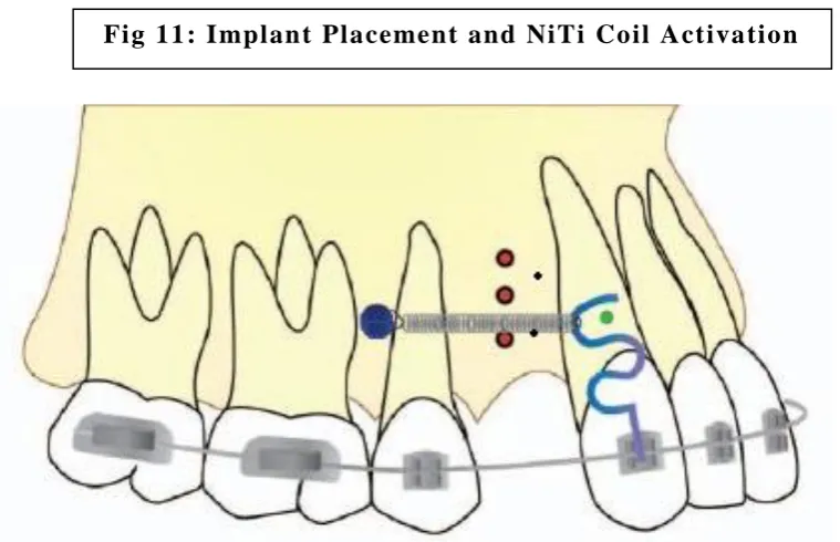
11. IMPL ANT PLACE MENT AND NITI COIL
SPRI NG ACT IVAT ION. 33
12. MO P PL ACE ME NT . 34
13. MEAS URE ME NT METHO D. 35
S.NO CONTE NT PAGE NO
1.
Compari son of M OP side vs control si de on
28t h, 56t h and 84t h days i n both m axillar y and
mandibul ar arch. 45
2.
Int er group com pari son between M OP s ide
and control side in maxillar y arch on 28t h
da y.
45
3.
Int er group com pari son between M OP s ide
and control side in maxillar y arch on 56t h
da y.
45
4.
Int er group com pari son between M OP s ide
and control side in maxillar y arch on 84t h
da y.
45
5.
Int er group com pari son between M OP s ide
and cont rol side in mandibul ar arch on 28t h
da y.
46
6.
Int er group com pari son between M OP s ide
and cont rol side in mandibul ar arch on 56t h
da y.
46
7.
Int er group com pari son between M OP s ide
and cont rol side in mandibul ar arch on 84t h
da y.
46
8.
Int er group comparison bet ween maxil lar y
and m andi bul ar arch On 28t h da y. 47
9.
Int er group comparison bet ween maxil lar y
and m andi bul ar arch On 56t h da y. 47
10. Int er group comparison bet ween maxil lar y
S.NO CONTE NT PAGE NO :
1.
Tests of normality.
412. Mann Whitne y U test – Com paris on of
control and experimental (MOP ) si de . 42
3.
Tes ts of si gni fi cance for Int er group
com paris on bet ween experim ent al
(MOP ) side and cont rol side.
43
4.
Tes ts of si gni fi cance for Int er group
com paris on bet ween maxill ar y and
mandibul ar arch on the experim ent al
(MOP ) side.
43
5.
Int er group com parison between
maxillar y and mandibular arch –
experim ent al (MOP) side.
AI M:
The aim of t he st ud y is to evaluat e the rat e of toot h movem ent b y
var ying t he frequency of mi cro osteo perforat ions and als o t o anal yz e
an y di fference in t he rat e of t ooth m ovement bet ween m axilla and
mandibl e at 28t h, 56t h, 84t h da y in indi vidual cani ne ret ract ion cas es.
MATE RI ALS AND METHO D:
Aft er approval from instit utional revi ew board and cl earance from
ethi cal committ ee ( IRB/ EC Ref No:2016 -MDS -BR.V -SUD-11/APDC H)
the st ud y was i niti at ed. Out pati ents who report ed to the departm ent of
orthodonti cs, APDC H, aft er compl eti ng thei r inform ed cons ent form , a
total of 20 pat ients havi ng class I m al occlusion, bim axill ar y prot rusi on
who s ati sfi ed the incl usion and excl usion crit eri a and requi red
therapeuti c extraction of both m axill ar y and m andi bul ar 1s t premol ars
were incl uded i n t he stud y.
In tot al 80 s am ples were obt ained 40 (20 cont rol & 20 experi ment al ) in
maxilla and 40 (20 cont rol & 20 experim ent al ) in mandibl e
respectivel y.
Pati ents were randoml y assi gned to one of the st ud y groups. The
experim ent al sites recei ved M OPs on ei ther t he ri ght or left side; t he
control group did not recei ve MOP s.
All the permanent teeth were bonded with 0.022” MBT
PRESCR IPT ION wit h auxiliar y verti cal slot cani ne brackets. Aft er
initi al al i gni ng and leveling, mi d treatm ent records were t aken . Fi nal
arch wire of 0.019*0.025 SS arch wi re was placed in bot h t he arche s
for one month and then the ret raction phase start ed.
Mini screws were us ed as the source of anchorage and the i ndivi dual
serp enti ne hook pl aced in the verti cal sl ot of canine bracket s to deliver
a force of 100g t o produce bodil y tooth movement whi ch was checked
periodicall y using dontrix gauge.
Five M OPs were perform ed using cust om made propuls er (impl ant
driver and mini s crew s, 3mm depth and 1.5mm wi dth). In that, 2 MOP s
were pl aced j ust di stal t o cani ne and 3 MOPs at the cent er of t he
extracti on socket.
The dist ance bet ween the canine and l ateral i nci sor was ass es sed before
and aft er cani ne ret racti on at 3 point s nam el y i ncis al, mi ddl e, and
cervical third of t he crown from pal at al as pect on the cast and
rechecked to reduce intra and i nt er exami ner error and th e res ults were
anal yz ed statisti call y.
RESULT :
The res ults of t he st ud y s howed a st atis t icall y si gni fi cant difference in
the rat e of toot h movem ent between the m icro-ost eo perforation
(
MOP ) and cont rol side at a ll 3 int erval of t i me period, t he maxi llar y archshowed 3 fold increas e and m andibul ar arch showed 2 fold increase.
When comparing the micro -osteo perforation (M OP) sit e al one i n both
the arches , maxill ary canines were ret racted fast er and m andibul ar
canine showed s li ght decrease in t he rat e of t oot h movement.
CO NCL USIO N:
Micro -osteo perforation (M OP) is an effecti ve, com fort abl e and s afe
procedure to accel erat e toot h movem ent and si gni fi cantl y reduce the
durati on of orthodontic t reatment ti me.
KE Y WORDS :
Micro -ost eo perforation (MOP ), individual canine retraction,
This is t o certi f y that t his dissertation work ti tled “CRITI CAL EVAL UAT ION O F MI CRO OSTE O PE RFO RATIO N (MO P) ASSISTED O RTHO DONT IC TOOTH MOVE ME NT BY VARYING THE FREQ UENCY OF MICRO OST EO PE RFO RAT IO N (MO P) - AN IN VIVO COMPARATIVE STUDY” Of the candidat e
Dr.VIJAYAS RI .M With Regi strati on number 241619452 For award of
MDS d egree In the branch of ORTHODO NTI CS AND DENTO FACI AL ORTHO PE DICS I personall y veri fied the unkund.com websi t e for t he purpose of plagiarism check. I found t hat
the uploaded thesi s file cont ains from int roduct ion to concl us ion pages
and result shows 5 P ercent age of pl agi ari sm in the di ss ert ati on.
MO P - Micro Ost eo Perforat ion
TAD - Tem porar y Anchorage Devi ce
RAP - Regi onal Accel eratory Phenom enon
PAOO - Peri odont all y Accel erat ed Ost eogeni c Ort hodonti c techni que
MBT - McLaughli n Bennet Trevis i bracket s ys t em
SS - Stainl es s St eel
Page 1
INTRODUCTION
The maj or concern of pat ients s eeki ng fixed appli ance
orthodonti c therap y was t he prolonged duration of t reatment. In order
to com plete thei r t reatm ent i n short er duration, pati ents opt ed for
alt ernat ive t reatm ent modaliti es. P ati ent start ed undergoi ng non ideal
treatm ent procedures like veneers, composit e restoration and fix ed
prosthesis , et c.
So, it became m andator y for orthodonti st to provide an ideal
treatm ent in a com parat ivel y shorter duration wi thout compensati ng the
treatm ent results .
The above scenario forced the ort hodonti sts to speed up the t oot h
movement b y usi ng alt ernat ive and adj uncti ve procedures which l ed to
the int roduction of Wilckodont ics b y Wi lcko brot hers9.
Wilcko dont ics invol ved the surgical int erventi on, where vert ical
cuts are given i n interradicul ar area, aft er rai sing full thi ckness
mucoperi ost eal fl ap b y utilizing t he rapid accel erat or y phenomenon
(RAP ) whi ch was initi all y t erm ed as periodontal l y accel erat ed
osteogeni c orthodontic (P AOO) t echni que9. This PAOO techni que
induced a localized infl amm ator y respons e, whi ch encourages local
recruitm ent and stim ulati on of ost eocl ast s and i ncreas ed remodeli ng.
Even though, periodont all y accel erated osteogeni c orthodontic
(PAOO ) t echnique yi el ded favorabl e r esults, it t urned out to be an
Page 2 vital structures, hi gh morbidit y, postoperative pai n, s wel ling, avas cul ar
necrosis and low patient acceptance. Thes e short com ings press uriz ed
the ort hodonti st s t o search for minim all y invasive technique t hat would
produce s am e amount of rapid accel erat ory phenom enon (RAP ) 7.
Lat er, minim all y i nvasive procedures like corti cis on, pi ezo ci son
were i nt roduced, which carried the risk of inj uri ng the roots , si nce th e
inci sions were bli ndl y perform ed3 3 , 3 4. Non -invasi ve t echniques like
low-level l as er therap y and vibration i mpuls es were al so t ried with
limited s uccess3 4.
Pharmacologi cal agents li ke vi tamin -D, prost agl andin,
interl eukin, parath yroid horm one, mi soprostol. etc. were t ried but,
undesi rabl e side effects like root resorption , increase in t he l evel of
LDH and CPK enz yme were result ed .
Bas ed on the well - known principl e that orthodonti c force
tri ggers i nfl amm atory pat hwa ys and ost eocl ast activit y, A li khani et
al( 2 ), h ypot hesized t hat cont roll ed m icro -t raum a in the form of Micro –
Ost eo Perforations (MOPs ) will amplif y t he expression of
infl amm ator y markers t hat are normal l y expres sed duri n g orthodontic
treatm ent and thi s respons e will accelerat e both bone res orption and
tooth movem ent .
Alikhani et al( 1 ), t es ted their h ypothes is in an anim al model and
in a human cli nical t rial . In adult rats, MOP t reatment si gnifi cantl y
Page 3 c yt okine expression, os t eocl astogenesis, and al veol ar b one
rem odelli ng.
Li kewi se, in hum an subj ects, Micro -Ost eo P erforations (MOPs)
increased the rat e of canine ret racti on concom itant wi th increased
TNF-al pha and IL- 1 bet a l evels i n the gi ngi val crevicular fluid.
Moreover, Mi cro –Ost eo Perforat ions (MOPs ) treatm ent di d not
produce additional pain or discom fort in the pati ent t est ed.( 1 ) The data
from t he above st udy conclude d that M OPs offered a s afe, minim all y
invasive, and eas y mechanism to accel erat e the orthodontic t oot h
movement. Micro -Osteo P erforation (MOP ) 1 3 is one of the l east
invasive surgi cal t echniques descri bed for use in conjunct ion with
orthodonti c treatm ent. It i nvol ves t he production of mult ipl e
transm ucos al perforat ions wi thin al veol ar bone, si ted in clos e
proximit y to the regi on of desired t ooth mo vem ent and i n speci fi c
confi gurati ons , depending on the t oot h m ovement requi red.
To dat e, the evi dence bas e for Micro –Osteo Perforations (M OP)
is small and contradictor y, wit h som e earl y dat a derived from anim al
models and a single cli ni cal t ri al i n hum ans t hat dem onst rated
si gni fi cant i ncrease s in t he rat e of ort hodonti c t ooth movem ent in
conjunction with t hi s technique. However, more recent evi dence was
less encouraging, suggesting that rat es of toot h m ovem ent were not
alt ered in the pres ence of M OP .
Addit ionall y, there is no current evidence regardi ng the
Page 4 treatm ent. A bett er underst anding of the clini cal effectivenes s of MOP
is therefore desi rabl e in ort hodonti cs.
Bas ed on the above findings, t his in -vivo cli ni cal t ri al was
desi gned t o eval uat e the increas e in t he rat e of toot h movem ent b y
increasing the number of Mi cro –Os teo Perforations (M OPs) from 3 to
5 b y pl acing 2 Micro –Os teo P erforati ons (M OPs ) just di s tal t o the
tooth to be moved and 3 at the cent er of t he extraction sit e.
This s tud y al so aim ed t o check, i f there is an y increas e in the
rat e of orthodonti c tooth m ovement on subs equent pl acem ent of t he
MOPs at regul ar i ntervals like 28t h, 56t h da y and an y possibl e
difference in the rat e of t ooth m ovement i n between m axilla and
mandibl e i s evident.
The current st ud y invest i gated MOP usi ng m ini -impl ant
support ed canine retracti on wi th fixed appliances . Thi s s pl it - mout h
random ized tri al focused o n cani ne retraction wit hin t he maxilla and
mandibl e following the extract ion of first premol ar t eeth, and the
effects of m ultipl e M OP carri ed out at specific tim e points duri ng 2 8t h,
Page 5
AIM AND OBJECTIVES
Ai m:
This stud y aims i n estim ati ng t he rat e of t ooth movem ent b y
mini-i mpl ant as sist ed cani ne ret ract ion t hrough Mi cro os teo perforati on
(MOP ), b y increasing t he frequenc y of Micro ost eo perforat ion ( MOP )
at an int erval of 28t h & 56t h da y.
Objective:
1. To evaluat e the changes in t he speed of too t h movement b y
repeati ng the Mi cro osteo perforati ons (MOP) in regular int erval .
2. To eval uate t he overall rate of tooth movement between the
Micro ost eo perforat ion (M OP) and cont rol side.
3. To check an y di fferences in t he rat e of M OP assi st ed tooth
Page 6
REVIE W O F LITE RAT URE
Hein rich kole et al 19603. In thei r stud y, the y have explai ned about the advant ages of t he corti cotom y. The y did onl y t he ost eotom y of the
corti cal l a yer, l eaving the spongios a int act, whi ch prevent ed the inj ur y
of t he peri odontium and p ocket formation. It als o prevent ed the
devital izing of a si ngl e tooth or of a group of t eeth. The nut ritive
functi on of the bone t o be displ aced is mai nt ained t hrough the
spongios a whi ch was left i nt act, even t hough the bone was expos ed.
The y als o s aid that Ost eot om y of t he corti cal l a yer and s ubs equent
healing s houl d prevent rel apse .
Nyman et al 1983 4 the st ud y done b y hi m was carri ed out on six dogs, three of whi ch were used as experi ment al anim als and three as
controls. Orthodonti c appli ances were fi tted t o the experim ental dogs
and duri ng a period of fi ve months the maxillar y incis ors on the ri ght
side were moved l abi all y, resulti ng i n loss of alveol ar bone at
approxim at el y t he mid -root l evel. During a further period of fi ve
months thes e teeth were moved back to their ori ginal positi on whil e the
corres ponding i nci sors on the l eft s ide were sim ult aneou sl y moved to a
positi on correspondi ng t o that previousl y reached b y the incis ors on the
ri ght side. The t eeth were then ret ained in this positi on for five mont hs
aft er whi ch the anim als were sacri fi ced. During t he entire cours e of the
stud y t he anim als wer e subj ect to meti cul ous pl aque cont rol. The stud y
showed that dehi scence or fenestrations can be produced in t he buccal
Page 7 will reform when the t eeth are m oved back t o t hei r ori ginal pos it ions.
It was als o demonst rat ed that such tooth movements are not neces saril y
accompani ed b y los s of connective tissue att achm ent .
Bridges et al 19885. A com pari son of tooth m ovem ent cycl es and changes in al veol ar tissue mi neral densit ies was m ade between young
(21 to 28 da ys ol d) and adult (90 to 100 da ys ol d) rats . An i nitial 60 -g
mesi al ti pping force was appli ed t o the maxillar y fi rst mol ars; tooth
movement was esti mat ed b y m easuring the openi ng between fi rst and
second mol ars, and tissue mineral densi t y b y sampl e as h wei ght per
cubic centim et er. A charact eri sti c three -part toot h movem ent c ycl e was
found i n both groups of rats. Thi s c ycl e consists of an earl y
"i nst ant aneous " m ovem ent that is a functi on of the vi scoel asti c
properti es of the ti ssues, a del a y peri od during whi ch l i ttle tooth
movement occurs due to h yal iniz ation and undermini ng resorption, and
a late period during whi ch bone rem odelli ng and t ooth movement
occur. C ompared wi th the adul t group, t he amount of "inst antaneous "
movement i n t he yo ung rats was great er (P < 0.001), the dela y peri od
was short er, and the rate of late tooth movem ent was fas ter (P <
0.001). The young rat s had si gni fi cant l y l ower (P < 0.05) mineral
densiti es before ort hodonti c treatm ent. The young rat s reached thi s
point b y da y 5 and the adults b y da y 7. The point of lowes t mineral
densit y occurred i n the treat ed alveol ar tissues t oward t he end of t he
del a y period for each group and was fol lowed b y a ret urn t o control
Page 8 The dat a i ndi cate that a great er am ount and rate of tooth movement
occur in younger ani mals b y alt erati ons i n all three phas es of t he toot h
movement c ycl e and that , wit h the ex ception of t he l at e movement
phase, thes e differences are refl ect ed by reducti ons in ti ss ue mi neral
densiti es.
H. M. Frost et al 19886. The y described about the bone healing process whi ch unit es fractures & ost eot omies. The proces s norm all y
precedes in succes s ive st ages nam el y t he fracture, granul ation, and
modelling/ remodel ling stages. A separate regi onal accelerator y
phenom enon speeds up each of t he ot her stages . The y are m ade b y l ocal
multi cel lul ar m ediator m echanisms t hat contain precursor and
supporti ng cells , capill ari es, l ymph, and innervati on, plus local
aut ocrine and paracrine regul ation. Under t he i nfl uence of local and
s yst emi c agents, t hese m edi ator m echanisms determi ne whether new
local ost eocl asts and ost eoblast will appear or not.
Bemard Gantes et al 1990 6.Thei r report des cribes the corti cotom y surgical t echnique used in conj unction with orthodonti c t herap y and its
effect on the periodont al st atus of t he involved t eeth. The surgi cal
procedure i n cl uded intracrevi cular i ncisions and el evation of buccal
and lingual mucoperiost eal fl aps . Buccal and lingual verti cal grooves
penet rating the corti cal bone were t hen made bet ween the roots. Thes e
grooves were extended from just bel ow t he interproximal al v eol ar bone
Page 9 horizont al grooves j oined the api cal extensions of the verti cal grooves .
The orthodonti c appliance was acti vat ed imm edi at el y upon wound
clos ure. Pl aque scores, probing depths, and probing at tachm ent l evels
were recorded before the surgical procedure and aft er t he compl etion
of the orthodont ic t reatment in 5 pati ent s. The results showed that t he
corti cotom y procedure caused mini mal changes in t he peri odont al
att achm ent apparatus .
Yaffe et al 19947 explai ned about t he RAP (regi onal accelerated phenom enon) in Wister rats and correl at ed as a reason for heali ng,
increased mobilit y following peri odont al surger y and bone dehiscence.
Rudolf et al 19978 did a pil ot st ud y to com pare the ra t e of maxillar y canine ret racti on int o heal ed recent extraction si tes and concluded the
stud y, t hat the canine on the recent extraction si de moved fast er than
on the heal ed si de, but also tipped
Shingo et al 19978 studi ed t he i nfl uence of age changes in the prol iferat ive activit y of P DL cells during experim ental tooth
movements in rats. There was a m arked difference i n the proliferat ive
activit y bet ween yo ung adult groups i n t ensi on and pres s ure areas
duri ng the earl y s tage of the toot h movem ent. Thus i t was prove, that
the proli ferative act ivit y of P DL cells has a substanti al influence on
Page 10
Wilck o et al 20019. The y pres ent ed Two cas e reports to dem onst rate a new orthodontic method t hat offers s hort t reatment tim es and the
abili t y t o si mult a neousl y reshape and increas e t he buccolingual
thickness of the support ing al veol ar bone. Thi s new surger y techni que
incl uded buccal and lingual full -thi cknes s fl aps , sel ective parti al
decorti cation of the corti cal plat es , concomitant bone
graft ing/ aug m ent ation, and prim ar y fl ap clos ure. Following t he
surger y, orthodont ic adjustm ents were made approxim atel y ever y 2
weeks. From bracket ing to debracketing, both cases were com plet ed i n
approxim at el y 6 months and 2 weeks. Pos t treat ment evaluation of bot h
pati ent s revealed good result s. At approximatel y 15 mont hs foll owi ng
surger y in one pati ent , a ful l - thi ckness fl ap was agai n refl ected.
Visual exami nation reveal ed good m ai ntenance of t he hei ght of t he
alveol ar crest and an increased thickness in the buccal bon e. The rapid
expansive toot h movem ents with no si gnifi cant apical root resorpt ion
ma y be attri but ed to the ost eocl asti c or cat aboli c phas e of t he regional
acceleratory phenomenon. Instead of bony “block” movement or
resorpti on/ appositi on, the degree of dem ineralizat ion/ remineraliz ation
mi ght be a more accurate explanati on of what occurs in the alveolar
bone during ph ys iol ogi c toot h m ovement i n thes e pati ents.
Haruyama et al 2002 1 0The purpose of this stud y was to i nvesti gat e whether there i s est rous -c ycl e -dependent vari ation in orthodontic toot h
movement, and, if so, t o det er mine t he m echanism . Ten -week -ol d
Page 11 duri ng specifi c phas es i n t he estrous c yc le. Tooth movem ent in anim als
that received force princi pal l y in est rus was about 33% great er t han
that in anim al s that received such force principall y i n pro estrus (p <
0.05). S erum est radi ol level s al so vari ed according t o the est rou s c ycl e,
with a peak during pro es trus and a nadi r duri ng est rus, and were
inversel y rel at ed to t ooth move m ent . Furt hermore, t hese res ult s suggest
that c ycli c changes in the est radiol l evel ma y be associ at ed with t he
est rous-c ycl e-dependent vari ati on i n t ooth movem ent through its
effects on bone resorption.
Kazero hayashi et al 2004 ( 1 2 ) his s tud y was to com pare the maxillar y canine ret raction wi th slidi ng m echani cs and a ret racti on spri ng: a
three – dim ens ional anal ys is bas ed on a midpalat al ort hodonti c
implant. The result dem onst rated that 30 anal ys is o f toot h movement
bas ed on a mid pal atal ort hodonti c implant provided detailed
inform ation on cani ne retracting force of 1N or l ess was more effective
spri ng. However, the s liding m echani cs approach was s uperior to the
ret raction spring wit h regard to rot ati onal cont rol.
Haluk et al 20051 3 rapid canine ret racti on and orthodont ic treatm ent with dentoalveolar distraction ost eogenes is and arri ved with conclusi on
that dentoalveol ar distraction t echnique is an i nnovati ve method that
reduces overall ort hodonti c treatment time nearl y 50% with no
Page 12
Chung-h och en et al 20061 3 their h ypot hesi s w as t o st ud y the mi cro implants of 1.2 mm diam et e r us ed in ort hodonti c anchorage and t hei r
success rel at ed to l ength. The ai m of t his s tud y is to det ermine t he
rel ati ons hip of micro impl ant l ength t o ret ention rate. The res ults
obt ained was 9 micro impl ants were rem oved and the overal l su ccess
rat e was 84.7% exploring t he causes for failure t he found si gnificant
differences bet ween the l ength and success rat e; 6mm was 72.2%and
8mm was 90.2% s uccess ful .
Wiech man et al 20071 3 did a pros pecti ve clini cal st ud y t o evaluat e the success rat e of mi cro im plants used for orthodontic anchorage. The
results i ndi cated t hat im plants can be imm edi at el y loaded b y
continuous forces when peak loads do not exceed an upper limit of
stress at the i mpl ant neck.
Nir shpack et al 20081 6 the y compared tipping m echanics (TM ) and bodil y movem ent (BM ) wi th respect to durat ion, angul ation, and
anchorage loss during canine ret racti on. It was concluded that
ret raction of m axill ar y canine into the first prem ol ar extraction sit e
using Ni ckel tit anium cl os ed coil springs occurred fast er with BM
brackets than wit h TM bracket s. The y found that great er time was
requi red to full y upri ght the cani ne wit h TM brackets . There was no
difference found i n the amount of mol ar anchorage loss bet ween t he
Page 13
Alexand er Dudi c et al 2008.1 7 The ai m of this st ud y was t o validat e the us e of di gi tized peri api cal radiographs in eval uati ng
orthodonti call y i nduced api cal root resorption agai nst mi cro -com put ed
tomograph y (mi cro -CT) scanning as a crit erion st andard test . In a
standardized experi me nt al prot ocol, 29 premol ars in 16 s ubjects were
tipped buccall y for 8 w eeks. Ninet een contral at eral premolars not
subj ected to ort hodonti c movem ent s erved as cont rols . Standardiz ed
peri api cal radiographs were tak en before and aft er t he experi ment al
period (Rx met hod). These t eeth were extracted and s canned using a
micro -CT technique . Two calibrated examiners as sessed bl indl y the
pres ence or abs ence of apical root resorption on di gitized radiographs
and t hree - dim ens i onal recons truct ions of t he scans. S igni f icant
differences were det ected between t he orthodonti call y m oved teeth and
controls: 86% of t he orthodonti call y moved t eeth and 21% of the
control t eeth showed apical root resorpti on when using mi cro -CT as a
vali dat ion m ethod. A total of 55% of the experi m ent al t eet h and 5% of
the cont rol t eeth showed resorpti on when as sess ed using R x method.
The Rx method showed a specifici t y of 78% and a s ens itivit y of 44%,
whi ch m eans that l ess than hal f of the cas es wi th root resorpt ion
identified us ing a C T s canner we re identifi ed b y radiography. Nearl y
all the orthodonticall y m oved t eeth showed api cal root resorpt ion.
Api cal root res orpti on m a y be underest imat ed when evaluated u si ng
Page 14
Thiruvenkatacheri et al 20081 8 com pared the rat e o f cani ne ret raction with conventional m olar anchorage and ti tani um impl ant anchorage and
proved t hat canine ret raction precedes at a fast er rat e than tit ani um
micro i mpl ants are used for anchorage.
Poggio et al 20081 9 provide an anatom ical m ap t o assist t he clinician i n mini screw pl acement in a s afe location between dental roots. Th e
results were on t he buccal si de, i n t he int erradi cul ar s pace bet ween the
first mol ar and s econd premol ar, from five to ei ght mm from the
alveol ar crest; i n mandibl e -int erradi cular space between t he first m olar
and s econd premol ar at 11mm from alveolar crest .
Yan ch en 20082 0 com pared t he influences of di fferent impl ant modaliti es on orthodonti c mi cro imp l ants and surrounding tis sues
biom echani cal l y and histologi call y and concl uded t hat s elf -drill ing
micron im pl ants can provide bett er anchorage and can be recommended
for use in the m andi ble.
Krishnan et al 20092 1 The results of this literature review emphas ize the fact that m echano responses and infl ammati on are both ess enti al for
achi eving tooth movem ent clini call y. If both are worki ng in concert,
orthodontist s mi ght be abl e t o accelerat e or decelerat e tooth movement
Page 15
Alexand er Dudi c et al 20092 2.Api cal root resorpt ion is an advers e si de effect of orthodont ic treatm ent. The y compared panorami c radiograph y
(OP T) with cone -beam computed tomograph y (C BCT) in eval uati ng
orthodonti call y i nduced api cal root resorpti on. Apical root resorpt ion
aft er orthodonti c tooth movem ent i s underestim at ed when evaluat ed on
OPT. C BCT mi ght be a us eful compl ement ar y di agnosti c method to
conventional radiograph y, to be applied when a decisi on on
continuati on or modifi cat ion of the orthodont ic t reat ment is necess ar y
becaus e of orthodo nt icall y induced root resorpt ion.
Theodosia et al 20092 3 evaluat ed the effect of m edi cation on the rat e of orthodont ic tooth movem ent . Medi cat ions mi ght have an important
infl uence on the rat e of tooth m ovement, like NS AIDS, ESTROGENS,
VITAM IN D3, D IETARY C ALC IUM decreas ed t oot h movem ent, NON
-NSA IDS such as paracet amol had no effect. Corticost eroid hormones ,
parath yroi d, t h yroxi n increases toot h m ovem ent .
Burrow et al 20102 4 compared canine ret racti on rat e wi t sel f –li gating brackets vs . conventional edge wis e bracket s and concluded t he
ret raction rat e is fast er wit h the conventional brackets , probabl y
becaus e of the narrower bracket widt h of the s el f -li gating brackets.
Aboul –Ela et al 20112 5 the purpose of the stud y was to clini call y evaluat e the mini screw im pl ant –support ed m axillar y canine ret raction
Page 16 dail y rat e of cani ne retraction was si gni fi cantl y hi gher on the
corti cotom y t hat the control si de b y 2 tim es b y 1s t 2 months: t he rat e of
tooth movement decl ined b y 1.6 tim es in the 3r d month and 1.06 tim es
in the 4t h mont h.
Wilck o, William Wilcko, M. Th omas 20132 9 concl uded that(1) the limited duration of the regional accel erat or y phenomenon, (2) th e
si gni fi cant additi onal expense, and (3) the lack of evidence of a
si gni fi cant reduction in orthodont ic t reat ment t ime lead us to question
the effici enc y of t his proced ure.
Alexand er Dudi c et al 2013 2 2The purpos e of this st udy was t o investi gat e the vari at ions of orthodont ical l y i nduced tooth movem ent in
the maxill ar y and m andibul ar arches bet ween pati ents and t he factors
such as age, s ex, and presence of an int erferen ce t hat mi ght infl uence
the amount of toot h displ acement. Younger pati ents showed great er
tooth movem ent vel ocit y than di d ol der ones. An int er arch or int ra
-arch obst acl e decreased the amount of tooth displ acement.
Noraina Hafizan Norman et al.20132 8. The y com pared t he clini cal perform ance of nickel t itanium (NiTi ) versus st ainl es s s teel (SS )
spri ngs duri ng orthodont ic space cl osure. Thei r st ud y s hows t hat
stai nless st eel springs are cli nicall y ineffective ; thes e spri ngs produce
as m uch l ess space clos ur e as thei r m ore expensive rivals, the Ni Ti
Page 17
Alikhani et al 20131. Test ed t he effect of mi cro ost eo perforat ion on the rate of tooth movement in rats and found it was effect ive
procedure.
Alikhani et al(2013)2 Thei r obj ecti ve was to stud y t he effe ct of m icro -osteo perforations on the rat e of toot h movement and the expression of
infl amm ator y m arkers; and obtained res ults as effective, comfort abl e,
and safe procedure to accelerate toot h movement and si gnifi cantl y
reduced t he duration of ort hodonti c treat ment .
Sean chung et al 20153 0 studi ed about photobiostimul ation as a modalit y to accel erat e ort hodonti c toot h movement usi ng l ow - level
las er therap y and li ght – emitting di odes (LED) and concl uded that the
positi ve effects of l aser therap y was about 0.5 mm/m onth in humans.
Hui Xue et al 20153 1 experi ment ed the effects of low int ensi t y puls ed ultras ound on the rat e of ort hodonti c tooth m ovement usi ng a rat
orthodonti c model showed that LIP US ma y promot e alveolar bone
rem odelli ng vi a increasing gene e xpressi on of HGF/R UNx2/ BMP -2 and
another benefit com es from the prevent ive effect of LIP US on root
resoprti on
Alans ari et al 20153 2 report ed t hat a succes sful approach to increase the rat e of tooth movement shoul d be bas ed on solid biological
Page 18 cells are well defi ned. The y also sai d that a good accel erating
techni que s h ould be affordabl e, repeat abl e, practi cal, and effi ci ent and
have no side effect s on periodontium, incl uding roots and al veolar
bone.
Serge Dib art et al 20153 3 cl aim ed that the pi ezocis ion is an orthodonti call y guided surgi cal al ternative to conventional
corti cotomies to a more s ophi sti cated philos oph y where t he
orthodontist is gi ven the tool to cont rol t he anchorage val ue of teeth b y
sel ectivel y alt ering the bone densit y surroundi ng t hem , using the
piezoel ectri c kni fe at ke y tim e int erval s in a effort to success full y
solve ort hodonti c chall enges
Donald .J.Fergu son et al 20153 4 gave a guidelines for tooth movement limits are m eant to help cli ni cians in treatm ent pl anning decisi ons ,
especially for ‘severe’ or ‘borderline’ adult malocclusions.
Peri odont all y accel erat ed ost eogeni c orthodont ics (P AOO) is a s urgical
techni que that accel erat es t ooth m ovem ent and expands t he scope of
conventional ort hodonti c treat ment in adult 2 -3 fol d in most spati al
dimensions.
Dubravko et al 20153 4 The y did a paral lel, doubl e -blind, prospect ive, random ized, cont roll ed t rial with the obj ective to ass ess the effect of a
defined low l evel c ycli c loading on t he rat e of orthodontic t ooth
Page 19 at 5Hz increases the rat e of tooth movem ent when appli ed as an adjunct
to orthodonti c t reatm ent .
Orton-gibbs et al 20153 5 This arti cl e demonst rated that the success ful incorporati on of Accel eDent int o an orthodonti c practi ce can
si gni fi cantl y reduce treatm ent time, maki ng it an attractive adjunct for
both pati ent s and cl inici ans. Because t his is a new technique, further
studi es and randomized controll ed t rials are needed to bett er
understand the opti mal us e of the devi ce. P ros pecti ve st udies will be
chall enging, however, a s the y need t o be adequat el y desi gned t o
accommodat e vari ations i n t reatm ent , patient com pliance, and
indivi dual ph ys iological respons e.
Kulsh res tha et al 20153 6 The aim of t his s yst em ati c review was to examine, in an evidence -based wa y, which kinds of ca nine ret raction
methods/t echniques are most effective and whi ch have the least side
effects. The s earch strat egy result ed i n 324 arti cl es , of whi ch 22 met
the i nclusi on criteri a. . All the m ethods were nearl y si mil ar to each
other for retraction of cani nes Most of the techniques lead to anchorage
loss in vari ous amounts depending on t he m ethods us ed. M ost of the
studi es had s erious probl ems with sm all s am ple size, confoundi ng
fact ors , l ack of method error anal ys is, and no bli nding in
Page 20
Noha Hus sein Abb as et al .20163 7. The purpos e of thi s stud y was t o evaluat e the effi ci enc y of cort icotom y -facilit at ed orthodontics and
piezocisi on in rapid canine ret racti on. The rates of canine crown ti p
were great er in the experim ent al sides than i n the cont rol sides in bot h
groups . Corti cotom ies produced greater rat es of canine movement t han
did pi ezocision at 4 time points. Canine root resorpti on was greater in
the cont rol si d es. The rem aini ng studied vari ables exhibited no
differences between the control and the experim ent al sides .
Conclusi ons: C ort icotom y-facilit at ed ort hodonti cs and piezocisi on ar e
effi ci ent treatm ent m odaliti es for accel erating canine ret racti on
Aliki Tsi chlaki et al.20163 8. There is lit tle agreem ent on the expected durati on of a cours e of orthodonti c treatm ent; however, a consensus
appears to have emerged that fixed appliance t reat ment is overl y
length y. This has s pawned num erous novel approaches di rec ted at
reducing the duration of t reatment, occasionall y wit h an acceptanc e
that occlusal out com es m a y be comprom i sed. The aim of this stud y was
to det erm ine the m ean durat ion and t he number of vi sits required for
com prehensive orthodonti c t reatment i nvolvi ng fixed appli ances. Bas ed
on prospective studi es carried out in uni versit y sett ings, com prehensi ve
orthodonti c t reat ment on average requires less t han 2 years to
com plet e.
Page 21 led to t he development of customi zed appliances ( Insi gni a®),
archwi res (Suresmil e®), and the producti on of devi ces to enhance tooth
movement (Accel edent®). Thi s revi ew pres ents a comprehens ive st ud y
of the li terature concerning t hese products, and anal yz es t he avail abl e
evi dence of their effici enc y. To date, one pi lot s tud y has evaluat ed the
effi ci enc y of t he Insi gni a® s ys t em, t hree ret ros pecti ve s tudies have
assess ed the effi ci enc y of the Sures mile® s ys t em, and a few
Accel edent® report s have des cri bed i ts effect on t reatm ent ti me.
Criti cal apprai sal of the revi ewed papers reveal ed t hat t he effi ci enc y of
the Ins i gni a® s yst em cannot be confi rmed based on t he avail abl e
evi dence, whil e t he us e of Suresmil e® can reduce overall treatm ent
time i n simpl e cases . The accel erati on of toot h movement b y
Accel edent® devi ces has not yet been confirmed.
Alfawal et al 20164 0 the objecti ve of this st ud y wa s to ass ess s yst em aticall y the avail abl e s ci ent ifi c evidence relating the effi ci enc y
of minim all y i nvasi ve surgi cal procedures in accel erating orthodonti c
tooth m ovem ent and the adverse effects as sociated with thes e
procedures. : Four RCTs (61 pati ent s) an d nine on-goi ng protocol s
were included in t his revi ew. Onl y t hree RC Ts were suitabl e for
quantit ative s ynt hesi s. Hi gher tooth m ovem ent rate was found wit h the
minim all y i nvasive s urgi cal procedures by a wei ght ed mean difference
of 0.65 mm for 1 m onth of c anine retraction (WMD = 0.65: 95 % C I
(0.54, 0.76), p < 0.001) and b y a wei ghted mean di fference 1.41 mm
Page 22 advers e effect s ass oci at ed with t hese procedures were report ed.
Although the current rev i ew indicat ed that M IS AO (mini mal l y invasi ve
surgicall y assis ted orthodonti c t reatm ent) can hel p in accelerating
canine retraction, further res earch i n thi s dom ain should be perform ed
before it can be recommended in ever yda y clini cal practice.
Chin-yang et al 20164 1compared the effects of micro -ost eo perforation and corti cision on t he rat e of orthodont ic t oot h m ovement i n rat s and
found no difference bet ween the t wo procedures.
Owen et al 20174 2 Elevat ion of a full -thickness mucoperi osteal fl ap alone (i e, without injur y to bone) decreases t he am ount and densit y of
medullar y bone surroundi ng the tooth and accelerat es t ooth movement.
Due to its li mit ed effects, el evati on of a flap alone to i ncrease tooth
movements ma y not be justi fi ed.
Yamile Zamora Es cobar et a 20174 3.R ecent publi cations done in wel l -recogniz ed journal s of orthodonti cs , assure t hat mi cro -ost eo
perforati ons (M OP) enhance t he process of bone remodeling and
accel erat e dental m ovement. Its appli cation is eas y, fast and can be
perform ed b y the or thodontist . C ani ne distaliz ation accel erat ion using
MOP i n pati ent s wit h ext racti ons leads t o hi ghl y effecti ve results; up to
41% fast er space clos ure. This resul ts in a shorter and m ore
Page 23
Makhlouf et al 20 184 4The current st ud y was carri ed out to compare the amount of tooth m ovement during canine ret ract ion compari ng two
different retraction mechani cs; frict ion mechani cs repres ented b y a
NiTi clos ed coil spri ng versus frict ionl es s m echani cs represented b y T
- loop, and t heir effect on root resorption using Cone Beam Comput ed
Tomograph y (C BC T). The Ni Ti coil s pring si de showed m ore dist al
movement more than the T -loop side. Both ret racti on mechanics with
controlled retraction force, do not cause root resorpti o n.
Sivarajan et al 20184 5 to inves ti gat e, using a spl it -m out h randomized clini cal desi gn, the effect of mi cro osteo perforati on (M OP) on mini
-implant s upported canine retraction using fixed appli ances. Thi rt y
subj ects (s even mal es and 23 females) with a mean age of 22.2 (3.72)
years were randomi zed int o three canine ret racti on groups: Group 1
(MOP 4 -weekl y m ax illa/ 8 -weekl y m andi ble; n ¼ 10); Group 2 (MOP
8weekl y m axilla/12 weekl y m andibl e; n ¼ 10) and Group 3 (MOP 12
-weekl y m axilla/ 4 --weekl y mandible; n¼ 10) m easured at 4 -week
intervals over 16 weeks . Subj ects. Mean overall canine ret raction was
4.16 (1.62) mm wit h MOP and 3.06 (1.64) mm without. MOP can
increase overall mi ni impl ant support ed canine ret raction over a 16
-week peri od of observation but this difference is unlikel y to be
Page 24
Alkegsi et al 20184 6 did a t hree dimensi onal ass essm ent of the effect of micro ost eo perforat ion on the rat e of t ooth m ovem ent duri ng canine
ret raction in adults with cl ass II mal occl usion: A randomiz e controlled
clini cal t ri al and found 3MOP s were not effecti ve for accelerating
Page 25
MATERIALS AND METHODS
An i n vivo comparat ive stud y was presented and approved b y the
sci enti fi c review board and institut ional ethi cal com mittee of
ADHIP ARAS AKTH I DENTAL C OLLEGE AND HOSP ITA L,
MELM AR UVATHUR (IR B/ EC R ef No: 2016 -M DS-BR.V
-SUD-11/AP DCH).
The stud y s ubjects were select ed from t he pati ent s who reported
as an out patient in the Departm ent of Ort hodonti cs and D ent ofacia l
Ort hopedics , Adhi parasakthi Dent al C oll ege and H ospit al. The pati ent s
were explai ned in det a il regardi ng t he s tud y and an i n form ed cons ent
was received from t hem and finall y 20 pati ent s who m et the following
incl usion and ex clusi on cri teri a were s el ect ed for our st ud y.
The inclusion and ex clus ion crit eria are as follows
Inclus ion crit eria:
• Pati ent in perm anent dent ition bet wee n age group of 15 - 25
years.
• Class 1 bi -m axillar y prot rusion, full y erupt ed m axillar y canine
with cl os ed apex .
• Cas es requi ring extraction of bot h m axillar y and m andibul ar first
premol ars.
• Pati ents wit h periodont all y sound dentiti on .
Page 26 Exclusi on crit eri a:
• Longt erm us e of anti bioti cs, phen ytoin, c yclos porine, anti
-infl amm ator y drugs , s ystem ic cort icosteroi ds, and calci um
channel blockers.
• Skel et al cl ass II t endenc y and ANB>2degree
• Skel et al cl ass III t endenc y and ANB< 2degree
• Cas es requi ri ng ort hognathi c surger y
• Histor y of s ys t emi c and m edi cal ill ness
• Contrai ndi cation of extracti on
• Previous hi stor y of orthodontic treatm ent
• Poor oral h ygi ene
• Smoking
• Nickel all ergy
A tot al of 20 pati ents were included in the stud y who ha d full y
erupt ed maxill ar y canines with class I m olar canine relationshi p and bi
-maxillar y prot rusi on that requi red t he removal of both max illar y and
mandibul ar 1s t premolars.
In t ot al, 80 sampl es were obtained;
40 (20 cont rol and 20 experiment al) in m axilla and
40 (20 cont rol and 20 experiment al) in m andibl e respectivel y.
The experi mental group received MOPs on either the ri ght or left
Page 27 habitual occlusion predom inantl y on one side. The cont rol group
received no MOP s.
The subjects and the doct or admi nist rat ing the t reatm ent were
aware of t he group assi gnm ent . To avoi d m easurem ent bi as , two more
investi gat ors t ook m easurements and those invest i gat ors performing t he
measurements and data anal ysi s were bli nded from t he group
assi gnm ent s.
Treatment was i niti ated b y bonding the fixed appliance i n both
arches with M BT 0.022 pres cripti on (DENTAUR UM EQU ILIBR IUM 2)
(Fi g.1) and with an auxiliar y verti cal slot i n the m axil lar y and
mandibul ar canine brackets. (AO, Ameri can ort hodonti cs .) (Fi g: 2 )
Pati ents were referred for extraction of the maxill ar y and
mandibul ar 1s t premolar b y the sam e surgeon to decrease variabilit y.
Aft er ali gni ng and l eveling was done usi ng the following
sequence of 0.016 Ni Ti, 0.017*0.25 Ni Ti , 0.019*0.025 NiTi wires and
0.019*0.025 SS arch wi re was pl aced for a peri od of 4 weeks and then
al gi nat e impressions were t aken as a record before ret racti on phas e.
At thi s ret ract ion st age, 19*25 SS arch wire was pl aced i n both
maxillar y and m andibul ar arch. A serpenti ne hook (Fi g: 4 ) was
Page 28 slot of canine (Fi g: 2), so that t he appli ed force was clos e to the cent r e
of resist ance .A temporar y anc horage device (1.5mm*9mm ) (Fi g: 5)
was placed between 2n d premolar and first molar on the buccal as pect
6mm from the int erdent al pa pill a.
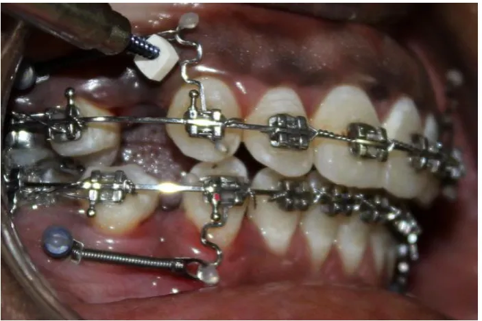
On the experim ent al sit e , l ocal anaes thesi a (2% li doca ine with
1:1,00,000 epinephrine) was infilt rat ed in t he 1s t prem olar regi on.
Micro os teo perforation was perform ed without an y fl ap elevation
using a hand -held devi ce (TAD, 1.5mm *9mm ) l oaded in the impl ant
driv er (S.K. SUR G ICALS, IND IA)(Fi g:6 ) wi th a rubber st opper pl aced,
at a depth of 3mm s o, that each perforat ion were of 3mm i n depth and
1.5mm in widt h.(Fi g: 10 ). 2 mi cro osteo perforati on (M OPs) were
perform ed dist al to cani ne and 3 i n t he m iddl e of the extracti on socket
before retraction . It was placed 5mm from the al veol ar cres t1 4, whereas the cont rol sit e di d not receive an y MOPs .
A Ni Ti cl os ed coil spri ng was pl aced bet ween TAD and the
serpenti ne hook (Fi g: 10). A 100g of force was appli ed f or indivi dual
canine ret racti on and was m easured using dont rix gauge on both
experim ent al and control s ide1 1. At each vis it, the force produced b y
the coil was checked, and appl iances were monitored for an y
Page 29 On the experim ent al side, a t 28t h, 56t hda y, 2 M OPs were
perform ed dist al to cani ne and 3 MOPs at the cent re of extract ion space
and no M OPs were perform ed in t he cont rol si de.
Al ginat e impressions were t aken at the beginni ng of the stud y,
immediat el y before canine ret raction, and at 28t h, 56t h, 84t h da y after
canine ret raction began, to m onitor the rat e of toot h movem ent in both
arches . The i mpress ions were poured i mmedi at el y wit h ort hokal. The
casts were l abel ed with the patient ’s num ber dat e and st ored.
Verti cal lines were drawn on t he cast over t he palatal surface of
the cani ne from middle of the cervical l ine. The dist ance between the
canine and the l at eral i ncisor was ass essed before and aft er canine
ret raction at 3 poi nts: incis al, middle, and cervi ca l thi rds of the
crowns. All t he cast meas urem ent s were made using an el ect roni c
di git al caliper with an accurac y of 0.01m m. (Fi g: 8)
The obtai ned m eas urem ents were tabulat ed and given for
Page 30
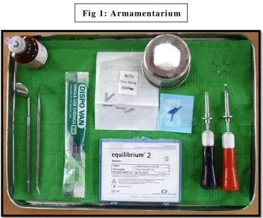
Fig 1: Armamen tari u m
1. Denta urum bracket kit
2. 2 s ets of canine brackets
3. LA Bottl e
4. S yringe
5. Titanium mini -im plants
6. NIT I Coil spring
7. Im pl ant drivers
8. Customiz ed propuls e r
Page 31
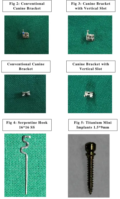
Fig 3: Canin e Brack et with Verti cal Slot Fig 2: Con ven tion al
Canin e B racket
Canin e B racket wi th Vertical Sl ot Conventional Canin e
Bracket
[image:44.595.75.483.75.746.2]Fig 5: Titaniu m Mi ni I mplan ts 1.5*9mm Fig 4: Serp entin e H ook
[image:44.595.300.470.75.478.2] [image:44.595.80.252.77.468.2]Page 32
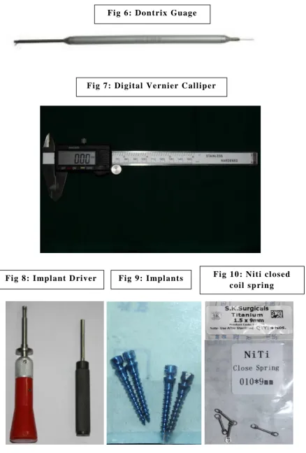
Fig 6: Don trix Guage
Fig 7: Di gital Verni er Callip er
[image:45.595.82.521.69.718.2]Page 33
[image:46.595.90.470.80.325.2]Page 34
[image:47.595.123.475.153.387.2]Page 35
[image:48.595.110.492.324.617.2]Page 36
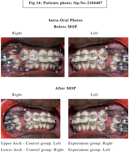
Fig 14 : Pati en ts ph oto; Op.No -2104407
Intra -Oral Photos Before MO P
Right Left
After MO P
Right Left
Upper Arch - Cont rol group: Left Experiment group: R ight
Lower Arch - Cont rol group: Ri ght Experiment group: Left
Cast Ph otos
[image:49.595.87.504.70.584.2]Page 37
Intra -Oral Photos - Day 28 Before MO P
Right Left
After MO P
Right Left
Upper Arch - Cont rol group: Left Experiment group: R ight
Lower Arch - Cont rol group: Ri ght Experiment group: Left
Cast Ph otos
Page 38
Intra -Oral Photos - Day 56 Before MO P
Right Left
After MO P
Right Left
Upper Arch - Cont rol group: Left Experiment group: R ight
Lower Arch - Cont rol group: Ri ght Experiment group: Left
Cast Ph otos
Page 39
Intra -Oral Photos - Day 84 Before MO P
Right Left
Upper Arch - Cont rol group: Left Experiment group: R ight
Lower Arch - Cont rol group: Ri ght Experiment group: Left
Cast Ph otos
Upper Lower
Page 40
STATISTI CAL ANALYSIS :
Dat a were tabul at ed in an excel sheet and anal yz ed usi ng S PSS
stati sti cal s oft ware (version 22). The dat a were ass ess ed for norm ali t y
using Shapi ro -wilk t est which reveal ed t hat the dat a were non -norm al
in dist ribution. Hence non -paramet ri c t es t (Mann Whit ne y-U-test ) was
empl o yed t o det ect t he si g nifi cant di fference bet ween MOP group and
control group. The sam e t est was empl o yed t o det ect t he s igni ficant
Page 41
RESULTS
A tot al of 20 pati ents were included in the stud y who had full y
erupt ed maxill ar y canines with class I m olar canine relationshi p and bi
-maxillar y prot rusi on that requi red t he removal of both max illar y and
mandibul ar 1s t premolars . In tot al, 80 sampl es were obtained , t he
experim ent al group received MOP s on either the ri ght or left si de,
whereas the cont rol group received no MOPs. Dat a were t abul at ed in an
excel sheet and anal yz ed using SPSS st atistical software (version 22).
The res ults are as fo l lows:
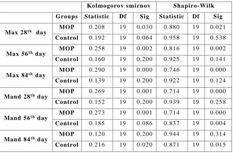
Table 1: T ests of n ormali ty
Kol mogorov smi rnov Shapiro -Wi lk Groups Statis tic Df Sig Statis tic Df Sig
Max 28t h day MO P 0.208 19 0.030 0.880 19 0.021
Control 0.192 19 0.064 0.958 19 0.538
Max 56t h day MO P 0.258 19 0.002 0.816 19 0.002
Control 0.160 19 0.200 0.925 19 0.141
Max 84t h day MO P 0.290 19 0.000 0.746 19 0.000
Control 0.139 19 0.200 0.922 19 0.124
Mand 28t h d ay MO P 0.269 19 0.001 0.714 19 0.000
Control 0.152 19 0.200 0.939 19 0.258
Mand 56t h d ay MO P 0.273 19 0.001 0.714 19 0.000
Control 0.185 19 0.086 0.837 19 0.004
Mand 84t h d ay MO P 0.120 19 0.200 0.944 19 0.314
Control 0.216 19 0.020 0.871 19 0.015
*MOP –micro osteo perforation; Max-maxillary arch; Mand -
[image:54.595.68.530.363.684.2]Page 42
Table 2: Mann Whitney U tes t – Comp arison of control and exp eri men tal (MO P) sid e
Groups Max
28t h day
Max 56t h day
Max 84t h day
Mand 28t h day
Mand 56t h day
Mand 84t h day
MO P Mean 0.6521 0.7374 0.8589 0.5679 0.6763 0.6705
Std d ev 0.20558 0.23154 0.26868 0.11970 0.20597 0.10368
N 19 19 19 0.19 0.19 0.19
Mini mu m 0.43 0.52 0.58 0.42 0.43 0.54
Med iu m 0.6100 0.6200 0.7900 0.5500 0.6400 0.6600
Maxi mu m 1.21 1.41 1.56 0.99 1.42 0.91
Control Mean 0.3700 0.4274 0.4742 0.3416 0.4011 0.4026
Std d ev 0.08825 0.10852 0.07932 0.8315 0.14594 0.12727
N 19 19 19 19 19 19
Mini mu m 0.20 0.26 0.36 0.21 0.25 0.25
Med iu m 0.3700 0.4300 0.4600 0.3600 0.3700 0.3900
Maxi mu m 0.52 0.62 0.59 0.48 0.86 0.61
*MOP – mi cro -os teo perforati on; Max -maxill ar y arch; Mand -
[image:55.595.53.550.182.485.2]Page 43
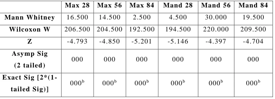
Table 3: T ests of s ignifican ce for In ter group comp ari son between exp eri men tal ( MO P) sid e and con trol si de .
Max 28 Max 56 Max 84 Mand 28 Mand 56 Mand 84 Mann Whitn ey 16.500 14.500 2.500 4.500 30.000 19.500
Wilcoxon W 206.500 204.500 192.500 194.500 220.000 209.500
Z -4.793 -4.850 -5.201 -5.146 -4.397 -4.704
Asymp Sig
(2 tai led ) 000 000 000 000 000 000
Exact Si g [2*(1
-tail ed Si g)] 000
b 000b 000b 000b 000b 000b
Table 4: Tests of si gnifican ce for In ter group comp arison betw een maxi llary and mand ibular arch on th e experi men tal (MO P) side .
MO P 28t h day
MO P 56t h day
MO P 84t h d ay
Mann Whitn ey 145.500 152.500 82.500
Wilcoxon W 335.500 342.500 272.500
Z -1.023 -818 -2.864
Asymp Sig (2 tail ed ) 0.306 0.413 0.004
[image:56.595.59.543.181.355.2] [image:56.595.107.490.501.654.2]