This is a repository copy of IL-7 induces rapid clathrin-mediated internalization and JAK3-dependent degradation of IL-7Rα in T cells.
White Rose Research Online URL for this paper: http://eprints.whiterose.ac.uk/88711/
Version: Accepted Version
Article:
Henriques, C.M., Rino, J., Nibbs, R.J. et al. (2 more authors) (2010) IL-7 induces rapid clathrin-mediated internalization and JAK3-dependent degradation of IL-7Rα in T cells. Blood, 115 (16). pp. 3269-3277. ISSN 0006-4971
https://doi.org/10.1182/blood-2009-10-246876
eprints@whiterose.ac.uk
Reuse
Unless indicated otherwise, fulltext items are protected by copyright with all rights reserved. The copyright exception in section 29 of the Copyright, Designs and Patents Act 1988 allows the making of a single copy solely for the purpose of non-commercial research or private study within the limits of fair dealing. The publisher or other rights-holder may allow further reproduction and re-use of this version - refer to the White Rose Research Online record for this item. Where records identify the publisher as the copyright holder, users can verify any specific terms of use on the publisher’s website.
Takedown
If you consider content in White Rose Research Online to be in breach of UK law, please notify us by
IL-7 induces rapid clathrin-mediated internalization and JAK3-dependent
degradation of IL-7R in T cells
Catarina M. Henriques1, José Rino2, Robert J. Nibbs3, Gerry G. Graham3, João T. Barata1*
1
Cancer Biology Unit, 2 BioImaging Unit, Instituto de Medicina Molecular, Lisbon
University Medical School, Lisbon, Portugal; 3 Division of Immunology, Infection &
Inflammation, Glasgow Biomedical Research Centre, Glasgow, UK.
* Correspondence to: João T. Barata, Cancer Biology Unit, Instituto de Medicina Molecular,
Lisbon University Medical School, Av. Prof. Egas Moniz, 1649-028 Lisboa, Portugal; Tel:
+351217999524; Fax: +351217999524; e-ma
Running title: IL-7R internalization, recycling and degradation
Word counts: 4427 (text) and 165 (abstract)
Number of Figures: 6
Number of References: 50
Abstract
Interleukin 7 (IL-7) is an essential cytokine for T-cell development and homeostasis. It
is well established that IL-7 promotes the transcriptional downregulation of IL7RA leading to
decreased IL-7R surface expression. However, it is currently unknown whether IL-7
regulates the intracellular trafficking and early turnover of its receptor upon ligand binding.
Here, we show that, in steady-state T-cells, IL-7R is slowly internalized and degraded, while
a significant fraction recycles back to the surface. Upon 7 stimulation, there is rapid
IL-7R endocytosis via clathrin-coated pits, decreased receptor recycling and accelerated
lysosome and proteasome-dependent degradation. In accordance, the half-life of IL-7R
decreases from 24h to approximately 3h after IL-7 treatment. Interestingly, we further
demonstrate that clathrin-dependent endocytosis is necessary for efficient IL-7 signal
transduction. In turn, pre-treatment of T-cells with JAK3 or pan-JAK inhibitors suggests that
IL-7R degradation depends on the activation of the IL-7 signaling effector JAK3. Overall,
our findings indicate that IL-7 triggers rapid IL-7R endocytosis, which is required for
Introduction
Interleukin 7 (IL-7) is a key pro-survival cytokine essential for T-cell proliferation,
development and homeostasis. IL-7 is produced by stromal cells in the thymus and bone
marrow, and also by vascular endothelial cells, intestinal epithelium, keratinocytes and
follicular dendritic cells1. The IL-7/ IL-7R signaling network was shown to be critical in
health and disease. IL-7 is essential in lymphoid development, since knockout mice for IL-7
2,3
, IL-7R 4, γc5, and JAK16 or JAK37suffer similar lymphoid developmental blocks. IL-7
was also shown to rescue primary T-cell acute lymphoblastic leukemia (T-ALL) cells from
spontaneous apoptosis in vitro8 and IL-7 transgenic mice develop lymphomas9. Furthermore,
IL-7 may be involved in chronic inflammation, osteoclast maturation and subsequent bone
destruction in rheumatoid arthritis10
IL-7 signals via the IL-7 receptor, composed by the IL-7R and the gamma common
chain (
.
c), which is shared by other interleukin receptors, namely IL-2, IL-4, IL-9, IL-15 and
IL-21. Upon ligand binding, IL-7R and c heterodimerize and are phosphorylated by
JAK1/31. In T-cells, this directs the activation of STAT5 and PI3K, leading to cell cycle
progression, increased viability and cell size1,8,11. In general, effective signal transduction
depends on the tight control of receptor membrane trafficking via internalization, degradation
and recycling mechanisms12, and may occur during receptor trafficking in intracellular
vesicles, rather than just at the cell surface13. Moreover, endocytosis is commonly required for
efficient receptor-mediated signal transduction, and it has been described for other cytokine
receptors such as IL-2R14 and IL-5R15. Receptor endocytosis can occur via clathrin-coated
pits and/or clathrin-independent routes. For example, assembly of high affinity IL-2R and
consequent signal transduction is dependent on lipid rafts14. Interestingly, the different chains
chains are targeted for degradation, the chain internalizes constitutively and recycles back to
the surface16. Other receptors, such as the IL-5R15, TGF 17 and EGFR18 internalize not only
via clathrin-coated pits but also via clathrin-independent routes. Independently of the
internalization route, it is thought that receptors traffic to early endosomes, where they can
then be sorted for degradation and/or recycling19,20. Whether an internalized receptor is
targeted for degradation or recycling depends on several factors, which differ between
receptor and cell type 19. Ubiquitination is usually associated with receptor degradation via
proteasomes, but in some cases efficient degradation of ubiquitinated receptor relies on both
proteasomes and lysosomes21,22
Dissecting how IL-7 regulates its cognate receptor membrane trafficking is crucial to
the in-depth understanding of the role of IL-7/IL-7R in lymphocyte function. Previous studies
have suggested that IL-7 stimulation of T-cells leads to surface down-modulation of IL-7R
within 30 minutes, possibly due to receptor internalization .
23
. At later time points (2-6h), IL-7
was shown to induce transcriptional downregulation of IL7RA24-26. However, the actual
dynamics of IL-7R internalization and the regulation of trafficking mechanisms by IL-7
remain to be elucidated. In the present study, we show that IL-7 induces rapid
clathrin-dependent IL-7R internalization in human T-cells. Moreover, our results suggest that
IL-7-induced signaling is dependent on IL-7R internalization and that subsequent receptor
degradation relies on JAK3 activity and is mediated by both proteasomes and lysosomes. Our
study explores for the first time the mechanisms of IL-7-induced IL-7Rα trafficking,
contributing to the understanding of the biology of the IL-7/IL-7R axis in lymphocyte
Materials & methods
Cell lines and primary human thymocytes. Human HPB-ALL cell line was cultured in
RPMI-1640 (Gibco) supplemented with 10% (vol/vol) heat inactivated FBS (Gibco) and
2mM L-glutamine (hereafter referred to as RPMI-10). Cells were maintained at 37ºC in 5%
CO2. Human thymocytes were isolated from thymic tissue obtained from children undergoing
cardiac surgery. Samples were obtained with informed consent after institutional review board
approval. The tissue was gently disrupted in RPMI and filtered through a cell strainer.
Thymocytes were enriched by density centrifugation over Ficoll-Paque (GE Healthcare) to at
least 95% purity.
Cell treatment with inhibitors. All cell culture experiments were performed in RPMI-10.
When indicated, cells were pre-treated or not (vehicle control), with inhibitors at a density of
106 cells/mL, and inhibitors were kept during the duration of experiment: hyperosmotic
sucrose (Sigma-Aldrich; 0.5M; 1h00 pretreatment), NH4Cl (Sigma-Aldrich; 50mM; 1h
pretreatment), JAK3 inhibitor WHI-P131 (Calbiochem; 150 M; 1h pretreatment), pan-JAK
inhibitor I (Calbiochem; 10 M; 1h pretreatment), Cycloheximide (Sigma-Aldrich; 500 M,
30min. pretreatment) Lactacystin (Calbiochem; 25 M, 1h pretreatment), Filipin
(Sigma-Aldrich; 2.5 g/ml, 1h pretreatment)
Flow cytometry analysis. For cell surface assessment of receptor expression, cells were
washed in ice cold PBS after the indicated stimulus/pre-treatment, resuspended in PBS/1%
BSA (Sigma-Aldrich), and incubated for 30 min. on ice with -human IL-7R - phycoerythrin
(PE)-conjugated antibody (R&D), or isotype and concentration matched IgG-PE control.
(Sigma-Aldrich), incubated 15 min. on ice, and analyzed using a FACScalibur flow cytometer
(Becton Dickinson) and FlowJo software (TreeStar). For intracellular assessment of receptor
expression, cells were washed in ice cold PBS after the indicated stimulus/pre-treatment,
resuspended in 4% paraformaldehyde/0.05M sucrose and incubated at 4ºC for 15 min. After
fixing, cells were washed in cold PBS as before, resuspended in 50mM NH4Cl in PBS for
quenching, and incubated at room temperature for 10 min. After quenching, cells were
washed with permeabilization buffer (PBS/1%BSA/0.05% saponin), resuspended in
permeabilization buffer and incubated at 37ºC for 15 min. After incubation with antibody for
30 min at 37ºC, cells were washed in PBS, resuspended in 2% PFA, incubated for 15 min. on
ice and analyzed by flow cytometry.
Antibody ‘chase’ for assessment of receptor co-localization. For confocal microscopy
analysis, the HPB-ALL cell line was plated on PDL (Poly-D-lysine) (Sigma Aldrich)-coated
coverslips for 30 min to 1h, at 37ºC for cells to adhere. To ‘chase’ the pool of surface
receptor, cells were incubated in RPMI-10 for 30 min. on ice, with -human IL-7R
unconjugated antibody. After primary antibody labeling, cells are washed and resuspended in
RPMI-10 and re-incubated on ice with -IgG- Alexa 633 or 488 (Molecular Probes). After
washing with PBS, cells were resuspended in RPMI-10 at the density of 106 cells/mL and
stimulated or not (vehicle control) with recombinant human IL-7 (Peprotech) (50ng/ml) at
37ºC, for the indicated time intervals. After stimulation, cells were washed, fixed, quenched
and permeabilized as for intracellular staining for flow cytometry analysis. Cells were then
incubated for 1h at 37ºC in permeabilization buffer with antibodies for different endosomal
compartments: clathrin heavy chain FITC-conjugated (Pharmingen); EEA1 FITC-conjugated
(Pharmingen); LAMP2 Alexa Fluor 488-conjugated (Ebioscience); rabbit unconjugated
antibodies: when using FITC-coupled antibodies, -FITC-Alexa Fluor 488-conjugated
antibody (Molecular Probes) was used to enhance signal detection. For Rab-11 detection, we
used -rabbit-Alexa Fluor 488-conjugated antibody. After secondary incubation, cells were
washed twice in permeabilization buffer and twice in PBS, followed by a 10 min incubation
with 4% PFA/sucrose at 4ºC. Cells were washed again with PBS and slides were inverted
over a drop of Vectashield/DAPI (Vector Laboratories) and sealed with varnish. Slides were
analyzed by confocal microscopy, as detailed below. Image acquisition was performed with
the pinhole aperture set to 1 Airy unit for the highest wavelength (633 nm) and adjusted for
the lower wavelengths to maintain the same optical slice thickness for all channels. Five to ten
different fields of view (~ 40 cells per field of view) were collected for quantification, which
was performed for a given pair of fluorophores. The percentage of co-localizing cells was
determined by counting the fraction of cells showing at least one co-localization event
(minimum 3x3 pixels) between the fluorophores from live cells presenting both fluorophores
only (cells in which only one of the fluorophores was present were discarded from the
analysis).
Antibody feeding and recycling. The HPB-ALL cell line was plated onto PDL-coated
coverslips as described for co-localization analysis. In parallel, 1M cells were plated on the
different conditions coverslips. Initially all cells were incubated at 4ºC, in RPMI-10, with
unconjugated -human IL-7R antibody for 30 min. After this, cells are washed gently 3x
with ice-cold PBS and transferred to 37ºC in the presence or absence of IL-7 (50ng/ml) for
antibody feeding for 1h. Cells are subsequently washed again and the positive time zero
control coverslip was further incubated at 4ºC with secondary -mouse Alexa 488 antibody,
in RPMI-10, for 30 min. Cells were fixed at the end with 2% PFA incubation for 15 min on
as described before. The negative control is obtained by performing an acid wash (RPMI-10
with Ph adjusted to 3), which removes all surface-bound antibody. After this acid wash, the
primary antibody/receptor complex was allowed to recycle to the cell surface by switching the
cells to 37ºC for the indicated time point, in pH 7 RPMI-10 (wash 2x with RPMI-10 after acid
wash to re-establish normal pH). Recycling is stopped by washing the cells with cold PBS.
Recycled IL-7Ra/unconjugated antibody complex is detected by incubating the cells with
anti-mouse IgG Alexa 488 secondary antibody, for 30 minutes on ice. Slides were analyzed
by confocal microscopy, as detailed below. Measurements of mean fluorescence intensity
were performed with the pinhole set to its maximum aperture. This ensures that the
fluorescence measured for each cell corresponds to the total surface intensity rather than a
single optical slice. At least 8 different fields of view were imaged for each condition (~ 40
cells per field of view), keeping the same excitation (10% laser transmission) and detection
settings throughout the acquisition. Image processing and quantification were performed with
ImageJ
thresholded to identify surface fluorescence signals whose mean intensity was then
determined for each image. The threshold level was determined based on histogram analysis
and was kept constant for each segmentation.
Confocal Microscopy. All images were acquired on a Zeiss LSM 510 META inverted
confocal laser scanning microscope (Carl Zeiss, Jena, Germany) using a PlanApochromat
63x/1.4 oil immersion objective. DAPI fluorescence was detected with a violet 405 nm diode
laser (30 mW nominal output) and a BP 420-480 filter. Both EGFP and Alexa Fluor 488
fluorescence were detected using the 488 nm laser line of an Ar laser (45 mW nominal
output) and a BP 505-550 filter. Alexa Fluor 633 fluorescence was detected using a 633 nm
Transmitted light imaging was performed using one of the available laser lines and the
transmitted PMT channel available in the LSM 510 META. Sequential multi-track/frame
imaging sequences were used to avoid any potential bleed-through from the different
fluorophores. All confocal images were acquired with a frame size of 512x512 pixels.
Time-Lapse Microscopy. For live time-lapse imaging, a procedure was used similar to the
antibody chase technique and co-localization analysis. However, cells were kept alive
throughout the procedure (no fixing or permeabilization). Cells were placed on ice until
immediately before imaging, at which point they were transferred to the Zeiss LSM 510
META system equipped with an incubator chamber, where they were kept at a controlled
37ºC and 5% CO2 environment. Cells were either imaged unstimulated or after addition of
IL-7 (50ng/ml). A total of 112 images were acquired with a time interval of 30 s between each
image and 488 nm laser intensity set to 3% to minimize photobleaching. Images were
background subtracted and further processed with ImageJ using a rigid body registration
algorithm to correct for cell displacement during image acquisition. Movies of cells were then
generated and time-annotated.
Immunoblotting. Following the indicated conditions and time intervals of culture, cell
lysates were prepared, and equal amounts of protein (50 g/sample) were analysed by 12%
sodium dodecyl sulphate–polyacrylamide gel electrophoresis (SDS-PAGE), transferred onto
nitrocellulose membranes, and immunoblotted with the indicated antibodies. pJAK3
(Sigma-Aldrich); pSTAT5a/b (Upstate); pAKT (Cell Signaling); Actin and ZAP 70 (Upstate). After
immunoblotting with antibodies, detection was performed by incubation with horseradish
Cruz) immunoglobulin (IgG, 1:5000 dilution), as indicated by the host origin of the primary
antibody and developed by chemiluminescence.
Statistics. Data were analyzed using Graph Pad Prism software (San Diego, CA). Statistical
analysis was performed using regular two-way ANOVA followed by Bonferroni post-tests
when comparing unstimulated VS IL-7 internalization and degradation rates throughout time.
When the unstimulated or IL-7-induced internalization or degradation was analyzed on its
own, we used regular one-way ANOVA followed by Bonferroni post-tests. Student T-test was
used to assess the effect of IL-7 with and without inhibitors, at a selected time point.
Results
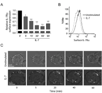
IL-7 induces rapid IL-7Rα internalization in T cells
To study the dynamics of IL-7Rα internalization we started by assessing cell surface
expression over time, in the presence or absence of IL-7. Flow cytometry analysis showed
that IL-7 induced rapid IL-7R surface downregulation, in normal human thymocytes (Supp.
Figure 1) and in the T-ALL cell lines TAIL7 27(data not shown) and HPB-ALL (Figure 1),
which we used in subsequent experiments. Interestingly, we found that IL-7Rα surface levels
decreased rapidly, with approximately 20% decrease upon 5 minutes, 30% after 15 minutes
(Figure 1A) and 40% after 30 minutes of IL-7 stimulation (Figure 1A,B). This indicates that
IL-7-mediated downregulation of IL-7R can occur more rapidly than previously
suggested23,25,26. In addition, rapid IL-7-mediated IL-7R surface downregulation was
dose-dependent and clearly observed at doses as low as 1ng/ml (Supp. Figure 2). To confirm that
decreased surface expression was due to receptor internalization, we stained HPB-ALL cells
with an unconjugated mouse anti-IL-7R antibody and an anti-mouse Alexa Fluor
488-conjugated secondary antibody on ice to label the IL-7R at the surface with minimal receptor
internalization 28. This strategy allowed us to follow the pool of surface receptor from a
defined stimulation point onwards. Consistent with the flow cytometry data, the levels of
surface receptor remained constant when cells were shifted to 37ºC in the absence of stimulus,
whereas upon IL-7 stimulation there was clear receptor clustering and internalization (Fig 1 C
and Supplementary video).
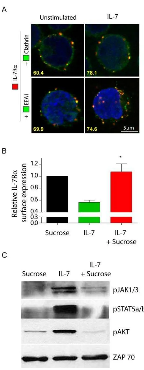
IL-7Rα internalization occurs via clathrin-coated pits and is required for IL-7-mediated
Receptor-mediated endocytosis can occur via coated pits and/or
clathrin-independent routes, such as lipid rafts. Irrespectively of the internalization route, it is thought
that internalized receptors traffic to an Early Endosome Antigen 1 (EEA-1)-positive early
endosome, where they are sorted for degradation and/or recycling12,19. IL-2Rα, a cytokine
receptor that shares γc with IL-7Rα, was shown to localize to lipid rafts and internalize in a
lipid raft-dependent manner 14. We therefore pre-treated the cells with Filipin, which is known
to specifically inhibit lipid raft-dependent internalization29
IL-7 can induce phosphorylation of clathrin heavy chain in mouse T cell precursors and assessed the impact on
constitutive and IL-7-dependent IL-7Rα internalization. Our data suggest that the
internalization of IL-7R is largely independent of lipid rafts (Supp. Figure 3).
30
,
highlighting the potential involvement of clathrin in IL-7R internalization. To assess this
hypothesis, we next analyzed the cellular localization of IL-7R , clathrin and EEA-1 by
confocal microscopy, in the absence or presence of IL-7. We found that IL-7R constitutively
co-localized with clathrin and with EEA-1 positive endosomes in 60.4% and 69.9% of the
cells analyzed, respectively (Figure 2A). This suggests that IL-7Rα is internalized via
clathrin-coated pits and traffics into early endosomes in a constitutive manner. Importantly,
after 5 minutes of IL-7 stimulation, IL-7Rα co-localized with clathrin in 78.1% and with
EEA-1 positive endosomes in 74.7% of the cells, suggesting that clathrin-dependent
internalization into early endosomes rapidly increased in the presence of IL-7 (Figure 2A). To
confirm these observations, we pre-treated the cells with hyperosmotic sucrose (0.5M), which
has been shown to inhibit clathrin-dependent endocytosis29. IL-7-induced receptor
internalization was abrogated by hyperosmotic sucrose pre-treatment (Figure 2B), indicating
that endocytosis of IL-7Rα relies on coated pits. Interestingly, inhibition of
clathrin-mediated endocytosis blocked IL-7-induced signaling, as determined by phosphorylation of
transmembrane receptors15,18,31, clathrin-dependent endocytosis is required for effective
triggering of IL-7-mediated signaling pathways, thereby underlining the functional relevance
of IL-7R endocytosis via clathrin-coated pits.
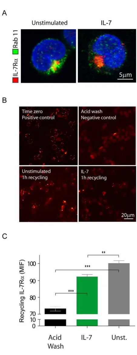
A pool of internalized IL-7R constitutively recycles back to the cell surface
Although IL-7Rα constitutively internalized via clathrin-coated pits, the overall
surface receptor levels were kept constant, and inhibition of clathrin-mediated endocytosis did
not affect the surface receptor levels in the absence of IL-7 (data not shown). These
observations raise the possibility that IL-7R may constitutively internalize and recycle back
to the cell surface. To test this assumption, we assessed whether IL-7R co-localized with
Rab-11-positive recycling endosomes32. We found that IL-7R co-localized with recycling
endosomes both in the absence and presence of IL-7 stimulus, suggesting constitutive
recycling of internalized receptor (Figure 3A). Next, we sought to quantify the amount of
recycling IL-7R in both conditions. To do this, we performed an antibody feeding and
recycling assay (adapted from Weber et al 33), as described in ‘Materials and Methods’, in
which the amount of antibody found at the surface corresponds to the amount of internalized
and subsequently recycled IL-7R . The results confirmed that there is constitutive IL-7Rα
recycling, which still occurs upon IL-7 stimulation, albeit somewhat less efficiently (Figure
3B,C). These data further indicate that there are no major differences in IL-7Rα recycling that
could account for decreased surface expression upon IL-7 treatment.
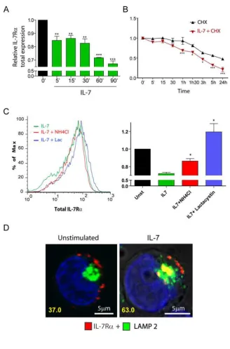
IL-7 induces rapid IL-7R degradation in a proteasome and lysosome-dependent
manner.
To further characterize the mechanisms involved in IL-7-mediated IL-7R surface
cytometry. We observed rapid downregulation of IL-7R upon IL-7 stimulation (Figure 4A),
which was consistent with immunoblot analysis (data not shown). In addition, pre-treatment
of cells with the translation inhibitor cycloheximide revealed that the kinetics of IL-7Rα
degradation was significantly accelerated upon 7 stimulation (Figure 4B). Accordingly,
IL-7Rα half-life was dramatically decreased, from roughly 24 hours in unstimulated cells to
around 3h in the presence of 7 (Figure 4B). These data indicate that 7-induced rapid
IL-7R internalization was followed by significant receptor degradation.
Protein degradation is thought to occur via two main mechanisms in the cell, namely
the ubiquitin-proteasome and the pH-dependent lysosomal degradation pathways34. To
identify the routes of IL-7-induced IL-7R degradation, we pre-treated the cells with the
specific proteasome inhibitor Lactacystin35 or the lysosome inhibitor NH4Cl, a lysomotropic
agent33,36, and evaluated total IL-7R expression. Our results show that NH4Cl partially
reversed, whereas Lactacystin completely prevented, IL-7-induced IL-7R degradation
(Figure 4C). This suggests that both lysosomes and proteasomes participated in the
degradation of the receptor. However, because NH4Cl displayed only a partial effect, we
sought to further demonstrate the involvement of lysosomes in the degradation of IL-7R . We
performed receptor ‘chase’ and co-localization experiments using Alexa Fluor 633-labelled
anti-IL7-R antibody and anti-LAMP-2 (Lysosome Associated Membrane Protein-2) Alexa
Fluor 488-conjugated antibody to detect lysosomes37. As shown in figure 4D, we found that a
significant portion of the receptor localized to lysosomes, after IL-7 stimulation (37%
co-localizing cells in unstimulated conditions vs. 63% at 1h post-IL-7 simulation). These results
suggest that, after IL-7 stimulation, a considerable fraction of the internalized IL-7Rα pool is
degraded by lysosomes, probably in coordination with the ubiquitin-proteasome pathway.
Thus, our data indicate that IL-7 induces IL-7Rα degradation by mechanisms that involve
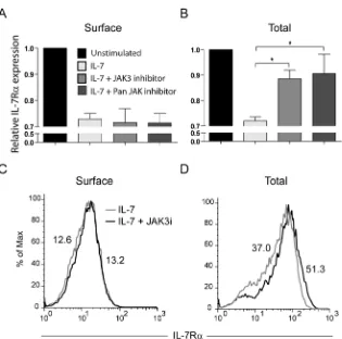
IL-7-induced IL-7R degradation relies on JAK3 activity
The JAK family of tyrosine kinases has been previously implicated in the regulation of
receptor trafficking38,39. In addition, IL-7 was shown to induce tyrosine phosphorylation of
clathrin heavy chain, possibly via activation of JAK3 30. Therefore, we asked whether JAK
kinases, and particularly JAK3, could be involved in IL-7R internalization and/or
degradation. Pre-treatment of HPB-ALL cells with a JAK3-specific inhibitor27 did not prevent
IL-7R internalization, as determined by the analysis of IL-7Rα surface expression (Figure
5A,C). In contrast, JAK3 inhibition significantly reversed IL-7-induced IL-7R degradation
(Figure 5B,C). Similar results were obtained using a pan-JAK inhibitor (Figure 5). Taken
together, our data suggest that IL-7R degradation, but not internalization, is largely
Discussion
The generalized importance of IL-7 and its receptor for T-cell biology, with critical
roles during development, homeostasis and differentiation, has raised particular interest on
how IL-7Rα expression is regulated. It is currently established that IL-7, and other
pro-survival cytokines, induce the transcriptional downregulation of IL7RA and consequently
decrease significantly the cell surface expression of IL-7Rα at late time points (2-6h) 24-26.
This has been proposed as an altruistic mechanism for optimizing the access of T-cells to
IL-7, which is expressed at limiting levels 26,40
Previous work suggested that IL-7R is downregulated at the T-cell surface within 30
minutes of IL-7 stimulation, possibly due to internalization
. Here, we focused for the first time on the
consequences of IL-7 stimulation regarding the early dynamics of IL-7R surface expression
and associated mechanisms of internalization, recycling and degradation. We propose that on
a steady state scenario, IL-7R at the cell surface takes part in a slow cycle of constitutive
clathrin-dependent internalization and recycling, with a pool of receptor eventually being
degraded and replaced by newly synthesized IL-7R . Upon IL-7 stimulation, endocytosis is
rapidly accelerated and the balance is dramatically shifted towards degradation, in a manner
that is dependent on JAK3 activation, which in turn appears to rely on IL-7Rα being
internalized (Figure 6).
23
. Alternatively, it has been
proposed that IL-7 leads to IL-7Rα membrane shedding, instead of endocytosis 41. However,
this has been consistently challenged by recent studies 42-44. In the present work, we describe
for the first time the kinetics and mechanisms of IL-7Rα membrane trafficking in human
T-cells, and confirm that IL-7 rapidly downregulates IL-7Rα surface levels. Importantly, both
flow cytometry and confocal microscopy analyses revealed that IL-7Rα surface
chain in mouse T-cells 30, which suggested that clathrin coated pits could be involved in
IL-7Rα endocytosis. In agreement, we found that pre-treatment with sucrose-containing
hypertonic media, which inhibits the formation of clathrin-coated pits29
Interestingly, we found that inhibition of clathrin-coated pits also largely prevented
IL-7-mediated signaling, suggesting that IL-7R clathrin-dependent endocytosis is essential for
optimal IL-7 signal transduction. This finding is not unprecedented. There is mounting
evidence that endocytosis is frequently necessary for efficient receptor-mediated signal
transduction
, abrogated
IL-7-induced IL-7R internalization.
45
. From a complementary point of view, the ability to transduce a signal is also
dependent on the availability of the receptor at the cell surface, which reflects the balance
between de novo synthesis, internalization, degradation and recycling46
IL-7-induced receptor degradation appears to be mediated by both proteasomes and
lysosomes. IL-7R co-localization with lysosomes is upregulated upon IL-7 stimulation, and
pre-treatment with the lysomotropic agent NH
. Our data suggest that
IL-7R constitutively internalizes and either recycles back to the surface or is degraded in a
process that is relatively slow – the half-life of the receptor is approximately 24h in
non-stimulated cells. Since the expression of IL-7Rα at the cell surface of cultured T-cells is
roughly constant, our observations implicate that, in steady state, de novo synthesis
compensates for the pool of IL-7Rα that is slowly degraded. In the presence of IL-7, the route
of IL-7Rα internalization remains clathrin-dependent. However, the kinetics is substantially
accelerated and the receptor surface expression is reduced by more than 30% within only 15
minutes of stimulation. Although recycling still occurs, there is a major shift towards
degradation. Consequently, the average half-life of IL-7Rα decreases dramatically to around
3h.
4Cl 36, which specifically inhibits pH-dependent
proteasome inhibitor Lactacystin completely prevented IL-7R degradation. Lactacystin did
not affect the internalization of the receptor (data not shown), suggesting that IL-7R
internalization is independent of ubiquitin modification, in contrast to what has been
described for some surface receptors47. There is evidence that the proteasome can play not
only a direct role in proteolysis but also an indirect crucial function upstream of the lysosomal
pathway, acting at the endosome level to sort proteins towards lysosome degradation48,49. Our
observations that NH4Cl and Lactacystin can both inhibit IL-7R degradation are in
agreement with the hypothesis that the proteasome acts upstream of the lysosome in
IL-7-mediated IL-7R proteolysis. The fact that Lactacystin is more effective may suggest that the
proteasome is involved not only in sorting of activated receptor towards the lysosome but also
directly in degradation. Alternatively, it is conceivable that NH4
Our present studies further indicate that activation of JAK3 is dispensable for
IL-7-mediated receptor internalization, while playing a crucial role in IL-7R degradation. This is
in line with IL-7 signaling, and therefore JAK3 activation, being evident only upon receptor
endocytosis. Furthermore, since JAK3 associates with the c, our data suggest the
requirement of c for efficient IL-7-mediated IL-7R degradation. Although we did not
analyze the fate of c after IL-7 stimulation, there is good evidence that IL-7R and c
co-localize. First, it is known that binding of IL-7R to c is dependent on JAK3
Cl is not completely effective
in inhibiting lysosomes, because it relies on a change in the pH to prevent lysosome protease
activity. This could explain why we were not able to rescue IL-7R degradation as efficiently
as by blocking an upstream step in the pathway.
50
Second,
similarly to IL-7R , c localizes to EEA-1 positive early endosomes and LAMP-1 positive
lysosomes together with JAK338. Notably, JAK3 is involved not only in triggering
7-mediated signaling but also in rapidly shutting it down by promoting the proteolysis of
restricted to, and can actually precede, the inhibition of IL7RA transcription 24-26. The
prevalence of these mechanisms is suggestive of the biological importance of limiting the
surface expression of IL-7Rα in T-cells stimulated with IL-7, and may constitute further
support to the ‘altruistic model’ 26,40. Independently of these considerations, our data clarify
how IL-7 stimulation impacts the trafficking of IL-7Rα in T-cells and thus may contribute to
a more complete understanding of T lymphocyte biology and of diseases in which IL-7 and its
receptor are thought to play a role, such as AIDS, multiple sclerosis, rheumathoid arthritis or
leukemia.
Acknowledgements
This work was supported by IMM start-up funds and by the grant
PTDC/SAU-OBD/104816/2008 from Fundação para a Ciência e a Tecnologia (to JTB). CMH has an
FCT-SFRH PhD fellowship. We thank Dr. Miguel Abecasis for providing thymic samples.
Authorship contributions
C.M.H. designed research, performed all the experiments, analyzed and interpreted
data, and wrote the manuscript; J.R. analyzed and interpreted data regarding microscopy
experiments; R.J.N. and G.G.G. designed research, and analyzed and interpreted data; J.T.B.
designed research, analyzed and interpreted data, and wrote the manuscript.
Disclosure of Conflicts of Interest
References
1. Jiang Q, Li WQ, Aiello FB, et al. Cell biology of IL-7, a key lymphotrophin. Cytokine Growth Factor Rev. 2005;16:513-533.
2. von Freeden-Jeffry U, Solvason N, Howard M, Murray R. The earliest T lineage-committed cells depend on IL-7 for Bcl-2 expression and normal cell cycle progression. Immunity. 1997;7:147-154.
3. von Freeden-Jeffry U, Vieira P, Lucian LA, McNeil T, Burdach SE, Murray R. Lymphopenia in interleukin (IL)-7 gene-deleted mice identifies IL-7 as a nonredundant cytokine. J Exp Med. 1995;181:1519-1526.
4. Peschon JJ, Morrissey PJ, Grabstein KH, et al. Early lymphocyte expansion is severely impaired in interleukin 7 receptor-deficient mice. J Exp Med. 1994;180:1955-1960.
5. Cao X, Shores EW, Hu-Li J, et al. Defective lymphoid development in mice lacking expression of the common cytokine receptor gamma chain. Immunity. 1995;2:223-238.
6. Rodig SJ, Meraz MA, White JM, et al. Disruption of the Jak1 gene demonstrates obligatory and nonredundant roles of the Jaks in cytokine-induced biologic responses. Cell. 1998;93:373-383.
7. Suzuki K, Nakajima H, Saito Y, Saito T, Leonard WJ, Iwamoto I. Janus kinase 3 (Jak3) is essential for common cytokine receptor gamma chain (gamma(c))-dependent signaling: comparative analysis of gamma(c), Jak3, and gamma(c) and Jak3 double-deficient mice. Int Immunol. 2000;12:123-132.
8. Barata JT, Cardoso AA, Boussiotis VA. Interleukin-7 in T-cell acute lymphoblastic leukemia: an extrinsic factor supporting leukemogenesis? Leuk Lymphoma. 2005;46:483-495.
9. Rich BE, Campos-Torres J, Tepper RI, Moreadith RW, Leder P. Cutaneous
lymphoproliferation and lymphomas in interleukin 7 transgenic mice. J Exp Med. 1993;177:305-316.
10. Churchman SM, Ponchel F. Interleukin-7 in rheumatoid arthritis. Rheumatology (Oxford). 2008;47:753-759.
11. Barata JT, Silva A, Brandao JG, Nadler LM, Cardoso AA, Boussiotis VA. Activation of PI3K is indispensable for interleukin 7-mediated viability, proliferation, glucose use, and growth of T cell acute lymphoblastic leukemia cells. J Exp Med. 2004;200:659-669.
12. Le Roy C, Wrana JL. Clathrin- and non-clathrin-mediated endocytic regulation of cell signalling. Nat Rev Mol Cell Biol. 2005;6:112-126.
13. Hoeller D, Volarevic S, Dikic I. Compartmentalization of growth factor receptor signalling. Curr Opin Cell Biol. 2005;17:107-111.
14. Matko J, Bodnar A, Vereb G, et al. GPI-microdomains (membrane rafts) and signaling of the multi-chain interleukin-2 receptor in human lymphoma/leukemia T cell lines. Eur J Biochem. 2002;269:1199-1208.
15. Lei JT, Martinez-Moczygemba M. Separate endocytic pathways regulate IL-5 receptor internalization and signaling. J Leukoc Biol. 2008;84:499-509.
16. Hemar A, Subtil A, Lieb M, Morelon E, Hellio R, Dautry-Varsat A. Endocytosis of interleukin 2 receptors in human T lymphocytes: distinct intracellular localization and fate of the receptor alpha, beta, and gamma chains. J Cell Biol. 1995;129:55-64.
17. Di Guglielmo GM, Le Roy C, Goodfellow AF, Wrana JL. Distinct endocytic pathways regulate TGF-beta receptor signalling and turnover. Nat Cell Biol. 2003;5:410-421.
19. Gonzalez-Gaitan M. The garden of forking paths: recycling, signaling, and degradation. Dev Cell. 2008;15:172-174.
20. Perrais D, Merrifield CJ. Dynamics of endocytic vesicle creation. Dev Cell. 2005;9:581-592.
21. Walrafen P, Verdier F, Kadri Z, Chretien S, Lacombe C, Mayeux P. Both proteasomes and lysosomes degrade the activated erythropoietin receptor. Blood. 2005;105:600-608.
22. van Kerkhof P, Strous GJ. The ubiquitin-proteasome pathway regulates lysosomal degradation of the growth hormone receptor and its ligand. Biochem Soc Trans. 2001;29:488-493.
23. Swainson L, Verhoeyen E, Cosset FL, Taylor N. IL-7R alpha gene expression is inversely correlated with cell cycle progression in IL-7-stimulated T lymphocytes. J Immunol. 2006;176:6702-6708.
24. Kim HR, Hwang KA, Kim KC, Kang I. Down-regulation of IL-7Ralpha expression in human T cells via DNA methylation. J Immunol. 2007;178:5473-5479.
25. Alves NL, van Leeuwen EM, Derks IA, van Lier RA. Differential regulation of human IL-7 receptor alpha expression by IL-7 and TCR signaling. J Immunol. 2008;180:5201-5210. 26. Park JH, Yu Q, Erman B, et al. Suppression of IL7Ralpha transcription by IL-7 and other prosurvival cytokines: a novel mechanism for maximizing IL-7-dependent T cell survival. Immunity. 2004;21:289-302.
27. Barata JT, Boussiotis VA, Yunes JA, et al. IL-7-dependent human leukemia T-cell line as a valuable tool for drug discovery in T-ALL. Blood. 2004;103:1891-1900.
28. Willingham IPaMC ed Endocytosis. Plenum Press, NY; 1985.
29. Ivanov AI. Pharmacological inhibition of endocytic pathways: is it specific enough to be useful? Methods Mol Biol. 2008;440:15-33.
30. Jiang Q, Benbernou N, Chertov O, Khaled AR, Wooters J, Durum SK. IL-7 induces tyrosine phosphorylation of clathrin heavy chain. Cell Signal. 2004;16:281-286.
31. Mills IG. The interplay between clathrin-coated vesicles and cell signalling. Semin Cell Dev Biol. 2007;18:459-470.
32. Horgan CP, Oleksy A, Zhdanov AV, et al. Rab11-FIP3 is critical for the structural integrity of the endosomal recycling compartment. Traffic. 2007;8:414-430.
33. Weber M, Blair E, Simpson CV, et al. The chemokine receptor D6 constitutively traffics to and from the cell surface to internalize and degrade chemokines. Mol Biol Cell. 2004;15:2492-2508.
34. Ciechanover A. Intracellular protein degradation from a vague idea through the lysosome and the ubiquitin-proteasome system and on to human diseases and drug targeting: Nobel Lecture, December 8, 2004. Ann N Y Acad Sci. 2007;1116:1-28.
35. Dick LR, Cruikshank AA, Grenier L, Melandri FD, Nunes SL, Stein RL. Mechanistic studies on the inactivation of the proteasome by lactacystin: a central role for clasto-lactacystin beta-lactone. J Biol Chem. 1996;271:7273-7276.
36. Houdebine LM, Djiane J. Effects of lysomotropic agents, and of microfilament- and microtubule-disrupting drugs on the activation of casein-gene expression by prolactin in the mammary gland. Mol Cell Endocrinol. 1980;17:1-15.
37. Eskelinen EL, Tanaka Y, Saftig P. At the acidic edge: emerging functions for lysosomal membrane proteins. Trends Cell Biol. 2003;13:137-145.
38. Hofmann SR, Lam AQ, Frank S, et al. Jak3-independent trafficking of the common gamma chain receptor subunit: chaperone function of Jaks revisited. Mol Cell Biol. 2004;24:5039-5049.
40. Mazzucchelli R, Durum SK. Interleukin-7 receptor expression: intelligent design. Nat Rev Immunol. 2007;7:144-154.
41. Vranjkovic A, Crawley AM, Gee K, Kumar A, Angel JB. IL-7 decreases IL-7 receptor alpha (CD127) expression and induces the shedding of CD127 by human CD8+ T cells. Int Immunol. 2007;19:1329-1339.
42. Blom-Potar MC, Bugault F, Lambotte O, Delfraissy JF, Theze J. Soluble IL-7Ralpha (sCD127) and measurement of IL-7 in the plasma of HIV patients. J Acquir Immune Defic Syndr. 2009;51:104-105.
43. Rose T, Lambotte O, Pallier C, Delfraissy JF, Colle JH. Identification and biochemical characterization of human plasma soluble IL-7R: lower concentrations in HIV-1-infected patients. J Immunol. 2009;182:7389-7397.
44. Faucher S, Crawley AM, Decker W, et al. Development of a quantitative bead capture assay for soluble IL-7 receptor alpha in human plasma. PLoS One. 2009;4:e6690.
45. Sorkin A, von Zastrow M. Endocytosis and signalling: intertwining molecular networks. Nat Rev Mol Cell Biol. 2009;10:609-622.
46. Zi Z, Klipp E. Steady state analysis of signal response in receptor trafficking networks. Genome Inform. 2007;18:100-108.
47. Yu A, Malek TR. The proteasome regulates receptor-mediated endocytosis of
interleukin-2. J Biol Chem. 2001;276:381-385.
48. Rocca A, Lamaze C, Subtil A, Dautry-Varsat A. Involvement of the
ubiquitin/proteasome system in sorting of the interleukin 2 receptor beta chain to late endocytic compartments. Mol Biol Cell. 2001;12:1293-1301.
49. Alwan HA, van Zoelen EJ, van Leeuwen JE. Ligand-induced lysosomal epidermal growth factor receptor (EGFR) degradation is preceded by proteasome-dependent EGFR de-ubiquitination. J Biol Chem. 2003;278:35781-35790.
Figure Legends
Figure 1. IL-7 induces rapid IL-7R surface downregulation due to receptor
internalization. (A) HPB-ALL cells were cultured in the presence or absence of IL-7
(50ng/ml) in culture medium for the indicated time points and subsequently analyzed by flow
cytometry for surface IL-7Rα expression, as described in ‘Materials and Methods’. Relative
IL-7Rα expression was calculated as the geometric mean intensity of fluorescence normalized
to the time zero control. Data represent mean±sem from at three independent experiments. *
P<0.05, *** P<0.001 (One-way ANOVA, with Bonferroni’s post-test). (B) Representative
flow cytometry histogram overlay of IL-7Rα surface expression in HPB-ALL cells stimulated
for 30 minutes with IL-7 (gray line) or left unstimulated (black line). (C) IL-7R
internalization visualized by time-lapse microscopy of HPB-ALL cells. IL-7Rα initially
expressed at the cell surface, was stained with -human IL-7R unconjugated antibody,
subsequently re-incubated with a secondary -mouse IgG-Alexa488-conjugated antibody, and
imaged for the indicated time points, with or without addition of IL-7 (50ng/ml), as described
in the ‘Materials and Methods’. See supplementary data for full time-lapse microscopy video.
Representative cells of each condition from two independent experiments are shown.
Figure 2. IL-7Rα internalization via clathrin-coated pits and trafficking into early
endosomes is required for efficient IL-7-mediated signaling. (A) Analysis of IL-7Rα
co-localization with clathrin and EEA1 was performed by confocal microscopy. HPB-ALL cells
were plated on PDL-coated coverslips, incubated at 4ºC with -human IL-7R unconjugated
antibody, and subsequently with a secondary -mouse IgG-Alexa-633 antibody (red). Cells
were then shifted to 37ºC in the presence or absence of IL-7 (50ng/ml) for 5 min, fixed,
antibodies for clathrin heavy-chain or EEA1, both enhanced by -FITC-Alexa Fluor 488
secondary antibody (green). The two bottom images are 3D projections and include
DAPI-stained nuclei. Representative cells of three independent experiments are shown. The
percentage of co-localizing cells is indicated and was determined by counting the fraction of
cells showing at least one co-localization event between red and green fluorescence. (B)
HPB-ALL cells were pretreated or not with 0.5M sucrose (hypertonic media) for 1h, to inhibit
formation of clathrin-coated pits, and then stimulated with 50ng/ml IL-7 or left untreated for
30 min. IL-7Rα surface expression was analyzed by flow cytometry and relative expression
was calculated by normalizing the geometrical mean of fluorescence of the cell population in
the presence of IL-7 stimulus, divided by the unstimulated condition at the same time point
(30 min). Data represent mean±sem from three independent experiments: * P<0.05. (C)
HPB-ALL cells were pretreated or not with 0.5M sucrose, and stimulated with 50ng/ml IL-7 for 30
min. Immunoblotting was performed and IL-7 mediated signaling assessed by detection of
phosphorylated JAK1/3, STAT5a/b and AKT. ZAP-70 was used as a loading control.
Figure 3. A pool of internalized IL-7R recycles to the cell surface. (A) Co-localization
between IL-7R and Rab-11 recycling endosomes was assessed after 5 min of stimulation
with IL-7 (50ng/ml). Total IL-7R expression was assessed by intracellular staining as for
flow cytometry. Rab-11 positive vesicles were further detected by incubation with rabbit
-human Rab-11 unconjugated antibody followed by secondary detection with -rabbit IgG
Alexa 488. Coverslips were mounted over Vectashield /DAPI. Cells representative of three
independent experiments are shown. (B) Co-localization between IL-7R and endosomes was
performed by antibody chase and co-localization assay, as described in Materials & Methods. Briefly,
all cells, plated onto coverslips, were initially incubated at 4ºC with unconjugated -human
or absence of IL-7 (50ng/ml) for 1h to allow for IL-7Rα internalization. Cells were washed
and the positive control (time zero) was generated by incubating the cells at 4ºC with -mouse
IgG Alexa 488 secondary antibody for 30 min. The remaining coverslips underwent an acid
wash treatment to remove surface-bound antibody. The negative control was not further
processed. The primary antibody/receptor complex was allowed to recycle to the cell surface
in the experimental conditions, by switching the cells to 37ºC again for 1h. Recycled
IL-7R /antibody complex was detected by incubating the cells with anti-mouse IgG Alexa 488
secondary antibody for 30 minutes on ice. Cells were fixed and the different coverslips
mounted with Vectashield/DAPI. Representative images of two independent experiments are
shown for each condition. (C) Microscopy images of the antibody feeding and recycling
assays were quantified by determining the Mean Intensity of Fluorescence (MIF) of at least 8
independent fields of view for each condition. ** P<0.01, *** P<0.001 (Students t-test,
two-tailed).
Figure 4. IL-7 stimulation accelerates IL-7R degradation, in a lysosome and
proteasome-dependent manner. (A,B,C) Total IL-7R expression (surface plus
intracellular) was assessed by flow cytometry analysis of fixed and permeabilized HPB-ALL
cells. Analysis of the data was performed as described in Figure 1. * P<0.05, ** P<0.01, ***
P<0.001 (One-way ANOVA, with Bonferroni’s post-test). (B) To determine IL-7R half-life
cells were treated with the translation inhibitor cycloheximide (CHX). (C) To determine the
pathways of degradation of IL-7R , cells were pre-treated for 1h with 50mM NH4Cl or 25 M
Lactacystin, and then stimulated or not with 50ng/ml IL-7 for 1h. The geometrical mean of
fluorescence of the population was determined by flow cytometry, as shown in the
representative histogram. * P<0.05 (Student T-test, two-tailed). Data in A, B and C represent
and lysosomes in the presence or absence of IL7 (1h; 50ng/ml) was performed using
-human IL-7R antibody and LAMP-2 as a lysosome marker. The percentage of co-localizing
cells is indicated and was determined as in Figure 2. Representative cells of three independent
experiments are shown.
Figure 5. IL-7-induced IL-7R degradation, but not internalization is JAK3 dependent.
(A,B) Surface (A) and total (B) IL-7R expression in HPB-ALL cells was assessed as
described by flow cytometry. Cells were pre-treated with 150 M of JAK3 inhibitor (WHI
-P131) or PAN-JAK inhibitor I, and then stimulated or not with IL-7 (50ng/ml) for 30 min (A)
or 1h (B). The geometrical mean of fluorescence for each population was determined by flow
cytometry. Data (mean±sem) are from three independent experiments. * P<0.05 (Student
T-test, two-tailed). (C,D) Representative flow cytometry histogram overlay of surface (C) or
total (D) IL-7Rα expression in HPB-ALL cells pre-treated (black line) or not (gray line) with
the JAK3 inhibitor WHI-P131, followed by IL-7 stimulation for 30 min (C) or 1h (D).
Figure 6. Proposed model for IL-7Rα trafficking. At the steady state, IL-7R is slowly
internalized via clathrin-coated pits and recycled back to the cell surface, with a pool of
receptor being degraded and replaced by newly synthesized IL-7R (not shown). Upon IL-7
stimulation, the balance is rapidly shifted towards receptor endocytosis and subsequent
degradation. JAK3 activation, which appears to rely on IL-7Rα being internalized, is critical