R E S E A R C H
Open Access
Mechanical ventilation enhances
extrapulmonary sepsis-induced lung injury:
role of WISP1
–
α
v
β
5 integrin pathway in
TLR4-mediated inflammation and injury
Xibing Ding
1,3,5,6†, Yao Tong
1†, Shuqing Jin
1,3, Zhixia Chen
1, Tunliang Li
2,3, Timothy R. Billiar
3, Bruce R. Pitt
4,
Quan Li
1,7*and Li-Ming Zhang
5*Abstract
Background:
High tidal volume ventilation of healthy lungs or exacerbation of existing acute lung injury (ALI) by
more moderate mechanical ventilation (MTV) produces ventilator-induced lung injury. It is less clear whether
extrapulmonary sepsis sensitizes the lung to MTV.
Methods:
We used a two-hit model of cecal ligation and puncture (CLP) followed 12 h later by MTV (10 ml/kg; 6 h)
to determine whether otherwise noninjurious MTV enhances CLP-induced ALI by contrasting wildtype and TLR4
−/−mice
with respect to: alveolar-capillary permeability, histopathology and intrapulmonary levels of WNT-inducible secreted protein
1 (WISP1) and integrin
β
5; plasma levels of cytokines and chemokines (TNF-
α
, IL-6, MIP-2, MCP-1) and intrapulmonary
neutrophil infiltration; and other inflammatory signaling via intrapulmonary activation of JNK, p38 and ERK. A separate
cohort of mice was pretreated with intratracheal neutralizing antibodies to WISP1, integrin
β
5 or IgG as control and the
presented phenotyping repeated in a two-hit model; there were 10 mice per group in these first three experiments. Also,
isolated peritoneal macrophages (PM) from wildtype and TLR4
−/−, MyD88
−/−and TRIF
−/−mice were used to identify a
WISP1
–
TLR4
–
integrin
β
5 pathway; and the requisite role of integrin
β
5 in WISP1-induced cytokine and chemokine
production in LPS-primed PM was examined by siRNA treatment.
Results:
MTV, that in itself did not cause ALI, exacerbated increases in alveolar-capillary permeability, histopathologic
scoring and indices of pulmonary inflammation in mice that previously underwent CLP; the effects of this two-hit model
were abrogated in TLR4
−/−mice. Attendant with these findings was a significant increase in intrapulmonary WISP1 and
integrin
β
5 in the two-hit model. Anti-WISP1 or anti-integrin
β
5 antibodies partially inhibited the two-hit phenotype. In PM,
activation of TLR4 led to an increase in integrin
β
5 expression that was MyD88 and NF-
κ
B dependent. Recombinant WISP1
increased LPS-induced cytokine release in PM that was inhibited by silencing either TLR4 or integrin
β
5.
Conclusions:
These data show for the first time that otherwise noninjurious mechanical ventilation can exacerbate ALI due
to extrapulmonary sepsis underscoring a potential interactive contribution of common events (sepsis and mechanical
ventilation) in critical care, and that a WISP1
–
TLR4
–
integrin
β
5 pathway contributes to this phenomenon.
Keywords:
Acute lung injury, Integrin, Lipopolysaccharide, Mechanical ventilation, Peritoneal macrophages, Sepsis, Toll-like
receptor 4, WISP1
* Correspondence:quanligene@126.com;Zhangl1@anes.upmc.edu
†Xibing Ding and Yao Tong contributed equally to this work. 1
Department of Anesthesiology, East Hospital, Tongji University School of Medicine, 150 Jimo Road, Pudong, Shanghai, China
5Department of Anesthesiology, University of Pittsburgh School of Medicine,
200 Lothrop St. UPMC MUH N467, Pittsburgh 15213, PA, USA Full list of author information is available at the end of the article
Introduction
Mechanical ventilation (MV) is well known to cause an
iatrogenic syndrome of ventilator-induced lung injury
(VILI). The pathophysiology of VILI includes
intrapulmon-ary inflammatory cell infiltrates, increased vascular
perme-ability and pulmonary edema, and it may occur in
ventilation of a healthy lung or worsening of preexisting and
coexisting injury [
1
]. Sensitization of VILI secondary to
pre-existing acute lung injury (ALI) due to pneumonia [
2
,
3
],
intratracheal endotoxin [
4
–
7
] or sterile injury [
8
–
10
] has
provided preclinical evidence of a two-hit model.
Although extrapulmonary endotoxemia combined with
noninjurious mechanical ventilation leads to VILI [
11
,
12
],
evidence of such a two-hit phenomenon in experimental
extrapulmonary bacterial sepsis is less clear. Ventilating
rodents after polymicrobial sepsis due to cecal ligation
and puncture (CLP) [
13
–
15
] has produced equivocal
results regarding sensitization to VILI. Mechanical
ventila-tion with injurious high VT (30
–
40 ml/kg) exacerbated
48 h of CLP-induced lung injury in rats [
16
]; shorter
periods of CLP were not associated with subsequent
ex-acerbation of VILI in mice [
17
] or rats [
9
,
18
] although
such overall injury was accelerated in the latter. Lower VT
(15
–
20 ml/kg) did not exacerbate CLP-induced ALI in
in-tact rats [
9
] or isolated perfused rat lungs [
19
]. Since both
CLP and VILI have a common TLR4-mediated pathway
to inflammation and injury [
5
,
20
–
22
] it seems plausible
that mechanical ventilation could exacerbate CLP-mediated
events within the lung and thus differences are likely to be
secondary to variables in experimental protocols (degree of
preexisting lung injury, volume and duration of ventilation)
as suggested by Yehya et al. [
18
].
In the current study, we examined the effect of prolonged
(6 h), otherwise noninjurious [
4
] moderate VT ventilation
(MTV) in mice with preexisting mild ALI after CLP (12 h).
We focused on a novel WNT1 inducible secreted protein
(WISP1 or CCN4; also referred to as WNT1 inducible
sig-naling protein-1)
–
integrin
β
5 pathway of TLR4-mediated
pulmonary inflammation and injury in this two-hit model
as we previously noted key roles for WISP1 and beta
integ-rins in VILI [
21
] and CLP [
22
,
23
] mediated ALI. The
mechanism by which WISP1 acts in CLP and/or VILI
re-mains unclear. WISP1 appears, however, to be a modulator
of TLR4
–
CD14 signaling and is known to signal through
integrins [
24
] although the precise integrin is unclear.
Al-though several integrins are important in ALI [
25
], integrin
β
5 is a central regulator of increased permeability in VILI
[
26
] and CLP [
27
].
Thus, the overall hypotheses of this study were that:
prolonged ventilation with otherwise noninjurious
mod-erate VT exacerbates ALI in extrapulmonary sepsis;
WISP1 and integrin
β
5 contribute to this two-hit model
(i.e., CLP + MTV); and TLR4 is central to the WISP1
–
TLR4
–
integrin
β
5 proinflammatory pathway.
Methods
Experimental protocols
Animal protocols were approved by the Animal Care
and Use Committee and experiments were performed in
strict adherence to NIH Guidelines and followed current
guidelines for preclinical models in research. Details of
materials and methods are provided in Additional file
8
:
Materials and Methods) and experimental protocols are
outlined for intact mice (Additional file
1
: Figure S1)
and cultured PM (Additional file
2
: Figure S2) including:
MTV and CLP-induced lung damage via TLR4-dependent,
WISP-1 and integrin
β
5 contributory fashion in two-hit
lung damage; MTV and CLP-induced changes in
circulat-ing levels of cytokines and chemokines, and an association
with increased neutrophil infiltration of the lungs of intact
mice; other inflammatory signaling (pJNK, p38, pERK)
pathways in lungs of mice after CLP and MV; the
mechanism of upregulation of integrin
β
5 in LPS-treated
PM isolated from wildtype, TLR4 null, Myd88
−/−and
TRIF
−/−mice; and the requisite role of integrin
β
5 in
WISP1-induced cytokine and chemokine production in
LPS-primed PM.
In-vivo experimental animal model
C57BL/6 mice (8
–
10 weeks old, male) were purchased
from Jackson Laboratory and TLR4
−/−mice were used
as described previously [
21
,
22
]. Forty wildtype mice were
prospectively randomized to one of four groups (
n
= 10
per group): spontaneous breathing (sham control),
spon-taneous breathing with CLP; mechanical ventilation; or
CLP and MTV. Mild sepsis was induced by CLP [
13
,
14
]
as modified by Ding et al. [
23
,
28
] and MTV (10 ml/kg;
150/min, zero positive end-expiratory pressure) was
per-formed in anesthetized mice alone or after CLP (12 h). A
cohort (
n
= 20) of TLR
−/−mice underwent sham and
spontaneous breathing (
n
= 10; control) or CLP and MTV
as already described (
n
= 10). In separate cohorts, mice
were intratracheally administered anti-WISP1 (
n
= 10),
anti-integrin
β
5 (
n
= 10) or serum IgG (
n
= 10; 0.5
μ
g/g in
50
μ
l PBS) after CLP but before MTV. Phenotypic
changes due to CLP, MTV or their combination included
histopathology (scored by a pathologist blinded to
experiments), alveolar-capillary permeability (Evans Blue
albumin [
21
]) and inflammation as assessed by plasma
levels of cytokines and chemokines, neutrophil
immigra-tion in the lung (flow cytometry) and intrapulmonary
MAP kinase activation (phosphorylation status of JNK,
ERK and p38).
In-vitro studies
WISP1 (rWISP1) and cytokine production was assessed
in conditioned medium of wildtype cells or after
trans-fection with 50 nM small interfering RNA (siRNA) for
integrin
β
5 (sc-35681; Santa Cruz) or scrambled siRNA.
The role of TLR4 signaling was assessed by comparing
the response of PM isolated from wildtype vs TLR4
−/−,
Myd88
−/−or TRIF
−/−mice.
Histological examination
Lung tissue samples were fixed in 4% paraformaldehyde
in PBS overnight at 4 °C and processed as described
pre-viously [
22
] including semiquantitative histopathology
(H&E; light microscopy) by a pathologist blinded to the
experimental group.
Western blot analysis of WISP-1 and integrin
β
5 was
performed as described previously [
21
,
22
]. Plasma or
con-ditioned medium were assayed for cytokines and
chemo-kines using commercially available ELISA reagents for
TNF-
α
, IL-6, MIP-2 and MCP-1.
Flow cytometry
The lung was enzymatically digested and mechanically
dissociated (MACS dissociator) and single cell
suspen-sions were isolated by passing the suspension through a
70-
μ
m filter. Cells were stained with mAbs specific to
Fixable Viability Dye eFluor® 506, CD45, CD11b, Ly6G
for 30 min at 4 °C and fixed with 2% paraformaldehyde
for 10 min at 4 °C. An LSR II (Becton Dickinson) was
used for flow cytometry and data were analyzed with
FlowJo software.
Alveolar-capillary permeability
Evans blue albumin (EBA; 0.5%, 25 mg/kg body weight)
was injected into the internal jugular vein 1 h before
euthanasia and lung harvesting. Blood samples and lung
tissue were obtained and processed as described
previ-ously [
21
,
22
] and the EBA permeability index was
calculated by dividing pulmonary EBA absorbance at
620 nm/g of lung tissue by plasma EBA absorbance at
620 nm.
Immunofluorescence staining of cells and florescence
microscopy
PM were cultured for a defined time period, fixed in 4%
paraformaldehyde in PBS for 15 min. Cells were washed
in PBS, permeabilized using 0.1% Triton X-100, blocked
with 5% BSA for 45 min and sequentially administered
primary antibody and secondary antibody
(Alexa-488-conjugated donkey anti rabbit secondary antibody).
Nuclei
were
stained
with
DAPI
(Thermo
Fisher
Scientific) and cells were examined and recorded using
EVOS FL fluorescence microscopy (immunofluoresence
analysis; Thermo Fisher Scientific).
Reagents are described in Additional file
8
: Materials
and Methods.
Data analysis and statistics
Data are presented as the mean ± SEM of the indicated
number of experiments and analyzed using one-way and
two-way ANOVA; post-hoc testing was performed using
the Bonferroni modification of the
t
test. The individual
studies performed throughout this work represent five
independent studies. Power analyses were performed by
using type I error probability of 0.05, with a power of
0.9, to determine the sample size necessary to reject the
null hypothesis. All statistical analyses were carried out
using the GraphPad Prism 5 program.
P
< 0.05 was
considered statistically significantly.
Results
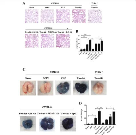
CLP alone led to modest lung injury as demonstrated by
histology (Fig.
1a, b
) and a significant increase in
alveolar-capillary permeability (Fig.
1c, d
). MTV alone
had no impact on lung injury or permeability, but when
applied after CLP it markedly enhanced both the lung
injury score and alveolar-capillary permeability. The
his-topathologic and permeability changes in the two-hit
model were completely abrogated in TLR4
−/−mice and
partially (but significantly) reduced in cohorts of mice
receiving antibodies to either WISP1 or integrin
β
5. In
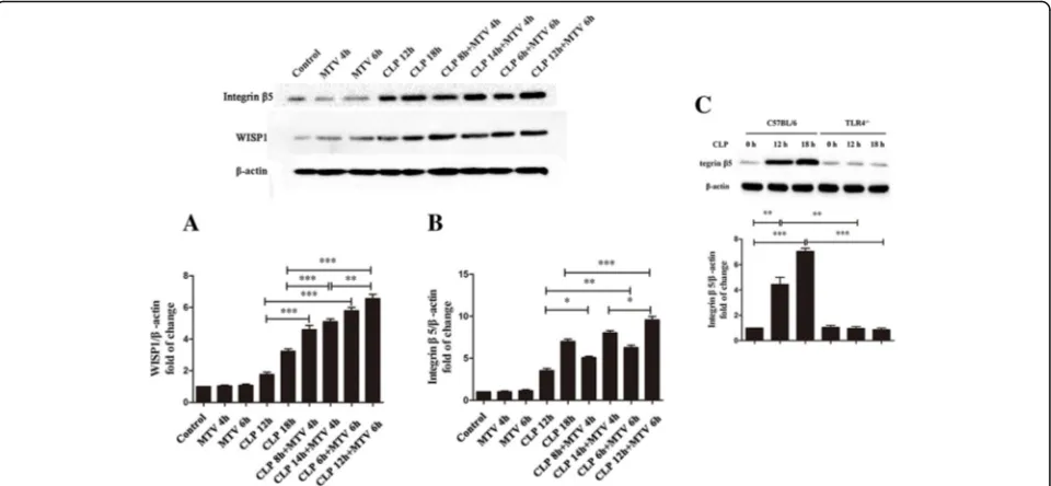
wildtype mice, we noted that: MTV did not affect
intra-pulmonary levels of either WISP1 or integrin
β
5; CLP
led to small but significant increases in either WISP1 or
integrin
β
5; and the two-hit model increased either of
these molecules 2
–
3× more than CLP alone (Fig.
2a, b
).
We previously noted that high VT ventilation increases
in WISP1 were abrogated in TLR4
−/−mice [
21
] and we
now note (Fig.
2c
) that CLP-induced increases in
integ-rin
β
5 are abrogated in TLR4
−/−mice.
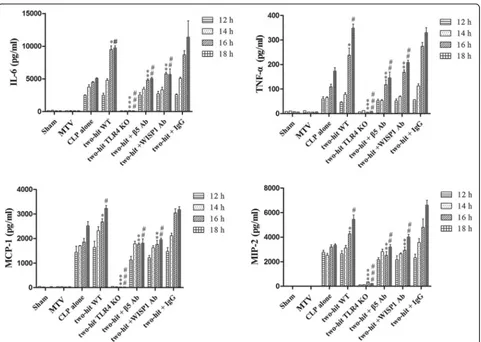
CLP increased circulating cytokines and chemokines
whereas MTV alone did not; the combination of MTV
and CLP, however, caused levels of all four mediators to
progressively rise above levels measured with CLP alone
(Fig.
3
). Deletion of TLR4 prevented increases in
cytokines and chemokines in the two-hit model and
inhibition of WISP1 or integrin
β
5 blocked further
increases induced by MTV (Fig.
3
).
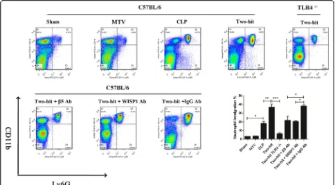
CLP significantly increased neutrophil influx in the
lung while MTV had no impact; CLP and MTV further
significantly increased neutrophil immigration (Fig.
4
)
that was abolished in TLR4 null mice. Blocking WISP1
or integrin
β
5 partially prevented the increase in the
per-centage of PMN induced in the two-hit model compared
to combined CLP and MTV in mice receiving control
IgG antibody.
levels of activated JNK, p38 and ERK MAP kinase. Six
hours of MTV had no effect on MAP kinase activation but
significantly promoted MAP kinase activation in mice
pre-viously subjected to CLP (Additional file
3
: Figure S3).
TLR4 deletion prevented the increases in MAPK activation
in CLP-treated and CLP + MTV-treated mice. Blocking
WISP1 or integrin
β
5 also prevented the increase in MAP
kinase phosphorylation induced by MTV in CLP mice.
To explore the mechanism of integrin
β
5 upregulation
by TLR4, we exposed peritoneal macrophages (PM) to
LPS and found that ultrapure LPS induced a time and
concentration-dependent increase in surface integrin
β
5
Fig. 1MTV enhances CLP-induced lung damage via TLR4-dependent WISP1–integrinβ5 pathway. As shown in Additional file1: Figure S1, mice lung tissue samples in eight mice groups were fixed and stained with hematoxylin–eosin for histological analysis (a) and lung injury score (b). Gross lung image in each group (c) and vascular permeability evaluated by Evans blue dye (d). Mice receiving combination of CLP + MTV (two-hit model) compared to mice subjected to CLP alone for 18 h or sham operation followed by 6 h of MTV. Two-hit model in wildtype mice compared to subgroup of TLR4−/−mice or wildtype mice that received intratracheally neutralizing antibodies to either integrinβ5 (β5 Ab) or WISP1 (WISP1 Ab) or a [image:4.595.62.538.89.560.2]expression (Additional file
4
: Figure S4). The increase in
integrin
β
5 expression was TLR4 and MyD88 dependent,
but TRIF independent (Additional file
5
: Figure S5). We
also found that integrin
β
5 upregulation by LPS was
NF-
κ
B dependent (Additional file
6
: Figure S6).
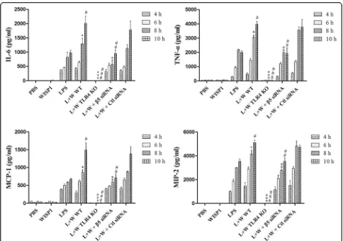
LPS-induced increases in IL-6, TNF-
α
, MIP-2 and
MCP-1 in medium of isolated PM were evident within
4
–
10 h and the addition of rWISP1 induced further
in-creases in all four mediators (Fig.
5
). No increase in any
of the mediators could be seen in PM from TLR4
−/−mice. Suppression of integrin
β
5 expression with siRNA
(Additional file
7
: Figure S7) prevented the WISP1-induced
enhancement of LPS-induced IL-6, TNF-
α
, MIP-2 and
MCP-1 released by PM (Fig.
5
).
Discussion
In the current study, MTV did not cause ALI, but
exa-cerbated CLP-mediated increases in alveolar-capillary
permeability and indices of pulmonary inflammation
(histopathology, cytokines, chemokines, neutrophil influx
and activation of MAPK) in wildtype mice: the effects of
this two-hit model were completely abrogated in TLR4
null mutants and partially inhibited by neutralizing
anti-bodies to either WISP1 or integrin
β
5. In PM, activation
of TLR4 led to an increase in integrin
β
5 expression and
rWISP1 increased LPS-induced cytokine release in PM
that could be inhibited by silencing either TLR4 or
integ-rin
β
5. Collectively, these data show: that prolonged MTV
ventilation exacerbates ALI caused by extrapulmonary
sepsis; and an important positive feedback role for the
WISP1
–
integrin
β
5 pathway in TLR4-mediated
exacerba-tions to this two-hit model.
[image:5.595.59.539.88.310.2]injury with CLP and the magnitude and duration of
mechanical ventilation, and speculated that prolonged
ventilation with lower VT may indeed enhance lung
in-jury after CLP. In this regard, we unequivocally note that
otherwise noninjurious MTV for a prolonged (6 h) time
exacerbated
underlying
CLP-induced
ALI.
Indeed,
whether CLP in itself causes ALI in mice is conjectural.
Iskander et al. [
32
] clearly showed that pulmonary
injury after CLP in mice cannot be considered the
etiology of death in the acute phase. The authors did
note that a severe model of sepsis with significant
mortality could show signs of lung injury. We
re-ported previously [
22
,
23
,
28
] that the current model
of CLP was associated with 80% mortality in 72 h
and this presumably accounted for the modest but
significant ALI noted at the earlier time period in
the current study. Furthermore, we noted that 6 h of
MTV (10 ml/kg) without PEEP was void of
signifi-cant lung injury. In this regard, our model of effects
of mechanical ventilation reproduces previous
ex-perience of lack of injury with MTV (10 ml/kg, zero
PEEP) [
4
,
28
], and was somewhat similar to the
results from Hegeman et al. [
33
] who noted little
evidence of ALI in mice after 5 h of mechanical
ven-tilation with 7 ml/kg and 3 cmH
2O of PEEP or 5 h
of MV with 15 ml/kg and zero PEEP. These authors
did report VILI under both conditions at a longer
time period (12 h) [
33
].
[image:6.595.59.540.85.427.2]We [
21
,
34
] and others [
5
,
35
] have noted the
import-ant role of TLR4 in experimental VILI. We also noted
[
22
,
23
,
28
] that a considerable component of
patho-physiology, inflammation and injury of CLP was due to
TLR4 activation (and CD14). The injury, neutrophil
se-questration and inflammation due to combined effects
of CLP and MTV in the current study were all
abrogated in TLR4 null mice.
WISP1 is a secreted matricellular protein involved in
cell adhesion, migration, differentiation, proliferation
and survival [
36
]. From an unbiased haplotype
associ-ation mapping in inbred strains of mice, we identified
WISP1 as a candidate gene associated with VILI [
21
].
We subsequently identified a role for WISP1 in
CLP-induced ALI [
22
,
23
]. WISP1 was first noted in the
lung to be a component of bleomycin-induced lung
injury and fibrosis [
37
], and subsequently has been
reported to be important in epithelial
–
mesenchymal
transition [
38
], airway remodeling [
39
] and proliferation
of fibroblasts in the context of lung fibrosis [
40
]. In-vitro
mechanical stretch of type II epithelial cells activated
innate immunity and increased WISP1 expression,
pro-viding fundamental support for its involvement in VILI
[
41
]. We noted that intrapulmonary WISP1 is elevated
in VILI [
21
], CLP [
22
,
23
], combined poly(I:C) and
mechanical ventilation [
42
], and in the current report in
combined CLP and MTV; neutralizing antibodies to
WISP1 partially reduced lung injury and inflammation
in all of these conditions. The potential convergence of
WNT/
β
-catenin signaling and WISP1 adds to its
import-ance in VILI [
43
] as well as WNT-mediated lung
epithe-lial cell repair [
44
].
The mechanism by which WISP1 acts in CLP and/or
VILI remains unclear. It appears, however, to be a
modulator of TLR4
–
CD14 signaling and is known to
signal through integrins [
24
]. WISP1
coimmunoprecipi-tated with active, glycosylated TLR4 in lungs of mice
subjected to high VT ventilation and rWISP1 augmented
LPS-induced TNF-
α
release in a TLR4
–
CD14-dependent
fashion in PM [
21
]. The RGD peptide-sensitive response
of intact mouse lungs to CLP and the
coimmunoprecipi-tation of WISP1
–
integrin
β
6 in lungs of these mice [
23
]
Fig. 4MTV increases PMN infiltration in lungs of septic mice. Neutrophil immigration in lung detected by flow cytometry. Mice receivingcombination of CLP + MTV (two-hit model) compared to mice subjected to CLP alone for 18 h or sham operation followed by 6 h of MTV. Two-hit model in wildtype mice compared to subgroup of TLR4 null mice or wildtype mice that received intratracheally neutralizing antibodies to either integrinβ5 (β5 Ab) or WISP1 (WISP1 Ab) (or control antibodies (IgG Ab)). Neutrophil influx by quantifying percent of CD11b+and Ly6G+
cells in CD45+cell population isolated from lung homogenates. CLP significantly increased percent of PMN while MTV had no impact on percent
[image:7.595.56.540.88.355.2]suggested the presence of the WISP1
–
integrin
β
6
pathway in mediating TLR4-dependent inflammation
and injury. We noted an obligatory role for integrin
β
3
in WISP1-mediated release of TNF-
α
in PM [
22
] and an
important role for integrin
β
3 in polymicrobial sepsis
and combined injury from poly I:C instillation and
mechanical ventilation [
42
]. In the current study we
noted that siRNA to integrin
β
5 reduced
WISP1-medi-ated release of multiple cytokines in PM and integrin
β
5
contributed to lung inflammation and injury with CLP
and MTV. Identifying the precise integrin
β
subunit
in-volved in complex lung injury and signaling in isolated
macrophages is complicated by: the multitude of
β
sub-units; the nondiscriminatory inhibition by RGD peptides;
and the promiscuity of WISP1 regarding interactions
with
α
V
β
integrin subunit receptors [
45
]. We focused on
the interaction of WISP1 and integrin
β
5 because: others
noted that WISP1 induces IL-6 production through
in-tegrin
β
5 receptor in human synovial fibroblasts [
46
];
and although several integrins are important in ALI [
25
],
integrin
β
5 is a central regulator of increased
permea-bility in VILI [
26
] and CLP [
27
].
We limited our phenotyping of lung injury to
inflamma-tion, alveolar-capillary permeability and histopathologic
changes, and did not assess lung mechanics or evolution
of changes in gas exchange. As such, fundamental issues
of alveolar overdistension and physiologic consequences
in the current study remain conjectural. It is noteworthy,
however, that we recently reported [
28
] that low VT
(6 ml/kg; 6 h) mechanical ventilation was protective after
Fig. 5WISP1-induced cytokine and chemokine production in LPS-primed peritoneal macrophages requires integrinβ5. Peritoneal macrophages (PM) treated with LPS (0.1μg/ml) followed by WISP1 (10μg/ml) exposure at 4 h. Supernatants collected at 2 h intervals from 4 to 10 h following WISP1 stimulation. Cytokines (TNF-αand IL-6) and chemokines (MIP-2 and MCP-1) in supernatant detected by ELISA. PM receiving combination of LPS + WISP1 (L + W) from wildtype mice (WT) compared to PM from subgroup of either TLR4−/−mice or wildtype mice receiving integrinβ5siRNA transfection (or control siRNA) subjected to LPS or WISP1. LPS-induced increases in IL-6, TNF-α, MIP-2 and MCP-1 evident within 4–10 h and addition of WISP1 induced further increases in all four mediators. Suppression of integrinβ5 expression prevented WISP1-induced enhancement of LPS induction in four mediators. *P< 0.05 compared with LPS alone at 8 h; **P< 0.05 compared with L + W WT at 8 h; ***P< 0.05 compared with L + W WT;
#
P< 0.05 compared with LPS alone at 10 h;##
P< 0.05 compared with L + W WT at 10 h;###
[image:8.595.56.540.85.425.2]CLP (6 h). We relied on neutralizing antibodies to assess
the role of WISP1 and integrin
β
5 in a two-hit model and
partial effects noted in the study may have been secondary
to incomplete deletion. For pragmatic reasons, we used
PM as a surrogate for the likely cell of interest (alveolar
macrophage) and future studies validating these
observa-tions with alveolar macrophages, a more challenging cell
to isolate in sufficient number, are necessary.
Conclusion
In the current study, we provide evidence that mild lung
in-jury secondary to extrapulmonary sepsis can sensitize intact
mouse lung to subsequent prolonged MTV. This two-hit
model is TLR4 sensitive and has important
proinflamma-tory contributions from both WISP1 and integrin
β
5. In
isolated PM, we further defined the nature of WISP1
sig-naling and identified a requisite TLR4-dependent activation
of integrin
α
Vβ
5 and MyD88
–
NF-
κ
B pathway inflammatory
mediator biosynthesis. This two-hit model provides
rele-vant new information regarding unresolved issues of a
common risk factor for ARDS (systemic sepsis) and
sensitization to often-used MTV.
Additional files
Additional file 1:Figure S1.Flow chart of two-hit animal model: CLP followed by MTV. Mice treated as Sham group (sham CLP and sham MTV), MTV group (sham CLP followed with 6-h MTV at 10 ml/kg), CLP group (12-h CLP followed with spontaneous breathing-sham MTV) and two-hit group (12-h CLP followed with 6-h MTV). Two-hit model established by mild sepsis induced by cecal ligation and puncture (CLP) with a 22-gauge needle for 12 h followed by mechanical ventilation with moderate tidal volume at 10 ml/kg (MTV; 50% O2) and 150 breaths/min
for 2–6 h. Two-hit model in wildtype mice compared to subgroup of TLR4 null mice (TLR4−/−) or wildtype mice that received intratracheally neutralizing antibodies to either integrinβ5 (β5 Ab) or WISP1 (WISP1 Ab) or a control antibody (IgG Ab) during mechanical ventilation (TIF 3849 kb)
Additional file 2:Figure S2.Schematic of experimental groups in peritoneal macrophages. Peritoneal macrophages (PM) obtained from wildtype, TLR−/−mice and treated with LPS (0.1μg/ml) and/or siRNA to integrinβ5 followed by WISP1 (10μg/ml) exposure at 4 h. Supernatants collected at 2-h intervals from 4 to 10 h (TIF 2044 kb)
Additional file 3:Figure S3.MTV increases inflammatory signaling in lungs of mice after CLP. Western blot for activated (phosphorylated) p-JNK (A), p-p38 (B) and p-Erk (C) MAP kinase expression in lung homogenates. Mice receiving the combination of CLP + MTV
(two-hit model) were compared to mice subjected to CLP alone for 18 h or sham operation followed by 6 h of MTV. Six hours of MTV alone had no effect on MAP kinase activation but significantly promoted MAP kinase activation in mice previously subjected to CLP, whereas TLR4 deletion prevented increases in MAPK activation in CLP-treated and CLP + MTV-treated mice and blocking WISP1 or integrinβ5 also prevented increase in MAP kinase phosphorylation induced by MTV in CLP mice. *P< 0.05; **P< 0.01 (TIF 7656 kb)
Additional file 4:Figure S4.LPS-induced increase in integrinβ5 expres-sion is time and concentration dependent. Integrinβ5 protein levels (A, C) and immunofluorescence for integrinβ5 (green) and nuclei with DAPI (blue) (B, D) in peritoneal macrophages (PM). Mouse PM isolated from C57BL/6 mice and stimulated with LPS (0, 0.01, 0.1, 1, 10μg/ml) in DMEM containing 10% FBS for 0–8 h. LPS induced time and
concentration-dependent increase in integrinβ5 protein levels and
increase in surface integrinβ5 expression maximal at 6 h. Corresponding actin identified for normalizing densitometry of integrinβ5 expression. **P< 0.01 (TIF 1689 kb)
Additional file 5:Figure S5.Upregulation of integrinβ5 requires TLR4/ MyD88 signaling. Western blot for integrinβ5 protein (A, B)and immunostaining of integrinβ5 (C) in peritoneal macrophages (PM). Increase in integrinβ5 protein levels and its surface expression was TLR4 and MyD88 dependent (A), but TRIF independent (B). Mouse PM isolated from C57BL/6 (wildtype), TLR4−/−, MyD88−/−and TRIF−/−mice and stimulated with LPS (0.1μg/ml) in DMEM containing 10% FBS for 0–6 h. Corresponding actin identified for normalizing densitometry.
Immunofluorescence for integrinβ5 stained green while nuclei stained with DAPI (blue). Images acquired using EVOSfl fluorescence microscopy. *P< 0.05; **P< 0.01; ***P< 0.001 (TIF 4019 kb)
Additional file 6:Figure S6.Integrinβ5 upregulation by LPS is NF-κB dependent. Western blot for nuclear and cytoplasma NF-κB p65 (A) and immunostaining of NF-κB p65 (C) in peritoneal macrophages (PM) induced by LPS over time. Increase in integrinβ5 protein levels induced by LPS at 4.5 h significantly decreased by inhibitor of NF-κB signaling, IKK-NBD (B). *P< 0.05; **P< 0.01; ***P< 0.001 (TIF 613 kb)
Additional file 7:Figure S7.siRNA to integrinβ5 suppressed LPS-induced increases in integrinβ5 levels in PM. Western blot for integrinβ5 levels in PM after treating with siRNA to integrinβ5 or control siRNA. Integrinβ5 siRNA dose-dependently suppressed LPS-induced increases in integrinβ5 levels compared to control siRNA. *P< 0.05; **P< 0.01; ***P< 0.001 (TIF 4824 kb)
Additional file 8Materials and MethodsEight to 10-week-old male C57BL/6 mice purchased from Jackson Laboratory. TLR4−/−, MyD88−/−, TRIF−/−mice obtained from Dr Billiar’s laboratory. All mice used were on
a C57BL/6 background with appropriate backcrossing for respective knockouts. Transgenic male mice confirmed to be desired genotype via standard PCR-based techniques. Animal protocols approved by the Animal Care and Use Committee of the University of Pittsburgh and experiments performed in strict adherence to National Institutes of Health Guidelines for the Use of Laboratory Animals. Mice bred and housed in specific pathogen-free conditions with free access to food and water (DOCX 21 kb)
Abbreviations
ALI:Acute lung injury; ARDS: Acute respiratory distress syndrome; CLP: Cecal ligation and puncture; EBA: Evans blue albumin; LPS: Lipopolysaccharide; MTV: Moderate tidal ventilation; MyD88: Myeloid differentiation factor 88; PM: Peritoneal macrophages; PMN: Polymorphonuclear leukocyte; RGD peptide: Arg–Gly–Asp peptide; TLR4: Toll-like receptor 4; TRIF: TIR-domain-containing adaptor inducing interferon beta; VILI: Ventilator-induced lung injury; WISP1: WNT1 inducible secreted protein
Acknowledgements
The authors thank Jing Xu, Li Xu, Zhengzheng Yan, Xubo Wu and Hong Liao for technical assistance.
Funding
This work was supported by National Institute of Health grants
(R01-GM-50441 to TRB and R01-GM-108639 to L-MZ) for the design of the study, collection and writing the manuscript, and by the National Natural Science Foundation (81270135 to QL and 81772114 to QL) for analysis and interpretation of data.
Availability of data and materials
The datasets generated and/or analyzed during the current study are available in the Dr Billiar laboratory of University of Pittsburgh, and the datasets are available from the corresponding author on request.
Authors’contributions
L-MZ contributed to manuscript preparation. XbD, YT, SqJ, ZxC, TlL, TRB, BRP, QL and L-MZ contributed to manuscript revision. All authors read and approved the final version of the manuscript.
Ethics approval
Animal protocols were approved by the Animal Care and Use Committee of the University of Pittsburgh and experiments were performed in strict adherence to the National Institutes of Health Guidelines for the Use of Laboratory Animals.
Consent for publication
Not applicable.
Competing interests
The authors declare that they have no competing interests.
Publisher
’
s Note
Springer Nature remains neutral with regard to jurisdictional claims in published maps and institutional affiliations.
Author details
1Department of Anesthesiology, East Hospital, Tongji University School of
Medicine, 150 Jimo Road, Pudong, Shanghai, China.2Department of Anesthesiology, Xiangya 3rd Hospital, Central South University, Hunan, China. 3Department of Surgery, University of Pittsburgh School of Medicine,
Pittsburgh, PA, USA.4Department of Environmental and Occupational Health, University of Pittsburgh Graduate School Public Health, Pittsburgh, PA, USA. 5Department of Anesthesiology, University of Pittsburgh School of Medicine,
200 Lothrop St. UPMC MUH N467, Pittsburgh 15213, PA, USA.6Department of Anesthesiology, Renji Hospital, Shanghai Jiaotong University School of Medicine, Shanghai, China.7Department of Anesthesiology, Cancer Hospital Chinese Academy of Medical Sciences, Shenzhen, China.
Received: 2 January 2018 Accepted: 15 October 2018
References
1. Slutsky AS, Ranieri VM. Ventilator-induced lung injury. N Engl J Med. 2013; 369:2126–36.
2. Dhanireddy S, Altemeier WA, Matute-Bello G, O'Mahony DS, Glenny RW, Martin TR, et al. Mechanical ventilation induces inflammation, lung injury, and extra-pulmonary organ dysfunction in experimental pneumonia. Lab Investig. 2006;86:790–9.
3. Muller-Redetzky HC, Will D, Hellwig K, Kummer W, Tschernig T, Pfeil U, et al. Mechanical ventilation drives pneumococcal pneumonia into lung injury and sepsis in mice: protection by adrenomedullin. Crit Care. 2014;18:R73. 4. Altemeier WA, Matute-Bello G, Gharib SA, Glenny RW, Martin TR, Liles WC.
Modulation of lipopolysaccharide-induced gene transcription and promotion of lung injury by mechanical ventilation. J Immunol. 2005;175:3369–76.
5. Hu G, Malik AB, Minshall RD. Toll-like receptor 4 mediates neutrophil sequestration and lung injury induced by endotoxin and hyperinflation. Crit Care Med. 2010;38:194–201.
6. Ding N, Wang F, Xiao H, Xu L, She S. Mechanical ventilation enhances HMGB1 expression in an LPS-induced lung injury model. PLoS One. 2013;8(9):e74633.
7. Rentsendorj O, Damarla M, Aggarwal NR, Choi JY, Johnston L, D'Alessio FR, et al. Knockdown of lung phosphodiesterase 2A attenuates alveolar inflammation and protein leak in a two-hit mouse model of acute lung injury. Am J Physiol Lung Cell Mol Physiol. 2011;301:L161–70.
8. Frank JA, Gutierrez JA, Jones KD, Allen L, Dobbs L, Matthay MA. Low tidal volume reduces epithelial and endothelial injury in acid-injured rat lungs. Am J Respir Crit Care Med. 2002;165:242–9.
9. Kuiper JW, Plotz FB, Groeneveld AJ, Haitsma JJ, Jothy S, Vaschetto R, et al. High tidal volume mechanical ventilation-induced lung injury in rats is greater after acid instillation than after sepsis-induced acute lung injury, but does not increase systemic inflammation: an experimental study. BMC Anesthesiol. 2011;11:26.
10. Makena PS, Luellen CL, Balazs L, Ghosh MC, Parthasarathi K, Waters CM, et al. Preexposure to hyperoxia causes increased lung injury and epithelial
apoptosis in mice ventilated with high tidal volumes. Am J Physiol Lung Cell Mol Physiol. 2010;299:1467–76.
11. Altemeier WA, Matute-Bello G, Frevert CW, Kawata Y, Kajikawa O, Martin TR, et al. Mechanical ventilation with moderate tidal volumes synergistically increases lung cytokine response to systemic endotoxin. Am J Physiol Lung Cell Mol Physiol. 2004;287:L533–42.
12. Bregeon F, Delpierre S, Chetaille B, Kajikawa O, Martin TR, Autillo-Touati A, et al. Mechanical ventilation affects lung function and cytokine production in an experimental model of endotoxemia. Anesthesiology. 2005;102:331–9. 13. Chaudry IH, Wichterman KA, Baue AE. Effect of sepsis on tissue adenine
nucleotide levels. Surgery. 1979;85:205–11.
14. Villar J, Ribeiro SP, Mullen JB, Kuliszewski M, Post M, Slutsky AS. Induction of the heat shock response reduces mortality rate and organ damage in a sepsis-induced acute lung injury model. Crit Care Med. 1994;22:914–21. 15. Dejager L, Pinheiro I, Dejonckheere E, Libert C. Cecal ligation and puncture:
the gold standard model for polymicrobial sepsis? Trends Microbiol. 2011;19:198–208.
16. Nin N, Lorente JA, Fernandez-Segoviano P, De Paula M, Ferruelo A, Esteban A. High-tidal volume ventilation aggravates sepsis-induced multiorgan dysfunction in a dexamethasone-inhibitable manner. Shock. 2009;31:429–34. 17. Uematsu S, Engelberts D, Peltekova V, Otulakowski G, Post M, Kavanagh BP.
Dissociation of inflammatory mediators and function: experimental lung injury in nonpulmonary sepsis. Crit Care Med. 2013;41:151–8.
18. Yehya N, Xin Y, Oquendo Y, Cereda M, Rizi RR, Margulies SS. Cecal ligation and puncture accelerates development of ventilator-induced lung injury. Am J Physiol Lung Cell Mol Physiol. 2015;308:L443–51.
19. Nakamura T, Malloy J, McCaig L, Yao LJ, Joseph M, Lewis J, et al. Mechanical ventilation of isolated septic rat lungs: effects on surfactant and
inflammatory cytokines. J Appl Physiol (1985). 2001;91:811–20. 20. Villar J, Cabrera N, Casula M, Flores C, Valladares F, Muros M, et al.
Mechanical ventilation modulates Toll-like receptor signaling pathway in a sepsis-induced lung injury model. Intensive Care Med. 2010;36:1049–57. 21. Li HH, Li Q, Liu P, Liu Y, Li J, Wasserloos K, et al. WNT1-inducible signaling
pathway protein 1 contributes to ventilator-induced lung injury. Am J Respir Cell Mol Biol. 2012;47:528–35.
22. Chen Z, Ding X, Jin S, Pitt B, Zhang L, Billiar T, et al. WISP1-alphavbeta3 integrin signaling positively regulates TLR-triggered inflammation response in sepsis induced lung injury. Sci Rep. 2016;6:28841.
23. Ding X, Wang X, Zhao X, Jin S, Tong Y, Ren H, et al. RGD peptides protects against acute lung injury in septic mice through Wisp1-integrin beta6 pathway inhibition. Shock. 2015;43(4):352–60.
24. Leask A, Abraham DJ. All in the CCN family: essential matricellular signaling modulators emerge from the bunker. J Cell Sci. 2006;119:4803–10. 25. Sheppard D. Modulation of acute lung injury by integrins. Proc Am Thorac
Soc. 2012;9:126–9.
26. Su G, Hodnett M, Wu N, Atakilit A, Kosinski C, Godzich M, et al. Integrin alphavbeta5 regulates lung vascular permeability and pulmonary endothelial barrier function. Am J Respir Cell Mol Biol. 2007;36:377–86. 27. Su G, Atakilit A, Li JT, Wu N, Luong J, Chen R, et al. Effective treatment of
mouse sepsis with an inhibitory antibody targeting integrin alphavbeta5. Crit Care Med. 2013;41:546–53.
28. Ding X, Jin S, Shao Z, Xu L, Yu Z, Tong Y, Chen Z, Turnquist H, Pitt BR, Billiar TR, Zhang LM, Li Q. Shock. 2018.https://doi.org/10.1097/SHK.
0000000000001260. [Epub ahead of print]. PMID:30192340.
29. Curley GF, Laffey JG, Zhang H, Slutsky AS. Biotrauma and ventilator-induced lung injury: clinical implications. Chest. 2016;150:1109–17.
30. Gattinoni L, Protti A, Caironi P, Carlesso E. Ventilator-induced lung injury: the anatomical and physiological framework. Crit Care Med. 2010;38:S539–48. 31. Matthay MA, Bhattacharya S, Gaver D, Ware LB, Lim LH, Syrkina O, et al.
Ventilator-induced lung injury: in vivo and in vitro mechanisms. Am J Physiol Lung Cell Mol Physiol. 2002;283:L678–82.
32. Iskander KN, Craciun FL, Stepien DM, Duffy ER, Kim J, Moitra R, et al. Cecal ligation and puncture-induced murine sepsis does not cause lung injury. Crit Care Med. 2013;41:159–70.
33. Hegeman MA, Hemmes SN, Kuipers MT, Bos LD, Jongsma G, Roelofs JJ, et al. The extent of ventilator-induced lung injury in mice partly depends on duration of mechanical ventilation. Crit Care Res Pract. 2013;2013:435236.
35. Vaneker M, Joosten LA, Heunks LM, Snijdelaar DG, Halbertsma FJ, van Egmond J, et al. Low-tidal-volume mechanical ventilation induces a toll-like receptor 4-dependent inflammatory response in healthy mice.
Anesthesiology. 2008;109:465–72.
36. Brigstock DR. The CCN family: a new stimulus package. J Endocrinol. 2003;178:169–75.
37. Konigshoff M, Kramer M, Balsara N, Wilhelm J, Amarie OV, Jahn A, et al. WNT1-inducible signaling protein-1 mediates pulmonary fibrosis in mice and is upregulated in humans with idiopathic pulmonary fibrosis. J Clin Invest. 2009;119:772–87.
38. Berschneider B, Konigshoff M. WNT1 inducible signaling pathway protein 1 (WISP1): a novel mediator linking development and disease. Int J Biochem Cell Biol. 2011;43:306–9.
39. Yang M, Du Y, Xu Z, Jiang Y. Functional effects of WNT1-inducible signaling pathway protein-1 on bronchial smooth muscle cell migration and proliferation in OVA-induced airway remodeling. Inflammation. 2016;39:16–29.
40. Klee S, Lehmann M, Wagner DE, Baarsma HA, Konigshoff M. WISP1 mediates IL-6-dependent proliferation in primary human lung fibroblasts. Sci Rep. 2016;6:20547.
41. Heise RL, Stober V, Cheluvaraju C, Hollingsworth JW, Garantziotis S. Mechanical stretch induces epithelial-mesenchymal transition in alveolar epithelia via hyaluronan activation of innate immunity. J Biol Chem. 2011; 286:14735–44.
42. Jin S, Chen Z, Ding X, Zhao X, Jiang X, Tong Y, et al. Mechanical ventilation augments poly(I:C)induced lung injury via a wisp1-integrin beta3 dependent pathway in mice. Mol Med. 2016;22:54–63.
43. Villar J, Cabrera NE, Casula M, Valladares F, Flores C, Lopez-Aguilar J, Blanch L, Zhang H, Kacmarek RM, Slutsky AS. WNT/beta-catenin signaling is modulated by mechanical ventilation in an experimental model of acute lung injury. Intensive Care Med. 2011;37:1201–9.
44. Zemans RL, et al. Role ofβ-catenin-regulated CCN matricullar proteins in epithelial repair after inflammatory lung injury. Am J Physiol. 2013;304:L415–27. 45. Chen CC, Lau LF. Functions and mechanisms of action of CCN matricellular
proteins. Int J Biochem Cell Biol. 2009;41:771–83.