Compensatory Mutations following Transmission of Simian
Immunodeficiency Virus
Benjamin J. Burwitz,a,bHelen L. Wu,a,gJason S. Reed,aKatherine B. Hammond,aLaura P. Newman,dBenjamin N. Bimber,b
Francesca A. Nimiyongskul,dEnrique J. Leon,aNicholas J. Maness,cThomas C. Friedrich,e,fMasaru Yokoyama,hHironori Sato,h
Tetsuro Matano,iDavid H. O’Connor,d,eJonah B. Sachaa,b,g
Vaccine & Gene Therapy Instituteaand Division of Pathobiology and Immunology, Oregon National Primate Research Center,bOregon Health & Science University,
Portland, Oregon, USA; Division of Microbiology, Tulane National Primate Research Center, Covington, Louisiana, USAc; Department of Pathology, University of
Wisconsin—Madison, Madison, Wisconsin, USAd; Wisconsin National Primate Research Center, Madison, Wisconsin, USAe; Department of Pathobiological Sciences,
University of Wisconsin School of Veterinary Medicine, Madison, Wisconsin, USAf; Molecular Microbiology & Immunology, Oregon Health & Science University, Portland,
Oregon, USAg; Laboratory of Viral Genomics, Pathogen Genomics Center, National Institute of Infectious Diseases, Musashi Murayama-shi, Tokyo, Japanh; AIDS Research
Center, National Institute of Infectious Diseases, Shinjuku, Tokyo, Japani
Compensatory mutations offset fitness defects resulting from CD8ⴙT lymphocyte (CD8TL)-mediated escape, but their im-pact on viral evolution following transmission to naive hosts remains unclear. Here, we investigated the reversion kinetics of Gag181–189CM9 CD8TLescape-associated compensatory mutations in simian immunodeficiency virus (SIV)-infected ma-caques. Preexisting compensatory mutations did not result in acute-phase escape of the SIVmac239 CD8TL epitope Gag181-189CM9 and instead required a tertiary mutation for stabilization in the absence of Gag181–189CM9 escape muta-tions. Therefore, transmitted compensatory mutations do not necessarily predict rapid CD8TLescape.
R
eversion of particular CD8⫹T lymphocyte (CD8TL) escapemutations is well documented following human immunode-ficiency virus (HIV) transmission between HLA-discordant indi-viduals and is dependent upon the fitness cost of the escape mu-tation. Escape mutations that exact a high fitness cost often revert following HIV transmission to an HLA-discordant individual, while those that impose a low fitness cost do not (1–3). Two epitopes targeted by CD8TLresponses associated with long-term
HIV control, HLA-B*27-bound Gag263–272KK10 and
HLA-B*57-bound Gag240 –249TW10, lie within a conserved region of Gag, and
escape mutations within these epitopes reduce viral fitness. How-ever, while the codon 242 threonine-to-asparagine (T242N) escape
mutation within Gag240 –249TW10 reverts readily following
trans-mission to an HLA-disparate host, the R264K escape mutation
within Gag263–272KK10 does not (3,4). This finding is surprising
given that the R264K mutation reduces viral replicative fitness to a
significantly greater extent than the T242N mutation (5).
Compensa-tory mutations that maintain viral fitness while allowing for intra-epitopic variation have been described for both Gag263–272KK10 and
Gag240 –249TW10, and the difference in reversion kinetics between
these epitopes is likely dependent on these compensatory mutations (4,6,7).
In simian immunodeficiency virus mac239 (SIVmac239)-in-fected rhesus macaques, the Mamu-A1*001:01-bound CD8TL
epitope Gag181–189CM9 is analogous to Gag263–272KK10 and
Gag240 –249TW10 in that it lies within a conserved region of Gag and
has associated compensatory mutations that increase the fitness of viral variants containing Gag181–189CM9 escape mutations. We, and
others, have previously shown that the canonical Gag181–189CM9
es-cape mutation T182A is frequently linked with the flanking
down-stream mutation I206V and reverts following transmission to
Mamu-A1*001:01-negative rhesus macaques (8–10). Additionally, the I206V
compensatory mutation is consistently associated with a subsequent upstream mutation, I161V. This mutation is not necessary for the
emergence of viruses with the T182A escape mutation, but it is
be-lieved to further increase viral fitness following mutations at both T182
and I206.
We hypothesized that preexisting I161V and I206V compensatory
mutations would facilitate rapid, acute-phase escape within the con-served Gag181–189CM9 CD8TLepitope upon transmission to
Mamu-A1*001:01-positive rhesus macaques. To test this hypothesis, we en-gineered an SIVmac239 variant harboring the I161V and I206V
compensatory mutations associated with Gag181–189CM9 escape
(SIVmac239-2V) by site-directed mutagenesis of SIVmac239 plas-mid DNA using the QuikChange XL kit (Stratagene, La Jolla, CA) and generated a virus stock as previously described (2). Following intravenous infection of aMamu-A1*001:01-positive rhesus ma-caque, r98003, with 100 ng Gag p27CA, SIVmac239-2V replicated
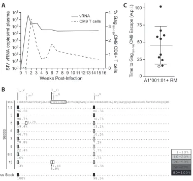
efficiently, peaking at 3.96⫻107viral RNA (vRNA) copies/ml plasma at 10 days postinfection (d.p.i.), as measured by quantitative reverse transcription-PCR (RT-PCR) using previously described techniques (11) (Fig. 1A). This level of viral replication is consistent with a large body of data collected from wild-type SIVmac239-infected Mamu-A1*001:01-positive rhesus macaques (12,13). Additionally, major histocompatibility complex (MHC) class I tetramer staining of freshly isolated peripheral blood mononuclear cells (PBMC) revealed that SIVmac239-2V elicited a strong CD8TLresponse
against Gag181-189CM9, with 3.5% of all circulating CD8TL
tar-Received12 November 2013Accepted22 December 2013 Published ahead of print26 December 2013
Editor:G. Silvestri
Address correspondence and reprint requests to Jonah B. Sacha, [email protected].
B.J.B. and H.L.W. contributed equally to this work.
Copyright © 2014, American Society for Microbiology. All Rights Reserved.
doi:10.1128/JVI.03304-13
on November 7, 2019 by guest
http://jvi.asm.org/
geting this epitope at 14 d.p.i. (Fig. 1A). Importantly, this Gag181-189CM9-specific CD8TLresponse remained at high
fre-quency in r98003 through 15 weeks p.i. (w.p.i.).
Next, we monitored viral evolution withingagby performing Roche/454 pyrosequencing on plasma samples as described pre-viously (8). Unexpectedly, instead of observing rapid, acute-phase escape of Gag181-189CM9, we detected high-frequency reversions
of both the I161V and I206V compensatory mutations (Fig. 1B).
The frequency of revertant viruses in plasma increased through 8.5 w.p.i., when 98.3% of plasma virus sequences were wild-type SIVmac239 at I206and 84% were wild-type SIVmac239 at I161.
Subsequently, we detected two Gag181-189CM9 escape mutations,
C181G and T182A, only after the I206V compensatory mutation had
reverted almost entirely to wild type, with I206V present in only
1.7% of all pyrosequencing reads (Fig. 1B). The C181G mutation is
rare but is a previously described Gag181-189CM9 escape mutation
that also associates with the I161V and I206V compensatory
muta-tions inMamu-A1*001:01-positive, SIVmac239-infected rhesus macaques (8,9). These three mutations increased in frequency together and were tightly linked, with 87% of sequences contain-ing one or both escape mutations also harborcontain-ing the I206V
com-pensatory mutation (data not shown). While we failed to observe rapid acute-phase CD8TLescape, r98003 did manifest the earliest
Gag181-189CM9 escape mutations of nine Mamu-A1*001:01-positive rhesus macaques that we have investigated previously via Roche/454 pyrosequencing (8) (Fig. 1C).
The SIV compensatory mutations Gag I161V and I206V do not always revert following transmission toMamu-A1*001:01 -neg-ative rhesus macaques. Given the surprising reversion of the compensatory mutations following infection of a naive host, we next investigated additional animals infected with SIVmac239 harboring the preengineered I161V and I206V mutations. Friedrich
0 1 2 3 4
0 1 2 3 4 5 6 7 8 9 10 11 12 13 14 15 16 100
101 102 103 104 105 106 107 108
Weeks Post-Infection
S
IV
vRNA
copies/
ml plas
ma
% G
ag
181-189
CM9 CD8+
T
c
ells
T
ime to Gag
181-189
CM9 Escape (w
.p.i.)
A
vRNA CM9 T cells
B
C
0 25 50 75 100
A1*001:01+ RM
[image:2.585.108.488.64.419.2]IEEKKFGAEVVPGFQALSEGCTPYDINQMLNCVGDHQAAMQIIRDIINEEAADWDLQHPQPAPQQGQLREPSGSDIAGTTSSVDEQIQWM
... ...
.... ... ...
... ...
... ...
... ...
... ...
... ... ... 1.5
3
5
6
7
8
8.5
r98003
w.p.i.
15
59.4% 46.7%
16% 22.4% 28.8% 90.7%
13%
... ...
96.6% 86.3%
13.1% 10.5%
1.7% 5.7% 7.2% 70.7%
10.3% 1.7%
8.9%1.4%
T182A I206V
I161V
F166I C181G
... ...
100% 98.5%
Virus Stock
1-10% 10-20% 20-50% 50-80%
80-100%
FIG 1Compensatory mutations in SIVmac239-2V revert rapidly to wild type despite a Gag181–189CM9-specific CD8TLresponse. (A) Replication of the
engineered SIVmac239 mutant SIVmac239-2V in r98003 was monitored in plasma over the first 15 weeks postinfection (w.p.i.). During the same period, the magnitude of the Gag181–189CM9-specific CD8TLresponse was assessed by Mamu-A1*001:01/Gag181–189CM9 tetramer staining. (B) The Gag181– 189CM9 region from the SIVmac239-2V-infected,Mamu-A1*00101-positive rhesus macaque r98003 was Roche/454 pyrosequenced at the indicated w.p.i. Each consensus sequence indicates all mutations present in 1% or more of total sequence reads (limit of detection). Amino acid substitutions are shown above the reference sequence; the frequencies of the mutations are shown both as percentages and as shaded boxes according to prevalence, as indicated by the key at the bottom right. Boxed amino acids within the reference sequence comprise the Gag181–189CM9 epitope. The consensus sequence of the SIVmac239-2V virus stock used to infect r98003 is shown below the sequences from r98003. (C) Times to Gag181–189CM9 escape (as defined by the first of two consecutive time points showing amino acid substitutions within the epitope at a frequency of⬎1% of total sequence reads) in nine Mamu-A1*00101-positive rhesus macaques (filled circles) and animal r98003 (open circle). The means and standard deviations of times to Gag181–189CM9 escape are shown.
on November 7, 2019 by guest
http://jvi.asm.org/
et al. previously described three rhesus macaques infected with 3XSIVmac239, a virus encoding the Gag181–189CM9 escape
muta-tion T182A and the two compensatory substitutions I161V and
I206V (2). We Roche/454 pyrosequenced virus from plasma
sam-ples obtained from these three animals, spanning the first 8 months of infection, to make direct comparisons with virus se-quences from r98003 and to glean additional insight into why
SIVmac239-2V reverted at positions I161V and I206V before
Gag181–189CM9 escape. Two of these rhesus macaques were
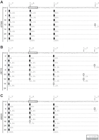
Mamu-A1*001:01negative (r98020, r96115), and one was Mamu-A1*001:01 positive (r97089). The 3XSIVmac239 circulating in r97089 retained all three (I161V, T182A, and I206V) preengineered
mutations, most likely due to low-frequency Mamu-A1*001:01-restricted, Gag181–189CM9-specific CD8TL(14) (Fig. 2A). In
con-1-10% 10-20% 20-50% 50-80%
80-100%
[image:3.585.126.456.67.532.2]IEEKKFGAEVVPGFQALSEGCTPYDINQMLNCVGDHQAAMQIIRDIINEEAADWDLQHPQPAPQQGQLREPSGSDIAGTTSSVDEQIQWM ... ... ...
... ... ... ...
... ... ...
... ... ...
... ... ...
... ...
... ... 3
6
8
12
16
24
30
59.8% 5.7% 98.7% 99.4% 100% 100%
100%
100%
100% 100% 99.5%
99.6% 1%
100%
100%
100%
100% 100% 99.5% 99.6%
r98020
w.p.i.
B
IEEKKFGAEVVPGFQALSEGCTPYDINQMLNCVGDHQAAMQIIRDIINEEAADWDLQHPQPAPQQGQLREPSGSDIAGTTSSVDEQIQWM ... ... ...
... ... ...
... ... ...
... ... ... ...
... ... ...
... ... ...
... ... ... 2
4
10
12
14
24
31
r97089
w.p.i.
99.6%
99.5%
100%
100% 100% 99.4% 98.6%
99.6%
99.5%
100%
100% 100% 100% 99.3%
100%
100%
100%
100% 100% 100% 100%
3.1%
A
I161V T182A I206V I247YC
IEEKKFGAEVVPGFQALSEGCTPYDINQMLNCVGDHQAAMQIIRDIINEEAADWDLQHPQPAPQQGQLREPSGSDIAGTTSSVDEQIQWM ... ... ...
... ... ...
... ... ... ...
... ... ...
... ... ... ...
... ... ... ...
... ...
... 2
4
6
8
10
12
14
r961
15
w.p.i.
24
99%
100%
98.8%
25.9% 86% 98.9% 99.4%
100%
99.3%
96.9%
5.2% 41.3% 66.5% 100% 100%
93.2%
52.2%
12.3% 24.5% 99.4%
2.1%
3.9%
2.5%
I236Y
T182A I206V
I161V I247Y
T182A I206V
I161V I247Y
FIG 2Consensus sequences of the Gag181–189CM9 region in rhesus macaques infected with 3XSIVmac239. The Gag181–189CM9 regions from three rhesus
macaques infected with 3XSIVmac239 were Roche/454 pyrosequenced at the indicated w.p.i. (A) r97089 (Mamu-A1*001:01positive); (B) r96115 ( Mamu-A1*001:01negative); (C) r98020 (Mamu-A1*001:01negative). Each consensus sequence indicates all mutations present in 1% or more of total sequence reads (limit of detection). Amino acid substitutions are shown above the reference sequence; the frequencies of the mutations are shown both as percentages and as shaded boxes according to prevalence, as indicated by the key at the bottom right. Boxed amino acids within the reference sequence comprise the Gag181–189CM9 epitope.
on November 7, 2019 by guest
http://jvi.asm.org/
trast, 3XSIVmac239 evolution diverged between the two Mamu-A1*001:01-negative animals. The 3XSIVmac239 circulating in r96115 reverted completely to wild type at all three positions (I161,
T182, and I206) by 24 w.p.i., supporting our data from r98003 (Fig.
2B). Surprisingly, 3XSIVmac239 in r98020 reverted completely to wild type at position T182by 24 w.p.i. but maintained the
compensa-tory mutations I161V and I206V in 100% of sequence reads through 30
w.p.i. (Fig. 2C). This finding is in contrast to our data from r98003 and r96115, in which compensatory mutations I161V and I206V
re-verted in the absence of a Gag181–189CM9 escape mutation (Fig. 1B
and2B).
The downstream Gag mutation A312P stabilizes SIV contain-ing compensatory mutations I161V and I206V in the absence of a Gag181–189CM9 escape mutation.The divergent viral evolution between r98003, r96115, and r98020 raised the possibility that a tertiary mutation was maintaining viral fitness while allowing for the retention of the I161V and I206V compensatory mutations. To
test this hypothesis, we analyzed Gag sequences from a cohort of 25 chronically SIV-infected rhesus macaques, including r98003, r97089, and r98020, at various time points postinfection (11
Mamu-A1*001:01-positive, 14 Mamu-A1*001:01-negative
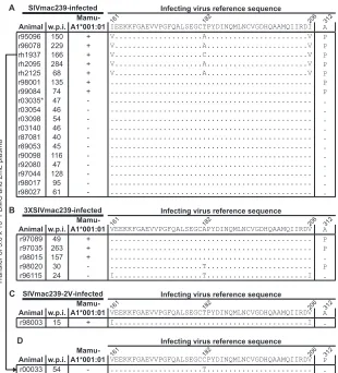
ma-caques) using consensus Sanger sequencing as previously de-scribed (15). We found that viral sequences from five of seven SIVmac239-infected, Mamu-A1*001:01-positive animals con-tained mutations I161V, T182A/C, I206V, and a downstream
muta-tion, Gag A312P (Fig. 3A). Virus from the two remaining
Mamu-A1*001:01-positive animals (r98001 and r99084) harbored the A312P mutation alone, suggesting that this mutation can precede
the I161V, T182A/C, and I206V mutations. In contrast, none of these
mutations were observed inMamu-A1*001:01-negative animals (Fig. 3A). Thus, it appeared that A312P was associated with
Gag181–189CM9 escape. Next, we examined the association of
A312P with the maintenance of the I161V and I206V compensatory
mutations following transmission of our engineered viruses to naive hosts. In two out of the threeMamu-A1*001:01-positive animals infected with 3XSIVmac239 (r97089 and r97035), the A312P mutation arose following infection, which again suggested
that A312P stabilizes the Gag181–189CM9 escape-associated
com-pensatory mutations (Fig. 3B). In the two Mamu-A1*001:01-neg-ative animals infected with 3XSIVmac239 (r98020 and r96115), A312P was present only in r98020, where the compensatory
muta-tions persisted in the absence of Gag181–189CM9 escape. In r98003,
161 182 206 312
161 182 206 312
161 182 206 312
B A
C
Transfer of 5.0 x 10
6 PBMC and 2mL plasma
161 182 206 312
D
Mamu-Animal w.p.i. A1*001:01 IEEKKFGAEVVPGFQALSEGCTPYDINQMLNCVGDHQAAMQIIRDI A
r95096 150 + V...A...V P
r96078 229 + V...A...V P
rh1937 166 + V...C...V P
rh2095 284 + V...A...V P
rh2125 68 + V...A...V P
r98001 135 + ... P
r99084 74 + ... P
r03035* 47 - ... .
r03054 46 - ... .
r03098 54 - ... .
r03140 46 - ... .
r87081 40 - ... .
r89053 45 - ... .
r90098 116 - ... .
r92080 47 - ... .
r97044 128 - ... .
r98017 95 - ... .
r98027 61 - ... .
Mamu-Animal w.p.i. A1*001:01 VEEKKFGAEVVPGFQALSEGCAPYDINQMLNCVGDHQAAMQIIRDV A
r97089 49 + ... P
r97035 263 + ... P
r98015 157 + ... .
r98020 30 - ...T... P
r96115 24 - I...T...I .
Mamu-Animal w.p.i. A1*001:01 VEEKKFGAEVVPGFQALSEGCTPYDINQMLNCVGDHQAAMQIIRDV A
r98003 15 + I...I .
Mamu-Animal w.p.i. A1*001:01 VEEKKFGAEVVPGFQALSEGCCPYDINQMLNCVGDHQAAMQIIRDV P
r00033 54 - ...T... .
Infecting virus reference sequence SIVmac239-infected
3XSIVmac239-infected
SIVmac239-2V-infected
Infecting virus reference sequence
Infecting virus reference sequence
Infecting virus reference sequence
FIG 3The Gag A312P mutation stabilizes the compensatory mutations Gag I161V and I206V. Consensus Sanger sequences spanning Gag I161through I206and the
tertiary mutation site Gag A312at the indicated w.p.i. are shown. (A) Eighteen SIVmac239-infected rhesus macaques (sevenMamu-A1*001:01positive and eleven
Mamu-A1*001:01negative). *, r03035 was infected with the SIVmac239 variant B*008:01 8⫻SIVmac239 described in reference21. (B) Five 3XSIVmac239-infected rhesus macaques (threeMamu-A1*001:01positive and twoMamu-A1*001:01negative). (C) OneMamu-A1*001:01-positive, SIVmac239-2V-infected rhesus macaque. (D) OneMamu-A1*001:01-negative rhesus macaque infected by transfer of plasma and PBMC from rh1937 at 166 w.p.i. (see the arrow). Reference sequences for respective infecting SIV strains are shown across the top.
on November 7, 2019 by guest
http://jvi.asm.org/
[image:4.585.140.450.65.407.2]theMamu-A1*001:01-positive animal infected with SIVmac239-2V, the lack of A312P was associated with reversion of the
compen-satory mutations to wild type (Fig. 3C).
To further test if the tertiary mutation A312P would stabilize the
I161V and I206V compensatory mutations during reversion of
Gag181–189CM9 epitope mutations, we infected aMamu-A1*001:
01-negative animal, r00033, with virus from rh1937 that con-tained the T182C Gag181–189CM9 escape mutation, the I161V and
I206V compensatory mutations, and the tertiary A312P mutation
(Fig. 3AandD). In agreement with our previous observations, the A312P mutation stabilized the I161V and I206V compensatory
mu-tations, while the T182C escape mutation reverted to wild type
(Fig. 3D). Cumulatively, these data suggest that A312P is necessary
for the maintenance of I161V and I206V compensatory mutations
in the absence of Gag181–189CM9 escape.
Gag A312P is a bona fide compensatory mutation.We ob-served that A312P consistently associated with the Gag181–189CM9
escape-associated compensatory mutations, but we wanted to fur-ther assess the compensatory characteristics of the A312P
muta-tion. We first investigated the possibility that A312P could be a
CD8TLescape mutation but found no evidence of T cells targeting
this region of Gag. Usingin silicobinding algorithms (http://www .cbs.dtu.dk/services/NetMHC/) we found that no 8-, 9-, or 10-mer peptides between SIVmac239 Gag residues 301 and 323 are predicted to bind Mamu-A1*001:01 with physiologically relevant affinity (50% inhibitory concentration [IC50]ⱕ500 nM) (16). In
addition, we detected no CD8TLresponses to the A312region by a
gamma interferon (IFN-␥) enzyme-linked immunosorbent spot assay (ELISpot) in 7Mamu-A1*001:01-positive rhesus macaques with detectable Gag181–189CM9-specific CD8TL responses, 4 of
A
A
B
C
A312 T182
I161 I206
SIVmac239 CA (Top view)
A312
I161 I206
T182
14 21
0 50 100 150
SIVmac239-2V SIVmac239-2V-P SIVmac239-P Uninfected
Days Post-Infection
F
re
q
u
e
n
c
y
SIV-inf
e
c
te
d
c
e
lls
(no
rma
liz
e
d
t
o
S
IV
m
ac239
)
Virus
D
0 102 103 104 105
0 20 40 60 80 100
Gag p27
% of Max Event Count
4.69 x 10³
5.64 x 10³ 1.04 x 10⁴
8.83 x 10³
Input virus 161 182 206 312
Amino acid position
DAY 14
I T I
V T I / V A
V T V P
I T I P
SIVmac239 CA (Side view - magnified) Peptides
SF
C
/1
0
6
PB
M
C
(b
a
c
kg
ro
u
n
d
su
b
tra
c
te
d
)
0 200 400 600 800 1600 1800 2000
rh2095
r25873 r97089
r26136 r24686 rh2125
r98001
SIVmac239
SIVmac239-2V
SIVmac239-2V-P
SIVmac239-P
E
SIVmac239-2V SIVmac239-2V-P SIVmac239-P Virus
SIVmac239 Gag p27 MFI
Gag CM9 Gag301-315Gag305-319Gag309-323
FIG 4Gag A312P is a tertiary compensatory mutation stabilizing SIVmac239 CA expressing I206V. (A) SevenMamu-A1*00101-positive rhesus macaques infected
with SIV were screened for T cell responses against three overlapping 15-mer peptides spanning the A312region by IFN-␥ELISpot. Animals harboring SIV with the A312P mutation are represented with open red symbols. SFC, spot-forming cells. The dashed line at 50 SFC is the limit of detection for this assay. (B) Frequency of Gag p27⫹CD4⫹T cell targets infected with engineered mutant virusesin vitro. Values were normalized to the frequency of Gag p27⫹targets infected with SIVmac239. (C) Table showing Sanger sequencing results from vRNA taken at day 14 of thein vitroreplication assay. (D) Structural modeling of SIVmac239 capsid, showing positions of the I161, T182, I206, and A312amino acid residues. (Top) Top view of the SIVmac239 CA hexamer, with one monomer colored light blue and a second monomer colored dark blue. (Bottom) Magnified side view of the potential interaction between A312(within the light-blue monomer) and I206(within the dark-blue monomer). (E) Representative Gag p27-staining histogram, showing the mean fluorescent intensity of intracellular Gag p27-fluorescein isothiocyanate (FITC) staining for each virus at day 21 of ourin vitroreplication assay. Results are indicative of separate assays performed with two different Gag p27-specific antibodies.
on November 7, 2019 by guest
http://jvi.asm.org/
which also had circulating virus harboring the A312P mutation
(Fig. 4A, red symbols). These results, in combination, support the conclusion that CD8TLpressure does not drive the emergence of
A312P inMamu-A1*001:01-positive animals.
Next, we performed anin vitroSIVmac239 replication assay to compare the growth kinetics and levels of stability of viruses con-taining the I161V, I206V, and A312P mutations. We engineered two
SIVmac239 mutants (in addition to SIVmac239-2V) containing the I161V, I206V, and A312P mutations together
(SIVmac239-2V-P) or the A312P mutation alone (SIVmac239-P). We infected
separate cultures of staphylococcal-enterotoxin B-stimulated rhesus macaque CD4⫹T cells with 2⫻107virus particles of either SIVmac239 or a mutant virus in complete medium containing 100 IU interleukin 2 (IL-2)/ml and cultured them for 7 days. On days 7 and 14, virus-containing cell supernatant was diluted 1:4 with medium and used to infect additional activated CD4⫹T cells. We compared the frequencies of infected targets for each mutant virus on days 14 and 21 by intracellular Gag p27 antibody staining, normalizing to the frequency of SIVmac239-infected targets (Fig. 4B). Mutant viruses showed replication kinetics similar to that of SIVmac239 over the first 14 days but exhibited a higher replicative capacity by day 21. We Sanger sequenced the mutant viruses from our replication assay at day 14 and found that SIVmac239-2V, but not SIVmac239-2V-P, showed a prominent double peak indicat-ing reversion of the I206V mutation (Fig. 4C).
Finally, we generated structural models of the SIVmac239 cap-sid (CA) hexamer as previously described (17) and assessed loca-tions and potential interacloca-tions of the I161, T182, I206, and A312
amino acid residues (Fig. 4D, top). The I161residue is found in
helix 1 of CA, and its side chain protrudes into helix 4, where the I206residue is located, supporting the observed linkage between
the I161V and I206V mutations. Importantly, the A312residue lies
within CA helix 9, near the I206-containing helix 4 of the adjacent
CA monomer (Fig. 4D, bottom). Thus, SIVmac239 CA structural data suggest that the A312P mutation may influence the
intermo-lecular interactions and stability of the CA hexamer. In support of this hypothesis, we consistently detected higher levels of intracel-lular Gag p27 CA in cells infected with viruses containing the A312P mutations (Fig. 4E). Taken together, our structural andin
vitrosequencing data strongly support our conclusion that A312P
is a tertiary compensatory mutation that stabilizes SIVmac239 expressing the Gag181–189CM9-associated compensatory
muta-tion.
The association of A312P with the I161V and I206V
compensa-tory mutations has not previously been described in studies of Gag181–189CM9 escape but is not altogether an unexpected
find-ing. In fact, the data presented herein contribute to a growing body of evidence that HIV and SIV variation within Gag is highly organized, both in terms of mutation kinetics and constrained patterns of escape (18–20). These structural restraints have con-tributed to the identification of Gag as a premier target for T cell-based HIV vaccine design.
As HIV circulates in human populations, there is concern that CD8TLepitopes will be “lost” as escape substitutions accumulate
in transmitted viruses. While the finding that such escape muta-tions frequently revert in the absence of selecting HLA alleles ame-liorates this concern, the persistence of compensatory mutations in transmitted viruses may facilitate rapid escape when the virus next encounters CD8TLrestricted by the selecting HLA molecule.
However, our data indicate that transmitted compensatory
muta-tions do not necessarily facilitate rapid, acute-phase immune es-cape from CD8TLresponses. Further study of additional
compen-satory mutations, in terms of their frequency in circulating HIV strains and their effect on posttransmission CD8TLresponses, is
warranted to fully understand their impact on the adaptation of HIV to HLA molecules present at high frequency within the hu-man population.
ACKNOWLEDGMENTS
This work was supported in part by the National Center for Research Resources (grants P51 OD011092 and R21 068586).
We gratefully acknowledge David Watkins for providing longitudinal plasma and PBMC samples. We thank Anusha Hoda for technical assis-tance. SIVmac239 plasmids p239SpSp5=and p239SpE3=nefopen were ob-tained from the NIH AIDS Reagent Program.
REFERENCES
1.Mudd PA, Ericsen AJ, Walsh AD, Leon EJ, Wilson NA, Maness NJ,
Friedrich TC, Watkins DI.2011. CD8⫹T cell escape mutations in simian
immunodeficiency virus SIVmac239 cause fitness defects in vivo, and many revert after transmission. J. Virol.85:12804 –12810.http://dx.doi .org/10.1128/JVI.05841-11.
2.Friedrich TC, Dodds EJ, Yant LJ, Vojnov L, Rudersdorf R, Cullen C,
Evans DT, Desrosiers RC, Mothe BR, Sidney J, Sette A, Kunstman K, Wolinsky S, Piatak M, Lifson J, Hughes AL, Wilson N, O’Connor DH,
Watkins DI.2004. Reversion of CTL escape-variant immunodeficiency
viruses in vivo. Nat. Med.10:275–281.http://dx.doi.org/10.1038/nm998.
3.Leslie AJ, Pfafferott KJ, Chetty P, Draenert R, Addo MM, Feeney M,
Tang Y, Holmes EC, Allen T, Prado JG, Altfeld M, Brander C, Dixon C, Ramduth D, Jeena P, Thomas SA, St John A, Roach TA, Kupfer B, Luzzi G, Edwards A, Taylor G, Lyall H, Tudor-Williams G, Novelli V,
Martinez-Picado J, Kiepiela P, Walker BD, Goulder PJ. 2004. HIV
evolution: CTL escape mutation and reversion after transmission. Nat. Med.10:282–289.http://dx.doi.org/10.1038/nm992.
4.Schneidewind A, Brumme ZL, Brumme CJ, Power KA, Reyor LL,
O’Sullivan K, Gladden A, Hempel U, Kuntzen T, Wang YE, Oniangue-Ndza C, Jessen H, Markowitz M, Rosenberg ES, Sekaly RP, Kelleher
AD, Walker BD, Allen TM.2009. Transmission and long-term stability
of compensated CD8 escape mutations. J. Virol.83:3993–3997.http://dx .doi.org/10.1128/JVI.01108-08.
5.Boutwell CL, Carlson JM, Lin TH, Seese A, Power KA, Peng J, Tang Y,
Brumme ZL, Heckerman D, Schneidewind A, Allen TM.2013. Frequent
and variable cytotoxic-T-lymphocyte escape-associated fitness costs in the human immunodeficiency virus type 1 subtype B Gag proteins. J. Virol.
87:3952–3965.http://dx.doi.org/10.1128/JVI.03233-12.
6.Brockman MA, Brumme ZL, Brumme CJ, Miura T, Sela J, Rosato PC,
Kadie CM, Carlson JM, Markle TJ, Streeck H, Kelleher AD, Markowitz M, Jessen H, Rosenberg E, Altfeld M, Harrigan PR, Heckerman D,
Walker BD, Allen TM.2010. Early selection in Gag by protective HLA
alleles contributes to reduced HIV-1 replication capacity that may be largely compensated for in chronic infection. J. Virol.83:11937–11949.
http://dx.doi.org/10.1128/JVI.01086-10.
7.Schneidewind A, Brockman MA, Yang R, Adam RI, Li B, Le Gall S,
Rinaldo CR, Craggs SL, Allgaier RL, Power KA, Kuntzen T, Tung CS, LaBute MX, Mueller SM, Harrer T, McMichael AJ, Goulder PJ, Aiken C,
Brander C, Kelleher AD, Allen TM.2007. Escape from the dominant
HLA-B27-restricted cytotoxic T-lymphocyte response in Gag is associated with a dramatic reduction in human immunodeficiency virus type 1 replication. J. Virol.81:12382–12393.http://dx.doi.org/10.1128/JVI.01543-07.
8.Burwitz BJ, Sacha JB, Reed JS, Newman LP, Norante FA, Bimber BN,
Wilson NA, Watkins DI, O’Connor DH.2011. Pyrosequencing reveals
restricted patterns of CD8⫹T cell escape-associated compensatory mu-tations in simian immunodeficiency virus. J. Virol.85:13088 –13096.http: //dx.doi.org/10.1128/JVI.05650-11.
9.Friedrich TC, Frye CA, Yant LJ, O’Connor DH, Kriewaldt NA, Benson
M, Vojnov L, Dodds EJ, Cullen C, Rudersdorf R, Hughes AL, Wilson N,
Watkins DI. 2004. Extraepitopic compensatory substitutions partially
restore fitness to simian immunodeficiency virus variants that escape from an immunodominant cytotoxic-T-lymphocyte response. J. Virol. 78:
2581–2585.http://dx.doi.org/10.1128/JVI.78.5.2581-2585.2004.
on November 7, 2019 by guest
http://jvi.asm.org/
10. Peyerl FW, Barouch DH, Yeh WW, Bazick HS, Kunstman J, Kunstman
KJ, Wolinsky SM, Letvin NL.2003. Simian-human immunodeficiency
virus escape from cytotoxic T-lymphocyte recognition at a structurally constrained epitope. J. Virol.77:12572–12578.http://dx.doi.org/10.1128 /JVI.77.23.12572-12578.2003.
11. Friedrich TC, Valentine LE, Yant LJ, Rakasz EG, Piaskowski SM,
Furlott JR, Weisgrau KL, Burwitz B, May GE, Leon EJ, Soma T, Napoe
G, Capuano SV, Wilson NA, Watkins DI.2007. Subdominant CD8⫹
T-cell responses are involved in durable control of AIDS virus replication. J. Virol.81:3465–3476.http://dx.doi.org/10.1128/JVI.02392-06.
12. Kim EY, Veazey RS, Zahn R, McEvers KJ, Baumeister SH, Foster GJ,
Rett MD, Newberg MH, Kuroda MJ, Rieber EP, Piatak MJ, Lifson JD,
Letvin NL, Wolinsky SM, Schmitz JE.2008. Contribution of CD8⫹T
cells to containment of viral replication and emergence of mutations in Mamu-A*01-restricted epitopes in simian immunodeficiency virus-infected rhesus monkeys. J. Virol. 82:5631–5635.http://dx.doi.org/10 .1128/JVI.02749-07.
13. Wilson NA, Reed J, Napoe GS, Piaskowski S, Szymanski A, Furlott J,
Gonzalez EJ, Yant LJ, Maness NJ, May GE, Soma T, Reynolds MR, Rakasz E, Rudersdorf R, McDermott AB, O’Connor DH, Friedrich TC, Allison DB, Patki A, Picker LJ, Burton DR, Lin J, Huang L, Patel D, Heindecker G, Fan J, Citron M, Horton M, Wang F, Liang X, Shiver
JW, Casimiro DR, Watkins DI.2006. Vaccine-induced cellular immune
responses reduce plasma viral concentrations after repeated low-dose challenge with pathogenic simian immunodeficiency virus SIVmac239. J. Virol.80:5875–5885.http://dx.doi.org/10.1128/JVI.00171-06.
14. Friedrich TC, McDermott AB, Reynolds MR, Piaskowski S, Fuenger S,
De Souza IP, Rudersdorf R, Cullen C, Yant LJ, Vojnov L, Stephany J,
Martin S, O’Connor DH, Wilson N, Watkins DI.2004. Consequences of
cytotoxic T-lymphocyte escape: common escape mutations in simian im-munodeficiency virus are poorly recognized in naive hosts. J. Virol.78:
10064 –10073.http://dx.doi.org/10.1128/JVI.78.18.10064-10073.2004.
15. O’Connor DH, McDermott AB, Krebs KC, Dodds EJ, Miller JE,
Gon-zalez EJ, Jacoby TJ, Yant L, Piontkivska H, Pantophlet R, Burton DR,
Rehrauer WM, Wilson N, Hughes AL, Watkins DI.2004. A dominant
role for CD8⫹-T-lymphocyte selection in simian immunodeficiency virus
sequence variation. J. Virol.78:14012–14022.http://dx.doi.org/10.1128 /JVI.78.24.14012-14022.2004.
16. Lundegaard C, Lamberth K, Harndahl M, Buus S, Lund O, Nielsen M.
2008. NetMHC-3.0: accurate web accessible predictions of human, mouse and monkey MHC class I affinities for peptides of length 8 –11. Nucleic Acids Res.36:W509 –W512.http://dx.doi.org/10.1093/nar/gkn202.
17. Inagaki N, Takeuchi H, Yokoyama M, Sato H, Ryo A, Yamamoto H,
Kawada M, Matano T.2010. A structural constraint for functional
inter-action between N-terminal and C-terminal domains in simian immuno-deficiency virus capsid proteins. Retrovirology7:90.http://dx.doi.org/10 .1186/1742-4690-7-90.
18. Dahirel V, Shekhar K, Pereyra F, Miura T, Artyomov M, Talsania S,
Allen TM, Altfeld M, Carrington M, Irvine DJ, Walker BD,
Chakraborty AK.2011. Coordinate linkage of HIV evolution reveals
re-gions of immunological vulnerability. Proc. Natl. Acad. Sci. U. S. A.108:
11530 –11535.http://dx.doi.org/10.1073/pnas.1105315108.
19. Schneidewind A, Brockman MA, Sidney J, Wang YE, Chen H,
Susco-vich TJ, Li B, Adam RI, Allgaier RL, Mothe BR, Kuntzen T, Oniangue-Ndza C, Trocha A, Yu XG, Brander C, Sette A, Walker BD, Allen TM.
2008. Structural and functional constraints limit options for cytotoxic T-lymphocyte escape in the immunodominant HLA-B27-restricted epitope in human immunodeficiency virus type 1 capsid. J. Virol.82:
5594 –5605.http://dx.doi.org/10.1128/JVI.02356-07.
20. Brockman MA, Schneidewind A, Lahaie M, Schmidt A, Miura T,
Desouza I, Ryvkin F, Derdeyn CA, Allen S, Hunter E, Mulenga J,
Goepfert PA, Walker BD, Allen TM.2007. Escape and compensation
from early HLA-B57-mediated cytotoxic T-lymphocyte pressure on hu-man immunodeficiency virus type 1 Gag alter capsid interactions with cyclophilin A. J. Virol. 81:12608 –12618.http://dx.doi.org/10.1128/JVI .01369-07.
21. Valentine LE, Loffredo JT, Bean AT, Leon EJ, MacNair CE, Beal DR,
Piaskowski SM, Klimentidis YC, Lank SM, Wiseman RW, Weinfurter JT, May GE, Rakasz EG, Wilson NA, Friedrich TC, O’Connor DH,
Allison DB, Watkins DI.2009. Infection with “escaped” virus variants
impairs control of simian immunodeficiency virus SIVmac239 replication in Mamu-B*08-positive macaques. J. Virol.83:11514 –11527.http://dx .doi.org/10.1128/JVI.01298-09.