Cellular Visualization of Macrophage Pyroptosis and Interleukin-1

Release in a Viral Hemorrhagic Infection in Zebrafish Larvae
Mónica Varela,aAlejandro Romero,aSonia Dios,aMichiel van der Vaart,bAntonio Figueras,aAnnemarie H. Meijer,bBeatriz Novoaa
Marine Research Institute, CSIC, Vigo, Spaina; Institute of Biology, Leiden University, Leiden, The Netherlandsb
ABSTRACT
Hemorrhagic viral diseases are distributed worldwide with important pathogens, such as dengue virus or hantaviruses. The lack of adequatein vivoinfection models has limited the research on viral pathogenesis and the current understanding of the under-lying infection mechanisms. Although hemorrhages have been associated with the infection of endothelial cells, other cellular types could be the main targets for hemorrhagic viruses. Our objective was to take advantage of the use of zebrafish larvae in the study of viral hemorrhagic diseases, focusing on the interaction between viruses and host cells. Cellular processes, such as tran-sendothelial migration of leukocytes, virus-induced pyroptosis of macrophages. and interleukin-1(Il-1) release, could be ob-served in individual cells, providing a deeper knowledge of the immune mechanisms implicated in the disease. Furthermore, the application of these techniques to other pathogens will improve the current knowledge of host-pathogen interactions and in-crease the potential for the discovery of new therapeutic targets.
IMPORTANCE
Pathogenic mechanisms of hemorrhagic viruses are diverse, and most of the research regarding interactions between viruses and host cells has been performed in cell lines that might not be major targets during natural infections. Thus, viral pathogenesis research has been limited because of the lack of adequatein vivoinfection models. The understanding of the relative pathogenic roles of the viral agent and the host response to the infection is crucial. This will be facilitated by the establishment ofin vivo
infection models using organisms such as zebrafish, which allows the study of the diseases in the context of a complete individ-ual. The use of this animal model with other pathogens could improve the current knowledge on host-pathogen interactions and increase the potential for the discovery of new therapeutic targets against diverse viral diseases.
E
ndothelial cells and leukocytes are at the front line of defense against pathogens. Most infectious pathogens have some rela-tionship with the endothelium, although not all pathogens are true endothelial invading organisms (1). Infections can produce large changes in endothelial cell function, particularly in relation to inflammation. Inflammatory stimuli can activate endothelial cells, which secrete cytokines and chemokines. Moreover, the im-portance of endothelial cells in the regulation of leukocyte trans-migration to the site of inflammation has given these cells a major role in immunity (2).Host-pathogen interactions are essential for the modulation of immunity (3). The presence of a pathogen alters the immune bal-ance prevailing in a host under normal conditions, causing the appearance of diseases, which lead to the death of the host if it is not able to control the infection. Clear examples of these effects are evident in viral hemorrhagic fevers, in which an understanding of the relative pathogenic roles of the viral agent and the host response to the infection is crucial (4). Hemorrhagic manifesta-tions are characteristic of these fevers, which have historically been associated with endothelium infections. Currently, many patho-gens causing hemorrhagic diseases do not infect endothelial cells as the primary target (1). Indeed, macrophages and dendritic cells are targets for some hemorrhagic viruses, such as Ebola virus (5) or dengue virus (6,7).
Most of the research regarding interactions between viruses and host cells has been performed with cell lines that might not be major targets during natural infections (8), making it difficult to characterize the behavior of pathogens in individuals and reduc-ing the likelihood of discoverreduc-ing an effective therapeutic target.
The lack of a good animal model in which the infection can be followed from the beginning and in the context of the whole or-ganism has contributed to these difficulties. Zebrafish (Danio rerio) provide advantages, since these organisms exhibit rapid de-velopment, transparency, and a high homology between their ge-nome and the human one (9). Furthermore, the existence of mu-tant and transgenic fish lines and the potential use of reverse-genetics techniques, such as the injection of antisense morpholino (MO) oligonucleotides or synthetic mRNA, make zebrafish a use-ful tool for the study of host-pathogen interactions.
No naturally occurring viral infections have been characterized for zebrafish (10). Moreover, the experimental susceptibility of zebrafish to viral infections has been demonstrated with other fish viruses (11–13) and also with mammalian viruses (14–16).
We used the rhabdovirus spring viremia of carp virus (SVCV) as a viral model to show the potential of the zebrafish in host-pathogen interaction studies. SVCV predominantly affects cy-prinid fish and causes important economic losses and mortalities
Received17 July 2014 Accepted2 August 2014
Published ahead of print6 August 2014
Editor:R. W. Doms
Address correspondence to Beatriz Novoa, beatriznovoa@iim.csic.es.
Supplemental material for this article may be found athttp://dx.doi.org/10.1128 /JVI.02056-14.
Copyright © 2014, American Society for Microbiology. All Rights Reserved.
doi:10.1128/JVI.02056-14
on November 7, 2019 by guest
http://jvi.asm.org/
worldwide (17,18). Affected fish exhibit destruction of tissues, such as kidney or spleen, leading to hemorrhage and death. How-ever, little is known about the infection mechanism of this envel-oped RNA virus.
Thus, the main objective of the present study was to improve the knowledge of the pathology generated by SVCV, as an example of a hemorrhagic virus, focusing on interactions between the virus and host cells at early stages of the infection in zebrafish larvae. Taking advantage of zebrafish transgenic lines, imaging tech-niques and, using morpholino-induced cell depletion, virus-in-duced pyroptosis and interleukin-1 (IL-1) release were ob-served in macrophages at the single-cell level.
MATERIALS AND METHODS
Ethics statement.The protocols for fish care and the challenge experi-ments were reviewed and approved by the CSIC National Committee on Bioethics under approval number 07_09032012. Experiments were con-ducted in fish larvae before independent feeding and therefore before ethical approval is required (EU directive 2010_63).
Fish.Homozygous embryos and larvae from the wild-type zebrafish, transgenic line Tg(Fli1a:eGFP)y1, in which endothelial cells are labeled, trans-genic line Tg(Mpx:GFP)i114, in which neutrophils are labeled, transtrans-genic line Tg(Mpeg1:eGFP)gl22, in which macrophages are labeled, and transgenic line Tg(Cd41:GFP)la2, in which thrombocytes are labeled, were obtained from our experimental facility, where the zebrafish were cultured using established protocols (19,20). The eggs were obtained according to protocols described inThe Zebrafish Book(19) and maintained at 28.5°C in egg water (5 mM NaCl, 0.17 mM KCl, 0.33 mM CaCl2, 0.33 mM MgSO4, and 0.00005% meth-ylene blue), and 0.2 mMN-phenylthiourea (PTU) (Sigma) was used to pre-vent pigment formation from 1 day postfertilization (dpf).
Virus and infection.The SVCV isolate 56/70 was propagated on epi-thelioma papulosum cyprini (EPC) carp cells (ATCC CRL-2872) and titrated in 96-well plates. The plates were incubated at 15°C for 7 days and examined for cytopathic effects each day. After the observation of the cells under the microscope, the virus dilution causing infection of 50% of the inoculated cell line (50% tissue culture infective dose [TCID50]) was
de-termined using the Reed-Müench method (21). The fish larvae were in-fected through microinjection into the duct of Cuvier as described by Benard et al. (22) at 2 or 3 dpf. SVCV was diluted to the appropriate concentration in phosphate-buffered saline (PBS) with 0.1% phenol red (as a visible marker to the injection of the solution into the embryo) just before the microinjection of 2 nl of viral suspension per larvae. The infec-tions were conducted at 23°C. SVCV was heat killed at 65°C during 20 min.
Imaging.Images of the signs and videos of the blood flow were ob-tained using an AZ100 microscope (Nikon) coupled to a DS-Fi1 digital camera (Nikon). Confocal images of live or fixed larvae were captured using a TSC SPE confocal microscope (Leica). The images were processed using the LAS-AF (Leica Application Suite Advanced Fluorescence) and ImageJ software programs. The three-dimensional (3D) reconstructions and the volume clipping were performed using the Image Surfer (http: //imagesurfer.cs.unc.edu/) and LAS-AF (Leica) programs, respectively.
Immunohistochemistry.Whole-mount immunohistochemistry was performed as previously described (23). The following primary antibodies and dilutions were used: L-plastin (rabbit antizebrafish, 1:500; kindly pro-vided by Paul Martin), caspase A (rabbit antizebrafish, 1:500; Anaspec); SVCV (mouse anti-SVCV, 1:500; BioX Diagnostics) and Il-1(rabbit anti-zebrafish, 1:500; kindly provided by John Hansen). The secondary antibodies used were Alexa 488 antirabbit, Alexa 546 antirabbit or Alexa 635 antimouse (Life Technologies), all diluted 1:1,000.
Real-time PCR.Total RNA was isolated from snap-frozen larvae using the Maxwell 16 LEV simplyRNA tissue kit (Promega) with the automated Maxwell 16 instrument according to the manufacturer’s instructions. cDNA synthesis using random primers and quantitative PCR (qPCR)
were performed as previously described (24). The 18S gene was used as a housekeeping gene to normalize the expression values. The gene expres-sion ratio was calculated by dividing the normalized expresexpres-sion values for infected larvae by the normalized expression values for the controls. Table S1 in the supplemental material shows the sequences of the primer pairs used. Two independent experiments, of 3 biological replicates each, were performed.
Microangiography.Tetramethylrhodamine (TMR) dextran (molecular weight [MW], 2⫻106; Life Technologies) was injected into the caudal vein of
anesthetized zebrafish Tg(Fli1a-GFP) embryos at 24 h postinfection (hpi). The images were acquired within 30 min after injection.
TUNEL assay.The terminal deoxynucleotidyltransferase-mediated dUTP-biotin nick end labeling (TUNEL) assay was performed using the Rochein situcell death detection kit, TMR red, or peroxidase (POD) (Roche) as previously described (25).
In vivoacridine orange staining.For acridine orange staining, larvae were incubated for 2 h in 10g/ml acridine orange solution, followed by washing for 30 min.
Macrophage depletion with PU.1-MO.For the PU.1 knockdown, the morpholino was injected into the yolk at the one-cell stage at 1 mM to block macrophage development as previously described (26). Macro-phage-depleted fish were infected at 2 dpf to ensure morpholino effective-ness until the end of the infection.
In vivoPI staining.For propidium iodide (PI) staining, larvae were incubated for 2 h in 10g/ml propidium iodide solution, followed by washing for 30 min.
Statistical analysis.Kaplan-Meier survival curves were analyzed with a log rank (Mantel-Cox) test. The neutrophil count and qPCR data were analyzed using one-way analysis of variance and Tukey’s test, and differ-ences were considered significant at aPvalue of⬍0.05. The results are presented as the means⫾standard errors of means (SEM).
RESULTS
Course of SVCV infection in zebrafish larvae.Zebrafish larvae are susceptible to SVCV through bath infection, but typically the results show high interindividual variations (12,27). Moreover, the long infection times required exclude the use of temporal knockdown techniques, such as morpholino microinjection. As with caudal vein injection (27), we found that SVCV injection into the duct of Cuvier was efficient, thereby providing a reproducible route of infection with fast kinetics. We injected 2 nl of an SVCV stock solution (3⫻106TCID
50/ml) per larva. Mortality began at
22 to 24 hpi, consistently reaching nearly 100% between 48 and 50 hpi (Fig. 1A). There were no differences in the development of the infection between the use of 2- or 3-dpf larvae (data not shown). To assess viral transcription, the expression of the SVCV nucleo-protein (N gene) was measured through quantitative RT-PCR (Fig. 1B). The samples were collected between 3 and 24 hpi (just prior to mortality). Efficient viral transcription was observed from 6 hpi, with an important increment between 12 and 18 hpi. More-over, we found that viral N gene transcription began quickly in the fish, since significant differences were found between heat-killed SVCV (HK-SVCV)-infected and SVCV-infected fish (Fig. 1B).
With regard to macroscopic signs, the first ones were visible after 18 h (Fig. 1C). A progressive decrease in the blood flow rate was observed, particularly in the tail (see Movies S1 and S2 in the supple-mental material). The circulation in the tail was completely stopped after 20 to 22 hpi, although it remained visible at the anterior part of the fish. The circulation in the head lasted longer but also eventually stopped (see Movie S3). At this point, blood was clearly visible inside the veins, with the exception of the hemorrhage areas (Fig. 1C). Hem-orrhages appeared in the anterior part of the larvae, mostly in areas with a huge amount of microvessels, but a great heterogeneity was
on November 7, 2019 by guest
http://jvi.asm.org/
FIG 1Infection course and loss of endothelium fluorescence in infected larvae. (A) Kaplan-Meier survival curve after infection with 2 nl of an SVCV solution of 3⫻106TCID
50/ml (P⬍0.001). (B) Quantification of SVCV N gene transcripts as a measure of the viral amount in the larvae over time. Heat-killed SVCV
was injected in order to separate the immune response against the initial viral load. At 3 hpi, significant differences in the quantification of SVCV N gene transcripts were found between heat-killed-SVCV-infected and SVCV-infected larvae. The samples were obtained from infected larvae at different times postinfection. Each sample (technical triplicates) was normalized to the 18S gene. To avoid differences due to the larval development stage, the samples were standardized with respect to an uninfected control of the same age. Three biological replicates (of 10 larvae each) were used to calculate the means⫾standard errors of means. Significant differences between infected and noninfected larvae at the same time point are displayed as “ⴱⴱ” (Pvalue of⬎0.001 and⬍0.01). qPCR results are representative of 2 independent experiments. (C) The common macroscopic symptoms observed at 24 hpi were hemorrhages and blood accumulation around the eyes and head (arrows). See also Movies S1 to S3 in the supplemental material. (D) Confocal images of zebrafish larval head (maximal projections from multiple z stacks) of infected 3-dpf Tg(Fli1a-GFP) larvae immunostained with L-plastin antibody at 24 hpi (magnification,⫻20), showing the loss of GFP fluorescence in infected fish. (E) Confocal images (maximal projections from multiple z stacks) of infected 3-dpf Tg(Fli1a-GFP) larvae immuno-stained with L-plastin antibody at 24 hpi (magnification,⫻20) of vessels in the yolk sac and trunk, showing the loss of GFP fluorescence in infected fish. (F) Higher-magnification images show morphological changes in L-plastin-positive cells in SVCV-infected fish.
on November 7, 2019 by guest
http://jvi.asm.org/
[image:3.585.51.538.53.600.2]observed in the occurrence of these hemorrhages, which varied in terms of location and time postinfection. The heartbeat was clearly less powerful over time, until it completely stopped, the moment at which the larvae were considered dead. The observed signs and times are consistent with those previously described for this virus using a similar infection model (27). Consistency in the infection results re-affirms the idea that SVCV and zebrafish larvae are an appropriate duo for an in-depth study of the innate immune response against that intracellular pathogen.
SVCV does not affect the integrity of the endothelium.The loss of circulation in the trunk and hemorrhages in the larval head were the most characteristic symptoms of infection. Therefore, we characterized the effect of the virus in the vascular system of the fish, as previously determined for another rhabdovirus, infectious hematopoietic necrosis virus (IHNV) (13). Using the transgenic line Tg(Fli1a-GFP) (28), which facilitates the visualization of the vascular system, we clearly observed a loss of green fluorescent protein (GFP) fluorescence in infected fish at 24 hpi (Fig. 1Dand E). This effect was particularly visible in the main veins of the head (Fig. 1D) and in the tail (Fig. 1E). In addition, we used this trans-genic line in combination with a whole-mount fluorescence im-munohistochemistry for L-plastin (leukocyte plastin) (29). The anti-L-plastin antibody was a useful tool for the detection of mac-rophages and neutrophils in the larvae. In addition to the loss of Fli1a-GFP fluorescence in the trunk, a decrease in the number of L-plastin-positive cells in the area corresponding to the caudal hematopoietic tissue was also observed at 24 hpi (Fig. 1E). More-over, a shape change in L-plastin-positive cells was clearly visible in infected fish. The morphology of the cells in infected fish changed from thin and elongated (with prolonged extensions) to rounded and lobulated (Fig. 1F). The morphological changes might be associated with alterations in cell motility or the infec-tion of the leukocytes themselves (30,31).
To confirm the integrity of the endothelium in the tail at 24 hpi, we injected tetramethylrhodamine dextran into the caudal veins of the larvae. This fluorescent dye remained inside the ves-sels, even when Fli1a-GFP fluorescence was absent (Fig. 2A). Shadows in tetramethylrhodamine dextran fluorescence corre-spond with blood cells that remain inside vessels even when cir-culation is stopped (Fig. 2AandB). Moreover, the results of the TUNEL assay and the acridine orange staining revealed neither DNA damage nor necrosis, respectively, in the endothelial cells of infected fish (Fig. 2C), suggesting that the cells were apparently healthy and the loss of fluorescence likely reflected a transcrip-tional regulation of the Fli1a gene and not cell death. This was verified by combining Fli1a-GFP fish with differential interference contrast (DIC) microscopy (Fig. 2D). Further, TUNEL-positive cells in the caudal hematopoietic tissue of infected larvae were observed, and the localization of these cells was consistent with the position of L-plastin-positive hematopoietic precursor cells in control fish (Fig. 2E). This observation was not surprising, since blood flow is essential for hematopoietic stem cell development (32). In addition, several studies have shown the relationship be-tween blood circulation and Fli1, since Fli1-deficient mice exhibit impaired hematopoiesis (33). Indeed, previous studies in mice found that both morphants and knockouts of Fli1 showed normal vessel development, with impaired circulation and hemorrhaging (33,34). This might suggest that the simple transcriptional regu-lation offli1acould be sufficient to cause the symptoms observed in SVCV-infected larvae.
To determine the mechanism underlyingfli1amRNA tran-scription and to associate it with the loss of blood flow in the larvae, we performed a time course analysis during SVCV infec-tion. Based on the signs of their morphants and on their implica-tion in the maintenance of vessel integrity, we also selected the v-ets erythroblastosis virus E26 oncogene homolog 1a (ets1a) and the vascular endothelial cadherin (ve-cad) genes for qPCR analysis (Fig. 2F). Fli1a and Ets1a are both transcription factors involved in the regulation of hematopoiesis and vasculogenesis (35). A statis-tically significant decrease in the level offli1agene transcripts was observed from 12 hpi. Similar results were observed for ets1a, since the time course profile for this transcription factor was iden-tical to that forfli1a. The interindividual variations inve-cad, a key regulator of endothelial intercellular junctions (36), made it diffi-cult to observe a clear expression pattern, particularly at the late stages of the infection. This effect might be associated with the observed heterogeneity in the appearance of hemorrhages, in terms of time, area, or larvae. Notably, the downregulation of
ve-cadat 9 hpi suggests an increase in the permeability of the endothelium at that infection point. Interestingly,fli1a down-regulation clearly occurred before the onset of hemorrhages and the loss of circulation, suggesting that hemorrhages were a conse-quence of this downregulation.
Zebrafish antiviral response to SVCV.To further study the mechanisms involved in the defense against SVCV, we analyzed the expression of some important genes associated with viral de-tection. Pattern recognition receptors, such as Toll-like receptors (TLRs) and RIG-1-like receptors (RLRs), are membrane-bound and cytosolic sensors, respectively (37). We observed a significant downregulation oftlr7,tlr22,tlr8a, andmda5at 3 hpi (Fig. 3A). As occurred with dengue virus (38) or influenza virus (39), this downmodulation could correspond to an immune evasion strat-egy.rig-1was not regulated during the first 24 h of the infection. Regardingtlr3, we also saw a slight but significant regulation at 12 hpi.
Pattern recognition receptors initiate antimicrobial defense mechanisms through several conserved signaling pathways (40). The activation of interferon-regulatory factors induces the pro-duction of several antiviral response-related genes. We observed that SVCV elicited a response in all the analyzed genes (Fig. 3B).
ifnphi1andifnphi2showed different response profiles.ifnphi1 re-mained downregulated between 3 and 9 hpi, the time in which
ifnphi2was regulated.ifnphi1was upregulated from 12 hpi, simi-lar tomx(a and b paralogues), which increased from 12 h and reached a maximum at 24 h.rarres3showed a biphasic response, with higher expression at 9 and 24 hpi. The recently characterized
ifitgene family in zebrafish (41, 42) was also present in the re-sponse, highlighting the response ofifit17a, which reached a 20-fold change.
SVCV-positive cells are not endothelial cells.To determine whether endothelial cells were the primary targets of SVCV in this
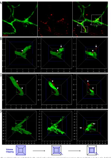
in vivomodel of infection, we performed immunohistochemistry using an antibody against SVCV (Fig. 4). Using Fli1a-GFP-in-fected larvae, SVCV was clearly visible in areas surrounding vessels (Fig. 4A). The 3D reconstruction and volume clipping of the Fli1a channel showed that the SVCV-positive cells were not endothelial cells (Fig. 4B,C, andD). We observed crossing and circulating SVCV-positive cells, suggesting that these cells might be leuko-cytes interacting with the endothelium.
Our hypothesis was confirmed by performing L-plastin
on November 7, 2019 by guest
http://jvi.asm.org/
ing (Fig. 5). L-plastin-positive cells were observed surrounding vessels, and many of these cells colocalized with SVCV. Again, the 3D reconstruction of the confocal images conferred information about the cells and their position relative to the vessels (Fig. 5A). In addition, we observed that SVCV-infected cells showed viral an-tigen-positive signal inside and on their surface (Fig. 5A). The signal detected on the cell surface might be associated with viral assembly, antigen presentation by infected cells, or both (6). We observed cells labeled with only L-plastin (interacting or not with the endothelium), double-positive cells (interacting or not with the endothelium), and cellular debris positive for one or both
markers. Large amounts of cellular debris were observed, always with L-plastin-positive cells phagocytosing SVCV-positive parti-cles in the same area (Fig. 5B). This cellular debris might play a role in the first line of defense against SVCV, as observed with other pathogens (43). Furthermore, defective debris clearance has been associated with persistent inflammatory diseases (44).
Cellular response to SVCV infection.The transendothelial migration of leukocytes during inflammation has been well char-acterized (45). In the present study, the role of leukocytes during infection was explored using the Mpx-GFP zebrafish transgenic line combined with L-plastin immunohistochemistry (Fig. 6).
Us-FIG 2Endothelium integrity is conserved 24 h postinfection. (A) Microangiography was performed at 24 hpi, injecting tetramethylrhodamine into the caudal vein of 3-dpf infected and control larvae. Tetramethylrhodamine remained inside vessels, even in infected fish without Fli1a-GFP fluorescence. Higher-magnification images are shown in the right panels. (B) DIC images showed the morphological integrity of the dorsal aorta (arrows) in 24-hpi and control larvae. Note that blood cells are visible inside dorsal aorta. (C) TUNEL assay and acridine orange staining were performed with 24-hpi and control larvae to detect cells with DNA damage and necrotic dead cells, respectively. Fli1a-GFP fluorescence is absent in infected fish, but TUNEL-positive cells were detected in the ventral part of the tail, which contains the caudal hematopoietic tissue. No differences were found between control and SVCV-infected larvae stained with acridine orange. (D) Fluorescent images of Fli1a-GFP of 3-dpf SVCV-infected and control larvae showing the loss of fluorescence in endothelial cells after 22 h of infection. Higher-magnification images show the intact morphology of the dorsal aorta in infected larvae even when the fluorescence has been lost (red arrowheads). (E) Localization of L-plastin-positive cells in caudal hematopoietic tissue is consistent with that of TUNEL-positive cells at 24 h postinfection. The TUNEL assay was performed in 24-h SVCV-infected and control larvae to detect cells with DNA damage. L-plastin-positive cells are absent in infected fish, but TUNEL-positive cells are detected in the ventral part of the tail, which contains the caudal hematopoietic tissue. (F) Expression qPCR measurements of vascular endothelium-related genes. The samples were obtained from infected larvae at different times postinfection. Each sample (technical triplicates) was normalized to the 18S gene. To avoid differences due to the larval development stage, the samples were standardized with respect to an uninfected control of the same age. Three biological replicates were used to calculate the means⫾standard errors of means. Significant differences between infected and noninfected larvae at the same time point are displayed as follows:ⴱⴱⴱ,Pvalue of⬎0.0001 and⬍0.001;ⴱⴱ,Pvalue of⬎0.001 and⬍0.01;ⴱ,Pvalue of⬎0.01 and⬍0.05.
Varela et al.
on November 7, 2019 by guest
http://jvi.asm.org/
[image:5.585.43.539.67.438.2]FIG 3Viral detection and antiviral response measure over time. (A) Expression qPCR measurements of TLRs and RLRs associated with viral detection. (B) Expression qPCR measurements of antiviral response-related genes. Samples were obtained from infected larvae at different times postinfection. Each sample (technical triplicates) was normalized to the 18S gene. To avoid differences due to the larval development stage, the samples were standardized with respect to an uninfected control of the same age. Three biological replicates were used to calculate the means⫾standard errors of means. Significant differences were displayed as follows:ⴱⴱⴱ,Pvalue of⬎0.0001 and⬍0.001;ⴱⴱ,Pvalue of⬎0.001 and⬍0.01;ⴱ,Pvalue of⬎0.01 and⬍0.05.
on November 7, 2019 by guest
http://jvi.asm.org/
[image:6.585.43.539.72.633.2]ing this system, we were able to differentiate between macro-phages (L-plastin⫹Mpx⫺) and neutrophils (L-plastin⫹Mpx⫹). After 24 h of infection, practically all macrophages were undetect-able (Fig. 6A), and a statistically significant migration of neutro-phils to the head was observed (Fig. 6A,B, andC). The loss of leukocytes in the caudal hematopoietic region was also confirmed (Fig. 6B). Using markers for both cellular types,marcofor
macro-phages andmpxfor neutrophils, we could check by qPCR the dynamics of that leukocyte population (Fig. 6D). The analysis of
marcogene expression suggested an initial decrease in the macro-phage population between 3 and 6 hpi, and then macromacro-phages remained stable until 18 hpi. Thereafter, the macrophage popula-tion markedly reduced to nearly 0 at 24 hpi (Fig. 6D). Regarding neutrophils,mpxrevealed no differences in the number of cells,
FIG 4SVCV-positive cells are distinct from endothelial cells. (A) Confocal images (maximal projections from multiple z stacks) of 3-dpf infected Tg(Fli1a-GFP) larvae immunostained with SVCV antibody at 20 hpi (magnification,⫻60). (B, C, and D) 3D reconstructions of the different zones shown in panel A. Sequential steps of volume clipping of the Fli1a channel are shown from left to right. We classified SVCV-positive cells as circulating (white arrows) and interacting (white arrowheads) cells. The axis numbers correspond tom.
Varela et al.
on November 7, 2019 by guest
http://jvi.asm.org/
[image:7.585.112.475.63.581.2]thus confirming the migration observed in confocal images of these cells during the infection. Complementary experiments us-ing other transgenic lines possessus-ing fluorescent macrophages (Mpeg-GFP), neutrophils (Mpx-GFP), or thrombocytes (Cd41-GFP) showed that only macrophages were SVCV positive at 18 hpi (data not shown). Moreover, given the importance of platelets and the coagulation system in hemorrhagic infections (46), we used the transgenic line with fluorescent thrombocytes (fish equivalent to mammalian platelets) to detect a possible thrombocytopenia or coagulopathy after SVCV infection. Since no differences were found in thrombocyte behavior or count between control and SVCV-infected larvae (data not shown), we decided to focus our attention on macrophages.
To further confirm the role of macrophages as a preferred tar-get for this hemorrhagic virus, at least during the first hours of the infection, we took advantage of the morpholino-mediated gene knockdown technique. We injected the morpholino (MO) PU.1 at a concentration of 1 mM to prevent the formation of macro-phages in embryos (47). A comparison of the normal fish and macrophage-depleted fish in these experimental infections showed a delay in the mortality of morphants (Fig. 6E), although eventually the mortality reached 100% in both groups. The dura-tion of the MO effect was analyzed at an equivalent time of 48 hpi in noninfected fish. Marco expression was completely inhibited,
as demonstrated using qPCR analysis (Fig. 6E). In contrast, the population of neutrophils was not affected by the morpholino, as demonstrated by thempxgene expression after 48 hpi (Fig. 6E). The approximate 9-hour delay in the mortality between both groups, suggested that macrophage presence is crucial for SVCV pathogenesis during the first hours of infection. Therefore, we analyzed the transcription levels of the SVCV N gene at 9 hpi (Fig. 6E). At this time, fish without macrophages had lower viral tran-scription levels than normal fish. Macrophage depletion delays the kinetics of viral transcription but is not enough to prevent the death of morphant fish. This effect likely reflects the high viru-lence of the infection. As an obligate intracellular pathogen, SVCV infects to survive, and even if the primary target is not present, the virus could still infect other cells. However, the efficiency of infec-tion is reduced in the first hours, resulting in a different timing of mortality. This, plus the fact that the only SVCV-positive cells detected at 18 hpi are macrophages, suggested that these cells could be the primary target of the virus.
SVCV elicits an inflammatory response that induces macro-phage pyroptosis and IL-1release.The fact that macrophages were primarily infected with SVCV prompted us to examine the “disappearance” of these cells and determine whether pyroptosis is involved. Pyroptosis might play a role in pathogen clearance, leading to inflammatory pathology, which is detrimental to the
FIG 5SVCV-positive cells are L-plastin positive and Fli1a negative. (A) Confocal image 3D reconstruction of different scenes observed during the infection of 3-dpf larvae. Double-positive cells are observed around vessels, interacting or not with the endothelium. (B) Confocal images (maximal projections from multiple z stacks) of infected Tg(Fli1a-GFP) larvae doubly immunostained with SVCV and L-plastin antibodies at 20 hpi (magnification,⫻60). Panels on the right show 3D reconstructions of the areas identified in the central panels.
on November 7, 2019 by guest
http://jvi.asm.org/
[image:8.585.135.451.66.395.2]Varela et al.
on November 7, 2019 by guest
http://jvi.asm.org/
host but positively affects the spread of the pathogen (3). This programmed cell death depends on inflammasome activation and consequent caspase-1 action (48). In zebrafish, Caspa associates with the apoptosis-associated speck-like protein containing a CARD domain (ASC), an inflammasome adaptor (49). Moreover, it has recently been demonstrated that this caspase is able to cleave Il-1in zebrafish (50). Using qPCR, we characterized the regula-tion of some important genes involved in the inflammatory re-sponse (Fig. 7A). Three typical proinflammatory cytokine genes,
il1,tnf␣, andil6, were regulated during infection.Il1was the first responder, and a clear biphasic profile was observed. The initial upregulation of cytokine expression might be associated with the recognition of SVCV by TLRs, followed by a second up-regulation of cytokine with a consequent inflammatory response (51). This inflammatory response also triggered the response of tumor necrosis factor alpha (TNF-␣) and Il-6. TNF-␣was in-duced from 12 to 24 hpi. In addition,il6showed a subtle expres-sion peak at 18 hpi. Strikingly, the response of this cytokine was delayed and weak. Similar results were observed in thecaspatime course. Complete inhibition ofascwas observed at 9 hpi, and then the expression ofascreached a maximum at 18 hpi (Fig. 7A).
Immunohistochemistry against Caspa and SVCV showed that the macrophages remaining at 20 hpi were positive for Caspa (Fig. 7B). These macrophages showed interactions with infected cells (Fig. 7B). In addition, Mpeg1-positive and Caspa-positive vesi-cles, presumably from macrophages, were observed in the same area near SVCV-positive cells. Consistent with previous experi-ments, the 3D reconstruction revealed that infected macrophages had external SVCV-positive signals (Fig. 7B). Significant differ-ences were found in Caspa levels between control and infected fish (Fig. 7C), showing the participation of that protein in the infec-tion.
Il-1regulation is essential for a proper acute inflammatory response to an infectious challenge. The implication of Caspa in Il-1secretion prompted us to investigate the presence of that proinflammatory cytokine in infected larvae. An antibody against zebrafish Il-1in combination with the Mpeg1-GFP transgenic line allowed detection of this cytokine in macrophages (Fig. 7D). A 3D reconstruction of the confocal images was generated to am-plify the details, and two types of Il-1-positive macrophages were observed in SVCV-infected larvae. The first type was SVCV-pos-itive macrophages (Fig. 7E). The second type of macrophage lo-calized near zones with SVCV signal was Il-1positive but was not positive for the virus (Fig. 7F). The difference between these two macrophage types was the amount of Il-1detected. Nonpermis-sive macrophages in areas with SVCV-positive signal contained a larger amount of Il-1. Furthermore, the Mpeg1 signal was lost in favor of Il-1signal, which could lead to the release of this cyto-kine. Il-1was detected in the macrophages of noninfected fish,
but the signal in these resting macrophages was clearly different from that observed in stimulated cells (Fig. 8A). Macrophages from infected fish showed a larger amount of Il-1, and what could be the release of this cytokine by microvesicle shedding (Fig. 8B) or directly through the cell membrane (Fig. 8C) was observed. To assess the membrane disruption produced during pyroptosis, we used a cell membrane-impermeant dye, PI. This dye was used
in vivo, and the uptake of PI by macrophages during SVCV infec-tion was confirmed (Fig. 8D). Furthermore, a TUNEL assay showed IL-1-releasing cells dying during infection (Fig. 8E).
DISCUSSION
In the present work, we obtained images of infection at the single-cell level using a zebrafish model, identifying macrophages as the preferred target of SVCV. We were able to demonstrate for the first time that pyroptosis and Il-1release were involved in the virus-induced cell death in a whole organism. The direct observa-tion of such processes in individual cells provides a deeper knowl-edge of the inflammatory mechanisms implicated in this viral dis-ease.
Hemorrhagic viral diseases are globally observed with impor-tant pathogens, such as dengue virus or hantaviruses (52). Al-though hemorrhages have been associated with the infection of endothelial cells (1), other cellular types could be the main targets for viral infection (4). The lack of adequatein vivoinfection mod-els has limited the research on viral pathogenesis. In SVCV-in-fected zebrafish larvae, we observed a loss of fluorescence of the endothelial marker Fli1a. However, this was not due to infection of endothelial cells or cytopathic effects, and vascular integrity remained intact, at least in the tail. The fact that hemorrhages always occur in the head of the larva suggests the possibility that these occur as a result of damage or permeability regulation in a particular kind of vessel, since it is known that the control of permeability may be different in different vessel types (53). The dysregulation of the vascular endothelium, observed as a loss of Fli1a fluorescence, could reflect strong immune activation during infection. In addition, the response induced in the rest of the or-ganism should also be considered. The downregulation of impor-tant transcription factors, such as Fli1a and Ets1a, in endothelial cells shows the importance of a response of the organism as a whole. It has been shown that the downregulation of Fli1 in re-sponse to lipopolysaccharide (LPS) is mediated through TLRs (54). Endothelial cells express different receptors, such as Tlr3, Rig-1, and Mda5, among others. Based on this fact and the qPCR results obtained in the present study, the activation of endothelial cells through Tlr3 might be the cause of the loss of Fli1a expres-sion, thereby inducing loss of circulation, hemorrhages, and inhi-bition in the development of hematopoietic precursors.
Recent studies have shown a relationship between Fli1 and
FIG 6Neutrophils and macrophages respond to SVCV. (A) Confocal images (maximal projections from multiple z stacks) of infected 3-dpf Tg(Mpx-GFP) larvae immunostained with L-plastin antibody 24 hpi (magnification,⫻20). the head shows the loss of L-plastin-positive cells in infected fish. MPX-positive cells increase in the same area as a consequence of the migratory wave from the yolk sac population. (B) View of yolk sac and tail of infected and control larvae. A yolk sac neutrophil population was observed in the control larvae. Macrophages are missing in infected larvae. (C) Neutrophil count in the head of control and 24-h SVCV-infected larvae (n⫽4). (D) qPCR results of the dynamics of macrophage and neutrophil population during SVCV infection. (E) PU.1 MO was used to block macrophage development. Macrophage-depleted fish showed a delay in mortality of 9 h after SVCV infection at 2 dpf compared to controls (P⫽0.0167). The results are representative of 3 independent experiments of 2 or 3 biological replicates each. qPCR ofmarcoandmpxfacilitated an assessment of the efficiency of PU.1 MO at 48 hpi. Regarding the SVCV N gene, the morphants showed less viral transcription during first hours of infection, suggesting that macrophages are the main target of SVCV during the first hours of infection. Significant differences were displayed as follows:ⴱⴱⴱ,Pvalue of⬎0.0001 and⬍0.001;ⴱⴱ,Pvalue of⬎0.001 and⬍0.01;ⴱ,Pvalue of⬎0.01 and⬍0.05.
on November 7, 2019 by guest
http://jvi.asm.org/
Varela et al.
on November 7, 2019 by guest
http://jvi.asm.org/
glycosphingolipid metabolism (55). Thus, we could consider Fli1a downregulation a mechanism for protection against viral infec-tion in endothelial cells, although, similar to the case with other hemorrhagic viral infections, we cannot rule out the possibility that endothelial cells could be infected during the final stages of infection (4). However, this situation would not be decisive for the pathogenesis of the disease.
Altogether, the qPCR results provide interesting data regarding the dynamics of the response. Viral transcription began to emerge from 9 hpi, the time at which we observed the first increase inil1
transcription and the strong downregulation ofasc. With regard to the complete inhibition ofasctranscription observed at 9 hpi, we cannot rule out the involvement of the virus. Pathogens have developed diverse strategies to block some of the components of
FIG 7SVCV elicits an inflammatory response and also macrophage pyroptosis. (A) Expression qPCR measurements of some important inflammatory response genes. Three biological replicates were used to calculate the means⫾standard errors of means. Significant differences between infected and noninfected larvae at the same time point were displayed as follows:ⴱⴱⴱ,Pvalue of⬎0.0001 and⬍0.001;ⴱⴱ,Pvalue of⬎0.001 and⬍0.01;ⴱ,Pvalue of⬎0.01 and⬍0.05. (B) Confocal images (maximal projections from multiple z stacks) of 3-dpf infected Tg(Mpeg1-GFP) larvae immunostained with SVCV and Caspa antibodies at 22 hpi (magnification,⫻60). The last macrophages in the larvae were Caspa positive. Some of these cells were infected (boxed area), and the other cells were interacting with a group of infected cells (arrowhead). (The arrow shows potential macrophage pyroptosis, observed as visible membrane vesicle shedding. The last panel shows a 3D reconstruction of the infected macrophage in the boxed area, with a viral antigen-positive area on its surface (arrowhead). (C) Resting macrophages show Caspa signal inside the cell. The level of Caspa was significantly less in resting macrophages than in the macrophages of infected larvae. (D) Confocal images (maximal projections from multiple z stacks) of infected Tg(Mpeg1-GFP) larvae immunostained with SVCV and Il-1antibodies at 20 hpi (magnification,⫻60). Regarding the proportion of Il-1and SVCV fluorescence observed, we distinguished two types of macrophages. (E) 3D reconstruction of infected macrophages in the boxed area is displayed. Il-1signal is visible inside cells, as the SVCV signal. SVCV antigens are also visible on the cell surface. (F) 3D reconstruction of noninfected macrophages positive for Il-1, box, is displayed. Mpeg1 fluorescence is lost in favor of the Il-1signal. Cellular debris positive for SVCV is visible around these macrophages.
FIG 8Interleukin-1release is induced by SVCV infection. (A) Resting macrophages showed Il-1signal inside the cell. The level of il-1was significantly less in resting macrophages than in the macrophages of infected larvae. (B) The macrophages in 3-dpf infected larvae showed shedding of Il-1-positive vesicles. (C) Il-1also could be released directly through membrane pores in the macrophages of infected larvae. (D)In vivoPI staining of infected larvae, showing macrophage PI uptake through membrane pores. (E) 3D reconstruction of macrophages in an SVCV-infected larva positive for IL-1and TUNEL is displayed.
on November 7, 2019 by guest
http://jvi.asm.org/
[image:12.585.75.511.65.442.2]the inflammasome (56). It has been shown that ASC transcription is downregulated during the infection of keratinocytes with some types of human papillomavirus (57). The host might also control this response (58); thus, it is likely that in this case, the host could control macrophage loss through the inhibition ofasc transcrip-tion. However, the potential blockade by the virus to favor viral spread cannot be excluded. Furthermore, the accumulation of Caspa as an unprocessed protein (procaspase) in the cells (59) would explain the weak transcriptional regulation observed for this gene during infection.
Recent studies have demonstrated the importance of the in-flammasome in Il-1generation upon infection with RNA viruses (60) and have shown that a biphasic response might be associated with the two signals necessary for Il-1release from macrophages (61). If we take into account the results also obtained at the protein level, the second upregulation observed at 12 hpi might be related to inflammasome activation. Inflammasome activation could be induced directly by the virus or cellular debris, but further inves-tigation is necessary to confirm the type or types of inflam-masomes implicated in this process. Moreover, as occurs with influenza virus (62) and based on the obtained qPCR results, we cannot exclude the involvement of Tlr7 in inflammasome activa-tion and IL-1release.
The zebrafish model facilitated the capture of images showing Il-1release from single macrophages during SVCV infection. The shedding of microvesicles containing fully processed Il-1 from the plasma membrane represents a major secretory pathway for the rapid release of this cytokine (63). We observed vesicles presumably derived from macrophages in areas with cellular de-bris or SVCV-positive cells. However, we also observed the possi-ble direct release of Il-1through the cellular membrane. Rarres3, more commonly known as an antiviral protein, has recently been associated with increased microtubule stability in cells (64), and cells with more stable microtubules displayed increased pyropto-sis (65). The fact that the expression profile ofrarres3overlaps with that of Il-1should be addressed in future studies.
The transcriptional regulation of some of the genes associated with pyroptosis and the complete disappearance of macrophages before the onset of mortality, being positive for Caspa and Il-1, suggested that pyroptosis is involved in SVCV infection. In recent years, there have been many advances in research concerning this type of cell death, particularly with regard to bacteria (66). In the case of viruses, our understanding remains a step behind. It is known that some viruses cause pyroptosis of macrophages in vitro, but so far, it is less clear whether this process occursin vivo
(67,68). Using the zebrafish model, we have provided the first description of virus-induced pyroptosis in the context of a whole organism. The fact that a hemorrhagic viral infection was able to induce macrophage pyroptosis in zebrafish, in the context of whole larvae, promotes the use of this model organism for study-ing host-pathogen interactions.
Future studies will be particularly important for an in-depth study of the role of the endothelium, the inflammasome, and pyroptosis in defense against the infection. Indeed, without losing sight of the possible effects that the virus might have on the regu-lation of the host response, the results obtained in the present study could be the starting point to understand the antiviral im-mune response as a whole. Furthermore, the application of these techniques and studies to other pathogens will improve the
cur-rent knowledge of host-pathogen interactions and increase the potential for the discovery of new therapeutic targets.
ACKNOWLEDGMENTS
This work was supported by the projects CSD2007-00002 (Aquagenom-ics) and AGL2011-28921-C03 from the Spanish Ministerio de Economía e Innovación and European structural funds (FEDER)/Ministerio de Cien-cia e Innovación (CSIC08-1E-102). Our laboratory is also funded by ITN 289209 (FISHFORPHARMA) (EU) and project 201230E057 from the Agencia Estatal Consejo Superior de Investigaciones Científicas (CSIC). Mónica Varela received predoctoral grants from the JAE Program (CSIC and European structural funds).
We thank Rubén Chamorro for technical assistance, Paul Martin (University of Bristol) for the provision of the L-plastin antibody, and John Hansen (Western Fisheries Research Center) for the provision of the Il-1antibody.
REFERENCES
1.Valbuena G, Walker DH.2006. The endothelium as a target for infec-tions. Annu. Rev. Pathol.1:171–198.http://dx.doi.org/10.1146/annurev .pathol.1.110304.100031.
2.Muller WA.2003. Leukocyte-endothelial cell interactions in leukocyte transmigration and the inflammatory response. Trends Immunol.24:
327–334.http://dx.doi.org/10.1016/S1471-4906(03)00117-0.
3.Lupfer CR, Kanneganti TD.2012. The role of inflammasome modulation in virulence. Virulence3:262–270.http://dx.doi.org/10.4161/viru.20266. 4.Paessler S, Walker DH.2013. Pathogenesis of the viral hemorrhagic fevers. Annu. Rev. Pathol.8:411– 440.http://dx.doi.org/10.1146/annurev -pathol-020712-164041.
5.Bray M, Geisbert TW.2005. Ebola virus: the role of macrophages and dendritic cells in the pathogenesis of Ebola hemorrhagic fever. Int. J. Biochem. Cell Biol. 37:1560 –1566. http://dx.doi.org/10.1016/j.biocel .2005.02.018.
6.Halstead SB, O’Rourke EJ, Allison AC.1977. Dengue viruses and mono-nuclear phagocytes. II. Identity of blood and tissue leukocytes supporting in vitro infection. J. Exp. Med.146:218 –228.
7.Aye KS, Charngkaew K, Win N, Wai KZ, Moe K, Punyadee N, Thiem-meca S, Suttitheptumrong A, Sukpanichnant S, Prida M, Halstead SB.
2014. Pathologic highlights of dengue hemorrhagic fever in 13 autopsy cases from Myanmar. Hum. Pathol.45:1221–1233.http://dx.doi.org/10 .1016/j.humpath.2014.01.022.
8.Mercer J, Greber UF.2013. Virus interactions with endocytic pathways in macrophages and dendritic cells. Trends Microbiol.21:380 –388.http://dx .doi.org/10.1016/j.tim.2013.06.001.
9.Howe K, Clark MD, Torroja CF, Torrance J, Berthelot C, Muffato M, Collins JE, Humphray S, McLaren K, Matthews L, McLaren S, Sealy I, Caccamo M, Churcher C, Scott C, Barrett JC, Koch R, Rauch GJ, White S, Chow W, Kilian B, Quintais LT, Guerra-Assunção JA, Zhou Y, Gu Y, Yen J, Vogel JH, Eyre T, Redmond S, Banerjee R, Chi J, Fu B, Langley E, Maguire SF, Laird GK, Lloyd D, Kenyon E, Donaldson S, Sehra H, Almeida-King J, Loveland J, Trevanion S, Jones M, Quail M, Willey D, Hunt A, Burton J, Sims S, McLay K, Plumb B, et al.2013. The zebrafish reference genome sequence and its relationship to the human genome. Nature496:498 –503.http://dx.doi.org/10.1038/nature12111.
10. Crim MJ, Riley LK.2012. Viral diseases in zebrafish: what is known and unknown. ILAR J.53:135–143.http://dx.doi.org/10.1093/ilar.53.2.135. 11. Encinas P, Rodriguez-Milla MA, Novoa B, Estepa A, Figueras A, Coll J.
2010. Zebrafish fin immune responses during high mortality infections with viral haemorrhagic septicemia rhabdovirus. A proteomic and tran-scriptomic approach. BMC Genomics11:518.http://dx.doi.org/10.1186 /1471-2164-11-518.
12. López-Muñoz A, Roca FJ, Sepulcre MP, Meseguer J, Mulero V.2010. Zebrafish larvae are unable to mount a protective antiviral response against waterborne infection by spring viremia of carp virus. Dev. Comp. Immunol.34:546 –552.http://dx.doi.org/10.1016/j.dci.2009.12.015. 13. Ludwig M, Palha N, Torhy C, Briolat V, Colucci-Guyon E, Brémont M,
Herbomel P, Boudinot P, Levraud JP.2011. Whole-body analysis of a viral infection: vascular endothelium is a primary target of infectious hematopoietic necrosis virus in zebrafish. PLoS Pathog.7:e1001269.http: //dx.doi.org/10.1371/journal.ppat.1001269.
Varela et al.
on November 7, 2019 by guest
http://jvi.asm.org/
14. Burgos JS, Ripoll-Gomez J, Alfaro JM, Sastre I, Valdivieso F. 2008. Zebrafish as a new model for herpes simplex virus type 1 infection. Zebrafish5:323–333.http://dx.doi.org/10.1089/zeb.2008.0552. 15. Hubbard S, Darmani NA, Thrush GR, Dey D, Burnham L, Thompson
JM, Jones K, Tiwari V.2010. Zebrafish-encoded 3-O-sulfotransferase-3 isoform mediates herpes simplex virus type 1 entry and spread. Zebrafish
7:181–187.http://dx.doi.org/10.1089/zeb.2009.0621.
16. Palha N, Guivel-Benhassine F, Briolat V, Lutfalla G, Sourisseau M, Ellett F, Wang CH, Lieschke GJ, Herbomel P, Schwartz O, Levraud JP.
2013. Real-time whole-body visualization of Chikungunya virus infection and host interferon response in zebrafish. PLoS Pathog.9:e1003619.http: //dx.doi.org/10.1371/journal.ppat.1003619.
17. Ahne W, Bjorklund HV, Essbauer S, Fijan N, Kurath G, Winton JR.
2002. Spring viraemia of carp (SVC). Dis. Aquat. Organ.52:261–272.http: //dx.doi.org/10.3354/dao052261.
18. Taylor NG, Peeler EJ, Denham KL, Crane CN, Thrush MA, Dixon PF, Stone DM, Way K, Oidtman BC.2013. Spring viraemia of carp (SVC) in the UK: the road to freedom. Prev. Vet. Med.111:156 –164.http://dx.doi .org/10.1016/j.prevetmed.2013.03.004.
19. Westerfield M.2000. The zebrafish book. A guide for the laboratory use of zebrafish (Danio rerio). University of Oregon Press, Eugene, OR. 20. Nüsslein-Volhard C, Dahm R. 2002. Zebrafish: a practical approach.
Oxford University Press, New York, NY.
21. Reed JL, Muench H.1938. A simple method of estimating fifty per cent end point. Am. J. Hyg. (Lond.)27:493– 497.
22. Benard EL, van der Sar AM, Ellett F, Lieschke GJ, Spaink HP, Meijer AH.2012. Infection of zebrafish embryos with intracellular bacterial pathogens. J. Vis. Exp.2012:e3781.http://dx.doi.org/10.3791/3781. 23. Cui C, Benard EL, Kanwal Z, Stockhammer OW, van der Vaart M,
Zakrzewska A, Spaink HP, Meijer AH.2011. Infectious disease modeling and innate immune function in zebrafish embryos. Methods Cell Biol.
105:273–308.http://dx.doi.org/10.1016/B978-0-12-381320-6.00012-6. 24. Varela M, Dios S, Novoa B, Figueras A.2012. Characterisation,
expres-sion and ontogeny of interleukin-6 and its receptors in zebrafish (Danio rerio). Dev. Comp. Immunol.37:97–106.http://dx.doi.org/10.1016/j.dci .2011.11.004.
25. Espín R, Roca FJ, Candel S, Sepulcre MP, González-Rosa JM.2013. TNF receptors regulate vascular homeostasis in zebrafish through a caspase-8, caspase-2 and P53 apoptotic program that bypasses caspase-3. Dis. Model. Mech.6:383–396.http://dx.doi.org/10.1242/dmm.010249.
26. Rhodes J, Hagen A, Hsu K, Deng M, Liu TX, Look AT, Kanki JP.2005. Interplay of pu.1 and gata1 determines myelo-erythroid progenitor cell fate in zebrafish. Dev. Cell8:97–108.http://dx.doi.org/10.1016/j.devcel .2004.11.014.
27. Levraud JP, Boudinot P, Colin I, Benmansour A, Peyrieras N, Her-bomel P, Lutfalla G.2007. Identification of the zebrafish IFN receptor: implications for the origin of the vertebrate IFN system. J. Immunol.178:
4385– 4394.http://dx.doi.org/10.4049/jimmunol.178.7.4385.
28. Lawson ND, Weinstein BM.2002.In vivoimaging of embryonic vascular development using transgenic zebrafish. Dev. Biol.248:307–318.http://dx .doi.org/10.1006/dbio.2002.0711.
29. Mathias JR, Dodd ME, Walters KB, Yoo SK, Ranheim EA, Hutten-locher A.2009. Characterization of zebrafish larval inflammatory macro-phages. Dev. Comp. Immunol.33:1212–1217.http://dx.doi.org/10.1016/j .dci.2009.07.003.
30. Herbomel P, Thisse B, Thisse C.1999. Ontogeny and behaviour of early macrophages in the zebrafish embryo. Development126:3735–3745. 31. Brannon MK, Davis JM, Mathias JR, Hall CJ, Emerson JC, Crosier PS,
Huttenlocher A, Ramakrishnan L, Moskowitz SM.2009.Pseudomonas aeruginosatype III secretion system interacts with phagocytes to modulate systemic infection of zebrafish embryos. Cell Microbiol.11:755–768.http: //dx.doi.org/10.1111/j.1462-5822.2009.01288.x.
32. Wang L, Zhang P, We Y, Gao Y, Patient R, Liu F. 2011. A blood flow-dependent klf2a-NO signaling cascade is required for stabilization of hematopoietic stem cell programming in zebrafish embryos. Blood118:
4102– 4110.http://dx.doi.org/10.1182/blood-2011-05-353235.
33. Spyropoulos DD, Pharr PN, Lavenburg KR, Jackers P, Papas TS, Ogawa M, Watson DK.2000. Hemorrhage, impaired hematopoiesis, and lethal-ity in mouse embryos carrying a targeted disruption of the Fli1 transcrip-tion factor. Mol. Cell. Biol. 20:5643–5652. http://dx.doi.org/10.1128 /MCB.20.15.5643-5652.2000.
34. Pham VN, Lawson ND, Mugford JW, Dye L, Castranova D, Lo B, Weinstein BM.2007. Combinatorial function of ETS transcription
fac-tors in the developing vasculature. Dev. Biol.303:772–783.http://dx.doi .org/10.1016/j.ydbio.2006.10.030.
35. Sumanas S, Lin S.2006. Ets1-related protein is a key regulator of vascu-logenesis in zebrafish. PLoS Biol.4:e10.http://dx.doi.org/10.1371/journal .pbio.0040010.
36. Vestweber D. 2008. VE-cadherin: the major endothelial adhesion molecule controlling cellular junctions and blood vessel formation. Arte-rioscler. Thromb. Vasc. Biol. 28:223–232. http://dx.doi.org/10.1161 /ATVBAHA.107.158014.
37. Kawai T, Akira S.2006. Innate immune recognition of viral infection. Nat. immunol.7:131–137.http://dx.doi.org/10.1038/ni1303.
38. Chang TH, Chen SR, Yu CY, Lin YS, Chen YS, Kubota T, Matsuoka M, Lin YL. 2012. Dengue virus serotype 2 blocks extracellular signal-regulated kinase and nuclear factor-B activation to downregulate cyto-kine production. PLoS One7:e41635.http://dx.doi.org/10.1371/journal .pone.0041635.
39. Keynan Y, Fowke KR, Ball TB, Meyers AFA.2011. Toll-like receptors dysregulation after influenza virus infection: insights into pathogenesis of subsequent bacterial pneumonia. ISRN Pulmonol.2011:142518.http://dx .doi.org/10.5402/2011/142518.
40. Broz P, Monack DM.2013. Newly described pattern recognition recep-tors team up against intracellular pathogens. Nat. Rev. Immunol.13:551– 565.http://dx.doi.org/10.1038/nri3479.
41. Liu Y, Zhang YB, Liu TK, Gui JF.2013. Lineage-specific expansion of IFIT gene family: an insight into coevolution with IFN gene family. PLoS One8:e66859.http://dx.doi.org/10.1371/journal.pone.0066859. 42. Varela M, Díaz-Rosales P, Pereiro P, Forn-Cuní G, Costa MM, Dios S,
Romero A, Figueras A, Novoa B.2014. Interferon-induced genes of the expanded IFIT family show conserved antiviral activities in non-mammalian species. PLoS One 9:e100015. http://dx.doi.org/10.1371 /journal.pone.0100015.
43. Norling LV, Perretti M.2013. Control of myeloid cell trafficking in res-olution. J. Innate Immun. 5:367–376. http://dx.doi.org/10.1159 /000350612.
44. Nathan C, Ding A.2010. Nonresolving inflammation. Cell140:871– 882. http://dx.doi.org/10.1016/j.cell.2010.02.029.
45. Muller WA.2009. Mechanisms of transendothelial migration of leuko-cytes. Circ. Res.105:223–230.http://dx.doi.org/10.1161/CIRCRESAHA .109.200717.
46. Zapata JC, Cox D, Salvato MS.2014. The role of platelets in the patho-genesis of viral hemorrhagic fevers. PLoS Negl. Trop. Dis.8:e2858.http: //dx.doi.org/10.1371/journal.pntd.0002858.
47. He S, Lamers GE, Beenakker JW, Cui C, Ghotra VP, Danen EH, Meijer AH, Spaink HP, Snaar-Jagalska BE.2012. Neutrophil-mediated experi-mental metastasis is enhanced by VEGFR inhibition in a zebrafish xeno-graft model. J. Pathol.227:431– 445.http://dx.doi.org/10.1002/path.4013. 48. Miao EA, Rajan JV, Aderem A.2011. Caspase-1-induced pyroptotic cell death. Immunol. Rev. 243:206 –214. http://dx.doi.org/10.1111/j.1600 -065X.2011.01044.x.
49. Masumoto J, Zhou W, Chen FF, Su F, Kuwada JY. 2003. Caspy, a zebrafish caspase, activated by ASC oligomerization is required for pha-ryngeal arch development. J. Biol. Chem.278:4268 – 4276.http://dx.doi .org/10.1074/jbc.M203944200.
50. Vojtech LN, Scharping N, Woodson JC, Hansen JD.2012. Roles of inflammatory caspases during processing of zebrafish interleukin-1in Francisella noatunensis infection. Infect. Immun.80:2878 –2885.http: //dx.doi.org/10.1128/IAI.00543-12.
51. Negash AA, Ramos HJ, Crochet N, Lau DT, Doehle B, Papic N, Delker DA, Jo J, Bertoletti A, Hagedorn CH, Gale MJ.2013. IL-1production through the NLRP3 inflammasome by hepatic macrophages links hepati-tis C virus infection with liver inflammation and Disease. PLoS Pathog.
9:e1003330.http://dx.doi.org/10.1371/journal.ppat.1003330.
52. Zapata JC, Cox D, Salvato MS.2014. The role of platelets in the patho-genesis of viral hemorrhagic fevers. PLoS Negl. Trop. Dis.8:e2858.http: //dx.doi.org/10.1371/journal.pntd.0002858.
53. Dejana E, Orsenigo F.2013. Endothelial adherens junctions at a glance. J. Cell Sci.126:2545–2549.http://dx.doi.org/10.1242/jcs.124529.
54. Ho HH, Ivashkiv LB.2010. Downregulation of friend leukemia virus integration 1 as a feedback mechanism that restrains lipopolysaccharide induction of matrix metalloproteases and interleukin-10 in human mac-rophages. J. Interferon Cytokine Res.30:893–900.http://dx.doi.org/10 .1089/jir.2010.0046.
55. Richard EM, Thiyagarajan T, Bunni MA, Basher F, Roddy PO, Siskind
on November 7, 2019 by guest
http://jvi.asm.org/
LJ, Nietert PJ, Nowling TK.2013. Reducing FLI1 levels in the MRL/lpr lupus mouse model impacts T cell function by modulating glycosphingo-lipid metabolism. PLoS One8:e75175.http://dx.doi.org/10.1371/journal .pone.0075175.
56. Taxman DJ, Huang MT, Ting JP.2010. Inflammasome inhibition as a pathogenic stealth mechanism. Cell Host Microbe8:7–11.http://dx.doi .org/10.1016/j.chom.2010.06.005.
57. Karim R, Meyers C, Backendorf C, Ludigs K, Offringa R, van Ommen GJ, Melief CJ, van der Burg SH, Boer JM.2011. Human papillomavirus deregulates the response of a cellular network comprising of chemotactic and proinflammatory genes. PLoS One.6:e17848.http://dx.doi.org/10 .1371/journal.pone.0017848.
58. Bedoya F, Sandler LL, Harton JA.2007. Pyrin-only protein 2 modulates NF-kappaB and disrupts ASC:CLR interactions. J. Immunol.178:3837– 3845.http://dx.doi.org/10.4049/jimmunol.178.6.3837.
59. Fernandes-Alnemri T, Wu J, Yu JW, Datta P, Miller B, Jankowski W, Rosenberg S, Zhang J, Alnemri ES.2007. The pyroptosome: a supramo-lecular assembly of ASC dimers mediating inflammatory cell death via caspase-1 activation. Cell Death Differ.14:1590 –1604.http://dx.doi.org /10.1038/sj.cdd.4402194.
60. Poeck H, Bscheider M, Gross O, Finger K, Roth S, Rebsamen M, Hannesschläger N, Schlee M, Rothenfusser S, Barchet W, Kato H, Akira S, Inoue S, Endres S, Peschel C, Hartmann G, Hornung V, Ruland J.
2010. Recognition of RNA virus by RIG-I results in activation of CARD9 and inflammasome signaling for interleukin 1 beta production. Nat. Im-munol.11:63– 69.http://dx.doi.org/10.1038/ni.1824.
61. Netea MG, Simon A, van de Veerdonk F, Kullberg BJ, van der Meer JW, Joosten LA.2010. IL-1processing in host defense: beyond the
inflam-masomes. PLoS Pathog. 6:e1000661. http://dx.doi.org/10.1371/journal .ppat.1000661.
62. Ichinohe T, Pang IK, Iwasaki A.2010. Influenza virus activates inflam-masomes via its intracellular M2 ion channel. Nat. Immunol.11:404 – 410. http://dx.doi.org/10.1038/ni.1861.
63. Piccioli P, Rubartelli A.2013. The secretion of IL-1and options for release. Semin. Immunol.25:425– 429.http://dx.doi.org/10.1016/j.smim .2013.10.007.
64. Scharadin TM, Jiang H, Martin S, Eckert RL.2012. TIG interaction at the centrosome alters microtubule distribution and centrosome function. J. Cell Sci.125:2604 –2614.http://dx.doi.org/10.1242/jcs.096495. 65. Salinas RE, Ogohara C, Thomas MI, Shukla KP, Miller SI, Ko DC.2014.
A cellular genome-wide association study reveals human variation in microtubule stability and a role in inflammatory cell death. Mol. Biol. Cell
25:76 – 86.http://dx.doi.org/10.1091/mbc.E13-06-0294.
66. Miae EA, Leaf IA, Treuting PM, Mao DP, Dors M, Sarkar A, Warren SE, Wewers MD, Aderem A.2010. Caspase-1-induced pyroptosis is an innate immune effector mechanism against intracellular bacteria. Nat. Immunol.11:1136 –1142.http://dx.doi.org/10.1038/ni.1960.
67. Aachoui Y, Sagulenko V, Miao EA, Stacey KJ.2013. Inflammasome-mediated pyroptotic and apoptotic cell death, and defense against infec-tion. Curr. Opin. Microbiol.16:319 –326.http://dx.doi.org/10.1016/j.mib .2013.04.004.
68. Tan TY, Chu JJH.2013. Dengue virus-infected human monocytes trigger late activation of caspase-1, which mediates proinflammatory IL-1 secre-tion and pyroptosis. J. Gen. Virol.94:2215–2220.http://dx.doi.org/10 .1099/vir.0.055277-0.
Varela et al.