Candidates by Inhibiting Viral mRNA Cap Methyltransferase
Yu Zhang,a,bYongwei Wei,aXiaodong Zhang,aHui Cai,aStefan Niewiesk,aJianrong Lia
Department of Veterinary Biosciences, College of Veterinary Medicine,aand Program in Food Science and Technology,bThe Ohio State University, Columbus, Ohio, USA
ABSTRACT
The paramyxoviruses human respiratory syncytial virus (hRSV), human metapneumovirus (hMPV), and human parainfluenza virus type 3 (hPIV3) are responsible for the majority of pediatric respiratory diseases and inflict significant economic loss, health care costs, and emotional burdens. Despite major efforts, there are no vaccines available for these viruses. The conserved region VI (CR VI) of the large (L) polymerase proteins of paramyxoviruses catalyzes methyltransferase (MTase) activities that typically methylate viral mRNAs at positions guanine N-7 (G-N-7) and ribose 2=-O. In this study, we generated a panel of recombinant hMPVs carrying mutations in theS-adenosylmethionine (SAM) binding site in CR VI of L protein. These recombinant viruses were specifically defective in ribose 2=-O methylation but not G-N-7 methylation and were genetically stable and highly attenu-ated in cell culture and viral replication in the upper and lower respiratory tracts of cotton rats. Importantly, vaccination of cot-ton rats with these recombinant hMPVs (rhMPVs) with defective MTases triggered a high level of neutralizing antibody, and the rats were completely protected from challenge with wild-type rhMPV. Collectively, our results indicate that (i) amino acid resi-dues in the SAM binding site in the hMPV L protein are essential for 2=-O methylation and (ii) inhibition of mRNA cap MTase can serve as a novel target to rationally design live attenuated vaccines for hMPV and perhaps other paramyxoviruses, such as hRSV and hPIV3.
IMPORTANCE
Human paramyxoviruses, including hRSV, hMPV, and hPIV3, cause the majority of acute upper and lower respiratory tract in-fections in humans, particularly in infants, children, the elderly, and immunocompromised individuals. Currently, there is no licensed vaccine available. A formalin-inactivated vaccine is not suitable for these viruses because it causes enhanced lung dam-age upon reinfection with the same virus. A live attenuated vaccine is the most promising vaccine strategy for human paramyxo-viruses. However, it remains a challenge to identify an attenuated virus strain that has an optimal balance between attenuation and immunogenicity. Using reverse genetics, we generated a panel of recombinant hMPVs that were specifically defective in ri-bose 2=-O methyltransferase (MTase) but not G-N-7 MTase. These MTase-defective hMPVs were genetically stable and suffi-ciently attenuated but retained high immunogenicity. This work highlights a critical role of 2=-O MTase in paramyxovirus repli-cation and pathogenesis and a new avenue for the development of safe and efficacious live attenuated vaccines for hMPV and other human paramyxoviruses.
H
uman metapneumovirus (hMPV) is a relatively new humanpathogen that was first described in 2001 in the Netherlands. It was isolated from infants and children experiencing respiratory tract infections with symptoms similar to those of infections caused by
human respiratory syncytial virus (hRSV) (1). Since its discovery in
2001, hMPV has been recognized worldwide to be one of the leading
causes of lower respiratory infections, second only to hRSV (2,3).
hMPV infections are observed in all age groups, with a high preva-lence and severity among infants, children, the elderly, and
immuno-compromised patients (2, 3). Clinical symptoms associated with
hMPV infection are similar to those caused by hRSV infection and range from asymptomatic infection to severe bronchiolitis and pneu-monia. hMPV is a nonsegmented negative-sense (NNS) RNA virus
belonging to the familyParamyxoviridae, the subfamily
Pneumo-virinae, and the genusMetapneumovirus(1). There are at least two lineages of hMPV circulating in human populations. These are designated A and B, which can be further divided into A1, A2, B1, and B2 on the basis of their surface glycoproteins and antigenicity
(4,5). The only other member in the genusMetapneumovirusis
avian metapneumovirus (aMPV), also known as avian pneumo-virus or turkey rhinotracheitis pneumo-virus, which causes respiratory
dis-eases in turkeys (6,7).
Despite major efforts, no vaccines or antiviral drugs with ac-tivity against hMPV are available. Generally, the two most com-mon strategies used in vaccine development are inactivated and live attenuated vaccines. For safety reasons, an inactivated vaccine is often preferred. However, inactivated vaccines developed for human paramyxoviruses have resulted in serious complications when tested in human clinical trials. In the early 1960s, not only did the vaccination of infants with a formalin-inactivated (FI) RSV vaccine fail to protect the recipients against RSV disease dur-ing the followdur-ing RSV season, but also many of the vaccine recip-ients developed enhanced disease upon infection with RSV,
re-sulting in increased rates of severe pneumonia and two deaths (8).
Enhanced lung damage was also observed when an FI vaccine for
Received27 March 2014Accepted14 July 2014 Published ahead of print23 July 2014 Editor:D. S. Lyles
Address correspondence to Jianrong Li, li.926@osu.edu.
Copyright © 2014, American Society for Microbiology. All Rights Reserved. doi:10.1128/JVI.00876-14
on November 7, 2019 by guest
http://jvi.asm.org/
human parainfluenza virus type 3 (PIV3), another important
hu-man paramyxovirus, was used (9). In 2007, Yim et al. reported
that an FI hMPV vaccine induced virus-specific immune re-sponses but resulted in enhanced lung damage upon reinfection in
cotton rats (10), similar to what had been previously described in
the hRSV clinical trial. These results demonstrate that an FI vac-cine is not advised for hMPV, RSV, or PIV3, all of which can cause serious respiratory tract infections in the same populations, namely, infants, children, the elderly, and the immunocompro-mised.
Live attenuated vaccines are the most promising vaccine can-didates against human paramyxoviruses, since enhanced lung damage has not been observed in animal models or human clinical
trials (11). Importantly, live attenuated vaccines are capable of
inducing robust and prolonged immune responses since they mimic a natural virus infection. Soon after the discovery of hMPV,
cold-passaged (cp) and temperature-sensitive (ts) hMPV strains
were isolated by randomly passing the virus in cell culture at
re-duced temperatures (12). ThesecpandtshMPVs showed
attenu-ation in replicattenu-ation in the upper and lower respiratory tracts and
induced protection against challenge with hMPV strains (12).
Re-cently, a reverse genetics system that has made manipulation of
the hMPV genome possible has been developed (13,14). A variety
of recombinant hMPVs have been generated by deleting
nones-sential genes, such as the G, SH, M2-1, and M2-2 genes (15–19). In
addition, chimeric hMPV and aMPV isolates were also
success-fully recovered from molecular clones (20). However, it has been
a challenge to identify an hMPV vaccine strain that has a satisfac-tory balance between attenuation and immunogenicity.
Recom-binant hMPV (rhMPV) with a G-gene deletion (rhMPV-⌬G)
demonstrates reduced immunogenicity because G protein is one of the major surface glycoproteins and plays an important role in
modulating the innate immune response (16,17,21). rhMPV with
an SH gene deletion (rhMPV-⌬SH) was not sufficiently
attenu-ated in an animal model (17). rhMPV with a an M2-1 gene
dele-tion (rhMPV-⌬M2-1) was highly attenuatedin vivo, but it was not
able to trigger a neutralizing antibody response or protective
im-munity in animal models (19). The only attenuated hMPV
cur-rently in phase I human clinical trials is a chimeric rhMPV in
which the P gene is derived from aMPV (20). The genetic stability,
safety, and immunogenicity of these vaccine candidates in hu-mans are currently not known.
Like all other NNS RNA viruses, hMPV produces capped, methylated, and polyadenylated mRNAs (reviewed in reference
22). Recent evidence suggests that the mRNA capping and
meth-ylation in NNS RNA viruses have evolved a mechanism that is distinct to their hosts. Using vesicular stomatitis virus (VSV), a prototypical NNS RNA virus, as a model, it was found that cap-ping of VSV mRNA is achieved by a novel polyribonucelotidyl-transferase (PRNTase), located in CR V of the large (L)
polymer-ase protein (Fig. 1A), which transfers a monophosphate RNA onto
a GDP acceptor through a covalent L-protein RNA intermediate (23–25). In contrast, cellular mRNA capping is catalyzed by an RNA guanylyltransferase (GTase) that reacts with GTP to form a covalent enzyme-GMP intermediate and transfers GMP to
diphosphate RNA via a 5=-5=triphosphate linkage (26). In
addi-tion, mRNA cap methylation in NNS RNA viruses is also unique,
in that both guanine N-7 (G-N-7) and ribose 2=-O (2=-O)
meth-yltransferase (MTase) activities are catalyzed by a single peptide,
CR VI, within the L protein (Fig. 1A) and both methylases share
the same binding site for the methyl donor,S
-adenosylmethio-nine (SAM) (27–30). In contrast, the cellular mRNA G-N-7 and
2=-O MTase activities are catalyzed by two separate enzymes, each
containing its own SAM binding site (31). Although a detailed
characterization of paramyxovirus capping and methylase activi-ties and the mechanism involved in these reactions has not been described, sequence alignment showed that the capping motif [GxxT(n)HR] and MTase signature motif are conserved in CR V and CR VI of the L proteins of all paramyxoviruses, respectively, suggest-ing that the general mechanism of cappsuggest-ing and methylation found in
VSV may be conserved in all paramyxoviruses (24,25).
It appears that the entire mRNA capping and methylation ma-chinery in NNS RNA viruses is distinct from that of their hosts. This difference, coupled with the fact that the replication of NNS RNA viruses occurs in the cytoplasm, makes mRNA cap forma-tion an excellent target for antiviral drug discovery and vaccine development. We hypothesize that inhibition of viral mRNA cap MTase can serve as a new approach to rationally attenuate hMPV for the development of live attenuated vaccines. The rationale for this hypothesis is that G-N-7 methylation is essential for efficient
protein translation, whereas 2=-O methylation is required to
es-cape the interferon (IFN)-mediated innate immune response (32,
33). Thus, inhibition of the viral MTase will likely downregulate
viral gene expression and, in turn, viral replication and spread, and ultimately, it will result in viral attenuation. To test this hy-pothesis, we generated a panel of recombinant hMPVs carrying mutations in the SAM binding site in the L protein. We found that
these recombinant viruses were specifically defective in 2=-O
methylation but not G-N-7 methylation. These MTase-defective
hMPVs were highly attenuatedin vitroandin vivoand provided
complete protection against challenge with wild-type rhMPV. Thus, our study highlights a new strategy to attenuate hMPV and perhaps other paramyxoviruses for vaccine purposes.
MATERIALS AND METHODS
Ethics statement.The animal study was conducted in strict accordance with USDA regulations and the recommendations in theGuide for the Care and Use of Laboratory Animalsof the National Research Council (34) and was approved by The Ohio State University Institutional Animal Care and Use Committee (IACUC; animal protocol no. 2009A0221). The ani-mals were housed within the University Laboratory Animal Resources (ULAR) facilities of The Ohio State University according to the guidelines of the Institutional Animal Care and Use Committee (IACUC). The ani-mal care facilities at The Ohio State University are AAALAC accredited. Every effort was made to minimize potential distress, pain, or discomfort to the animals throughout all experiments.
Cell lines.LLC-MK2 (ATCC CCL-7) cells were maintained in Opti-MEM medium (Life Technologies, Bethesda, MD) supplemented with 2% fetal bovine serum (FBS). Vero E6 cells (ATCC CRL-1586) and BHK-SR19-T7 cells (kindly provided by Apath, LLC, Brooklyn, NY) were grown in Dulbecco’s modified Eagle’s medium (DMEM; Life Technolo-gies) supplemented with 10% FBS. The medium for the BHK-SR19-T7 cells was supplemented with 10g/ml puromycin (Life Technologies) during every other passage to select for T7 polymerase-expressing cells.
Plasmids and site-directed mutagenesis. Plasmids encoding the hMPV minigenome, the full-length genomic cDNA of hMPV strain NL/ 1/00, and support plasmids expressing hMPV N protein (pCITE-N), P protein P), L protein L), and M2-1 protein (pCITE-M2-1) were kindly provided by Ron A. M. Fouchier at the Department of Virology, Erasmus Medical Center, Rotterdam, The Netherlands (14). The F cleavage site in the genome of hMPV NL/1/00 was modified to a trypsin-independent F cleavage site, as described previously (35). The
on November 7, 2019 by guest
http://jvi.asm.org/
L-protein CR VI mutations were introduced into the full-length genome of wild-type hMPV NL/1/100 carrying the F-cleavage-site mutations. A QuikChange site-directed mutagenesis kit (Stratagene, La Jolla, CA) was utilized following the manufacturer’s recommendations. Mutations were confirmed by DNA sequencing.
Minigenome assay.The minigenome assay was performed in BHK-SR19-T7 cells, which stably express the T7 RNA polymerase. Briefly, con-fluent BHK-SR19-T7 cells were transfected with 1.0g of the minige-nome plasmid together with 0.8g of pCITE-N, 0.4g of pCITE-P, 0.4
g of pCITE-M2-1, and 0.4g of pCITE-L or pCITE-L mutants using the Lipofectamine 2000 reagent (Life Technologies). Transfections were per-formed overnight following the manufacturer’s recommendations. At 24 h posttransfection, the medium was replaced with DMEM containing 5% FBS. At day 3 posttransfection, the expression of green fluorescent protein (GFP) in the transfected cells was visualized by use of a fluorescence mi-croscope.
Recovery of rhMPVs from the full-length cDNA clones.rhMPVs were rescued using a reverse genetics system as described previously (13, 14). Briefly, BHK-SR19-T7 cells (kindly provided by Apath LLC), which stably express T7 RNA polymerase, were transfected with 5.0g of plas-mid phMPV carrying the full-length hMPV genome, 2.0g of pCITE-N, 2.0g of pCITE-P, 1.0g of pCITE-L, and 1.0g of pCITE-M2-1 using Lipofectamine 2000 (Life Technologies). At day 6 posttransfection, the
cells were harvested using scrapers and were cocultured with LLC-MK2 cells at 50 to 60% confluence. When an extensive cytopathic effect (CPE) was observed, the cells were subjected to three freeze-thaw cycles, followed by centrifugation at 3,000⫻gfor 10 min. The supernatant was subse-quently used to infect new LLC-MK2 cells. The successful recovery of the rhMPVs was confirmed by immunostaining, agarose overlay plaque as-say, and reverse transcription (RT)-PCR.
Purification of hMPV.The rhMPV stocks used in animal studies were grown in LLC-MK2 cells and further purified by ultracentrifugation. Briefly, confluent LLC-MK2 cells in 10 T150 flasks were infected with each rhMPV at a multiplicity of infection (MOI) of 0.01 in a volume of 2 ml of DMEM. After 1 h of adsorption with constant shaking, 20 ml of Opti-MEM medium (supplemented with 2% FBS) was added to the cultures, and infected cells were incubated at 37°C for 6 days. When an extensive CPE was observed, cells were harvested by scraping. The cell suspension was clarified by low-speed centrifugation at 3,000⫻gfor 20 min at 4°C in a Beckman Coulter Allegra 6R centrifuge. The cell pellet was resuspended in 2 ml of Opti-MEM medium and was subjected to three freeze-thaw cycles. The mixture was clarified by low-speed centrifugation, and the supernatants were combined. The virus was pelleted by ultracentrifuga-tion at 28,000⫻gin a Beckman Ty 50.2 rotor for 2 h. The final virus pellet was resuspended in 0.3 ml of Opti-MEM medium, aliquoted, and stored FIG 1Sequence alignment of conserved domain VI of L proteins and modeling with two known 2=-O MTase structures, VP39 and RRMJ. (A) CRs in the L proteins of paramyxoviruses. Amino acid sequence alignment identified six CRs, numbered I to VI, in L proteins. The function of CRs III, V, and VI has been mapped to the VSV L protein. (B) Sequence alignment of CR VI in L proteins. STR, structure of RRMJ and VP39. Predicted or known alpha-helical regions are shown as cylinders, and the-sheet regions are shown as arrows. The conserved motifs (motifs X and I to VIII) corresponding to the SAM-dependent MTase superfamily are indicated. The predicted MTase active site (K-D-K-E tetrad) is shown by yellow boxes. The predicted SAM binding site (GXGXG . . . D) is shown by gray boxes. The conserved aromatic amino acid resides are shown by red boxes. The sequences of representative members of theParamyxoviridae(HMPV, human metapneumovirus; AMPVC, avian metapneumovirus subtype C; HRSV, human respiratory syncytial virus; BRSV, bovine respiratory syncytial virus; PVM, pneumonia virus of mice; PIV3, parainfluenza virus type 3; NDV, Newcastle disease virus),Filoviridae(EBOM, Ebola virus), andRhabdoviridae(VSIV, vesicular stomatitis virus Indiana serotype) are shown.
on November 7, 2019 by guest
http://jvi.asm.org/
[image:3.585.42.540.66.388.2]at⫺80°C. One vial of virus was thawed, and the titer was determined by an immunostaining assay.
RT-PCR and sequencing.All plasmids, viral stocks, and virus isolates from the nasal turbinates and lungs of cotton rats were sequenced. Viral RNA was extracted from 200l of each recombinant virus using an RNeasy minikit (Qiagen, Valencia, CA) following the manufacturer’s rec-ommendations. A 1.4-kb DNA fragment spanning CR VI of the hMPV L-protein gene was amplified by RT-PCR using primers designed to an-neal to nucleotide positions 11759 and 13199 (the numbering is based on the genome sequence of hMPV strain NL/1/00), primers
hMPV-L-11759-Foward (5=-TATATAGGGTTTAAGAATTGG-3=) and
hMPV-L-13199-Reverse (5=-ATCATTTTTTACTTACAAGC-3=), respectively.
The PCR products were purified and sequenced at The Ohio State University Plant Microbe Genetics Facility to confirm the presence of the designed mutations using a sequencing primer, hMPV-L-12113-Forward (5=-GCTAAAGGAAAGCTAAC-3=). To eliminate possible contamina-tion with the original plasmid DNA, the initially recovered virus stocks were digested with DNase I (Invitrogen), followed by RNA extraction and RT-PCR. In addition, PCR alone (without the RT step) was performed to confirm the complete digestion of plasmid DNA.
Immunostaining plaque assay.Vero E6 cells were seeded in 24-well plates and infected with serial dilutions of rhMPV. At day 6 postinfection, the supernatant was removed and cells were fixed in a prechilled acetone-methanol solution (ratio, 3:2) at room temperature for 15 min. Cells were permeabilized in phosphate-buffered saline (PBS) containing 0.4% Tri-ton X-100 at room temperature for 10 min and blocked at 37°C for 1 h using 1% bovine serum albumin (BSA) in PBS. The cells were then labeled with an anti-hMPV N-protein primary monoclonal antibody (Millipore, Billerica, MA) at a dilution of 1:1,000, followed by incubation with horse-radish peroxidase (HRP)-labeled rabbit antimouse secondary antibody (Thermo Scientific, Waltham, MA) at a dilution of 1:5,000. After incuba-tion with 3-amino-9-ethylcarbazole (AEC) chromogen substrate (Sigma, St. Louis, MO), positive cells were visualized under a microscope. The viral titer was calculated as the number of PFU per ml.
Viral replication kinetics in LLC-MK2 cells.Confluent LLC-MK2 cells in 35-mm dishes were infected with wild-type rhMPV or mutant rhMPV at an MOI of 0.01. After 1 h of adsorption, the inoculum was removed and the cells were washed three times with PBS. Fresh DMEM (supplemented with 2% FBS) was added, and the infected cells were in-cubated at 37°C. At different time points postinfection, the supernatant and cells were harvested by three freeze-thaw cycles, followed by centrif-ugation at 1,500⫻gat room temperature for 15 min. The virus titer was determined by an immunostaining assay in Vero E6 cells.
Analysis of hMPV N-protein expression by Western blotting. Con-fluent LLC-MK2 cells were infected with each rhMPV mutant at an MOI of 1.0. At days 3, 5, and 7 postinfection, the cell culture supernatant was removed and the cells were lysed in 200l of radioimmunoprecipitation assay lysis buffer (25 mM Tris-HCl [pH 7.6], 150 mM NaCl, 1% NP-40, 1% sodium deoxycholate, 0.1% SDS). Twenty microliters of the cell lysate was denatured at 99°C for 5 min and analyzed on a 12% polyacrylamide bis-Tris gel. The separated protein was transferred to a Hybond-P polyvi-nylidene difluoride membrane (Amersham Biosciences, Pittsburgh, PA) using a Trans-Blot SD semidry transfer cell (Bio-Rad, Hercules, CA). Membranes were blocked with 5% skim milk in PBS and subsequently probed with anti-hMPV N-protein monoclonal antibody (Millipore) di-luted 1:200 in PBS-milk, followed by incubation with horseradish perox-idase-conjugated anti-mouse IgG monoclonal antibody (Thermo Scien-tific) diluted to 1:2,000 in PBS-milk. Membranes were developed with a chemiluminescence substrate (Thermo Scientific) and exposed to Biomax MR film (Kodak) for visualization of the hMPV N protein.
Quantification of viral genomic RNA replication and mRNA synthe-sis by real-time RT-PCR.Confluent LLC-MK2 cells were infected with each rhMPV mutant at an MOI of 1.0. At days 1, 3, 5, and 7 postinfection, total RNA was isolated from virus-infected cells using the TRIzol reagent (Life Technologies). Viral genomic RNA copies were quantified by
real-time RT-PCR using two primers specifically targeting the hMPV leader sequence and N-protein gene. Poly(A)-containing viral mRNA was iso-lated from total RNA using a Dynabeads mRNA isolation kit (Life Tech-nologies) according to the manufacturer’s recommendations. Using the viral mRNA as the template, the N-protein mRNA copies were quantified by real-time RT-PCR using two primers targeting the viral N-protein gene.
Genetic stability of rhMPV mutants in cell culture.Confluent LLC-MK2 cells in 150-mm dishes were infected with each rhMPV mutant at an MOI of 0.1. At day 3 postinfection, the cell culture supernatant was har-vested and used for the next passage in LLC-MK2 cells. Using this method, each rhMPV mutant was repeatedly passaged 10 times in LLC-MK2 cells. At each passage, CR VI of the L-protein gene was amplified by RT-PCR and sequenced. At passage 10, the entire genome of each recombinant virus was amplified by RT-PCR and sequenced.
In vitro trans-methylation assay for hMPV.Confluent Vero E6 cells in 150-mm dishes were mock infected or infected with wild-type or mu-tant rhMPV at an MOI of 0.1. At day 3 postinfection, total RNA was isolated from virus-infected cells using the TRIzol reagent (Life Technol-ogies) and dissolved in 10 mM Tris-HCl buffer (pH 7.5). Subsequently, poly(A)-containing RNA was isolated from total RNA using a Dynabeads mRNA isolation kit (Life Technologies) according to the manufacturer’s recommendations. hMPV-specific mRNA (N mRNA) and cellular mRNA (GAPDH mRNA) were then quantified by real-time RT-PCR. To determine whether rhMPV mutants were defective in G-N-7 methylation, 500 ng of mRNA was incubated with 10 units of vaccinia virus G-N-7 MTase supplied by an m7G capping system (Cellscript, Madison, WI) in the presence of 15Ci [3H]SAM (85 Ci/mmol; PerkinElmer) for 4 h. After the methylation reaction, RNA was purified using an RNeasy minikit (Qiagen), and the methylation of the mRNA cap structure was measured by determination of the level of3H incorporation using a 1414 series scintillation counter (PerkinElmer). The3H incorporation (CCPM) from RNA from mock-infected cells was subtracted from the CCPM of wild-type and mutant hMPV samples.3H incorporation was normalized to the level of viral mRNA quantified by real-time RT-PCR. Similarly, a trans-ribose 2=-O methylation assay was performed by incubating 500 ng of mRNA with 10 units of vaccinia virus 2=-O MTase supplied by a vaccinia virus 2=-O-methyltransferase kit (Cellscript, Madison, WI) in the pres-ence of 15Ci [3H]SAM (85 Ci/mmol; PerkinElmer). After the methyl-ation reaction, RNA was purified, and the level of 2=-O methylation was measured by determination of the level of3H incorporation using a scin-tillation counter. The CCPM was normalized by virus-specific mRNA. The ratio of3H-SAM incorporation between each mutant and wild-type virus was calculated.
In vitro trans-methylation assay for VSV.VSV mRNAs synthesized
fromin vitrotranscription using highly purified VSV or VSV mRNAs puri-fied from VSV-infected cells were used for thetrans-methylation assay.
(i) Reconstitution of VSV mRNA synthesisin vitro.Transcription of VSV mRNAin vitrowas performed using 10g of purified virus (recom-binant VSV [rVSV] or rVSV with the K1651A, G1670A, G1674A, and D1735A mutations) as described previously (27,28,36). Reactions were performed in the presence of 1 mM ATP; 0.5 mM CTP, GTP, and UTP; 1 mM SAM; and 25% (vol/vol) rabbit reticulocyte lysate (Promega). Where indicated, reaction mixtures were supplemented with 1 mM S-adenosyl-homocysteine (SAH). After incubation at 30°C for 5 h, RNA was purified with an RNeasy minikit (Qiagen). Poly(A)-containing VSV mRNA was isolated from total RNA using a Dynabeads mRNA isolation kit (Invitro-gen).
(ii) Isolation of VSV RNA from virus-infected cells. Confluent BHK-21 cells in 150-mm dishes were mock infected or infected with wild-type or mutant rVSVs at an MOI of 0.1. At 10 h postinfection, total RNA was isolated from virus-infected cells using the TRIzol reagent (Invitro-gen). Poly(A)-containing VSV mRNA was isolated from total RNA using a Dynabeads mRNA isolation kit. Equal amounts of mRNAs synthesized
on November 7, 2019 by guest
http://jvi.asm.org/
in vitroor purified from VSV-infected cells were used for the trans-meth-ylation assay, as described above for hMPV.
Replication and pathogenesis of rhMPV in cotton rats.For the rep-lication and pathogenesis study, 25 4-week-old female specific-pathogen-free (SPF) cotton rats (Harlan Laboratories, Indianapolis, IN) were ran-domly divided into five groups (5 cotton rats per group). The cotton rats were housed within the ULAR facilities of The Ohio State University ac-cording to IACUC policies and guidelines (animal protocol no. 2009A0221). Each inoculated group was separately housed in rodent cages under biosafety level 2 conditions. Prior to virus inoculation, the cotton rats were anesthetized with isoflurane. The cotton rats in group 1 were inoculated with 2.0⫻105PFU of wild-type rhMPV and served as positive controls. The cotton rats in groups 2 to 5 were inoculated with 2.0⫻105PFU of three MTase-defective rhMPV mutants (the rhMPV G1696A, D1755A, and D1700A mutants). The cotton rats in group 6 were mock infected with 0.1 ml of Opti-MEM medium and served as unin-fected controls. Each cotton rat was inoculated intranasally with a volume of 100l. After inoculation, the animals were evaluated on a daily basis for mortality and the presence of any respiratory symptoms. At day 4 postin-fection, the cotton rats were sacrificed, and lungs and nasal turbinates were collected for both virus isolation and histological analysis.
Immunogenicity of rhMPVs in cotton rats.For the immunogenicity study, 30 cotton rats (Harlan Laboratories, Indianapolis, IN) were ran-domly divided into six groups (5 cotton rats per group). The cotton rats in group 1 were mock infected with Opti-MEM medium and used as an uninfected control, and those in groups 2 to 6 were intranasally inoculated with 2.0⫻105PFU of wild-type rhMPV or MTase-defective hMPVs in 0.1 ml Opti-MEM medium. The cotton rats in group 7 were inoculated with DMEM and served as the unimmunized challenged control. After immu-nization, the cotton rats were evaluated daily for mortality and the pres-ence of any symptoms of hMPV infection. Blood samples were collected from each rat weekly by facial vein retro-orbital bleeding, and serum was isolated for neutralizing antibody detection. At week 4 postimmuniza-tion, the cotton rats in groups 2 to 7 were challenged intranasally with wild-type rhMPV at a dose of 1.0⫻106PFU per cotton rat. After chal-lenge, the animals were evaluated twice every day for mortality and the presence of any symptoms of hMPV infection. At day 4 postchallenge, all cotton rats from each group were euthanized by CO2asphyxiation. The lungs and nasal turbinates from each cotton rat were collected for virus isolation and histological evaluation. The immunogenicity of the MTase-defective hMPVs was evaluated using the following methods: (i) humoral immunity was determined by analysis of serum neutralization of virus using an endpoint dilution plaque reduction assay. (ii) The viral titers in the nasal turbinates and lungs were determined by an immunostaining plaque assay, and viral genomic RNA was quantified by real-time RT-PCR. (iii) Pulmonary histopathology and viral antigen distribution were determined using the procedure described below. The protection was evaluated with respect to viral replication, antigen distribution, and pul-monary histopathology.
Pulmonary histology.After sacrifice, the right lung of each animal was removed, inflated, and fixed with 4% neutral buffered formaldehyde. Fixed tissues were embedded in paraffin and sectioned at 5m. Slides were then stained with hematoxylin-eosin (H&E) for the examination of histological changes by light microscopy. The pulmonary histopatholog-ical changes were reviewed by 2 to 3 independent pathologists. Histo-pathological changes were scored to include the extent of inflammation (focal or diffuse), the pattern of inflammation (peribronchiolar, perivas-cular, interstitial, alveolar), and the nature of the cells making up the infiltrate (neutrophils, eosinophils, lymphocytes, macrophages).
Immunohistochemical (IHC) staining.The right lung of each animal was fixed in 10% neutral buffered formaldehyde and embedded in paraf-fin. Five-micrometer sections were cut and placed onto positively charged slides. After deparaffinization, sections were incubated with target re-trieval solution (Dako, Carpinteria, CA) for antigen rere-trieval. After anti-body block, a primary mouse anti-hMPV monoclonal antianti-body (Virostat,
Portland, ME) was added and incubated for 30 min at room temperature, followed by incubation with a biotinylated horse antimouse secondary antibody (Vector Laboratories, Burlingame, CA). Slides were further in-cubated with ABC Elite complex to probe biotin (Vector Laboratories), and the slides were then developed using a 3,3=-diaminobenzidine (DAB) chromogen kit (Dako) and hematoxylin as a counterstain. Lung sections from hMPV-infected and uninfected samples were used as positive and negative controls, respectively.
Determination of hMPV-neutralizing antibody. hMPV-specific neutralizing antibody titers were determined using a plaque reduction neutralization assay. Briefly, cotton rat sera were collected by retro-orbital bleeding weekly until challenge. The serum samples were heat inactivated at 56°C for 30 min. Twofold dilutions of the serum samples were mixed with an equal volume of DMEM containing approximately 100 PFU/well hMPV NL/1/00 in a 96-well plate, and the plate was incubated at room temperature for 1 h with constant rotation. The mixtures were then trans-ferred to confluent Vero E6 cells in a 96-well plate in triplicate. After 1 h of incubation at 37°C, the virus-serum mixtures were removed and the cells were overlaid with 0.75% methylcellulose in DMEM and incubated for another 4 days before virus plaque titration. The plaques were counted, and 50% plaque reduction titers were calculated as the hMPV-specific neutralizing antibody titers.
Determination of viral titer in lung and nasal turbinate.The nasal turbinate and the left lung from each cotton rat were removed, weighed, and homogenized in 1 ml of PBS solution using a Precellys 24 tissue homogenizer (Bertin, MD) following the manufacturer’s recommenda-tions. The presence of infectious virus was determined by an immuno-staining plaque assay in Vero cells, as described above.
Sensitivity of recombinant hMPVs to IFN-␣and IFN-treatment. Confluent Vero E6 cells in 24-well plates were treated with DMEM or DMEM containing the indicated amounts (2 or 20 U) of alpha interferon (IFN-␣) and IFN-(PBL, Piscataway, NJ) at 37°C for 4 h. The cells were then washed three times with DMEM and infected with wild-type rhMPV or the rhMPV mutants at an MOI of 0.5. After 1 h adsorption, cells were washed three times with DMEM before adding fresh medium. At 48 h postinfection, the supernatant from each well was harvested and the viral titers were determined by immunostaining using Vero E6 cells.
Statistical analysis.Quantitative analysis was performed by either densitometric scanning of autoradiographs or by using a phosphorimager (Typhoon; GE Healthcare, Piscataway, NJ) and ImageQuant TL software (GE Healthcare, Piscataway, NJ). Statistical analysis was performed by one-way multiple comparisons using SPSS (version 8.0) statistical analy-sis software (SPSS Inc., Chicago, IL). APvalue of⬍0.05 was considered statistically significant.
RESULTS
Sequence analysis of the putative MTase domain in hMPV L protein. To begin to utilize MTase as the target to attenuate hMPV, we first performed a detailed bioinformatics analysis of CR VI within the hMPV L protein. Our aim was to predict critical amino acids that may be essential for mRNA cap methylation by comparing the amino acid sequence of the hMPV L protein with those of other MTases and the structural homology of the hMPV L protein with the available crystal structures of MTases, such as
those of vaccinia virus VP39 (37) andEscherichia coliRRMJ (38).
TheS-adenosylmethionine (SAM)-dependent MTase
superfam-ily contains six motifs involved either in SAM binding (motifs I, III, and IV) or in the catalytic reaction (motifs IV, VI, VIII, and X) (Fig. 1B) (39,40). Sequence alignment and structural modeling
identified these motifs in CR VI of paramyxovirus L proteins (Fig.
1B). Typically, a ribose 2=-O MTase contains a K-D-K-E tetrad
whose residues function as the catalytic residues for methylation (Fig. 1B). As predicted, this K-D-K-E tetrad is highly conserved in CR VI of the L proteins of all known NNS RNA viruses, with the
on November 7, 2019 by guest
http://jvi.asm.org/
exception of Borna disease virus, which replicates in the nucleus. Amino acid sequence alignments suggest that residues K1673, D1779, K1817, and E1848 of the hMPV L protein correspond to the catalytic K-D-K-E tetrad. In methylation reactions, a G-rich motif and an acidic residue (D) are involved in binding the methyl donor SAM. Indeed, this GXGXG . . . D motif is conserved in all paramyxo-viruses and other NNS RNA paramyxo-viruses. Sequence alignments suggested that the SAM binding site residues of hMPV L protein include G1696,
G1698, G1700, and D1755 (Fig. 1B).
Examination of the function of mutants with mutations in the MTase region of L protein using a minigenome assay.We performed mutagenesis in the MTase region of the hMPV L pro-tein, individually changing each of the amino acids in the pre-dicted KDKE motif and in the SAM binding site to alanine by site-directed mutagenesis. A minigenome assay was performed to examine whether these L-protein mutants were functional. To do this, BHK-SR19-T7 cells were transfected with the hMPV minige-nome (pSP72-hMPV-GFP) together with support plasmids pCITE-N, pCITE-P, pCITE-M2-1, pCITE-L, or pCITE-L carrying a mutated L protein. At 48 h postinfection, the cells expressing
GFP were observed by fluorescence microscopy. As shown inFig.
2, strong GFP expression was observed when wild-type pCITE-L
was used for transfection. No GFP signal was detected when pCITE-L was omitted from the transfection. Cotransfection of pCITE-L carrying any of the mutations in the predicted MTase catalytic and SAM binding sites decreased GFP expression. This result suggests that the amino acids predicted to be important in MTase catalytic and SAM binding sites were critical because the mutations conferred a defect in replication, gene expression, or both.
Recovery of rhMPV carrying mutations in the SAM binding sites.Previously, we demonstrated that rVSV carrying mutations in the KDKE tetrad had 2- to 3-log-unit reductions in viral titer, whereas rVSV carrying mutations in the SAM binding site had
1-to 2-log-unit reductions (27,28). Thus, we decided to focus on the
SAM binding site with the goal of generating rhMPVs that are attenuated but grow reasonably well in cell culture. Since
L-pro-tein mutants were functional in the minigenome replication assay, it was assumed to be feasible to recover recombinant hMPVs car-rying mutations in SAM binding sites. To do this, the amino acids in the predicted GXGXG . . . D/E motif were individually mutated to alanine in the full-length genomic cDNA of hMPV lineage A strain NL/1/00 (phMPV). Using a reverse genetics system, we re-covered three recombinant viruses (the rhMPV G1696A, G1700A, and D1755A mutants) carrying mutations in the SAM binding
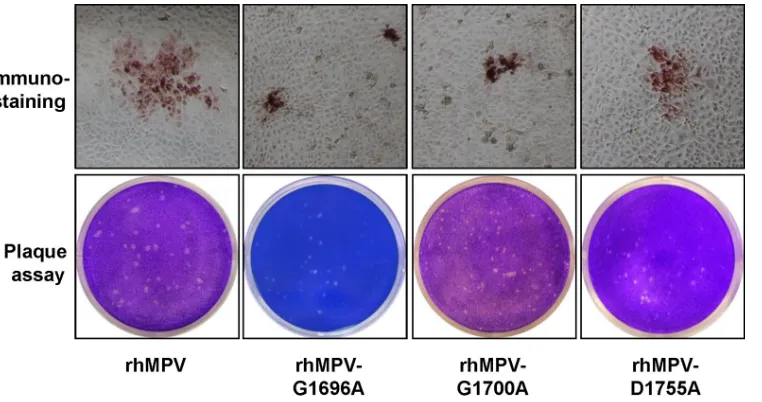
site. As shown inFig. 3, all rhMPV mutants were positive for viral
N-protein expression in the immunostaining assay using a mono-clonal antibody against N protein. However, the immunospots formed by these rhMPV mutants in LLC-MK2 cells were much smaller than those formed by wild-type rhMPV. To further con-firm the phenotype of these recombinant hMPVs, a direct agarose overlay plaque assay was performed in LLC-MK2 cells. As shown inFig. 3, the plaque sizes of the rhMPV G1696A, G1700A, and
D1755A mutants were 0.6⫾0.1 mm, 0.5⫾0.1 mm, and 0.7⫾0.2
mm, respectively, which were significantly smaller than the plaque
size of wild-type rhMPV (1.2⫾0.1 mm) (P⬍0.05). This suggests
that rhMPVs carrying mutations in the SAM binding site had defects in cell-cell spread and/or viral replication. To confirm that the mutant rhMPVs contained the desired mutations, virus stocks were treated with DNase I to remove possible contamination with plasmid DNA from transfection, followed by RNA extraction and RT-PCR. A 1.4-kb DNA fragment spanning CR VI of the L gene was amplified by RT-PCR from each rhMPV mutant (data not shown). However, no DNA band was observed when PCR was performed without the RT step (data not shown), suggesting that the plasmid DNA was completely removed by DNase treatment. Sequencing results showed that all rhMPV mutants contained the desired mutation in CR VI of the L-protein gene. Finally, the en-tire genomes of these recombinants were sequenced to confirm that no additional mutation(s) had been introduced.
Recombinant hMPVs carrying mutations in the SAM bind-ing site have a delay in viral replication.The replication kinetics of these rhMPV mutants in LLC-MK2 cells was determined. Briefly, LLC-MK2 cells were infected with each recombinant virus
FIG 2Mutations to CR VI of the hMPV L gene diminished GFP expression in a minigenome assay. Confluent BHK-SR19-T7 cells were transfected with the minigenome plasmid together with pCITE-N, pCITE-P, pCITE-M2.1, pCITE-L, or pCITE-L mutant using Lipofectamine 2000. GFP expression was visualized by a fluorescence microscope at 48 h postinfection.
on November 7, 2019 by guest
http://jvi.asm.org/
[image:6.585.78.507.67.265.2]at an MOI of 0.01. At the indicated time points, the supernatant of infected LLC-MK2 cell cultures was collected and the virus yield was determined on LLC-MK2 cells, using the immunostaining
assay.Figure 4Ashows the growth curves of these rhMPV
mu-tants. The rhMPV G1696A, G1700A, and D1755A mutants were significantly delayed in viral replication compared to wild-type
rhMPV (P⬍0.05). The peak viral titer of the hMPV G1755A
mutant was 4.6 log10PFU/ml, which was 1.4 log units lower than
that of wild-type rhMPV. The peak titers of the rhMPV G1696A
and D1700A mutants were 5.5 and 5.6 log10PFU/ml at day 6
postinfection, respectively; these values were only 0.5 log unit less
than the value for wild-type rhMPV (Fig. 4A). Since the rhMPV
D1755A mutant had a 1.5-log-unit reduction in titer, we also
tested the growth of this rhMPV mutant at higher MOIs. As
shown inFig. 4B, the viral titer significantly increased when
LLC-MK2 cells were infected with the rhMPV D1755A mutant at a
higher MOI (P⬍0.05). At an MOI of 1.0, the titer of the rhMPV
D1755A mutant was approximately 0.4 log unit less than that of wild-type rhMPV. The progression of the CPE was recorded in LLC-MK2 cells. Wild-type rhMPV caused an extensive CPE at day 3 postinoculation. The CPEs caused by the rhMPV mutants were significantly delayed. The onset of the CPE caused by the rhMPV G1696A mutant was delayed by 3 days, whereas that of the CPE caused by the rhMPV G1700A and D1755A mutants was delayed
by 1 day (Fig. 5). Collectively, these results demonstrate that
rhMPVs carrying mutations in the SAM binding site had a delay in viral replication and were attenuated in cell culture.
Recombinant hMPVs carrying mutations in the SAM bind-ing site have defects in viral protein synthesis.We determined whether these recombinant viruses had defects in viral protein
synthesis by using Western blotting for the N protein.Figure 6A
shows the kinetics of viral N-protein synthesis. The rhMPV G1700A and D1755A mutants had a significant delay in N-protein
synthesis (P ⬍ 0.05). Quantitative analysis of protein bands
showed that the rhMPV G1700A and D1755A mutants had ap-proximately 70 to 80% and 50 to 70% reductions in N-protein expression, respectively, compared with that of wild-type rhMPV at days 1 and 3 postinfection. In comparison, the rhMPV G1696A mutant showed moderate (10 to 30%) reductions in N-protein expression compared to wild-type rhMPV at days 1 and 3
postin-fection (Fig. 6A). The level of N protein in wild-type
rhMPV-infected cells decreased at days 5 and 7 postinfection because many cells started to lyse. In contrast, N-protein expression in rhMPV G1700A and D1755A mutant-infected cells continued to
increase at days 5 and 7 postinfection (Fig. 6A).
Recombinant hMPVs carrying mutations in the SAM bind-ing site have defects in viral genomic RNA replication and mRNA synthesis.We also determined viral genomic RNA
repli-FIG 3Recovery of recombinant hMPVs carrying mutations in the SAM binding site. (Top) Immunostaining spots formed by recombinant hMPVs. LLC-MK2 cells were infected with recombinant hMPV mutants and incubated at 37°C for 1 h. At day 4 postinfection, the supernatant was removed and the cells were fixed. The cells were then labeled with an anti-hMPV N-protein primary monoclonal antibody, followed by incubation with HRP-labeled rabbit antimouse secondary antibody. After incubation with AEC chromogen substrate, positive cells with immunostaining spots were visualized under a microscope. (Bottom) Plaque morphology of recombinant hMPVs. An agarose overlay plaque assay was performed with an LLC-MK2 cell monolayer. Viral plaques were developed at day 6 postinfection.
FIG 4Single-step growth curve of recombinant hMPVs carrying mutations in the SAM binding site. LLC-MK2 cells in 35-mm dishes were infected with each recombinant hMPV at an MOI of 0.01. After adsorption for 1 h, the inocula were removed and the infected cells were washed 3 times with Opti-MEM medium. Then, fresh Opti-MEM medium containing 2% FBS was added and the cells were incubated at 37°C for various times. Aliquots of the cell culture fluid were removed at the indicated intervals. The viral titer was determined by an immunostaining assay in Vero E6 cells. DPI, day postinfection. (A) Growth curves for wild-type rhMPV and the rhMPV G1696A, G1700A, and D1755A mutants; (B) viral titers for wild-type rhMPV and the rhMPV D1755A mutant at different MOIs.
on November 7, 2019 by guest
http://jvi.asm.org/
[image:7.585.104.485.65.266.2] [image:7.585.40.285.505.618.2]cation and mRNA synthesis for each recombinant virus. Since we used a primer annealing to the hMPV leader sequence, viral genomic RNA could be directly quantified by real-time RT-PCR using total RNA isolated from virus-infected cells. At day 5 postin-fection, the amount of genomic RNA in rhMPV G1696A, G1700A, and D1755A mutant-infected cells was 1.6-, 0.8-, and
1.7-fold less than that in wild-type rhMPV, respectively (Fig. 6B).
We also separated viral mRNA from total RNA by poly(A) bind-ing, since viral genomic RNA is not polyadenylated. At day 5 postinfection, the rhMPV G1696A, G1700A, and D1755A mu-tants had 1.1-, 0.5-, and 3.3-fold less N-protein mRNA than
wild-type rhMPV, respectively (Fig. 6C). Thus, all three rhMPV
mu-tants had significant defects in viral mRNA synthesis and genomic
RNA replication (P⬍0.05). In addition, the ratio between mRNA
and genomic RNA for each hMPV mutant was calculated. As
shown inFig. 6D, the ratio of viral mRNA/genomic RNA for each
mutant was not significantly altered from that for wild-type
rhMPV (P⬎0.05).
Genetic stability of recombinant hMPVin vitro.To deter-mine whether the rhMPV mutants were genetically stable in cell cul-ture, each rhMPV mutant was passaged 10 times in LLC-MK2 cells. The region containing the respective mutation in the virus from each passage was sequenced. It was found that all rhMPV mutants retained their mutation in CR VI of the L gene. At passage 10, the entire genome of each recombinant virus was sequenced. No muta-tions other than the desired mutation in the MTase region in the L gene were found in the genome. This result suggests that these rhMPV mutants are genetically stable.
Recombinant hMPVs carrying mutations in the SAM bind-ing site are defective in 2=-O methylation but not G-N-7 meth-ylation.Analysis of mRNA cap methylation of paramyxoviruses has been hampered because most of them, including hMPV,
can-not synthesize viral mRNAin vitro. We have developed atrans
-methylation assay that allows us to analyze the cap -methylation of viral mRNA in virus-infected cells. Briefly, Vero E6 cells were mock infected or infected with wild-type or mutant rhMPVs at an MOI of 0.1, and mRNAs for each recombinant virus were har-vested and purified. To determine whether the rhMPV mutants were defective in G-N-7 methylation, equal amounts of mRNA from each mutant were incubated with 10 units of vaccinia virus
G-N-7 MTase in the presence of 15Ci [3H]SAM. After the
meth-ylation reaction, RNA was purified, and the methmeth-ylation of the
mRNA cap structure was quantified by determining the level of3H
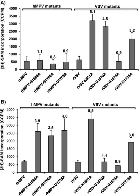
incorporation with a scintillation counter. As shown inFig. 7A,
left, rhMPV mRNAs were not methylated by vaccinia virus G-N-7 MTase, which is consistent with this site already being methylated
by the rhMPV enzymes. Similarly, the level of [3H]SAM
incorpo-ration of mRNAs produced by the rhMPV G1696A, G1700A, and D1755A mutants was indistinguishable from that of mRNA
pro-duced by wild-type rhMPV (P⬎0.05), suggesting that the mRNA
of these mutants was not methylated by the vaccinia virus G-N-7 MTase. Therefore, this result indicates that the rhMPV G1696A, G1700A, and D1755A mutants are not defective in G-N-7 meth-ylation.
A similar strategy was used to determine 2=-O methylation in
mRNA. Since G-N-7 methylation is required for 2=-O
methyl-FIG 5CPEs caused by hMPVs carrying mutations in the SAM binding site. LLC-MK2 cells were infected with each recombinant hMPV at an MOI of 1.0. The CPE was monitored on a daily basis. Pictures were taken at days 3, 4, 5, 6, and 7 postinfection.
on November 7, 2019 by guest
http://jvi.asm.org/
[image:8.585.95.492.66.365.2]ation in vaccinia virus (37,41), we first incubated the RNA with cold SAM and the vaccinia virus G-N-7 MTase to methylate the G-N-7 position. In this way, if there was a lack of G-N-7 methyl-ation in the original RNA, it would not inhibit the methylmethyl-ation of
the 2=-O position. The mRNAs were purified and further
methyl-ated by 10 units of vaccinia virus 2=-O MTase in the presence of 15
Ci [3H]SAM. After the methylation reaction, the RNA was
pu-rified and the level of 2=-O methylation was measured by
determi-nation of the level of3H incorporation using a scintillation
coun-ter. As shown in Fig. 7B, left, the rhMPV mRNAs were not
methylated by vaccinia virus 2=-O MTase, consistent with the fact
that rhMPV produces 2=-O-methylated mRNA. The level of3H
incorporation of the rhMPV G1696A, G1700A, and D1755A
mu-tants was 3.5- to 4-fold higher than that of wild-type rhMPV (P⬍
0.05), suggesting that the rhMPV G1696A, G1700A, and D1755A
mutants had defects in 2=-O methylation. Therefore, these results
indicate that rhMPVs carrying mutations in the SAM binding site
are specifically defective in 2=-O methylation but not G-N-7
meth-ylation.
To verify the results for hMPV described above, we performed atrans-methylation assay using VSV as a control. VSV mRNA cap MTase is the best-characterized NNS RNA virus MTase because
purified VSV can reconstitute viral mRNA synthesisin vitro.
Fur-thermore, purified VSV L protein can methylate VSV-specific mRNAin vitro. We chose the following four VSV L-protein mu-tants for this assay: the rVSV K1651A mutant, carrying a point mutation in the MTase catalytic site which abolishes both G-N-7
and 2=-O methylations, and the rVSV G1670A, rVSV G1674A,
and rVSV D1735A mutants, each of which carries a mutation in the SAM binding site equivalent to the rhMPV G1696A, G1700A, and D1755A mutations, respectively. Previously, we had found that the rVSV G1670A mutant is specifically defective in G-N-7
methylation but not 2=-O methylation, the rVSV G1674A mutant
is fully methylated at G-N-7 and 2=-O at high (1 mM) SAM
con-centrations, and the rVSV D1735A mutant inhibits both G-N-7
and 2=-O methylations by approximately 70% (27–29).
Using the exact sametrans-methylation assay developed for
hMPV, we determined the methylation status of VSV mRNAs
isolated from virus-infected cells. As shown inFig. 7A, right, rVSV
and rVSV G1674A mutant mRNAs were not methylated by vac-cinia virus G-N-7 MTase, which is consistent with the fact that they produce fully G-N-7-methylated mRNA in the presence of 1 mM SAM. In contrast, the mRNAs of the rVSV K1651A, G1670A, and D1735A mutants were efficiently methylated by vaccinia virus
G-N-7 MTase. The level of3H incorporation of the rVSV K1651A,
G1670A, and D1735A mutants was approximately 5.1-, 4.5-, and 3.2-fold higher than that of wild-type rVSV, respectively. This is consistent with the fact that the rVSV K1651A and G1670A mu-tants lacked G-N-7 methylation and the rVSV D1735A mutant
had a 70% deficiency in G-N-7 methylation (27,28). Using the
pre-G-N-7-methylated mRNAs as the substrates, we performed a
trans-2=-O methylation assay. As shown inFig. 7B, right, mRNAs synthesized from the rVSV K1651A and D1735A mutants but not those synthesized from wild-type rVSV or the rVSV G1674A and rVSV G1670A mutants could be efficiently methylated by vaccinia
virus 2=-O MTase. The level of3H incorporation of the rVSV
FIG 6Recombinant hMPVs carrying mutations in the SAM binding site were defective in gene expression and RNA synthesis. (A) N-protein synthesis. LLC-MK2 cells were infected with wild-type rhMPV or rhMPV mutants at an MOI of 1.0 for various periods of time, as indicated. Total cell lysates were harvested and subjected to Western blotting using a monoclonal antibody against hMPV N protein. (B) N mRNA synthesis. Viral mRNA was further purified using a Dynabeads mRNA isolation kit and subsequently quantified by real-time PCR using primers annealing to the N gene. (C) Viral genomic RNA replication. LLC-MK2 cells were infected with wild-type rhMPV or rhMPV mutants at an MOI of 0.1 for various periods of time. Total RNA was isolated using the TRizol reagent, and genomic RNA was quantified by real-time RT-PCR using specific primers annealing to the hMPV leader sequence and N gene. The results are the averages of three independent experiments and are expressed as means⫾SEs of the numbers of copies of the transcribed N mRNA or viral genomic RNA. (D) Ratio between mRNA and genomic RNA. The ratio between N mRNA and genomic RNA was calculated for each recombinant hMPV. Data are the averages of three independent experiments.
on November 7, 2019 by guest
http://jvi.asm.org/
[image:9.585.97.493.66.309.2]K1651A and D1735A mutants was approximately 5.5- and 3.0-fold higher than that of wild-type rVSV, respectively. This is con-sistent with our previous results showing that the rVSV K1651A
mutant completely lacked both G-N-7 and 2=-O methylations, the
rVSV G1670A mutant lacked G-N-7 methylation but not 2=-O
methylation, and the rVSV D1735A mutant was partially defective
in both G-N-7 and 2=-O methylations (27,28).
Replication and pathogenesis of MTase-defective rhMPVs in cotton rats. (i) Viral replication in cotton rats.To determine
whether the MTase-defective rhMPVs are attenuatedin vivo,
cot-ton rats were inoculated with recombinant wild-type and mutant viruses and viral replication and pathogenesis were examined. None of the cotton rats inoculated with recombinant viruses ex-hibited detectable clinical symptoms of respiratory tract infection. No distinguishable body weight change was observed in any treat-ment group (data not shown). At day 4 postinfection, cotton rats were sacrificed, and viral replication in nasal turbinates and lungs and pulmonary histology were determined.
As shown inTable 1, wild-type rhMPV replicated efficiently in
the nasal turbinates and lungs of all five cotton rats. Average viral
titers of 5.24⫾0.35 log10PFU/g and 3.02⫾0.46 log10PFU/g were
found in the nasal turbinate and lung, respectively. All MTase-defective rhMPVs had reduced viral replication in the nasal turbi-nate and lung. For the rhMPV G1700A mutant, 4 out of 5 cotton rats had detectable virus in the nasal turbinate, with an average
titer of log 2.69⫾0.34 log10PFU/g, and only 1 out of 5 rats had
detectable virus in lung tissue, with a titer of log 2.02 log10PFU/g.
For the rhMPV G1696A and G1755A mutants, there was no de-tectable infectious virus in either nasal turbinate or lung tissue. This result demonstrated that the rhMPV G1696A and G1755A mutants were completely defective in viral replication in both the upper and lower respiratory tracts in cotton rats, whereas the rhMPV G1700A mutant had low levels of viral replication only in the upper respiratory tract. These results demonstrate that
MTase-defective hMPVs are attenuated in viral replicationin vivo.
Since no infectious virus was detected in some of the MTase-defective rhMPV-inoculated animals, real-time RT-PCR was per-formed to determine whether viral genomic RNA was present in nasal turbinate and lung tissues. All virus-infected cotton rats had detectable viral genomic RNA in their tissues, even though they
were negative for infectious virus (Table 1). In cotton rats
inocu-lated with wild-type rhMPV, 7.54 and 8.54 log10genomic RNA
copies/g tissue were detected in nasal turbinate and lung tissues, respectively. The viral RNA level was significantly reduced in the upper and lower respiratory tracts of all animals infected with the MTase-defective rhMPVs. In animals infected with the rhMPV
G1696A, G1700A, and D1755A mutants, 6.34, 6.95, and 5.27 log10
genomic RNA copies/g tissue were detected in the nasal
turbi-nates, respectively. In addition, 6.72, 7.46, and 6.07 log10genomic
RNA copies per gram of lung tissue were detected in animals in-fected with the rhMPV G1696A, G1700A, and D1755A mutants, respectively. These levels of genomic RNA production are approx-imately 10- to 100-fold lower than the level for wild-type rhMPV. Therefore, all MTase-defective rhMPVs had significant defects in viral genome replication in cotton rats.
(ii) Pulmonary histopathology.Half of the lung tissue was inflated with formaldehyde and subjected to H&E staining and histopathological examination. Each lung tissue specimen was
scored on a scale of from 0 (no change) to 3 (severe change) (Table
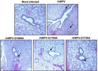
1). Wild-type rhMPV caused moderate pulmonary
histopatho-logical changes, including interstitial pneumonia, peribronchial lymphoplasmocytic infiltrates, mononuclear cell infiltrates, and
edematous thickening of the bronchial submucosa (Fig. 8). The
rhMPV G1700A mutant similarly induced interstitial pneumonia but caused fewer epithelial changes and less bronchial exudate and
mononuclear cell infiltration (Fig. 8). The rhMPV G1696A and
D1755A mutants caused mononuclear inflammation similar to that caused by wild-type rhMPV but caused significantly fewer pulmonary histopathological changes. No pathological changes were found in the lungs of cotton rats inoculated with DMEM.
FIG 7Analysis of hMPV and VSV mRNA cap methylation byin vitro trans-methylation assay. (A)trans-G-N-7 methylation assay for hMPV and VSV. Five hundred nanograms of mRNA was isolated from virus-infected Vero E6 cells and wastrans-methylated by vaccinia virus G-N-7 MTase in the presence of 15Ci [3H]SAM as described in Materials and Methods.
The number of corrected counts per minute (CCPM) of [3H]SAM
incor-poration from RNA from mock-infected cells was subtracted from the CCPM of each hMPV mutant. Subsequently, the CCPM was normalized by the level of viral mRNA quantified by real-time RT-PCR. The number on the top of each column indicates the ratio of [3H]SAM incorporation of
viral mRNAs between mutant and wild-type virus. The averages of four independent experiments are shown. (B)trans-2=-O methylation assay for hMPV and VSV. Five hundred nanograms of mRNA was isolated from virus-infected cells and was subsequently premethylated by vaccinia virus G-N-7 MTase in the presence of 1 mM cold SAM. The RNAs were purified and furthertrans-methylated by vaccinia virus 2=-O MTase in the presence of 15Ci [3H]SAM. The number of CCPM of [3H]SAM incorporation for
each virus is shown. The number on the top of each column indicates the ratio of [3H]SAM incorporation of viral mRNAs between mutant and
wild-type virus. The averages of four independent experiments are shown.
on November 7, 2019 by guest
http://jvi.asm.org/
[image:10.585.42.286.67.410.2]These results similarly indicate that MTase-defective hMPVs are attenuated in cotton rats.
(iii) Viral antigen distribution in lung tissues.To determine the viral antigen distribution in lung tissues, IHC was performed using an antibody against the hMPV matrix protein. As shown in
Fig. 9andTable 1, large numbers of viral antigen-positive cells were detected at the luminal surfaces of the bronchial epithelial
[image:11.585.38.554.77.170.2]cells in the lungs of wild-type rhMPV-infected cotton rats. The pattern of viral antigen was discontinuous, and antigen appeared in clusters of adjacent cells. In some cases, luminal cellular debris that may have included both epithelial cell debris and macro-phages also stained positive for hMPV antigen. Significantly fewer viral antigen-positive cells were detected in the bronchial epithe-lial cells of the rhMPV G1700A mutant-infected group. No or
TABLE 1Replication of MTase-defective rhMPVs in cotton ratsh
rhMPV mutanta
Viral replication in nasal turbinateb Viral replication in lungc
Lung histology scored
Lung IHC scoree % of infected
animals
Viral titer (log10
PFU/g)
Viral RNA load (log10GRCf/g)
% of infected animals
Viral titer (log10
PFU/g)
Viral RNA load (log10GRC/g)
Wild type 100 5.24⫾0.35A 7.54⫾0.32A 100 3.02⫾0.46A 8.54⫾0.34A 1.5A 3.0A
G1696A 0 NDg 6.34⫾0.72B 0 ND 6.72⫾0.24B 0.3B 0.2B
G1700A 80 2.69⫾0.34B 6.95⫾0.18B 60 2.27⫾0.28B 7.46⫾1.10B 0.5B 0.5B
D1755A 0 NDA 5.27⫾0.77B 0 ND 6.07⫾1.55B 0.4B 0.2B
DMEM 0 ND ND 0 ND ND 0 0
aCotton rats were inoculated intranasally with DMEM or 2⫻105PFU of wild-type rhMPV or an rhMPV mutant. At day 4 postimmunization, animals were euthanized for
pathology study.
bThe viral titer was determined by an immunostaining assay. For the rhMPV G1700A mutant, 4 out of 5 cotton rats had detectable virus with an average titer of 2.69 log units. Viral
RNA was determined by real-time RT-PCR.
cFor the rhMPV G1700A mutant, 3 out of 5 cotton rats had detectable virus with a titer of 2.27 log units.
d
The severity of lung histology was scored for each lung tissue specimen. The average score for each group is shown. 0, no change; 1, mild change; 2, moderate change; 3, severe change.
e
The amount of hMPV antigen expressed in lung tissue was scored. The average score for each group is shown. 0, no antigen; 1, small amount of antigen; 2, moderate amount of antigen; 3, large amount of antigen.
f
GRC, genomic RNA copies. gND, not detected.
h
Five cotton rats were tested in each group. Values within a column followed by different capital letters (A and B) are significantly different (P⬍0.05).
FIG 8Lung histological changes in cotton rats infected with MTase-defective rhMPVs. The right lung from each cotton rat was preserved in 4% (vol/vol) phosphate-buffered paraformaldehyde. Fixed tissues were embedded in paraffin, sectioned at 5m, and stained with H&E for the examination of histological changes by light microscopy. Arrows, lymphocyte infiltrates.
on November 7, 2019 by guest
http://jvi.asm.org/
[image:11.585.93.494.401.692.2]little viral antigen was detected in lung tissues from the groups infected with the rhMPV G1696A and D1755A mutants. Consis-tently, these data demonstrate that MTase-defective hMPVs had significant defects in viral replication and antigen expression in the lower respiratory tract.
(iv) Genetic stability of MTase-defective rhMPVsin vivo.To determine whether MTase-defective rhMPVs were genetically sta-ble in cotton rats, we amplified CR VI of the L gene from total RNA extracted from each lung. Sequencing of these PCR products showed that all MTase-defective rhMPVs retained the desired mutation in CR VI of the L gene. No additional mutation was found in this region. This result suggests that MTase-defective rhMPVs are genetically stable in cotton rats.
Immunogenicity of MTase-defective hMPVs in cotton rats.
Since MTase-defective rhMPVs were attenuatedin vitroandin
vivo, we subsequently determined their immunogenicity in cotton
rats. Briefly, cotton rats were inoculated intranasally with the rhMPV mutants. Serum samples were collected weekly for detec-tion of a humoral immune response. At week 4 postinoculadetec-tion,
animals were challenged with 106PFU of rhMPV. At day 4
post-challenge, all the animals were sacrificed and nasal turbinate and lung tissue samples were collected for virus detection and patho-logical examination.
Serum antibody was determined by a plaque reduction
neu-tralization assay. As shown inFig. 10, all of the MTase-defective
hMPVs elicited high levels of neutralizing antibody in cotton rats. Antibody was detectable at week 1 postimmunization, and the levels gradually increased during weeks 2 to 4. Overall, the
anti-bodies generated by MTase-defective hMPVs were comparable to
those generated after wild-type rhMPV immunization (P⬎0.05).
In contrast, no hMPV-specific antibody was detected in the un-vaccinated control. This result demonstrates that MTase-defective rhMPVs are not only sufficiently attenuated but also capable of triggering high levels of antibody.
Viral replication in the nasal turbinates and lungs of cotton rats after challenge was examined by immunostaining. As shown in
FIG 9IHC staining of lung tissue from cotton rats infected with MTase-defective rhMPVs. Lung tissues were fixed in 4% (vol/vol) phosphate-buffered paraformaldehyde. Deparaffinized sections were stained with monoclonal antibody against hMPV matrix protein (Virostat, Portland, ME) to determine the distribution of viral antigen.
FIG 10Recombinant hMPVs triggered a high level of neutralizing antibody titer in cotton rats. Cotton rats were immunized with each recombinant hMPV intranasally at a dose of 2.0⫻105PFU per rat. Blood samples were collected
from each rat weekly by retro-orbital bleeding. The hMPV-neutralizing anti-body titer was determined using a plaque reduction neutralization assay, as described in Materials and Methods.
on November 7, 2019 by guest
http://jvi.asm.org/
[image:12.585.95.493.65.357.2] [image:12.585.334.508.532.663.2]Table 2, cotton rats vaccinated with wild-type rhMPV or MTase-defective rhMPVs did not have any detectable infectious virus in either the nasal turbinate or lung tissue after challenge with rhMPV. In contrast, unvaccinated challenged controls had
aver-age titers of 5.59⫾0.49 and 4.76⫾0.29 log10PFU/g in the nasal
turbinate and lung, respectively. These results demonstrate that infection with the rhMPV G1696A, G1700A, and D1755A mu-tants provided complete protection from challenge with wild-type rhMPV, preventing viral replication in both the upper and lower respiratory tracts.
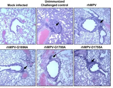
After challenge, lung histology was evaluated for each cotton rat. As expected, unvaccinated challenged controls had moderate pulmonary histological changes, characterized by interstitial pneumonia, mononuclear cell infiltration, and edematous thick-ening of the bronchial submucosa, and bronchial epithelium
changes (Fig. 11andTable 2). In contrast, significantly fewer
his-tological changes were found in the lungs of cotton rats vaccinated with wild-type rhMPV and the rhMPV G1696A, G1700A, and
D1755A mutants (Table 2;Fig. 11). No enhanced lung damage
was observed in the vaccinated and challenged groups. No histo-logical changes were found in the unvaccinated and unchallenged controls. These results demonstrate that MTase-defective rhMPV provides protection against lung damage from virus challenge.
For lung tissues from unvaccinated challenged controls, large numbers of viral antigens were found at the luminal surface of the
bronchial epithelial cells (Fig. 12). Interestingly, lung tissues from
the vaccinated and challenged groups exhibited different antigen
distribution patterns (Fig. 12). Antigen was found within
bron-chial tissue but not on the luminal surface of bronbron-chial epithelial cells, which may be related to viral clearance. No antigen was found in the unvaccinated unchallenged group.
2=-O MTase-defective hMPVs are highly sensitive to IFN-␣ and IFN-treatment.Recent work on West Nile virus (33),
den-gue virus (42), mouse hepatitis virus (MHV) (32), and severe
acute respiratory syndrome coronavirus (43) found that the
mechanism of attenuation of the 2=-O MTase-defective viruses is
attributable to enhanced sensitivity to the antiviral action of IFN. However, whether this mechanism is conserved in nonsegmented negative-sense RNA viruses (such as hMPV) is not known. We
have now determined the sensitivity of 2=-O MTase-defective
hMPVs to IFN treatment in Vero E6 cells, which are defective in type I interferon production but express IFN receptors. Thus, Vero E6 cells can respond to exogenous type I IFN. Briefly, Vero E6 cells were pretreated with the different amounts (2 and 20
units) of IFN-␣or IFN-for 4 h, followed by washing the treated
cells with DMEM three times, and the cells were then infected with each hMPV mutant at an MOI of 0.5. At 48 h postinfection, su-pernatant was harvested and the viral titer was determined by immunostaining assay. Treatment of Vero E6 cells with 2 or 20
units of IFN-␣(Fig. 13A) or IFN-(Fig. 13B), followed by
infec-tion with each recombinant hMPV mutant, resulted in a
reduc-tion in the viral titer of 2 to 4 log10PFU. However, there was no
significant reduction for wild-type hMPV when Vero E6 cells were
treated with IFN-␣or IFN-at the same doses (P⬎0.05).
There-fore, all 2=-O MTase-defective hMPVs were highly sensitive to
both IFN-␣and IFN-inhibition, whereas wild-type hMPV was
not sensitive to IFN inhibition. This result suggests that the
mech-anism of attenuation of 2=-O defective viruses may also be
con-served in hMPV.
DISCUSSION
Paramyxoviruses, such as RSV, hMPV, and PIV3, are responsible for the majority of pediatric respiratory tract disease and inflict significant economic loss, health care costs, and emotional bur-dens. Despite tremendous efforts, no vaccine is currently available for these viruses. In this study, we found that MTase-defective rhMPVs were sufficiently attenuated but retained high immuno-genicity in cotton rats. These results demonstrate that MTase-defective rhMPVs are excellent live vaccine candidates.
MTase as a new target for attenuation of hMPV.A live atten-uated vaccine is the most promising vaccine for human paramyxoviruses. Live attenuated vaccines offer many advantag-es: (i) enhanced lung damage has not been observed either after vaccination with live attenuated viruses or after natural
reinfec-tion (44, 45), (ii) intranasal administration of live vaccines
[image:13.585.39.546.77.170.2]duces balanced immune responses that closely resemble those in-duced after natural virus infection, and (iii) intranasal vaccination induces better local immunity than the intramuscular injection of
TABLE 2Immunogenicity of MTase-defective rhMPVs in cotton ratsh
rhMPV mutanta
Nasal turbinateb Lungc
Lung histology scored
Lung IHC scoree
Protection ratef(%) % of infected
animals
Viral titer (log10
PFU/g)
% of infected animals
Viral titer (log10
PFU/g)
Viral RNA load (log10
GRC/g)
DMEM 100 5.59⫾0.49 100 4.76⫾0.29 5.65⫾0.22A 1.6A 3.0A 0
Wild type 0 NDg 0 ND 2.54⫾0.35B 0.6B 0.5B 100
G1696A 0 ND 0 ND 2.68⫾0.22B 0.8B 0.5B 100
G1700A 0 ND 0 ND 2.50⫾0.28B 0.5B 0.5B 100
D1755A 0 ND 0 ND 2.46⫾0.23B 0.3B 0.5B 100
aAnimals were immunized intranasally with DMEM or 2⫻105PFU of wild-type rhMPV or an rhMPV mutant. At day 28 postimmunization, animals were challenged with 106
PFU of wild-type rhMPV.
bThe viral titer was determined by an immunostaining assay.
c
The viral RNA titer was determined by real-time RT-PCR. GRC, genomic RNA copies.
dThe severity of lung histology was scored for each lung tissue specimen. The average score for each group was shown. 0, no change; 1, mild change; 2, moderate change; 3, severe
change.
eThe amount of hMPV antigen expressed in lung tissue was scored. The average score for each group is shown. 0, no antigen; 1, small amount of antigen; 2, moderate amount of
antigen; 3, large amount of antigen.
fThe protection rate was calculated on the basis of the level of viral replication in lungs and nasal turbinates.
g
ND, not detected.
hFive cotton rats were tested in each group. Values within a column followed by different capital letters (A and B) are significantly different (P⬍0.05).


