Antisense-Derived HIV-1 Cryptic Epitopes Are Not Major
Drivers of Viral Evolution during the Acute Phase of Infection
Binghao J. Peng,
aJonathan M. Carlson,
bMichael K. P. Liu,
c*
Feng Gao,
dNilu Goonetilleke,
c*
Andrew J. McMichael,
cPersephone Borrow,
cJill Gilmour,
eSonya L. Heath,
aEric Hunter,
f,gAnju Bansal,
aPaul A. Goepfert
aaDepartment of Medicine, University of Alabama at Birmingham, Birmingham, Alabama, USA
bMicrosoft Research, Redmond, Washington, USA
cNuffield Department of Medicine, University of Oxford, Oxford, United Kingdom
dDepartment of Medicine, Duke Human Vaccine Institute, Duke University Medical Center, Durham, North Carolina, USA
eIAVI Human Immunology Laboratory, Imperial College London, London, United Kingdom
fEmory Vaccine Center at Yerkes National Primate Research Center, Emory University, Atlanta, Georgia, USA
gDepartment of Pathology and Laboratory Medicine, Emory University, Atlanta, Georgia, USA
ABSTRACT
While prior studies have demonstrated that CD8 T cell responses to
cryptic epitopes (CE) are readily detectable during HIV-1 infection, their ability to
drive escape mutations following acute infection is unknown. We predicted 66 CE in
a Zambian acute infection cohort based on escape mutations occurring within or
near the putatively predicted HLA-I-restricted epitopes. The CE were evaluated for
CD8 T cell responses for patients with chronic and acute HIV infections. Of the 66
predicted CE, 10 were recognized in 8/32 and 4/11 patients with chronic and acute
infections, respectively. The immunogenic CE were all derived from a single
anti-sense reading frame within
pol
. However, when these CE were tested using
longitu-dinal study samples, CE-specific T cell responses were detected but did not
consis-tently select for viral escape mutations. Thus, while we demonstrated that CE are
immunogenic in acute infection, the immune responses to CE are not major drivers
of viral escape in the initial stages of HIV infection. The latter finding may be due to
either the subdominant nature of CE-specific responses, the low antigen sensitivity,
or the magnitude of CE responses during acute infections.
IMPORTANCE
Although prior studies demonstrated that cryptic epitopes of HIV-1
induce CD8 T cell responses, evidence that targeting these epitopes drives HIV
es-cape mutations has been substantially limited, and no studies have addressed this
question following acute infection. In this comprehensive study, we utilized
longitu-dinal viral sequencing data obtained from three separate acute infection cohorts to
predict potential cryptic epitopes based on HLA-I-associated viral escape. Our data
show that cryptic epitopes are immunogenic during acute infection and that many
of the responses they elicit are toward translation products of HIV-1 antisense
read-ing frames. However, despite cryptic epitope targetread-ing, our study did not find any
evidence of early CD8-mediated immune escape. Nevertheless, improving cryptic
epitope-specific CD8 T cell responses may still be beneficial in both preventative and
therapeutic HIV-1 vaccines.
KEYWORDS
CD8 T cells, acute infection, alternative reading frames, cryptic epitopes,
human immunodeficiency virus, viral evolution
C
D8 T cells play a vital role in controlling intracellular pathogens by recognizing
epitopes presented on infected cells. For human immunodeficiency virus type 1
(HIV-1) infections, CD8 T cells have been shown to be essential in control of viremia and
Received3 May 2018Accepted7 July 2018
Accepted manuscript posted online18 July 2018
CitationPeng BJ, Carlson JM, Liu MKP, Gao F, Goonetilleke N, McMichael AJ, Borrow P, Gilmour J, Heath SL, Hunter E, Bansal A, Goepfert PA. 2018. Antisense-derived HIV-1 cryptic epitopes are not major drivers of viral evolution during the acute phase of infection. J Virol 92:e00711-18.https://doi.org/10.1128/JVI .00711-18.
EditorFrank Kirchhoff, Ulm University Medical Center
Copyright© 2018 American Society for Microbiology.All Rights Reserved.
Address correspondence to Paul A. Goepfert, pgoepfert@uabmc.edu.
*Present address: Michael K. P. Liu, Center for Immunology and Vaccinology, Imperial College London, London, United Kingdom; Nilu Goonetilleke, Department of Microbiology and Immunology, University of North Carolina at Chapel Hill, Chapel Hill, North Carolina, USA.
crossm
on November 6, 2019 by guest
http://jvi.asm.org/
disease progression, and part of this evidence comes from the fact that they drive HIV
escape mutations that may impact overall viral fitness (1). However, due to the mutable
nature of HIV, achieving a broader range of epitope recognition by targeting beyond
the scope of the nine conventional proteins of the virus may be necessary to augment
the efficacy of both preventative and therapeutic vaccines (2, 3). Several studies have
described a lesser-known form of epitopes (termed cryptic epitopes [CE]) within several
viruses, including HIV and simian immunodeficiency virus (SIV) (4–24). These epitopes
are predominantly derived from the translation of two sense and three antisense
alternative open reading frames. Translation products of any of these five alternate
reading frames (ARF) were found to be recognized by CD8 T cells during the acute and
chronic stages of infection (6, 21, 25–29). While CE usually induce T cell responses of
lower magnitude than that of responses induced by their main reading frame
coun-terparts, past studies have demonstrated that immune responses to these ARF epitopes
are observed in many infected individuals or are immunoprevalent during the course
of the disease (25, 27). Furthermore, HIV was demonstrated to escape CE-specific CD8
T cell responses in chronic infection, demonstrating that they exert significant selective
pressure on viral replication
in vivo
. However, the contributions made by such
re-sponses in driving viral evolution during acute and early HIV-1 infections have not been
addressed adequately.
In the present study, we utilized a variety of methods to predict CE that potentially
induced CD8 T cell responses during the acute phase of infection. By validating the
epitope predictions for one of the acute-phase cohorts through immunogenicity
screening assays, we identified 10 previously undescribed CE to be immunogenic in
both acute and chronic infections. However, no reliable CE responses could be detected
using similar methods of epitope prediction for two additional acute-phase cohorts.
From this study, we conclude that CE are indeed present during early stages of
infection and can be targeted by T cells. Nevertheless, due to the overall subdominant
T cell response to these epitopes, they appear to contribute minimally to driving viral
adaptation.
RESULTS
Cryptic epitope predictions and T cell responses.
To analyze the role of CE
targeting during acute infections, we adopted an approach similar to that of previous
studies, utilizing viral sequence data obtained from a serodiscordant cohort (30, 31). We
first identified sites of HLA-I-associated polymorphisms within or near previously
pre-dicted CE that may have evolved to escape CD8 T cell responses during the first 2 years
of infection in 78 acutely infected Zambian patients. Due to the limited Zambian
sample availability and to facilitate analysis of HIV subtype B-infected patients, we
narrowed the list of all predicted epitopes down to 66 putative CE for testing (Table 1).
The CE were first tested as seven distinct peptide pools (6 to 10 peptides/pool) for
recognition by peripheral blood mononuclear cells (PBMCs) from chronically HIV-1
subtype B-infected patients in a gamma interferon-specific enzyme-linked
immunosor-bent spot (IFN-
␥
ELISpot) assay. Thirty-two patient samples were used. Seventy-eight
percent of patients (25/32 patients) were not on active antiretroviral therapy (ART); the
median viral load of the cohort was 3,380 RNA copies/ml, and the median absolute CD4
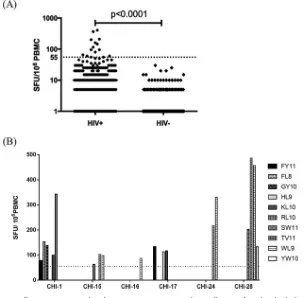
count (per milliliter) was 646 (Table 2). We observed T cell responses to the CE peptide
pools for 25% of the subjects tested (8/32 patients) (Table 2, shaded rows, and Fig. 1A).
In contrast, CE pools did not elicit T cell responses for any of the 16 HIV-seronegative
donors (
P
⬍
0.0001) evaluated. Next, we deconvoluted the positive pool responses to
the single-peptide level. Single-peptide responses were successfully deconvoluted for
six of the eight CE pool responders, which allowed us to identify 10 novel immunogenic
CE (Table 3) with no significant homology to HIV-1 proteins encoded by the main
reading frames (Table 4). Interestingly, all immunogenic CE were derived from the
antisense reverse frame 3 (RF3) of
pol
. The magnitudes of the responses to these CE for
the six patients tested varied, ranging from 63 to 487 spot-forming units (SFU)/10
6PBMCs (median magnitude of 133 SFU/10
6PBMCs) (Fig. 1B).
on November 6, 2019 by guest
http://jvi.asm.org/
TABLE 1Cryptic epitopes predicted from HLA-I-associated polymorphisms
ARFa Gene Predicted HLA-I allele(s)
Peptide sequence
Posterior probability
No. of escapes/no.
of reversions Conservationb
FF2 gag A74, A3301 PTRGGQGISFR 0.69 3/3 0.81
FF3 gag A0205, B0801 EMWKRTPNERL 0.64 3/8 0.87
gag B39, B4201 FPQITLWQRPL 0.79 6/6 0.97
gag A74 NLAFPQGEAR 0.51 3/3 0.88
RF1 gag A340201 EPVYIVSK 0.68 1/4 0.94
gag A3001 FPRGPAIFGY 0.87 2/4 0.85
gag A0201 GLMYHLPL 0.94 0/7 0.97
gag C0802 HPCYLFLKV 0.86 1/4 0.80
gag B5101 EPVYIVSKGF 0.56 0/4 0.93
RF2 gag B57 HAFKVLGDMAW 0.66 2/2 0.84
gag B58 HAFKVLGDMDW 0.78 4/2 0.78
gag C0804 HAVIISSNVA 0.77 3/1 0.90
gag B4201 LPQLKHLTIL 0.56 3/4 0.82
gag B1402 WRKFPGLPL 0.66 5/2 0.88
gag C0401, C17 YCFFPPGLNL 0.63 4/6 0.78
RF3 gag A0202 FLLCHFPLV 0.83 1/7 0.97
gag B5703 LLLLPMHSKF 0.80 0/3 0.87
gag A2402 SFPLALTFF 0.66 1/6 0.82
gag A4301 WLLDVPPLY 0.96 0/3 0.84
RF2 nef A2402 GYLTPGPGVF 0.59 2/2 0.89
FF2 pol B5702 IAGLSMIYRSW 0.70 2/1 0.81
pol B5301 IPLPNYGTSW 0.91 0/5 0.88
pol B5703 ITAIGEQW 0.88 0/6 0.84
pol A33 KIWPSHKGR 0.73 7/2 0.95
pol A0301, A340201, A66 QIRRLNYK 0.68 4/3 0.84
pol A2402 QYWMWGMHI 0.99 0/4 0.85
pol A0202, A2402, A6801, B1402, B3001 WMWGMHIFQFL 0.82 2/17 0.79
pol A3201 YIMTHQKTW 0.61 1/12 0.76
FF3 pol A33 GLPVMGTSTQR 0.66 0/3 0.82
RF1 pol B1801 CPFVRMEF 0.76 1/5 0.85
pol B15 FITASYYFFCY 0.62 2/5 0.88
pol A0201 FLFPCWVWYSL 0.80 0/4 0.81
pol B15 FLHCLCLFY 0.53 5/2 0.94
pol A0301, A68 FLMFFVWCGK 0.74 1/5 0.92
pol B4201 FPSLRKHIIL 0.80 1/3 0.78
pol B0801, C18 FYKYLIIL 0.63 1/2 0.85
pol A2902 GLDFCHATLEY 0.56 1/7 0.85
pol C0501 HIQYCHFF 0.83 0/6 0.83
pol A2902 LYFCSMLLY 0.86 1/3 0.86
pol B1801 NPWCFIVYTRY 0.65 0/7 0.84
pol A4301 QIIHILIDDY 0.95 1/2 0.76
pol A4301 QLLYFCSMLPY 0.70 0/3 0.79
pol A29 WTTVYLSMY 0.85 1/2 0.87
pol C18 YCHLYCCLKVL 0.87 2/3 0.80
pol B18 YPQVDPFLFY 0.93 0/10 0.76
pol A24 YYYAFHGYF 0.94 1/2 0.84
RF2 pol A2902 AMGSFSSWY 0.73 0/6 0.76
pol B5301 APAMGSFSSW 0.73 1/5 0.78
pol B3501 KPSSKGTEKY 0.58 5/1 0.79
pol A30, A3002 KSDPTYKSSIY 0.73 2/6 0.90
pol A0201 SIACNSVFFV 0.54 1/3 0.89
pol B3501 YPAFPILVSL 0.52 5/1 0.89
RF3 pol C18 DYSLLVHVWL 0.68 2/3 0.87
pol A0201 ELPKSLEFFYV 0.70 0/4 0.81
pol A26 FLLILYHLLLY 0.54 0/4 0.94
pol B1801, B4201 FPWLFFAL 0.66 1/5 0.79
pol A0101, A29, A2902, A4301 GTIILLGEGY 0.63 6/14 0.81
pol C0501 HNILHVLVL 0.82 1/3 0.84
pol A02, A0201, B15:16 KLLLMLLFFL 0.81 5/8 0.86
pol B35 LPRSLEFFY 0.59 0/4 0.86
pol B42 RPFPFLLIVL 0.71 0/4 0.77
pol A0202 SLIFSFLTLL 0.65 0/6 0.76
pol A3201, A6801, A6802, B58 STMSPMFLFGW 0.80 5/15 0.76
pol A02 TMFPMFLFGWV 0.81 1/4 0.79
pol A0202 WLFFALPLL 0.74 0/5 0.92
pol B5702 YQTPTQESRW 0.52 0/5 0.95
aFF, forward reading frame; RF, reverse reading frame.
bScore based on the average percentage of occurrence for the listed amino acids within the HIV subtype C-infected cohort.
on November 6, 2019 by guest
http://jvi.asm.org/
Polyfunctionality of T cells responding to CE.
Since T cell responses to cryptic
epitopes WL9 and FY11 were frequently observed in the ELISpot assays, intracellular
cytokine staining (ICS) assays were employed to determine CD4/CD8 restriction of
CE-reactive T cell responses. The CE-specific CD8 T cells produced IFN-
␥
and
upregu-lated perforin expression in an
ex vivo
intracellular cytokine staining assay, confirming
that CE could elicit antiviral responses (Fig. 2A). To demonstrate the ability of these cells
to kill virally infected targets, we generated CD8 T cell lines by using two rounds of
peptide-pulsed monocytes as antigen-presenting cells and tested their effector
func-tions in an ICS assay as described in Materials and Methods. CD8 T cells specific for WL9
and FY11 were able to expand after the 2-week culture and were highly specific for CE,
based on effector molecule expression after restimulation (Fig. 2B). Furthermore, these
CE-specific cell lines were capable of killing HIV (NL4.3)-infected HLA-I-matched target
TABLE 2Demographics and clinical parameters of chronically and acutely HIV clade B-infected individualsa
ID Gender
Age (yr)
CD4 countb
Viral
loadc Risk factord ART status
CHI-1 M 54 705 47 IVDU No
CHI-2 F 31 606 4,684 Heterosexual No
CHI-3 M 51 468 6,900 Heterosexual No
CHI-4 F 45 829 132 Heterosexual No
CHI-5 F 38 772 12,700 Heterosexual No
CHI-6 F 46 650 33,543 Heterosexual No
CHI-7 F 63 827 47 Heterosexual No
CHI-8 F 42 717 47 Heterosexual No
CHI-9 M 62 593 5,857 Heterosexual No
CHI-10 M 52 474 36,538 MSM No
CHI-11 M 44 848 561 MSM No
CHI-12 M 26 454 435 IVDU No
CHI-13 M 52 1,155 47 MSM No
CHI-14 M 35 522 3,860 MSM No
CHI-15 F 47 667 132 Heterosexual No
CHI-16 M 34 681 2,359 MSM No
CHI-17 M 43 686 277 Heterosexual No
CHI-18 M 52 711 112 MSM No
CHI-19 F 27 641 1,260 Heterosexual No
CHI-20 M 42 632 305 Heterosexual No
CHI-21 F 22 656 47 Heterosexual No
CHI-22 F 37 731 324 Heterosexual No
CHI-23 F 51 550 14,100 Heterosexual No
CHI-24 M 52 567 8,424 Heterosexual No
CHI-25 M 31 289 5,258 MSM No
CHI-26 F 46 357 119 Heterosexual Yes
CHI-27 F 46 1,562 47 Heterosexual Yes
CHI-28 F 52 445 49 Heterosexual Yes
CHI-29 F 53 360 187 Heterosexual Yes
CHI-30 M 42 892 229 MSM Yes
CHI-31 F 24 619 13,371 IVDU Yes
CHI-32 M 43 240 5,934 IVDU Yes
USAHI-1 M 34 503 2,090,000 IVDU No
USAHI-2 F 31 619 492 No
USAHI-3 M 19 Yes
USAHI-4 M 28 Yes
USAHI-5 M 33 Yes
USAHI-6 M 28 868 1,083 MSM Yes
USAHI-7 M 33 534 461 Yes
USAHI-8 M 30 995 64 Heterosexual Yes
USAHI-9 M 29 1,429 683 Yes
USAHI-10 M 49 1,129 152 MSM Yes
USAHI-11 M 50 541 1,580 MSM Yes
aShaded rows indicate patients that tested positive for at least one CE used in the study. CHI, chronic infection; USAHI, acute infection; F, female; M, male; MSM, men
who have sex with men; IVDU, intravenous drug user.
bAbsolute CD4 count per microliter of blood. cCopies of HIV RNA detected per microliter of blood. dRoute of virus acquisition.
on November 6, 2019 by guest
http://jvi.asm.org/
cells
in vitro
, demonstrating their effectiveness at eliminating HIV-infected target cells
(Fig. 2C) (
P
⬍
0.001).
CD8 T cell responses to
pol
CE in acute infection.
We next analyzed samples from
the Zambian cohort to determine whether T cell responses to the 10
pol
CE epitopes
could drive viral escape during acute and early infection. With limited quantities of
[image:5.585.52.358.72.370.2]FIG 1T cell responses to predicted cryptic epitopes (CE) in chronically HIV-infected individuals. (A) PBMCs obtained from 32 chronically HIV-infected individuals (HIV⫹) were evaluated for responses to 10 predicted CE by use of an IFN-␥ELISpot assay. Samples obtained from HIV-seronegative (HIV⫺) donors served as a control group. PBMCs were stimulated overnight with CE peptide pools (6 to 10 peptides/ pool), and the number of cytokine-producing cells was enumerated and is shown as the number of spot-forming units (SFU) per million PBMCs. Each dot represents the average magnitude of response to a single peptide pool for one individual. A positive response is defined asⱖ55 SFU/106PBMCs (dotted line) and a 3 times greater magnitude than that for the unstimulated control (medium wells). The Mann-Whitney U test was used to determine the significance of the difference between the two groups. (B) Deconvoluted responses to single peptides for six chronic patients. Controls and criteria for positive responses were identical to those for panel A.
TABLE 3Novel immunogenic CE identified during acute and chronic HIV-1 infectionsc
CE
Amino acid
sequence HLA-I allele(s)a
Frequency (%)b
Acute Chronic
FY11 FLLILYHLLLY A*26 18 6
FL8 FPWLFFAL B*1801, B*4201 0 3
GY10 GTIILLGEGY A*0101, A*29, A*4301 18 3
HL9 HNILHVLVL Cw*0501 0 3
KL10 KLLLMLLFFL A*02 18 6
RL10 RPFPFLLIVL B*42 18 3
SW11 STMSPMFLFGW A*3201, A*6801, A*6802, B*58 9 3
TV11 TMFPMFLFGWV A*02 9 9
WL9 WLFFALPLL A*0202 0 13
YW10 YQTPTQESRW B*5702 9 3
aEpiPred-predicted HLA-I restriction.
bFrequencies of responses to each CE for acute (n⫽11) and chronic (n⫽32) U.S. patient cohorts. cAll 10 epitopes described here are translated from antisense reverse frame 3 of thepolcoding region.
on November 6, 2019 by guest
http://jvi.asm.org/
[image:5.585.41.371.588.712.2]PBMC samples remaining from the cohort, we were able to identify only two Zambian
patients with PBMCs available who showed a sequence-based mutation in or around
any of the CE. These patients each had a synonymous mutation in
pol
, which generated
a fixed amino acid change near the CE-KL10 epitope. For patient ZA1, the mutation did
not occur until 24 months after the estimated date of infection, and for patient ZA2 the
mutation occurred very early, being detectable by month 3 after transmission of the
virus (Fig. 3A). No T cell responses for patient ZA1 were detected by IFN-
␥
ELISpot assay
at either 590 or 841 days postinfection (dpi). The magnitude of the IFN-
␥
response for
patient ZA2 at 280 days postinfection was below our criterion for a positive response,
but the IFN-
␥
level was subsequently increased to 150 SFU/10
6PBMCs at 389 days
postinfection (Fig. 3B). To examine whether CE-specific T cell responses were indeed
stronger in earlier stages of infection, we examined 11 subtype B-infected individuals
with PBMC samples collected within the first 3 months of infection (Table 2). Four of 11
patients showed T cell responses to at least one of the novel CE identified, with the
highest magnitude of response being 443 SFU/10
6PBMCs (Fig. 3C). CE antigen
sensi-tivity testing was further performed using acute-phase patient samples (Fig. 4A and B).
Although they had higher 50% effective concentrations (EC
50), the CE tested show
avidities comparable to those of some main reading frame epitopes tested in the past
(32, 33). To strengthen our finding, we examined whether the emergence of
synony-mous mutations in early HIV infections resulted from CD8 T cell targeting of cryptic
epitopes in two additional acute-phase cohorts (Rwandan/Ugandan and CHAVI001
cohorts) (see Table S1 in the supplemental material). T cell responses to putative
autologous virus CE were tested using IFN-
␥
ELISpot assays as described previously (
ex
vivo
and cultured). No CE-specific T cell responses were detected during acute/early
infection for the 16 individuals studied (data not shown), suggesting that although CE
can be targeted by CD8 T cells, it may not be common for these responses to drive viral
escape during early stages of infection.
DISCUSSION
[image:6.585.41.373.92.214.2]Beginning with the discovery of antisense reading frame protein-specific antibodies
in the sera of HIV-1-infected individuals nearly 2 decades ago (22), alternative reading
frame transcription and translation have remained exciting and active facets of viral
immunology research (8, 9, 19, 23, 34). Several groups have since reported the
immunogenicity of peptides derived from the ARF of HIV-1 that can trigger CD8 T cell
activation and effector functions. Although some studies have attempted to unravel the
biological roles of these alternate reading frame-derived proteins and their derivative
epitopes, the exact role of cryptic epitopes and their relationship with host immunity
remain enigmas. The potential ability of HIV to produce up to six different amino acid
combinations per single genomic coding region remains a tantalizing idea for
broad-ening T cell responses in vaccine designs. Although many have described the ability of
TABLE 4Amino acid sequence homologies of cryptic epitopes to HXB2-derivedsequences
CE sequence
HXB2 sequencea
HXB2 protein
regionb % homologyc
FLLILYHLLLY LDLWIYHTQG Nef 36
FPWLFFAL FLWMGYEL Pol 38
GTIILLGEGY VTVLDVGDAY Pol 30
HNILHVLVL HNVWATHACV Env 36
KLLLMLLFFL RILQQLLFIH Vpr 40
RPFPFLLIVL QPIPIVAIVA Vpu 40
STMSPMFLFGW QAISPRTLNA Gag 36
TMFPMFLFGWV VRYPL-TFGW Nef 36
WLFFALPLL WEFVNTPPL Pol 44
YQTPTQESRW FKLPIQKETW Pol/RT 30
aClosest-matching HXB2 aa sequence, based on the LANL HIV sequence locator. bHIV-1 protein in which the HXB2 sequence is located.
cPercent identity between CE and HXB2 sequences.
on November 6, 2019 by guest
http://jvi.asm.org/
FIG 2Ex vivo and in vitrofunctional responsiveness of T cells responding to cryptic epitopes. (A) Representative flow cytometry data for an individual who tested positive for CE-WL9 in the IFN-␥ELISpot assay. PBMCs were incubated with peptide for 12 h before antibody staining for flow cytometry-based analysis. Cell populations shown in the panel were pregated on the live CD3⫹lymphocyte population
and excluded cells expressing CD14, CD19, and CD4. (B) WL9- and FY11-specific cells were expandedin vitroby culture for 2 weeks with peptide-pulsed monocyte stimulation (see Materials and Methods). Cell populations were pregated in the same format as that for panel A, with the top, middle, and bottom panels depicting secretion of IFN-␥, TNF-␣, and perforin, respectively. (C) A CD8⫹T cell line specific for
CE-FY11 was generated using peptide-pulsed monocyte stimulation and cocultured with HLA A* 02-matched and mis02-matched, NL4.3-infected CD4⫹T cell targets (see Materials and Methods). The
percent-age of killing was determined by use of the following equation: % killing⫽[1 – (% p24⫹staining with
effectors/% p24⫹staining without any effector)]⫻100.
on November 6, 2019 by guest
http://jvi.asm.org/
[image:7.585.47.352.66.586.2]FIG 3Analysis of cryptic epitope targeting in acutely HIV-1-infected individuals. (A) Amino acid (aa) sequences of CE-KL10 are shown for two subtype C-infected Zambian patients (ZA1 and ZA2) who were predicted to respond to this epitope. Longitudinal changes in aa sequence at the polymorphic site within each CE are shown for each of these individuals. (B) IFN-␥ ELISpot assays were performed on PBMCs cryopreserved at the indicated number of days after the estimated day of infection (dpi) for patients ZA1 and ZA2, using 10M CE-KL10 as the antigen. Error bars represent mean magnitudes of responses in triplicate wells. A positive response was defined asⱖ55 SFU/106PBMCs (dotted line) and a 3 times greater magnitude than that for the unstimulated control (medium alone). (C) Eleven acutely subtype B-infected individuals were tested for T cell responses to 10 CE, and 4/11 had one or more positive T cell responses. The magnitudes of the responses to each of the 10 peptides in these four individuals are shown as numbers of SFC/106PBMCs. All samples used here were cryopreserved within 3 months of the estimated date of infection.
on November 6, 2019 by guest
http://jvi.asm.org/
[image:8.585.37.378.69.591.2]CE to trigger CD8 T cell responses in both acute and chronic stages of HIV-1 and SIV
infections (20, 25, 27, 35, 36), our findings here do not indicate that CE-specific CD8 T
cells select for HIV escape mutations. This conclusion is based on longitudinal data
obtained for 16 patients following acute infection whereby we did not demonstrate
evolutionary pressure from cryptic epitope-specific CD8 T cells. This finding is in sharp
contrast to what was observed for CD8 T cells targeting epitopes in the main reading
frame in the CHAVI001 cohort (37). Eleven participants of this cohort demonstrated an
FIG 4Antigen sensitivities of immunogenic cryptic epitopes. (A) Serial 10-fold dilutions of cryptic peptides were used to stimulate USAHI-3 PBMCs in an ELISpot assay. Means and standard deviations of IFN-␥levels (xaxes) at different concentrations of each peptide (yaxes) are displayed. Dotted lines represent the limit of positive responses. (B) Data from panel A normalized to show the percent maximal IFN-␥response induced at each peptide concentration. Dotted lines show the EC50.
on November 6, 2019 by guest
http://jvi.asm.org/
[image:9.585.46.543.75.587.2]average of 4 CD8 T cell responses (range, 2 to 11 responses) to the main reading frame
epitopes that were temporally associated with escape. In a similar fashion, the two
Rwandan individuals had 12 and 13 total T cell responses to the main reading frame
(38). When all immune data were combined, a median of 3 main reading frame CD8 T
cell responses associated with viral escape were detected for these cohorts. These
numbers are significantly different from those seen for the cryptic epitopes tested, for
which we showed only one possible detectable T cell response to the 176 cryptic
epitope regions tested (Fisher’s exact test;
P
⬍
0.0001).
The Zambian sequence data allowed us to identify 10 novel cryptic epitopes, each
derived from antisense reverse frame 3 in the
pol
coding region of HIV. Perhaps the
most intriguing and perplexing observation in our study is the fact that half of the novel
immunogenic CE do not appear to be translation products of ORFs initiated from the
typical AUG
metstart codon. In addition,
in silico
analysis of the HIV-1 consensus
sequence shows an enrichment of CUG
leu-initiated ORFs within reverse frame 3 of the
virus (data not shown). With
pol
being the largest gene of the virus, this observation
may help explain why all of our immunogenic CE were derived from reverse frame 3 of
pol
. Nevertheless, this phenomenon is similar to what was previously observed by
Berger et al. (27), where an immunogenic CE of HIV from the alternative forward frame
was also found to be initiated by a leucine (e.g., CUG
leu) start codon. As in that prior
study, we too showed killing of virus-infected cells by a CD8 T cell line specific to a
cryptic epitope translated from a leucine codon-initiated ORF (Fig. 2C). Furthermore,
the studies of Shastri et al. in recent years have provided evidence in both cell lines and
animal models of how CUG
leustart codons are frequently used by cells to generate
epitopes that can trigger T cell activation (39–42). Moreover, using
in silico
analysis, we
found an enrichment of CUG-initiated ORFs within the antisense RF3 of the HIV-1
consensus sequence (data not shown), providing a further anecdotal explanation for
our data. In conjunction with our observation of multiple CE being translated from a
CUG-initiated ORF, we hypothesize that the production of HIV CE relies heavily on the
utilization of alternative start codons. It has been shown that the translation efficiency
of CUG-initiated ORFs appears to depend heavily on other molecular mechanisms and
that environmental factors can impact the protein translation process. In particular,
inflammation and the cytokine milieu seem to have a dramatic effect on the translation
efficiency of CUG
leu-initiated ORFs (43). When proinflammatory cytokines, such as type
I and type II interferons and tumor necrosis factor alpha (TNF-
␣
), were added
exoge-nously to cell lines, CUG
leu-initiated ORF-encoded peptides were, in turn, more
effi-ciently produced. We speculate that CE production from CUG
leu-initiated ORFs may
therefore be modulated by the levels of inflammatory cytokines produced during
different stages of infection.
We previously showed that the magnitude of CD8 T cell responses to CE is often
lower during acute than during chronic HIV-1 infection (25). Unfortunately, with the
limited availability of acute-phase Zambian samples, we were unable to directly test the
magnitude of T cell responses to the predicted CE immediately after infection.
Never-theless, with the longitudinal Zambian samples obtained, we definitively captured
archival T cell responses to CE-KL10 for one subject (ZA2) but not for the other (ZA1).
The lone T cell responses to CE-KL10 were detected at time points that were well into
the chronic stage of infection. The kinetics of selection of T cell escape mutations
during acute and early HIV-1 infection is strongly dependent on both the
immunodomi-nance of the epitope-specific T cell response and the fitness costs associated with
acquisition of escape-conferring mutations in and around the epitope sequence (37,
44–47). Although acquisition of escape mutations in epitopes derived from alternative
open reading frames may potentially incur lower average costs to viral fitness than
those for mutations in epitopes derived from the main reading frames, the fact that
responses to CE are typically of very low magnitude during acute infection likely
explains why virus escape is slower and not detected until later in infection. Another
potential explanation for the lack of escape is the lower antigen avidity noticed for
some of the CE in acute-phase samples (Fig. 4). In line with all observations, an
on November 6, 2019 by guest
http://jvi.asm.org/
SIV-macaque study published by Harris et al. provided evidence for strong CD8 T cell
responses to CE mainly when the infecting SIV strain has an accumulation of mutations
in epitopes derived from the main reading frame (48). This indicates that HIV does
evolve to escape CE-specific T cell responses but does so more during the chronic
stages of infection, when CE are generated more frequently due to the accumulation
of inflammatory stimuli and when viral escape from responses to immunodominant
epitopes encoded by the main reading frame has occurred (43, 48).
Together our data support the notion that cryptic epitopes are translated by HIV
during acute stages of infection, but due to their subdominant nature during natural
infections, selection pressure on these epitopes is likely minimal. However, with
vac-cination strategies that specifically target cryptic epitopes, the frequencies of these
responses would likely increase, augmenting the immune pressure targeted at these
ARF regions of the virus. Future studies are needed to determine if vaccine-boosted HIV
CE immune responses can enhance the overall quality of response directed against the
virus.
MATERIALS AND METHODS
Patient cohorts. Autologous HIV-1 sequence and HLA-I allelic data from three separate acute infection cohorts were used in this study for cryptic epitope prediction. These included data from a small cohort of two Rwandans and two Ugandans, Zambian (n ⫽ 78) patients enrolled in the International AIDS Vaccine Initiative (IAVI) protocol C study, and patients enrolled in the CHAVI001 study (n⫽12). Putative CE predicted based on information from the Zambian cohort (see below) were tested for 32 patients with chronic infection and 11 with acute infection enrolled at the 1917 HIV-1 clinic at the University of Alabama at Birmingham (UAB). Healthy donor PBMCs from the Alabama Vaccine Research Clinic were used as HIV-1-seronegative controls. Institutional review board (IRB) approval was obtained, and all participants consented to this study. Demographic details of the 1917 clinic cohort can be found in Table 2.
CE prediction and peptide synthesis.For the patients in the Rwandan/Ugandan and CHAVI001 cohorts, HIV-1 single-genome amplification (SGA) and sequencing were performed at serial postin-fection time points as previously described (37, 38) (CHAVI001 cohort sequences can be accessed via the Los Alamos HIV sequence database [http://www.hiv.lanl.gov/components/sequence/HIV/search/ search.html]), and viral sequences were evaluated for the emergence of synonymous mutations (with respect to the main reading frame). Peptides were designed that overlapped the region containing each synonymous mutation, and putative optimal epitopes in this region were also predicted using either Microsoft EpiPred (https://phylod.research.microsoft.com/) or LANL ELF (http://www.hiv.lanl.gov/ content/sequence/ELF/epitope_analyzer.html) software. Following acute HIV infection of the patients in the Zambian cohort, bulk plasma viral sequences were previously analyzed at the time of infection and every 3 to 6 months after that for 2 years (30, 31). We used prior predicted CE, obtained using HLA-I-associated polymorphisms in the ARF of HIV from chronically subtype C-infected patients (25). We then applied these CE predictions to the longitudinal sequence data obtained for 78 patients in a linked transmission cohort in Zambia to determine if synonymous mutations were fixed in or within 3 amino acids of these epitopes during the first 2 years of follow-up. A fixed mutation was defined as being present for at least the penultimate and final sequencing time points. A list of likely previously predicted epitopes (25) containing the fixed mutation were generated based on the HLA-I type of the recipient and the sequence flanking the fixed mutation. Additional criteria were adopted for selecting CE, including predicted CE that had a posterior probability of⬎50%,⬎80% peptide homology to the subtype B consensus sequence at the amino acid level, and the mutation observed in least three or more individuals. Based on these criteria, 66 epitopes were selected for testing in the Zambian cohort. All peptides were synthesized in a 96-well peptide array format (New England Peptides, Gardner, MA) or as previously described (37). The peptides were reconstituted in dimethyl sulfoxide (DMSO) and stored at⫺80°C. The peptides were tested in pools (6 to 10 peptides/pool), and responses were further mapped to the single-peptide level.
IFN-␥ELISpot assay.ELISpot assays were performed as previously described by our group and others (25, 26, 28, 37, 38, 45, 49, 50). In brief, 96-well nitrocellulose plates were first coated with anti-IFN-␥ monoclonal antibody. PBMCs, rested for 2 to 6 h after thawing, were then incubated in the plates (100,000 to 200,000 cells/well) in at least duplicates with the appropriate antigen for 20 to 24 h at 37°C in 5% CO2. The final concentrations of CE used were 2M (peptide pools) and 10M (single peptides). PBMCs were washed off (phosphate-buffered saline [PBS] and Tween 20) after overnight culture, and biotinylated anti-IFN-␥antibody (Mabtech) was added to the plates for 2 h at room temperature. Another round of washing was performed, followed by the addition of streptavidin-conjugated alkaline phos-phatase (Mabtech) for 45 min. Finally, nitroblue tetrazolium/5-bromo-4-chloro-3-indolylphosphate (NBT/ BCIP) (Southern Biotech) was added to develop spots as the readout. An ELISpot reader (C.T.L.) counted individual spot-forming units (SFU), and the counts were then normalized to numbers of SFU per 106 PBMCs.
Criteria for positive ELISpot responses were described previously (25, 26, 37, 38, 50).
on November 6, 2019 by guest
http://jvi.asm.org/
ICS assay. Cryopreserved PBMCs were stained in an intracellular cytokine staining (ICS) assay following stimulation with appropriate CE peptides at a final concentration of 10M. In brief, costimu-latory anti-CD28 and anti-CD49d monoclonal antibodies, each at 2 g/ml, were added to tubes containing 106PBMCs. Cells were then stimulated with the appropriate peptide, monensin, and brefeldin A for 12 h at 37°C. The cells were then washed and stained with Live/Dead cell dye (Invitrogen) for 30 min, washed again, and stained for surface markers for 30 min. Surface labeling of cells was done using anti-CD3 (Pacific Blue conjugated), anti-CD8 (V500 conjugated), and anti-CD4 (Alexa Fluor 780 conju-gated) antibodies. Anti-CD14 and anti-CD19 labeled with peridinin chlorophyll protein (PerCP)-Cy5.5 were used as dump channels. Cells were washed with PBS before being treated with Cytofix/Cytoperm reagent (BD) for 20 min at room temperature. Following permeabilization, cells were labeled with intracellular antibodies for 30 min at room temperature. Intracellular staining was performed using anti-IFN-␥(Alexa Fluor 700 conjugated), anti-interleukin-2 (anti-IL-2) (allophycocyanin [APC] conjugated), anti-TNF-␣(phycoerythrin [PE]-Cy7 conjugated), and anti-perforin (PE conjugated) antibodies. Stained cells were fixed with 2% paraformaldehyde and acquired on a BD LSRII flow cytometer. Data collected were analyzed using FlowJo software, version 9.9.7 (Tree Star, Inc.).
CD8 T cell line generation.Epitope-specific CD8⫹T cell lines were generated from patient PBMCs
initially identified by a positive response to CE based on the IFN-␥ELISpot assay. As previously described by our group, cell lines were expandedin vitroover the course of 14 days, using two rounds of irradiated monocytes pulsed with peptide as antigen-presenting cells (26, 45). In brief, at the end of 2 weeks, CD8 T cell lines were restimulated with cognate antigen in the presence of costimulatory antibodies and intracellular transport inhibitors (monensin and brefeldin A) for 12 h and analyzed via flow cytometry for effector function, as described above.
In vitroviral killing assay (iVKA).To obtain target cells needed for viral infection, PBMCs from HIV-seronegative donors were first depleted of CD8⫹ T cells by magnetic bead separation (CD8
Dynabeads; Invitrogen). CD8⫺PBMCs were then activated with phytohemagglutinin (PHA) (5g/ml) and
IL-2 (50 U/ml) for 2 days before infection with HIV NL4.3 at a multiplicity of infection (MOI) of 0.5. At 2 days postinfection, 1⫻105cells of the CE-FY11-specific CD8⫹T cell line (HLA-A*0201/A*0201, -B*1503/
B*3501, and -C*0210/0401) were added to infected HLA-matched (match target 1, HLA-A*0201/A*0201, -B*5501/B*5801, and -C*0303/C*0701; and match target 2, HLA-A*0201/A*0201, -B*4402/B4402, and -C*0501/0501) and completely HLA-mismatched targets (HLA-A*0301/A*3101, -B*0702/B*4001, and -C*0304/C*0702) at 0:1, 1:1, and 3:1 effector/target (E/T) ratios and cocultured in duplicate for 48 h at 37°C in 5% CO2. Cells were then stained with Live/Dead stain and for surface markers by use of anti-CD3 (Pacific Blue conjugated), anti-CD8 (V500 conjugated), anti-CD4 (Alexa Fluor 780 conjugated), and anti-CD14 (PerCP-Cy5.5 conjugated) antibodies. Following a PBS wash, cells were first permeabilized with Perm A reagent (Invitrogen) and then stained intracellularly with a Gag p24-PE antibody (Beckman Coulter) diluted in Perm B buffer (Invitrogen) according to the manufacturer’s instructions. Gates for p24 expression were set based on uninfected targets. The percentage of killing for each E/T ratio was determined by use of the following formula: % killing⫽[1 – (% p24⫹staining with effectors/% p24⫹
staining without any effector)] ⫻100. For the purpose of calculating statistical significance, values obtained for the two HLA-matched targets were combined (Fig. 2C).
Antigen sensitivity. Serial 10-fold dilutions of peptides were used in IFN-␥ ELISpot assays to stimulate functional responses. CE antigen sensitivity was measured as the peptide concentration eliciting 50% of the maximal IFN-␥response (EC50), which was calculated and graphed using GraphPad Prism software (version 7.0). Additional evaluations were based on the response magnitude (number of SFU per 106PBMCs).
Statistics.Fisher’s tests, paired tests, and the nonparametric Mann-Whitney U test were used to determine statistical significance in this study. An area-under-the-curve-based pairedttest was used to determine the significance of differences in p24 expression of matched and mismatched targets in iVKA. GraphPad Prism (version 7.0) was used to perform these tests, and significance was defined byPvalues of⬍0.05.
SUPPLEMENTAL MATERIAL
Supplemental material for this article may be found at
https://doi.org/10.1128/JVI
.00711-18
.
SUPPLEMENTAL FILE 1,
XLSX file, 0.1 MB.
ACKNOWLEDGMENTS
We acknowledge the International AIDS Vaccine Initiative (IAVI) and the U.S. Agency
for International Development (USAID) for support and funding of the IAVI cohort. This
work was supported by NIH grants RO1 A1 084772 (P.A.G.), RO1 A1 112566 (P.A.G.), R56
A1 098551 (P.A.G.), and RO1 A1 064060 (P.A.G. and E.H.), by Center for HIV/AIDS Vaccine
Immunology grant A1 067854 (A.J.M.), and by the Grand Challenges in Global Health
Program of the Bill and Melinda Gates Foundation, under grant 37874 (P.B.). Parts of the
work were performed in the UAB CFAR BSL-3 facilities and by the UAB CFAR Flow
Cytometry Core/Joint UAB Flow Cytometry Core, which are funded by NIH/NIAID grant
P30 AI027767 and NIH grant 5P30 AR048311.
on November 6, 2019 by guest
http://jvi.asm.org/
We are grateful to Karen Conrod for her contribution to screening for T cell
responses to cryptic peptides for the acutely subtype A-infected patients.
B.J.P., P.B., N.G., A.B., E.H., P.A.G., J.G., and A.J.M. conceived the study and designed
the experiments. J.M.C., P.B., M.K.P.L., N.G., and F.G. produced the peptide predictions.
S.L.H. contributed to provisioning of samples and interpretation of results. B.J.P. and
M.K.P.L. performed the experiments. B.J.P., A.B., P.A.G., P.B., M.K.P.L., and N.G. analyzed
the data. B.J.P., A.B., and P.A.G. wrote the manuscript.
We declare that we have no financial conflicts of interest for this study.
REFERENCES
1. Goepfert PA, Lumm W, Farmer P, Matthews P, Prendergast A, Carlson JM, Derdeyn CA, Tang J, Kaslow RA, Bansal A, Yusim K, Heckerman D, Mulenga J, Allen S, Goulder PJ, Hunter E. 2008. Transmission of HIV-1 Gag immune escape mutations is associated with reduced viral load in linked recipients. J Exp Med 205:1009 –1017.https://doi.org/10.1084/jem .20072457.
2. Liu J, O’Brien KL, Lynch DM, Simmons NL, La Porte A, Riggs AM, Abbink P, Coffey RT, Grandpre LE, Seaman MS, Landucci G, Forthal DN, Monte-fiori DC, Carville A, Mansfield KG, Havenga MJ, Pau MG, Goudsmit J, Barouch DH. 2009. Immune control of an SIV challenge by a T-cell-based vaccine in rhesus monkeys. Nature 457:87–91.https://doi.org/10.1038/ nature07469.
3. Wilson NA, Keele BF, Reed JS, Piaskowski SM, MacNair CE, Bett AJ, Liang X, Wang F, Thoryk E, Heidecker GJ, Citron MP, Huang L, Lin J, Vitelli S, Ahn CD, Kaizu M, Maness NJ, Reynolds MR, Friedrich TC, Loffredo JT, Rakasz EG, Erickson S, Allison DB, Piatak M, Jr, Lifson JD, Shiver JW, Casimiro DR, Shaw GM, Hahn BH, Watkins DI. 2009. Vaccine-induced cellular responses control simian immunodeficiency virus replication after heterologous challenge. J Virol 83:6508 – 6521.https://doi.org/10 .1128/JVI.00272-09.
4. Bukrinsky MI, Etkin AF. 1990. Plus strand of the HIV provirus DNA is expressed at early stages of infection. AIDS Res Hum Retroviruses 6:425– 426.https://doi.org/10.1089/aid.1990.6.425.
5. Bullock TN, Patterson AE, Franlin LL, Notidis E, Eisenlohr LC. 1997. Initiation codon scanthrough versus termination codon readthrough demonstrates strong potential for major histocompatibility complex class I-restricted cryptic epitope expression. J Exp Med 186:1051–1058.
https://doi.org/10.1084/jem.186.7.1051.
6. Cardinaud S, Moris A, Fevrier M, Rohrlich PS, Weiss L, Langlade-Demoyen P, Lemonnier FA, Schwartz O, Habel A. 2004. Identification of cryptic MHC I-restricted epitopes encoded by HIV-1 alternative reading frames. J Exp Med 199:1053–1063.https://doi.org/10.1084/jem.20031869. 7. Cardinaud S, Starck SR, Chandra P, Shastri N. 2010. The synthesis of
truncated polypeptides for immune surveillance and viral evasion. PLoS One 5:e8692.https://doi.org/10.1371/journal.pone.0008692.
8. Dolan BP, Bennink JR, Yewdell JW. 2011. Translating DRiPs: progress in understanding viral and cellular sources of MHC class I peptide ligands. Cell Mol Life Sci 68:1481–1489.https://doi.org/10.1007/s00018-011-06 56-z.
9. Dolan BP, Li L, Veltri CA, Ireland CM, Bennink JR, Yewdell JW. 2011. Distinct pathways generate peptides from defective ribosomal products for CD8⫹T cell immunosurveillance. J Immunol 186:2065–2072.https:// doi.org/10.4049/jimmunol.1003096.
10. Garrison KE, Champiat S, York VA, Agrawal AT, Kallas EG, Martin JN, Hecht FM, Deeks SG, Nixon DF. 2009. Transcriptional errors in human immunodeficiency virus type 1 generate targets for T-cell responses. Clin Vaccine Immunol 16:1369 –1371.https://doi.org/10.1128/CVI.00410-08. 11. Gaur A, Green WR. 2005. Role of a cytotoxic-T-lymphocyte
epitope-defined, alternative gag open reading frame in the pathogenesis of a murine retrovirus-induced immunodeficiency syndrome. J Virol 79: 4308 – 4315.https://doi.org/10.1128/JVI.79.7.4308-4315.2005.
12. Ho O, Green WR. 2006. Cytolytic CD8⫹T cells directed against a cryptic epitope derived from a retroviral alternative reading frame confer dis-ease protection. J Immunol 176:2470 –2475. https://doi.org/10.4049/ jimmunol.176.4.2470.
13. Ho O, Green WR. 2006. Alternative translational products and cryptic T cell epitopes: expecting the unexpected. J Immunol 177:8283– 8289.
https://doi.org/10.4049/jimmunol.177.12.8283.
14. Landry S, Halin M, Lefort S, Audet B, Vaquero C, Mesnard JM, Barbeau B.
2007. Detection, characterization and regulation of antisense transcripts in HIV-1. Retrovirology 4:71.https://doi.org/10.1186/1742-4690-4-71. 15. Larocque E, Halin M, Landry S, Marriott SJ, Switzer WM, Barbeau B. 2011.
Human T-cell lymphotropic virus type 3 (HTLV-3)- and HTLV-4-derived antisense transcripts encode proteins with similar Tax-inhibiting func-tions but distinct subcellular localization. J Virol 85:12673–12685.
https://doi.org/10.1128/JVI.05296-11.
16. Laverdure S, Gross A, Arpin-Andre C, Clerc I, Beaumelle B, Barbeau B, Mesnard JM. 2012. HIV-1 antisense transcription is preferentially acti-vated in primary monocyte-derived cells. J Virol 86:13785–13789.
https://doi.org/10.1128/JVI.01723-12.
17. Lefebvre G, Desfarges S, Uyttebroeck F, Munoz M, Beerenwinkel N, Rougemont J, Telenti A, Ciuffi A. 2011. Analysis of HIV-1 expression level and sense of transcription by high-throughput sequencing of the in-fected cell. J Virol 85:6205– 6211.https://doi.org/10.1128/JVI.00252-11. 18. Li C, Goudy K, Hirsch M, Asokan A, Fan Y, Alexander J, Sun J, Monahan
P, Seiber D, Sidney J, Sette A, Tisch R, Frelinger J, Samulski RJ. 2009. Cellular immune response to cryptic epitopes during therapeutic gene transfer. Proc Natl Acad Sci U S A 106:10770 –10774.https://doi.org/10 .1073/pnas.0902269106.
19. Malarkannan S, Horng T, Shih PP, Schwab S, Shastri N. 1999. Presentation of out-of-frame peptide/MHC class I complexes by a novel translation initiation mechanism. Immunity 10:681– 690. https://doi.org/10.1016/ S1074-7613(00)80067-9.
20. Maness NJ, Valentine LE, May GE, Reed J, Piaskowski SM, Soma T, Furlott J, Rakasz EG, Friedrich TC, Price DA, Gostick E, Hughes AL, Sidney J, Sette A, Wilson NA, Watkins DI. 2007. AIDS virus specific CD8⫹T lymphocytes against an immunodominant cryptic epitope select for viral escape. J Exp Med 204:2505–2512.https://doi.org/10.1084/jem.20071261. 21. Maness NJ, Walsh AD, Piaskowski SM, Furlott J, Kolar HL, Bean AT, Wilson
NA, Watkins DI. 2010. CD8⫹T cell recognition of cryptic epitopes is a ubiquitous feature of AIDS virus infection. J Virol 84:11569 –11574.
https://doi.org/10.1128/JVI.01419-10.
22. Miller RH. 1988. Human immunodeficiency virus may encode a novel protein on the genomic DNA plus strand. Science 239:1420 –1422.
https://doi.org/10.1126/science.3347840.
23. Saulquin X, Scotet E, Trautmann L, Peyrat MA, Halary F, Bonneville M, Houssaint E. 2002.⫹1 frameshifting as a novel mechanism to generate a cryptic cytotoxic T lymphocyte epitope derived from human interleu-kin 10. J Exp Med 195:353–358.https://doi.org/10.1084/jem.20011399. 24. Zook MB, Howard MT, Sinnathamby G, Atkins JF, Eisenlohr LC. 2006.
Epitopes derived by incidental translational frameshifting give rise to a protective CTL response. J Immunol 176:6928 – 6934.https://doi.org/10 .4049/jimmunol.176.11.6928.
25. Bansal A, Carlson J, Yan J, Akinsiku OT, Schaefer M, Sabbaj S, Bet A, Levy DN, Heath S, Tang J, Kaslow RA, Walker BD, Ndung’u T, Goulder PJ, Heckerman D, Hunter E, Goepfert PA. 2010. CD8 T cell response and evolutionary pressure to HIV-1 cryptic epitopes derived from antisense transcription. J Exp Med 207:51–59.https://doi.org/10.1084/jem.2009 2060.
26. Bansal A, Mann T, Sterrett S, Peng BJ, Bet A, Carlson JM, Goepfert PA. 2015. Enhanced recognition of HIV-1 cryptic epitopes restricted by HLA class I alleles associated with a favorable clinical outcome. J Acquir Immune Defic Syndr 70:1– 8.https://doi.org/10.1097/QAI.00000000000 00700.
27. Berger CT, Carlson JM, Brumme CJ, Hartman KL, Brumme ZL, Henry LM, Rosato PC, Piechocka-Trocha A, Brockman MA, Harrigan PR, Heckerman D, Kaufmann DE, Brander C. 2010. Viral adaptation to immune selection pressure by HLA class I-restricted CTL responses targeting epitopes in
on November 6, 2019 by guest
http://jvi.asm.org/
HIV frameshift sequences. J Exp Med 207:61–75.https://doi.org/10.1084/ jem.20091808.
28. Bet A, Sterrett S, Sato A, Bansal A, Goepfert PA. 2014. Characterization of T-cell responses to cryptic epitopes in recipients of a noncodon-optimized HIV-1 vaccine. J Acquir Immune Defic Syndr 65:142–150.
https://doi.org/10.1097/QAI.0b013e3182a9917e.
29. Champiat S, Raposo RA, Maness NJ, Lehman JL, Purtell SE, Hasenkrug AM, Miller JC, Dean H, Koff WC, Hong MA, Martin JN, Deeks SG, Spotts GE, Pilcher CD, Hecht FM, Kallas EG, Garrison KE, Nixon DF. 2012. Influence of HAART on alternative reading frame immune responses over the course of HIV-1 infection. PLoS One 7:e39311.https://doi.org/ 10.1371/journal.pone.0039311.
30. Carlson JM, Schaefer M, Monaco DC, Batorsky R, Claiborne DT, Prince J, Deymier MJ, Ende ZS, Klatt NR, DeZiel CE, Lin TH, Peng J, Seese AM, Shapiro R, Frater J, Ndung’u T, Tang J, Goepfert P, Gilmour J, Price MA, Kilembe W, Heckerman D, Goulder PJ, Allen TM, Allen S, Hunter E. 2014. HIV transmission. Selection bias at the heterosexual HIV-1 transmission bottleneck. Science 345:1254031. https://doi.org/10 .1126/science.1254031.
31. Monaco DC, Dilernia DA, Fiore-Gartland A, Yu T, Prince JL, Dennis KK, Qin K, Schaefer M, Claiborne DT, Kilembe W, Tang J, Price MA, Farmer P, Gilmour J, Bansal A, Allen S, Goepfert P, Hunter E. 2016. Balance between transmitted HLA preadapted and nonassociated polymorphisms is a major determinant of HIV-1 disease progression. J Exp Med 213: 2049 –2063.https://doi.org/10.1084/jem.20151984.
32. Carlson JM, Du VY, Pfeifer N, Bansal A, Tan VY, Power K, Brumme CJ, Kreimer A, DeZiel CE, Fusi N, Schaefer M, Brockman MA, Gilmour J, Price MA, Kilembe W, Haubrich R, John M, Mallal S, Shapiro R, Frater J, Harrigan PR, Ndung’u T, Allen S, Heckerman D, Sidney J, Allen TM, Goulder PJ, Brumme ZL, Hunter E, Goepfert PA. 2016. Impact of pre-adapted HIV transmission. Nat Med 22:606 – 613. https://doi.org/10 .1038/nm.4100.
33. Du VY, Bansal A, Carlson J, Salazar-Gonzalez JF, Salazar MG, Ladell K, Gras S, Josephs TM, Heath SL, Price DA, Rossjohn J, Hunter E, Goepfert PA. 2016. HIV-1-specific CD8 T cells exhibit limited cross-reactivity during acute infection. J Immunol 196:3276 –3286. https://doi.org/10.4049/ jimmunol.1502411.
34. He Y, Vogelstein B, Velculescu VE, Papadopoulos N, Kinzler KW. 2008. The antisense transcriptomes of human cells. Science 322:1855–1857.
https://doi.org/10.1126/science.1163853.
35. Cardinaud S, Consiglieri G, Bouziat R, Urrutia A, Graff-Dubois S, Fourati S, Malet I, Guergnon J, Guihot A, Katlama C, Autran B, van Endert P, Lemonnier FA, Appay V, Schwartz O, Kloetzel PM, Moris A. 2011. CTL escape mediated by proteasomal destruction of an HIV-1 cryptic epitope. PLoS Pathog 7:e1002049.https://doi.org/10.1371/journal.ppat .1002049.
36. Walsh AD, Bimber BN, Das A, Piaskowski SM, Rakasz EG, Bean AT, Mudd PA, Ericsen AJ, Wilson NA, Hughes AL, O’Connor DH, Maness NJ. 2013. Acute phase CD8⫹ T lymphocytes against alternate reading frame epitopes select for rapid viral escape during SIV infection. PLoS One 8:e61383.https://doi.org/10.1371/journal.pone.0061383.
37. Liu MK, Hawkins N, Ritchie AJ, Ganusov VV, Whale V, Brackenridge S, Li H, Pavlicek JW, Cai F, Rose-Abrahams M, Treurnicht F, Hraber P, Riou C, Gray C, Ferrari G, Tanner R, Ping LH, Anderson JA, Swanstrom R, CHAVI Core B, Cohen M, Karim SS, Haynes B, Borrow P, Perelson AS, Shaw GM, Hahn BH, Williamson C, Korber BT, Gao F, Self S, McMichael A, Goonetilleke N. 2013. Vertical T cell immunodominance and epitope entropy determine HIV-1 escape. J Clin Invest 123:380 –393.https://doi .org/10.1172/JCI65330.
38. Yue L, Pfafferott KJ, Baalwa J, Conrod K, Dong CC, Chui C, Rong R, Claiborne DT, Prince JL, Tang J, Ribeiro RM, Cormier E, Hahn BH, Perelson AS, Shaw GM, Karita E, Gilmour J, Goepfert P, Derdeyn CA, Allen SA, Borrow P, Hunter E. 2015. Transmitted virus fitness and host T cell responses collectively define divergent infection outcomes in two HIV-1 recipients. PLoS Pathog 11:e1004565. https://doi.org/10.1371/journal .ppat.1004565.
39. Schwab SR, Shugart JA, Horng T, Malarkannan S, Shastri N. 2004. Unan-ticipated antigens: translation initiation at CUG with leucine. PLoS Biol 2:e366.https://doi.org/10.1371/journal.pbio.0020366.
40. Starck SR, Jiang V, Pavon-Eternod M, Prasad S, McCarthy B, Pan T, Shastri N. 2012. Leucine-tRNA initiates at CUG start codons for protein synthesis and presentation by MHC class I. Science 336:1719 –1723.https://doi .org/10.1126/science.1220270.
41. Starck SR, Ow Y, Jiang V, Tokuyama M, Rivera M, Qi X, Roberts RW, Shastri N. 2008. A distinct translation initiation mechanism generates cryptic peptides for immune surveillance. PLoS One 3:e3460.https://doi.org/10 .1371/journal.pone.0003460.
42. Starck SR, Shastri N. 2011. Non-conventional sources of peptides pre-sented by MHC class I. Cell Mol Life Sci 68:1471–1479.https://doi.org/ 10.1007/s00018-011-0655-0.
43. Prasad S, Starck SR, Shastri N. 2016. Presentation of cryptic peptides by MHC class I is enhanced by inflammatory stimuli. J Immunol 197: 2981–2991.https://doi.org/10.4049/jimmunol.1502045.
44. Dong T, Stewart-Jones G, Chen N, Easterbrook P, Xu X, Papagno L, Appay V, Weekes M, Conlon C, Spina C, Little S, Screaton G, van der Merwe A, Richman DD, McMichael AJ, Jones EY, Rowland-Jones SL. 2004. HIV-specific cytotoxic T cells from long-term survivors select a unique T cell receptor. J Exp Med 200:1547–1557. https://doi.org/10.1084/jem.200 32044.
45. Akinsiku OT, Bansal A, Sabbaj S, Heath SL, Goepfert PA. 2011. Interleukin-2 production by polyfunctional HIV-1-specific CD8 T cells is associated with enhanced viral suppression. J Acquir Immune Defic Syndr 58:132–140.https://doi.org/10.1097/QAI.0b013e318224d2e9. 46. Liu D, Zuo T, Hora B, Song H, Kong W, Yu X, Goonetilleke N, Bhattacharya
T, Perelson AS, Haynes BF, McMichael AJ, Gao F. 2014. Preexisting compensatory amino acids compromise fitness costs of a HIV-1 T cell escape mutation. Retrovirology 11:101.https://doi.org/10.1186/s12977 -014-0101-0.
47. Song H, Hora B, Bhattacharya T, Goonetilleke N, Liu MK, Wiehe K, Li H, Iyer SS, McMichael AJ, Perelson AS, Gao F. 2014. Reversion and T cell escape mutations compensate the fitness loss of a CD8⫹T cell escape mutant in their cognate transmitted/founder virus. PLoS One 9:e102734.
https://doi.org/10.1371/journal.pone.0102734.
48. Harris M, Burns CM, Becker EA, Braasch AT, Gostick E, Johnson RC, Broman KW, Price DA, Friedrich TC, O’Connor SL. 2013. Acute-phase CD8 T cell responses that select for escape variants are needed to control live attenuated simian immunodeficiency virus. J Virol 87:9353–9364.
https://doi.org/10.1128/JVI.00909-13.
49. Bansal A, Yue L, Conway J, Yusim K, Tang J, Kappes J, Kaslow RA, Wilson CM, Goepfert PA. 2007. Immunological control of chronic HIV-1 infection: HLA-mediated immune function and viral evolution in adoles-cents. AIDS 21:2387–2397. https://doi.org/10.1097/QAD.0b013e3282f 13823.
50. Bet A, Maze EA, Bansal A, Sterrett S, Gross A, Graff-Dubois S, Samri A, Guihot A, Katlama C, Theodorou I, Mesnard JM, Moris A, Goepfert PA, Cardinaud S. 2015. The HIV-1 antisense protein (ASP) induces CD8 T cell responses during chronic infection. Retrovirology 12:15.https://doi.org/ 10.1186/s12977-015-0135-y.
on November 6, 2019 by guest
http://jvi.asm.org/