Identification of Essential Genes in the
Salmonella
Phage SPN3US
Reveals Novel Insights into Giant Phage Head Structure and Assembly
Julie A. Thomas,aAndrea Denisse Benítez Quintana,aMartine A. Bosch,aAdriana Coll De Peña,aElizabeth Aguilera,b Assitan Coulibaly,bWeimin Wu,cMichael V. Osier,aAndré O. Hudson,aSusan T. Weintraub,dLindsay W. Blacke
Thomas H. Gosnell School of Life Sciences, Rochester Institute of Technology, Rochester, New York, USAa
; Natural and Physical Sciences, Baltimore City Community College, Baltimore, Maryland, USAb
; National Institute of Arthritis and Musculoskeletal and Skin Diseases, National Institutes of Health, Bethesda, Maryland, USAc ; University of Texas Health Science Center at San Antonio, San Antonio, Texas, USAd
; University of Maryland School of Medicine, Baltimore, Maryland, USAe
ABSTRACT
Giant tailed bacterial viruses, or phages, such asPseudomonas aeruginosaphageKZ, have long genomes packaged into large, atypical virions. Many aspects ofKZ and related phage biology are poorly understood, mostly due to the fact that the functions of the majority of their proteins are unknown. We hypothesized that theSalmonella entericaphage SPN3US could be a useful model phage to address this gap in knowledge. The 240-kb SPN3US genome shares a core set of 91 genes withKZ and related phages,⬃61 of which are virion genes, consistent with the expectation that virion complexity is an ancient, conserved feature. Nucleotide sequencing of 18 mutants enabled assignment of 13 genes as essential, information which could not have been deter-mined by sequence-based searches for 11 genes. Proteome analyses of two SPN3US virion protein mutants with knockouts in64
and241provided new insight into the composition and assembly of giant phage heads. The64mutant analyses revealed all the genetic determinants required for assembly of the SPN3US head and a likely head-tail joining role for gp64, and its homologs in related phages, due to the tailless-particle phenotype produced. Analyses of the mutation in241, which encodes an RNA poly-merasesubunit, revealed that without this subunit, no other subunits are assembled into the head, and enabled identification of a “missing”=subunit domain. These findings support SPN3US as an excellent model for giant phage research, laying the groundwork for future analyses of their highly unusual virions, host interactions, and evolution.
IMPORTANCE
In recent years, there has been a paradigm shift in virology with the realization that extremely large viruses infecting prokaryotes (giant phages) can be found in many environments. A group of phages related to the prototype giant phageKZ are of great in-terest due to their virions being among the most complex of prokaryotic viruses and their potential for biocontrol and phage therapy applications. Our understanding of the biology of these phages is limited, as a large proportion of their proteins have not been characterized and/or have been deemed putative without any experimental verification. In this study, we analyzed
Salmo-nellaphage SPN3US using a combination of genomics, genetics, and proteomics and in doing so revealed new information
re-garding giant phage head structure and assembly and virion RNA polymerase composition. Our findings demonstrate the suit-ability of SPN3US as a model phage for the growing group of phages related toKZ.
“
G
iant” and “jumbo” bacterial viruses are terms used to de-scribe tailed phages with long double-stranded DNA (dsDNA) genomes (⬎200 kb) and large, structurally complex vi-rions (1,2). For many years, giant phages were considered elabo-rate oddities of little general relevance due to their perceived rarity (1). However, this viewpoint has changed radically in the last de-cade with the realization that standard phage isolation techniques were biased against larger phages and that giant phages can be readily isolated from a diversity of environmental samples (3–5). The first giant phage genome sequenced was the 280-kb genome ofPseudomonas aeruginosaKZ (2). There has been much interest inKZ and related phages for their clinical use in the treatment of multidrug-resistant bacteria, i.e., phage therapy (6–9). The virion ofKZ is of great interest, as it is one of the most complex of known prokaryotic viruses, having a large T⫽27 capsid (10,11) and a complex baseplate (12). Notably, in theKZ virion, there is an unusual structural feature, known as the inner body (IB), that is not seen in other phage types. The IB forms a large cylindrical proteinaceous structure within the head around which the dsDNA is spooled (13). The three-dimensional reconstruction of theKZ IB enabled estimation that it is composed of a 15- to 20-MDaprotein, all of which is likely ejected into the host cell with the genome (14). This indicates that the IB likely has a novel role(s) in host takeover, as well as virion assembly and stability (15,16). Mass spectrometry (MS) of theKZ head identified candidates for the IB proteins, none of which can be assigned a function using bioinformatics (15).
Received26 July 2016 Accepted26 August 2016
Accepted manuscript posted online7 September 2016
CitationThomas JA, Benítez Quintana AD, Bosch MA, Coll De Peña A, Aguilera E, Coulibaly A, Wu W, Osier MV, Hudson AO, Weintraub ST, Black LW. 2016. Identification of essential genes in theSalmonellaphage SPN3US reveals novel insights into giant phage head structure and assembly. J Virol 90:10284 –10298.
doi:10.1128/JVI.01492-16.
Editor:R. M. Sandri-Goldin, University of California, Irvine Address correspondence to Julie A. Thomas, jatsbi@rit.edu.
Supplemental material for this article may be found athttp://dx.doi.org/10.1128 /JVI.01492-16.
Copyright © 2016 Thomas et al. This is an open-access article distributed under the terms of theCreative Commons Attribution 4.0 International license.
on November 7, 2019 by guest
http://jvi.asm.org/
Another unusual feature ofKZ and related phages is the pres-ence of two multisubunit RNA polymerases (RNAPs)— quite dif-ferent from the single-subunit RNAPs encoded by phages T7 and N4. The existence of enoughand=subunits to account for two separate RNAPs was first demonstrated forPseudomonas chloro-raphisphage 2012-1 (which belongs to theKZ-like phage ge-nus) using bioinformatics and mass spectrometry analyses (17). One of the RNAPs of these giant phages, the virion RNAP (vR-NAP), is packaged into the phage head and is ejected into the host cell with the phage DNA for the transcription of early phage genes (17). The second RNAP, the nonvirion RNAP (nvRNAP), was recently purified from cells infected withKZ and shown to be responsible for the transcription of late genes (18). There is great interest in the unusual RNAPs of these phages, asKZ is able to infect its host in the presence of rifampin, indicating that phage transcription could be completely independent of the host tran-scriptional machinery (19).
The first phages related to KZ whose genomes were se-quenced infected pseudomonads (9,17,20,21), but recent studies have shown a broader host diversity of related phages, such as Salmonellaphage SPN3US,Vibriophage JM-2012, andErwinia amylovoraphage Ea35-70 (9,17,20,22,23). Recently, the giant Bacillus subtilisphage AR9, whose 251-kb genome includes 292 open reading frames (ORFs), was also described asKZ related, as it shares a core set of orthologous genes with sequenced phages related toKZ, including homologs of the vRNAP and nvRNAP (24). The isolation of increasing numbers of giant phages indicates that further evolutionary and taxonomic clarification of these phages is required. Importantly, the existence of these phages raises many questions pertaining to what makes a giant phage a “giant,” their host interactions (and how we can exploit them most effectively for phage therapy), their roles in the environ-ment, and their evolution.
These questions are not straightforward to address, as the ge-nomes of the giant phages related toKZ range in length from approximately 167 kb (JM-2012) to 316 kb (2012-1) and include many genes that are highly divergent in nature compared to those of other phage groups (17,25). This divergence makes functional annotation of proteins very difficult, even for proteins that are essential and well conserved in many tailed phage types and whose identification is normally straightforward—for instance, it was 10 years afterKZ was first sequenced that its portal protein and split subunit DNA polymerase were identified (22), as well as its pro-head protease (15). Comparative genome analyses have revealed an unusually high degree of genome rearrangement, especially inversions, between different members of theKZ-related phages (17,22,25). Obviously, with many genes whose functions remain undetermined, theKZ-related phages are likely to encode many novel molecular mechanisms, and the challenge is how to unravel them.
One approach to understanding phage infections is to use om-ics approaches, including transcriptomom-ics and metabolomom-ics (26,
27); however, seeing a clear link between any individual phage gene and its functions is not always straightforward. Historically, the genetic system based on the T4 phage has been one of the most paradigm-shifting model systems in biology, and it is critical for our understanding of the fine structure of the gene and many of the founding principles of molecular genetics (28–31). The T4 system continues to be employed by researchers around the world for purposes ranging from addressing questions pertaining to
fun-damental molecular interactions (such as the molecular motor that drives DNA into phage heads [32]) to the creation of novel vaccines and therapeutics (33,34). We hypothesized that a similar model system for a giant phage would also be an extremely pow-erful tool for understanding their biology and potentially for de-veloping novel therapeutics, especially if we could blend classical genetics with omics approaches. The isolation of phage SPN3US by Lee et al. (35) created an opportunity for us to test our hypoth-esis, as SPN3US infectsSalmonella, a genetic workhorse with its well-characterized genome and suppressor strains (36,37). We were particularly interested in testing our hypothesis to identify a phage-host system in which we could study giant phage head as-sembly and structure.
In this study, we show that SPN3US contains a set of genes homologous to those ofKZ and other giant phages. We demon-strate that SPN3US amber mutants that have mutations in these conserved genes can be isolated, indicating that the analysis of such mutants will have broader relevance. The proteomes of two SPN3US head mutants have revealed novel findings pertaining to giant phage head assembly and structure and to transcription.
MATERIALS AND METHODS
Identification of homologs of SPN3US proteins in other giant phages.
Proteins with similarity to those encoded by SPN3US (GenBank accession no.JN641803) and other long-genome phages were identified using Core-Genes (38,39) and PsiBlast searches (40). CoreGene version 3.5 matches were determined using software available online (http://binf.gmu.edu:80
80/CoreGenes3.5/BatchCoreGenes.html) with the default BLASTP
threshold score of 75. The phage genome GenBank accession numbers used for the CoreGenes analyses were as follows: PhiEaH2,JX316028; CR5,JX094500;KZ,AF399011.1; 2012-1,EU197055.1; andPA3,HQ 630627.1. PSI-BLAST (40) matches to SPN3US proteins were generated using a locally implemented version of the software with the entire NCBI nonredundant (nr) and environmental protein (env_nr) databases.
Bacteria and phages.The wild-type SPN3US phage was provided by Sangryeol Ryu (Seoul National University). TheSalmonella enterica sero-var Typhimurium LT2 nonsuppressor (sup⫺) TT9079 [genotype:
sty(LT2)hisC527(UAG) leuA414(UAG)srl-202::Tn10 recA1] and sup-pressor (supD) TT6675 [genotype: sty(LT2) hisC527(UAG)
leuA414(UAG) supD10(UAG, ser) srl-202::Tn10 recA1] strains were kindly provided by John Roth (University of California Davis). These strains and others in the Roth strain collection can be searched online (http://rothlab.ucdavis.edu/textStrainer).S. enterica serovar Typhimu-rium LT2 strain UB0015 (sup⫺) was provided by Sherwood Casjens (University of Utah). The bacterial stocks and phage were propagated using LB medium. Phages were propagated in overlays containing 0.34% agar at 30°C. Phage dilutions were prepared in SM buffer (50 mM Tris-HCl, pH 7.5, 100 mM sodium chloride, 10 mM magnesium sulfate, 0.01% [w/v] gelatin).
Isolation of SPN3US amber mutant phage candidates. Hydroxyl-amine (HA) mutagenesis of a high-titer stock (⬃1⫻1012PFU/ml) of SPN3US was performed as described for the T4 phage (41). The phage sample was treated at 37°C in the presence of 0.05 M sodium phosphate buffer (pH 6.0), 0.4 M hydroxylamine, and 1 mM EDTA. A control was included in which HA was replaced with SM buffer. At 23 h, aliquots of HA-treated and control suspensions were diluted 100-fold in LB broth supplemented with 1 mM EDTA. HA-treated samples determined to have an approximately 1,000-fold reduction in titer were either enriched by growth in a suppressor host (supD) and plated to obtain 100 to 200 plaques per overlay or plated directly to obtain individual plaques. Indi-vidual plaques were stabbed and transferred to suppressor and nonsup-pressor host overlays, and after overnight incubation, they were examined for amber mutant candidate phages (i.e., those able to propagate only on the permissive [sup⫹] host). These isolates were retested to ensure the
on November 7, 2019 by guest
http://jvi.asm.org/
desired plating characteristics held, and high-titer stocks were prepared from individual plaques.
DNA extraction of SPN3US mutant candidates.SPN3US mutant phage DNA was extracted from high-titer stocks (typically 1011to 1012 PFU/ml). DNA extracted from the mixture of SPN3US mutants was pu-rified using organic solvent extraction followed by ethanol precipitation (42). DNA used for sequencing of the individual and two double mixtures of amber mutant phages was purified using a phage DNA isolation kit (Norgen).
Sequencing of SPN3US mutant candidates.DNA sequencing of the mixture of 50 DNAs was undertaken at LC Sciences (Houston, TX) on an Illumina HiSeq. Analyses to identify amber mutations in this mixture were performed by Accura Science. Individual mutant samples and two mixtures comprising two mutants each were prepared using the NexteraXT workflow. Three individual genomes and the two mixtures were sequenced on an Illumina HiSeq-2500 machine. Eleven other indi-vidual mutant phage genomes were sequenced on an Illumina MiSeq (150-bp paired-end reads). These 14 individual mutants and two mix-tures were sequenced at the University of Rochester Genomics Research Center. Assemblies and single nucleotide polymorphism (SNP) analyses were performed using SeqMan NGen and SeqMan Pro, respectively (DNAStar). The wild-type sequence (GenBank accession no.JN641803.1) was used as the reference genome.
Purification of SPN3US mutants.Liquid cultures ofS. enterica sero-var Typhimurium LT2 TT6675 and TT9079 were grown to an optical density at 600 nm (OD600) of 0.3 at 30°C, infected at a multiplicity of infection (MOI) of 10 with an amber mutant phage, and propagated over-night. Samples were treated with lysozyme (0.5 mg/ml) on ice for 1 h and then spun at 4,300⫻gfor 10 min. The supernatant was decanted and then spun at 39,000⫻gfor 30 min at 4°C. The pellets were resuspended in SM buffer overnight at 4°C, and samples were further purified by ultracentrif-ugation on CsCl step and buoyant-density gradients. Phage samples (typ-ically 300l) were layered onto CsCl step gradients composed of the following concentrations of CsCl: 1.59 g/ml (1 ml), 1.52 g/ml (1 ml), 1.41 g/ml (0.9 ml), 1.30 g/ml (0.9 ml), and 1.21 g/ml (0.9 ml). The buffer used throughout the gradient was 10 mM Tris-HCl (pH 7.5) and 1 mM MgCl2. The tubes were spun at 31,000 rpm for 3 h at 6°C in an SW50.1 rotor (Beckman Coulter ultracentrifuge), and the resulting bands were har-vested by side tube puncture. The refractive index of each sample was measured using a refractometer, and then the sample was added to a freshly prepared solution of 10 mM Tris-HCl (pH 7.5) and 1 mM MgCl2 containing CsCl at the refractive index of each sample. The buoyant-density gradients then underwent overnight centrifugation at 31,000 rpm at 4°C. Samples were again collected by side tube puncture, and the re-fractive index was recorded and then dialyzed against three changes of 50 mM Tris-Cl (pH 7.5), 200 mM NaCl, and 10 mM MgCl2.
Mass spectrometry of SPN3US mutants.Samples from SPN3US mu-tants grown onS. entericaserovar Typhimurium LT2 strains TT6675 and TT9079 that had undergone purification and dialysis were boiled for 10 min in SDS sample buffer (Bio-Rad). The samples then underwent elec-trophoresis on Criterion XT MOPS (morpholinepropanesulfonic acid) SDS-12% PAGE reducing gels (Bio-Rad) and subsequent protein visual-ization by staining with Coomassie blue. The gel lanes were divided into six slices (seeFig. 4). Efforts were made to avoid transecting visibly stained bands. After destaining, proteins in the gel slices were reduced with TCEP [tris(2-carboxyethyl)phosphine hydrochloride] and then alkylated with iodoacetamide before digestion with trypsin (Promega). The gel slices were destained in 40 mM NH4CO3-50% acetonitrile, dehydrated in ace-tonitrile, and digested overnight at 37°C with trypsin (Promega; sequenc-ing grade) in 40 mM NH4CO3-10% acetonitrile. The tryptic peptides were extracted with 0.1% trifluoroacetic acid (TFA) followed by 0.1% TFA-50% acetonitrile. The combined extracts were dried by vacuum centrifu-gation and resuspended in 0.5% TFA for analysis by high-performance liquid chromatography– electrospray ionization–tandem mass spectrom-etry (HPLC–ESI–MS-MS). HPLC–ESI–MS-MS was performed on a
Thermo Fisher LTQ Orbitrap Velos Pro mass spectrometer. An Eksigent NanoLC-Ultra 2-D HPLC system was used, with separation accomplished with a PicoFrit column (New Objective; 75-mm inside diameter [i.d.]) packed to 15 cm with C18adsorbent (Vydac; 218MS; 5 mm; 300 Å). Pre-cursor ions were acquired on the Orbitrap at a resolution of 60,000 (m/z
400). Data-dependent collision-induced dissociation spectra of the six most intense ions in the survey scan were acquired from the linear trap while the precursor ion spectra were being collected. Mascot (Matrix Sci-ence, London, United Kingdom) was used to search the MS files against a locally generated SPN3US protein database that had been concatenated with the Swiss-Prot database (version 51.6). Carbamidomethylation was considered a fixed modification and methionine oxidation a variable modification; semitrypsin was specified as the proteolytic agent. Subset searching of the Mascot output with X! Tandem (The Global Proteome Machine Organization; http://www.thegpm.org/tandem/), determina-tion of probabilities of peptide assignments and protein identificadetermina-tions, and cross-correlation of the Mascot and X! Tandem identifications were accomplished with Scaffold (Proteome Software), using the MudPIT op-tion to combine the results from the six slices in each lane.
Transmission electron microscopy (TEM).Purified wild-type and mutant particles were adsorbed to 400-mesh carbon-coated grids and negatively stained with phosphotungstic acid (PTA), ammonium molyb-date, or uranyl formate. Samples were examined at 80.0 kV using an FEI Tecnai T12 transmission electron microscope.
RESULTS
SPN3US shares homologous proteins with other giant phages, includingKZ.Proteins from two long-genome phages,Erwinia phage PhiEaH2 (243,050 bp) (43) and Cronobacterphage CR5 (223,989 bp) (44), were repeatedly observed to have matches to SPN3US proteins with the highest percent identity and lowest E value, as determined by PSI-BLAST, of any phage or nonphage proteins in the nr or env_nr databases. Overall a total of 215 (81%) and 156 (59%) SPN3US proteins were determined to be homologs of proteins in PhiEaH2 and CR5, respectively. CoreGenes (38,39) confirmed that 147 of these SPN3US homologs were shared by both PhiEaH2 and CR5 (Fig. 1; see Table S1 in the supplemental material).
CoreGenes identified 69 proteins in SPN3US, PhiEaH2, and CR5 as being homologs of proteins in the phageKZ (Fig. 1; see Table S1 in the supplemental material). PSI-BLAST searches iden-tified an additional 22 SPN3US gene products as having homologs inKZ (see Table S1 in the supplemental material). Among these homologs, 61 SPN3US proteins are similar toKZ proteins that are part of its virion (15) (see Table S1 in the supplemental mate-rial). This number of similar virion proteins is supported by the similarity in the protein profiles of SPN3US,KZ, and 2012-1
(Fig. 2). Of the SPN3US virion proteins with similarity to those in
KZ and other related phages, only 10 have been assigned specific functions. SPN3US and CR5 are classified as myoviruses (phages with contractile tails) in GenBank; however, the entry for PhiEaH2 (JX316028) notes that it is a siphovirus. Our search re-sults indicate that essentially all of the structural genes of SPN3US are extremely similar to genes in PhiEaH2, including the tail sheath, indicating PhiEaH2 is clearly a myovirus.
Isolation of SPN3US amber mutant phage candidates. Mu-tant phage candidates were isolated from hydroxylamine-treated SPN3US and identified by growth on the permissive suppressor (supD) strain ofSalmonellaand inability to grow on nonpermis-sive strains. These stocks typically had titers of 1011to 1012PFU/ml and low reversion rates as determined by tests on two different nonpermissive hosts (see Table S2 in the supplemental material).
on November 7, 2019 by guest
http://jvi.asm.org/
Most of the amber mutants grew equally well on a nonsuppressor strain (TT9079) containing a plasmid-borne tRNASer(generously
supplied by David Peabody, University of New Mexico), confirm-ing the amber identification and amino acid insertion. Most were also able to propagate on the nonpermissive strain with a plasmid-borne tRNAGly, but only 23 were able to propagate in the strain
with a plasmid-borne tRNAAla. These plating experiments were performed to ensure that, for each mutant, the amber phenotype was reproducible on different suppressor strains. In addition, we performed cross-plating experiments with the amber mutants to enable us to select mutants for DNA sequencing that were able to rescue one another by complementation and/or recombination
(Fig. 3) to reduce the likelihood of sequencing repeat mutations.
Genome sequencing of SPN3US amber mutant phage popu-lations.To assay at the genome level the success of our mutant isolation, we initially sequenced a mixture of all SPN3US amber mutant phages on an Illumina HiSeq machine. This first popula-tion sequencing included all 50 mutant phage candidates mixed in approximately equal numbers prior to DNA extraction. More than 77 million sequencing reads (100-bp paired ends) were ac-quired for the mixture, 99.7% of which mapped to the SPN3US genome. Samtools mpileup (http://www.htslib.org/) was applied, and variants were called at an alternative allele frequency of ⬎0.5%. Since hydroxylamine treatment causes C-to-T transition mutations, only TGG-to-TAG or CAG-to-TAG nonsense
muta-tions were expected. Thirty-one different amber mutamuta-tions were identified in annotated SPN3US coding regions (Table 1). Con-sistent with hydroxylamine mutagenic specificity, 25 of these tations resulted from a mutation in a glutamine codon and 6 mu-tations resulted from a mutation in a tryptophan codon. The frequency of each alternate allele within the mixture ranged from 0.4 to 12.9% (Table 1). Despite the fact that every effort was made to pick only originalde novomutants prior to growth of the mu-tagenized phage population, it remains a possibility that amber mutant phages multiplied and migrated in the early top agar-containing plate to generate by growth and spreading repeat mu-tant plaques in the plate. Regardless of whether this was their origin, the number of different mutations identified was lower than expected. This, in conjunction with the⬃30-fold range of variant frequencies, suggested that some mutant phages might contain repeats of individual mutations (resulting in a high fre-quency) or that some mutations may have been missed if they had a frequency too low to be identified or occurred in a coding region that was not annotated. Further sequencing confirmed that repeat mutations did exist among the mutants and also that low-fre-quency mutations could be missed (see below).
We decided to test the effectiveness of sequencing just two amber mutant candidates mixed in amounts that varied by a fac-tor of 6.25, as theoretically such an approach would be more eco-nomical. These mixtures were sequenced on an Illumina HiSeq
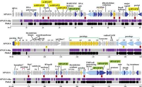
FIG 1Map of the SPN3US genome highlighting genes with homologs. Homologs identified in both phages PhiEaH2 and CR5 are shown in purple and homologs ofKZ in black. Newly identified essential genes are indicated by red-shaded Es, and genes whose essential status requires confirmation are indicated by question marks. In the SPN3US track, genes colored dark blue have a homolog of a head protein inKZ, genes colored light blue have homology with a tail protein inKZ, and genes colored yellow have homology with a virion protein inKZ whose head/tail location is unassigned. Subunit names of the vRNAP are shaded in green. Subunit names of the nvRNAP are shaded in yellow.
on November 7, 2019 by guest
http://jvi.asm.org/
[image:4.585.43.544.67.374.2](150-bp paired-end reads), and SNPs were identified using Seq-ManPro (Table 2). Four amber mutations were identified using this approach, and their presence in each phage was confirmed by Sanger sequencing of the region of interest after PCR amplifica-tion. Sanger sequencing was performed by Genewiz Inc. These four mutation sites had been identified in the mixture of 50 amber mutants. In each double mixture, the frequency (percentage) of each SNP causing an amber mutation was close to that expected based on the numbers of particles added to each mixture. How-ever, for other mutation sites (such as synonymous and nonsyn-onymous missense sites), it was demonstrated that there was a greater range in SNP frequencies, resulting in difficulty in con-cluding with certainty to which mutant these nonamber muta-tions belonged. These data also indicated that for some mutants there could be a considerable number of nonamber mutations; for
instance, a total of 115 mutations were detected in mixture 2, which could potentially have an impact on the phenotype of the mutant. This led us to conclude that for further studies on mu-tants it would be desirable to have their genomes individually sequenced to ensure that all the mutations in each mutant are clearly documented.
Genome sequencing of individual SPN3US amber mutant phages.Fourteen individual SPN3US mutant genomes were bar-coded and underwent genome sequencing. Three genomes (am1, am6, and am26) were sequenced using HiSeq technology (Table 3). Each of the genomes had excessive depth of coverage, and it was deduced that it would be more appropriate to sequence indi-vidual genomes using MiSeq technology, and subsequently, 11 mutant candidates were sequenced with this technology (see Ta-ble S3 in the supplemental material). In the 14 genomes, 15 dif-ferent amber mutations were detected in SPN3US coding regions at a SNP percentage of 99.5% or higher. Thirteen of these amber mutation positions had been identified in the mixture of 50 mu-tants, with the same mutation position detected in 2 mutants (am11 and am43), supporting our expectation of repeat muta-tions. Twelve mutants had a single amber mutation per genome, making the classification of the genes in which they occurred as “essential” straightforward (Table 4).
Overall, eight genes of the 12 individually sequenced mutants were identified as being essential. This is because for three genes there were two mutants in which a single amber mutation was identified, but at a different base pair. The genes in which there were two mutation sites were64(am2 and am27),186(am39 and am50), and203(am18 and am19). This is not surprising, as the mutagenesis is random (41) and the probability of a mutation occurring in a particular gene increases with increased gene length. Gene64is 1,314 bp,186is 2,511 bp, and203is 1,380 bp. Cross-plating of each pair of intragenic mutants under nonper-missive conditions was successful and showed that the
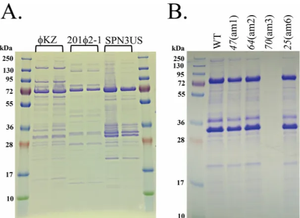
recombi-FIG 2SDS-PAGE profiles of SPN3US,KZ, and 2012-1 phages. (A) Profiles of CsCl step gradient-purifiedP. aeruginosaphageKZ,P. chlororaphisphage 2012-1, andS. entericaserovar Typhimurium phage SPN3US. (B) Profiles of wild-type (WT) and amber mutant phages of SPN3US after propagation on nonpermissiveS. entericaserovar Typhimurium (strain TT9079).
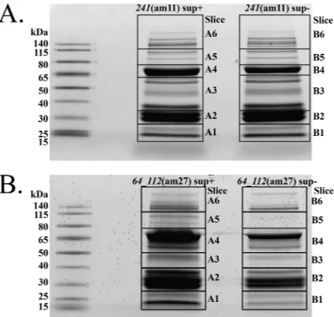
FIG 3Cross-plating of SPN3US amber mutants in the nonpermissiveS. en-tericaserovar Typhimurium strain (TT9079). Approximately 107particles of
29(am22) (A) and64_112(am27) (B) were seeded into the bacterial overlay (the boxed areas show no plaques/lysis in the lawn). Mutant phages were spotted onto the overlay, with approximate numbers of PFU in each spot indicated. Note that when29(am22) and64_112(am27) were spotted onto the overlays containing themselves, there was only clearing caused by lysis from without in the presence of a large number of particles.
on November 7, 2019 by guest
http://jvi.asm.org/
[image:5.585.141.444.65.286.2] [image:5.585.43.288.528.650.2]nation frequency of SPN3US is proportional to the distance be-tween mutations (data not shown).
Two mutant phages, am26 and am27, each had two amber mutations in their respective genomes. The amber mutation in 112of am27 was not detected previously in the sequencing of the mutant population, although the amber mutation in64had been, raising the possibility of additional undetected amber mutations in the mutant population. Since64had an individual amber mu-tation in am2, it was assigned as essential. The assignment of the three other genes mutated in double mutants,19,112, and168, is less straightforward, as one or both mutations in each genome could feasibly be essential.
Of the 20 amber mutations identified, only two (in203and 168in am18 and am27, respectively) resulted from the muta-tion of a tryptophan codon; the rest resulted from the mutamuta-tion of a glutamine codon. To ensure there were no unusual codon usage patterns within the SPN3US genome causing tryptophan codons to be rarer than glutamine codons, we determined the codon usage of the entire genome and of the individual genes mutated in this study using the Countcodon software (available
athttp://www.kazusa.or.jp/codon/countcodon.html). These
analy-ses showed there are similar total numbers of Gln-CAG (1,398) and Trp-UGG (1,441) codons throughout the SPN3US genome and revealed no special biases for these codons within the mutated genes (data not shown). It is possible that our observation of nearly 10-fold more mutated glutamine than tryptophan codons in the mutants could be due to the relatively small number of mutants sequenced. Alternatively, it might be an unintentional bias resulting from our use of a serine suppressor strain and the fact that serine replaces glutamine more effectively than trypto-phan with its large aromatic side chain. It will be interesting to determine if this pattern holds by the sequencing of more mutants and to test it further by using glutamine and tryptophan suppres-sor strains for future mutant isolations.
Newly identified SPN3US essential genes.Thirteen SPN3US genes were demonstrated to be essential in this study (Table 4). Of these, only two genes encoded proteins with previously assigned specific functions, gp186 and gp241. gp186 is a predicted SbcC subunit of an SbcCD complex (35), and gp241 is thesubunit of a predicted vRNAP subunit. In addition to gp241, six other SPN3US essential genes,25,64,171,203,214, and238, were pre-dicted to encode proteins that were part of the virion based on their similarity to proteins inKZ that had been identified as virion proteins. While it has not been conclusively demonstrated that168is an essential gene, we have designated this gene “ex-pected” essential, as it is a member of a paralog family that is found in each member of thePseudomonasphages that are related to KZ. gp168 is similar toKZ gp131, which was demonstrated to be located at the periphery of the baseplate and also possibly asso-ciates with fibers that emanate from the baseplate by immune-gold labeling (12).
Proteomes of phage particles resulting from mutations in SPN3US 241 and 64. Two mutants, 241(am11) and 64_112(am27), with mutations in the genes encoding well-con-served and predicted virion genes were selected for propagation in the nonpermissive and permissive host strains to determine the effect of knocking out their respective genes. Purified particles from both mutants grown on the permissive host purified at a buoyant density consistent with that of the wild-type phage, which is similar to that obtained for bothKZ and 2012-1 (1.38 g/ml). The241(am11) particles produced in the nonpermissive host
pu-TABLE 2Amber mutation sites in mixtures containing two SPN3US amber mutants whose particles were combined in amounts differing by 6.25-fold
Mixture Mutant
Mutation positiona SNP %
SPN3US ORF
DNA changeb
Amino acid changec
1 am21 186856 85.70 214 c.682C¡T p.Q228.
am28 203975 13.80 238 c.1186C¡T p.Q396.
2 am24 159099 82.30 171 c.361C¡T p.Q121.
am30 190698 15.70 219 c.271C¡T p.Q91.
aPosition relative to that of GenBank accession numberJN641803.1. Amber mutation positions were confirmed in each mutant by Sanger sequencing.
b“c.” indicates the coordinate of the mutation within the open reading frame and its reference to called base change.
[image:6.585.40.285.89.438.2]cPeriods after position numbers indicate that the glutamine at that position in the polypeptide chain is not replaced, as its codon has been mutated to a nonsense codon and the protein product truncated.
TABLE 1Identification of mutations resulting in amber codons in the SPN3US genome in a mixture of 50 mutant candidatesa
Genome positionb
Gene location
Reference base
Alternate base
No. for reference basec
No. for alternate based
Alternate frequency (%)
215999 241 C T 28,278 3,645 12.9
23427 29 C T 17,315 1,776 10.3
195232 225 C T 33,887 2,181 6.4
169242 186 C T 22,789 1,374 6.0
186856 214 G A 22,375 1,240 5.5
37455 39 C T 31,428 1,439 4.6
192318 222 G A 25,506 995 3.9
33261 35 C T 35,090 1,295 3.7
30811 34 C T 37,702 1,370 3.6
63849 64 C T 40,330 1,415 3.5
46330 47 C T 32,816 1,092 3.3
201073 235 C T 36,575 1,189 3.3
66658 70 C T 28,668 916 3.2
153240 169 C T 25,241 785 3.1
23982 30 C T 34,630 1,054 3.0
230169 258 G A 17,353 499 2.9
13997 19 C T 24,577 597 2.4
203975 238 G A 21,902 513 2.3
159924 171 C T 18,225 420 2.3
20159 25 C T 28,444 612 2.2
190698 219 C T 34,501 731 2.1
146047 168 C T 31,761 633 2.0
159099 171 C T 48,284 898 1.9
63957 64 C T 41,325 638 1.5
170989 186 G A 30,758 416 1.4
179577 203 C T 26,162 322 1.2
179287 203 G A 29,504 352 1.2
77022 78 C T 33,542 239 0.7
81829 82 G A 37,167 219 0.6
229576 257 C T 33,223 183 0.6
170544 186 C T 30,232 126 0.4
a
Mutations subsequently identified in individual mutant phage genomes (Tables 2and 3) are in boldface.
b
Position relative to that of GenBank accession numberJN641803.1. cNumber of times reference base observed.
d
Number of times alternate base observed.
on November 7, 2019 by guest
http://jvi.asm.org/
[image:6.585.296.546.98.183.2]rified at a slightly lower buoyant density (1.392 g/ml) than those produced from the permissive host (1.396 g/ml), and that, in ad-dition to small observable differences in the protein profile of each sample (Fig. 4A, slices A6 and B6), indicated there might be dif-ferences in their proteomes that could be detected by mass
[image:7.585.40.548.78.292.2]spec-trometry. Conversely, the particles produced by growth of 64_112(am27) in the nonpermissive hosts had very different buoyant densities (1.4240 to 1.4298 g/ml), so it was assumed there would be a marked difference in their proteomes. Consequently, samples of 241(am11) and 64_112(am27) propagated on both host strains were subjected to gel separation and analysis by LC– MS-MS (GeLCMS) (Fig. 4).
TABLE 3Amber mutations identified in SPN3US individual mutant phage genomes
Mutant
Genome reference positiona
Reference base
Called
base SNP % SPN3US ORF DNA changec
Amino acid changed
am1b 46630 C T 99.8 47 c.1432C
¡T p.Q478.
am2 63849 C T 99.6 64 c.763C¡T p.Q255.
am3 77022 C T 99.6 78 c.1459C¡T p.Q487.
am6b 20159 C T 99.5 25 c.184C
¡T p.Q62.
am11 215999 C T 100.0 241 c.2011C¡T p.Q671.
am13 66658 C T 99.7 70 c.181C¡T p.Q61.
am18 179577 C T 99.8 203 c.377G¡A p.W126.
am19 179287 G A 99.8 203 c.667C¡T p.Q223.
am22 23427 C T 100.0 29 c.358C¡T p.Q120.
am26b 13997 C T 99.5 19 c.70C
¡T p.Q24.
146047 C T 99.8 168 c.4673G¡A p.W1558.
am27 63957 C T 99.5 64 c.871C¡T p.Q291.
100963 C T 99.9 112 c.100C¡T p.Q34.
am39 169242 C T 99.5 186 c.343C¡T p.Q115.
am43 215999 C T 100.0 241 c.2011C¡T p.Q671.
am50 170544 C T 99.5 186 c.1645C¡T p.Q549.
a
Position relative to GenBank accession numberJN641803.1. bMutant sequenced on an Illumina Hi-Seq machine. c
“c.” indicates the coordinate of the mutation within the open reading frame and its reference to called base change.
[image:7.585.39.285.449.698.2]dPeriods after position numbers indicate that the glutamine in that position in the polypeptide chain is not replaced, as its codon has been mutated to an amber stop codon and the protein product truncated.
TABLE 4Features of SPN3US proteins encoded by essential genes
Gene product
Mass
(kDa) Essentiala KZ
homolog Function/commentb
19 10.6 ND
25 14.6 Yes ORF62 STR; low-copy-number tail
protein
29 24.5 Yes ORF67
47 62.8 Yes STR; head/neck protein
64 48.9 Yes ORF101 STR; head, possible neck protein
70 32.2 Yes
78 60.3 Yes
112 10.9 ND
168 188.1 Expected ORF145 STR; baseplate/fiber protein 171 47.1 Yes ORF130 STR; possible baseplate/fiber
protein
186 95.7 Yes ORF165 SbcC subunit
203 51.9 Yes ORF157 STR; tail protein
214 28.1 Yes ORF153 STR; head protein
219 28.6 Yes ORF147
238 82.1 Yes ORF182 STR; tail protein, possible host membrane-targeting function (predicted N-terminal transmembrane domain residues 152 and 174)
241 159.1 Yes ORF178 STR; vRNAPN
aND, the “essential” status of proteins encoded by genes with amber mutations in double mutants was not determined.
bSTR, protein detected as part of the virion by mass spectrometry.
FIG 4SDS-PAGE gels used for mass spectrometry of SPN3US amber mu-tants. Shown are241(am11) (A) and64_112(am27) (B). sup⫹, mutant prop-agation on the permissive host; sup⫺, mutant propagation on the nonpermis-sive host.
on November 7, 2019 by guest
http://jvi.asm.org/
[image:7.585.300.544.450.682.2]Peptides from 83 different SPN3US proteins were detected by mass spectrometry in each mutant propagated on the permissive host (see Table S4 in the supplemental material). Sixty of these proteins are similar toKZ virion proteins (Fig. 5), as determined by both the CoreGenes analyses and additional PSI-BLAST searches. In each mutant, it was of interest to determine whether a partial product of the mutated gene would be incorporated into the mature particle. For instance, in241(am11), the amber muta-tion in241would result in the truncation of the normally 1,401-residue protein at 1,401-residue 670, and in64_112(am27), the muta-tion in64would result in a product of only 290 residues compared to the normal 437 residues. However, in each mutant, no peptides from the mutated gene product were detected. In addition, other proteins detected in particles produced from the permissive host were not detected in the particles propagated on the nonpermis-sive host, 7 and 28 different proteins for am11 and am27, respec-tively. This indicates that the incorporation of both gp241 and gp64 into the virion is essential for the incorporation of the full complement of proteins in the virion.
The fact that gp241 was not included in particles propagated on the nonpermissive host was very definitive, with no spectra de-tected for tryptic peptides in this large protein. This is in contrast to the 110 total spectra detected for gp241 in the sample
propa-gated on the permissive host. In addition to gp241, six other SPN3US proteins were detected in 241(am11) and 64(am27) propagated in the permissive strain but were not detected when 241(am11) was propagated on the nonpermissive strain (see Table S4 in the supplemental material). They are gp37, gp42, gp158, gp218, gp240, and gp244. Three of these proteins, gp42, gp218, and gp240, along with gp241, are all predicted to be subunits of the vRNAP (Table 5), so they are expected to be part of the phage head.
The gp64 gene encodes a low-abundance protein found in the SPN3US virion, as determined by the detection of only nine mass spectra in the sample from the64_112(am27) particles propagated on the permissive host. This is consistent with the number of spectra identified for gp64 in241(am11) grown on both hosts. Previous mass spectral studies also showed the homologous pro-tein in bothKZ and 2012-1 to be low-abundance proteins in their respective virions (15,17). Notably, in addition to no mass spectra being detected for gp64 in64_112(am27) grown on the nonpermissive host, none were identified for an additional 28 proteins normally seen in the mutant grown on the permissive host (Fig. 5; see Table S4 in the supplemental material). Among the “missing” proteins were those associated with the phage tail, such as the major tail tube protein (gp255) and the tape measure
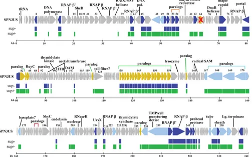
FIG 5Map of SPN3US genome comparing proteomes of mutant64_112(am27) when grown on permissive (sup⫹) and nonpermissive (sup⫺) hosts. The proteome of64_112(am27) propagated on the sup⫹or permissive host is indicated in green, and its proteome when propagated on the sup⫺or nonpermissive mutant host is indicated in blue. The gene encoding gp64 (marked with a red cross) is essential, as determined by sequencing of a mutant (am2) with a single amber mutation in its genome, in the gp64 gene. gp112 is not a virion protein, and its essential status requires clarification. In the SPN3US track, genes colored dark blue have homology with a head protein inKZ, genes shaded light blue have homology with a tail protein inKZ, and genes colored yellow have homology with a virion protein inKZ whose head/tail location in unassigned.
on November 7, 2019 by guest
http://jvi.asm.org/
[image:8.585.43.546.72.385.2]protein (gp239) (Fig. 5). Among the 52 proteins identified in the 64_112(am27) sample propagated under nonpermissive condi-tions were proteins with funccondi-tions associated with the head, such as the major capsid protein (gp75) and the portal protein (gp81)
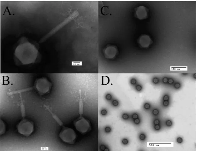
(Fig. 5). Examination of the gp64 samples by TEM revealed tailless
particles, indicating that the proteins identified in this sample are located in the SPN3US head or neck (Fig. 6). The latter function is indicated by the fact that the heads contained DNA, implying any “plug” proteins that normally function to prevent the DNA from exiting the head after packaging had also been added to these particles. Further suggesting that the normal neck structure was nearly complete was the observance of a small number of spectra for tryptic peptides from the tail sheath protein, gp256 (8 spectra versus 686 spectra in the mutant grown on the permissive host). From these observations, we conclude that putative functions of
gp64 are an aid in tail attachment to the head or a neck protein or that they have an essential role in tail assembly.
The identification of SPN3US virion proteins in the analyses of 241(am11) and64_112(am27) enabled the general delineation of all the essential genes identified in this study as encoding either nonvirion or virion proteins. Three essential proteins are not as-sociated with the virion, gp29, gp70, and gp78. gp19, whose essen-tial status requires clarification, is also not associated with the virion. No functions can be assigned to any of these proteins based on sequence-based searches, and further investigations will be re-quired to resolve their roles. It is interesting that several genes encoding essential proteins were determined to be located close to an expected essential gene, suggesting there may be clustering of essential genes throughout the genome. For instance, the gp78 gene is located immediately downstream of the gene encoding the N-terminal fragment of the nvRNAP(77). We hypothesize that gp78 may have a regulatory function, as apparently many proteins are not produced when this protein is knocked out, as determined by the lack of detectable virion proteins being produced when 78(am3) is propagated under nonpermissive conditions (Fig. 3B). In addition to the two head proteins, gp64 and gp241, the pro-teome analyses facilitated the delineation of the products of six other newly identified SPN3US essential genes into those encod-ing tail-associated (gp25, gp171, gp203, and gp238) and head-associated (gp47 and gp214) proteins. gp168, whose gene’s amber mutation was detected in a double mutant, is also a tail protein.
DISCUSSION
SPN3US, a candidate type phage of a new giant phage genus,
“SPN3USlikevirus.”SPN3US was determined to share 55.7% of
[image:9.585.40.286.97.214.2]its proteins with easily identifiable homologs in both Erwinia phage PhiEaH2 andCronobacterphage CR5 by both the Core-Genes program and PSI-BLAST. This set of homologs in these
TABLE 5Mass spectral counts detected for64_112(am27) and
241(am11) propagated on permissive (sup⫹) and nonpermissive (sup⫺) hostsa
Gene product
Length (aa)
Total spectrum count
vRNAP subunit
am27 am11
sup⫹ sup⫺ sup⫹ sup⫺
37 127 6 0 6 0
42 431 37 36 27 0 =M
158 171 3 0 2 0
218 222 23 28 24 0 C
240 519 56 38 14 0 =N
241 1,401 203 152 110 0 N
244 240 12 13 10 0 =Cb
a
64_112(am27) grown on the nonpermissive host formed tailless particles. bgp244 was identified as a candidate for the C-terminal region of=in this study (see the text).
FIG 6Transmission electron microscopy of SPN3US wild type and amber mutant phage64_112(am27). (A) Wild-type phage in a nonpurified lysate stained with PTA. (B) CsCl-purified wild-type phage stained with ammonium molybdate. (C and D) CsCl gradient-purified particles of64_112(am27) grown on the nonpermissive host stained with ammonium molybdate (C) and uranyl formate (D).
on November 7, 2019 by guest
http://jvi.asm.org/
[image:9.585.136.450.451.692.2]phages is comparable to the percentage shared byKZ,PA3, and 2012-1 (members of the genusPhiKZlikevirus), and according to the recommendations of Lavigne et al. (45), SPN3US, PhiEaH2, and CR5 could be considered a genus, which we refer to as “SPN3USlikevirus.” The fact that SPN3US shares 26% of its pro-teins with identifiable homologs inPseudomonasphageKZ, as determined by CoreGenes, and 37.8% when PSI-BLAST matches are included, also leads us to suggest that SPN3US could be con-sidered a diverged member of a tentative “KZ-related” phage subfamily. This conclusion is based on the taxonomic status of phages EL and OBP. It was recommended that EL be considered a separate genus within a common family based on a smaller num-ber of easily identified homologs (30% of EL’s proteins were found to be similar to those inKZ). The addition of OBP to the EL-like genus enabled an extensive tabulation of more divergedKZ ho-mologs (22). It also enabled the determination that, while global synteny is disrupted by a series of inversions in the genomes of phages in this subfamily, local synteny confirmed the orthologous status of mostKZ homologs (22). This finding made it clear that a group of phages related toKZ is supported by vertical descent of a large number of genes, which include those specifying a char-acteristic head and tail morphology, as well as a charchar-acteristic gene expression and replication strategy (22).
The existence of homologs ofKZ in an expanding group of giant phages was the incentive to study a representative by using genetics, which would first enable us to test if there was a relation-ship between vertically descended genes and essential genes in these phages and, second, enable the study of currently function-ally unassigned proteins. We isolated amber mutants of SPN3US, the first such mutants for these giant phages. Initial sequencing of a mixture of the candidates showed the presence of amber muta-tions in 33 SPN3US genes, 24 with homologs inKZ. Numbers of these 24 genes would also be expected to be essential based on studies of other phages; for instance, the SbcC subunit of T4 was demonstrated to be essential using genetics, and the subunits of theKZ multisubunit RNAPs would be expected to be essential
based on the replication of the phage in the presence of rifampin (19). However, to confirm that any of the detected SPN3US amber mutations were actually in an essential gene, each mutation must be identified as the sole amber mutation in an individual genome. Toward this goal, we sequenced 18 individual mutant phage ge-nomes, 2 of which had double amber mutations and 16 of which had a single amber mutation per genome, enabling us to identify 13 essential genes in SPN3US.
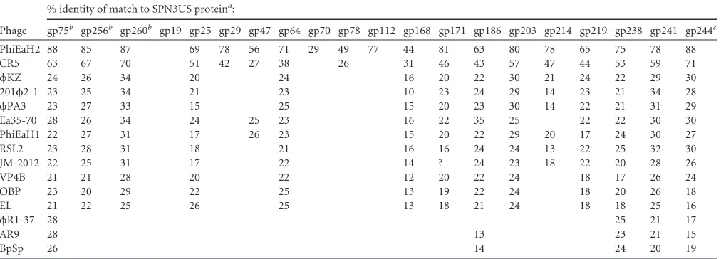
[image:10.585.41.547.88.270.2]Of the 13 SPN3US genes newly classified as essential, 9 (25,78, 171,186,203,214,219,238, and241) were also determined to have homologs inKZ, 2012-1,PA3, EL, and OBP (Table 6), as does the “expected” essential tail gene,168. Further searches showed that the 10 proteins encoded by these genes in SPN3US also have homologs in other recently identifiedKZ-related phages, such as Vibriophage JM2012 (25) andErwiniaphage Ea35-70 (46), and other giant phages (Table 6), supporting the broader relevance of utilizing a genetic system to study SPN3US. Examples of several proteins well conserved in all myoviruses (major head, sheath, and terminase proteins [47]) are also included inTable 6to illustrate the intrinsically divergent nature of homologous proteins in these phages. The homologs in the table also highlight a dichotomy in the literature as to what is referred to as a “KZ-related” phage. TheB. subtilisphage AR9 was recently described as belonging to theKZ-related phage group (24). As illustrated inTable 6, while AR9 has homologs of several SPN3US essential genes, such as the SbcC subunit, terminase, and vRNAP subunits, there is clearly an absence of easily detectable homologs in AR9 of other essential proteins in SPN3US, which have counterparts inKZ and most other phages described as beingKZ related in the literature. No-tably, there is no easily identifiable homolog in AR9 of two highly abundant virion proteins, the major capsid and tail sheath, in SPN3US orKZ, although obviously AR9 is a myovirus and has these proteins. These differences highlight a need for clearer defi-nitions of a taxon for giant phages. Ideally, such defidefi-nitions would incorporate both predicted core genes and experimentally identi-fied essential genes, now that we have a genetic system to prove
TABLE 6Proteins in giant phages with similarity to SPN3US proteins that are encoded by genes identified as having an amber mutation in this study or expected to be essential
Phage
% identity of match to SPN3US proteina:
gp75b gp256b gp260b gp19 gp25 gp29 gp47 gp64 gp70 gp78 gp112 gp168 gp171 gp186 gp203 gp214 gp219 gp238 gp241 gp244c
PhiEaH2 88 85 87 69 78 56 71 29 49 77 44 81 63 80 78 65 75 78 88
CR5 63 67 70 51 42 27 38 26 31 46 43 57 47 44 53 59 71
KZ 24 26 34 20 24 16 20 22 30 21 24 22 29 30
2012-1 23 25 34 21 23 10 23 24 29 14 23 21 34 28
PA3 23 27 33 15 25 15 20 23 30 14 22 21 31 29
Ea35-70 28 26 34 24 25 23 16 22 35 25 22 22 30 30
PhiEaH1 22 27 31 17 26 23 15 20 22 29 20 17 24 30 27
RSL2 23 28 31 18 21 16 16 24 24 13 22 25 32 30
JM-2012 22 25 31 17 22 14 ? 24 23 18 22 20 28 26
VP4B 21 21 28 20 22 12 20 22 24 18 17 26 24
OBP 23 20 29 22 25 13 19 22 24 18 20 26 18
EL 21 22 25 26 25 13 18 21 24 18 18 25 16
R1-37 28 25 21 17
AR9 28 13 23 21 15
BpSp 26 14 24 20 19
aSimilarity was determined using PSI-BLAST with a maximum of three iterations. b
Three proteins were included as positive controls, as they must be essential in all myoviruses and are expected to be detectable in related phages. They are gp75 (major capsid), gp256 (sheath protein), and gp260 (terminase).
c
The newly identified candidate vRNAP subunit, gp244, in SPN3US and its homologs are also included.
on November 7, 2019 by guest
http://jvi.asm.org/
that status. The differences among these giant phages also high-light exciting and likely complex evolutionary pathways that re-quire unraveling.
The identification of 13 essential SPN3US gene products em-phasizes the potential of the SPN3US genetic system for protein function determination, as only two proteins, gp186 and gp241, previously had assigned functions based on sequence similarity. However, even for these two proteins, there is a lack of specific knowledge pertaining to their functions and interactions with other proteins, indicating the potential of their mutants. For in-stance, the SPN3US gp186 gene encodes a putative SbcC subunit. InEscherichia coli, SbcCD has roles in DNA repair and replication via its ATP-dependent double-stranded DNA exonuclease activity and ATP-independent single-stranded DNA endonuclease activ-ity. The SbcC subunit is larger (1,048 amino acids [aa]), with ATPase activity, whereas the smaller (400-aa) SbcD subunit has nuclease activity.E. coliSbcC belongs to the structural mainte-nance of chromosomes (SMC) family, whose members are highly conserved in bacteria and have structural homology with the eu-karyotic Mre11/Rad50 proteins that function to repair breaks in dsDNA (48). In light of this conservation, it is not surprising that there are homologs of the SbcCD proteins inKZ and other giant phages, and as has now been shown for SPN3US gp186 and in-ferred for gp27 (the SbcD subunit), these proteins are also essen-tial. InE. coli, the SbcD gene is upstream of that encoding SbcC in thesbcDCoperon (48). Similarly, the T4 phage homolog gp46 and gp47 genes are clustered, although separated by two small hypo-thetical ORFs, gp46.1 and gp46.2. In contrast, in SPN3US (Fig. 1) and inKZ and other related phages, the SbcC and SbcD genes have been drastically separated within the genome. This splitting of functionally grouped genes is a feature of the genomes of phages related toKZ, even for genes typically grouped together in a phage genome (such as the morphogenesis genes [e.g., head-re-lated genes]), and is suggestive of unusual and interesting evolu-tionary mechanisms.
The SbcCD complex of SPN3US likely has a role similar to that of T4 gp46 and gp47. Mutations in either of the T4 genes encoding these proteins result in deficiencies in recombination and DNA replication and no host DNA degradation (28). However, the sim-ilarities and differences in the roles of this complex in SPN3US require confirmation, as it is extremely divergent from that in T4 —there is no sequence similarity between the proteins of the two phages that can be detected by a simple BLASTP search. The SPN3US SbcC protein is larger (95.7 kDa) than that of T4 (63.6 kDa), also suggesting potential functional differences between the two proteins. As inE. coli, both phage SbcC proteins have ATP-binding cassette domains split into an N-terminal domain con-taining a Walker A motif and a C-terminal domain concon-taining an ABC transporter signature motif, Walker B motif, D loop, and H loop/switch region. However, the region between the split do-mains in SPN3US gp186 is more than 600 residues, approximately 300 residues longer than that of T4 gp46. In T4, the gp46-gp47 complex was determined to be a membrane protein and was sug-gested to be anchored to the membrane by gp47.1 by Miller et al. (28). T4 gp47.1 is a small protein (46 aa) with a predicted trans-membrane domain (28). The gene immediately downstream of the SPN3US SbcC gene, the gp187 gene, encodes a small (63-amino-acid) protein with a transmembrane domain predicted by TMHMM (49), so it will be of interest to determine the other
proteins with which the SPN3US SbcCD complex interacts and its cellular location.
Using mutant proteomes to resolve the structure and assem-bly of a complex virion. Two-thirds of the newly identified SPN3US essential genes encode predicted virion proteins, which is consistent with a significant proportion of the genome encoding virion proteins (⬃46%), although what percentage of this is es-sential remains to be determined. Our examination of the pro-teomes of two mutants [241(am11) and 64_112(am27)] with knockouts in virion protein genes was unexpectedly fruitful. The 64_112(am27) mutant was selected for proteome analysis because the product of its mutated essential gene has homologs inKZ and related phages (Table 6). In addition, the gene encoding gp64 is situated at the end of a module of apparently functionally clus-tered genes in these phages. This is in striking contrast to the many other genes, such as the SbcCD and RNAP genes discussed above, that are split and have undergone genomic rearrangement. This gene region is important inKZ, as four of its genes encode abun-dant internal head proteins, and hence, it was named the IB region (15). TheKZ homolog of SPN3US gp64, gp101, was not identi-fied in the head; however, the genetic basis for thets13 mutant upon which that study was based is unknown.
Fortuitously, the SPN3US 64_112(am27) mutant produced tailless particles, and based on these particles containing packaged DNA, we inferred that the heads were likely mostly complete and that gp64 is potentially a neck protein. Importantly, the purifica-tion of these particles enabled the delineapurifica-tion of the more than 80 proteins associated with the SPN3US particle into general catego-ries of head (53 proteins) and tail (28 proteins). Clearly, the exact composition of each substructure within the SPN3US virion will need further investigation, but our strategy of applying proteomic and structural studies to additional virion gene mutants will facil-itate the piecing together of this complex structural jigsaw. An unexpected finding was that a large, 17-member paralog family (gp138 to gp154) was identified as being part of the head, poten-tially the largest family of paralogs identified in any single phage genome.
Comparison of the number of different SPN3US head proteins (⬃53) to the number in the T4 phage head (10) highlights the extent of the challenge in assigning functions to virion proteins and the importance of the mutants in doing so. Excitingly, such analyses will have broader relevance to theKZ structural-ho-mology group, as we determined that 60 SPN3US virion proteins are similar toKZ virion proteins (Fig. 4) by CoreGenes and/or PSI-BLAST searches.
Multimerization of the vRNAP prior to incorporation into the phage head.gp241 is the second SPN3US essential gene iden-tified in this study with a known predicted function, as a subunit of a multisubunit RNAP, prior to the studies. Two multisubunit RNAPs are considered hallmark features of phages related toKZ, with every member containing two sets of subunits (19), one set presumably arising from a gene duplication event(s) after becom-ing phage borne. This is quite remarkable considerbecom-ing that the two subunits (and=) themselves are hypothesized to have arisen from an ancient duplication event (50). The phageand= sub-units have extremely divergent homologies with prokaryotic RNAPs, as if they have been evolving independently from cellular RNAPs for a considerable time. The recent identification of ho-mologous subunits in the phage AR9 (24) supports this, as the presence of the RNAP genes in phages infecting both
on November 7, 2019 by guest
http://jvi.asm.org/
itive and Gram-negative hosts suggests that if these genes under-went little horizontal exchange they may have descended from an ancestral phage prior to the Gram-positive and Gram-negative split, estimated to have occurred over 3 billion years ago (51).
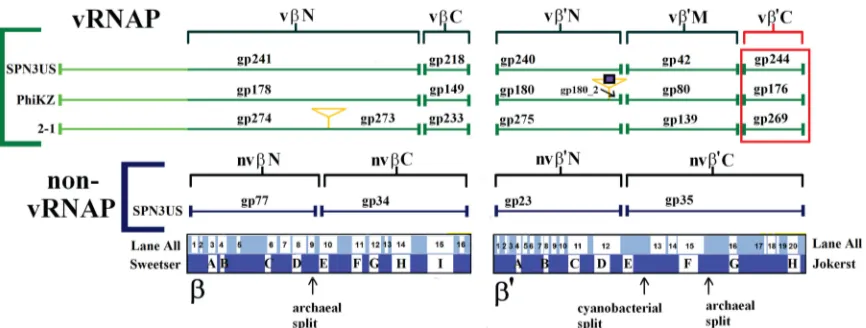
Remarkably, inKZ, one of the RNAPs, the vRNAP, is pack-aged into the phage head among the densely packpack-aged DNA (15), and in this study, we have confirmed that the SPN3US vRNAP is head associated, as it was identified in the tailless mutant. In the SPN3US complex, gp241 represents the N-terminal fragment of thesubunit and gp218 represents a smaller, C-terminal frag-ment (Fig. 7). The other predicted subunits of the SPN3US vRNAP are gp240 and gp42, which represent the N- and C-termi-nal subunits of the=subunit, respectively. The absence of all vRNAP subunits in the proteome of 241(am11) grown under nonpermissive conditions indicates that gp241 has a crucial role in the incorporation of the other subunits into the phage head. The enzyme complex likely assembles prior to its incorporation into the head, and we hypothesize that the N-terminal domain of gp241 targets the complex into the head, as it has a long N-termi-nal domain (⬃400 residues) that has no homology with prokary-otic RNAP. These findings provide further support for the no-tion that the vRNAP of SPN3US (and, we infer, related phage vRNAPs) has extremely unique features, including the following: (i) the enzyme assembles from gene products whose transcripts are from very different locales in the genome; (ii) the vRNAP subunits apparently assemble/multimerize prior to incorporation into the head; (iii) the vRNAP subunits must transition from within the head through the connector complex and tail tube (the tube diameter is 4.5 nm inKZ [10]) into the host cell, which hints that the proteins cannot be in a native form to do this; and (iv) the subunits must reassemble (based on point iii) into a func-tional complex once in the host cell. Addifunc-tionally, the state of the vRNAP within the mature head is completely unknown, but based on the fact that the DNA concentration in tailed phage heads is typically about 500 mg/ml (52,53), it is likely that structurally the vRNAP is very different from that of its active form or that of any active prokaryotic RNAP. It has been proposed that the high-force packaging enzyme compresses native proteins within the prohead
into their mature head state, thereby allowing exit through the narrow-diameter portal and tail tube (54).
Finding a “missing” vRNAP subunit.The SPN3US proteins gp241, gp218, gp240, and gp42 and their homologs in other giant phages that were previously identified as forming the vRNAP complex (19) account for all of the highly conserved regions in prokaryotic RNAP, with the exception of several hundred amino acids in the C-terminal region of=(approximately residues 1260 to 1524 of the RNAP=ofThermus thermophilusHB8). The C-terminal region of=was initially recognized as being conserved in both prokaryotic and eukaryotic RNAPs by Jokerst et al. and designated region H (55). Due to the massive increase in sequence data deposited in the databases, Lane and Darst clarified the con-served regions and lineage-specific domain insertions in theand =subunits of eukaryotes, eukaryotic viruses, bacteria, and ar-chaea (56). They identified four motifs (a17 to a20) at the C ter-minus of the=subunit that are conserved in all RNAPs, with a20 equating to the previously defined region H (56). Structurally, this region has an important role in the clamp, and a20 in particular serves as a hinge to mediate clamp movement (57).
There has been no identified counterpart to region=a17 to a20 in anyKZ-related phage vRNAP, although a counterpart to the region is present at the C terminus of the nvRNAP=subunit, SPN3US gp35 (Fig. 7). Of all the conserved regions inand=of prokaryotic and eukaryotic RNAPs,=a17 to a20 is the most weakly conserved (58). In addition, between different bacterial taxa, there is considerable variation in=a17 to a20, as the region is associated with two sites in which domains of various lengths have been inserted:=In6, which occurs immediately after a16, and=In7, which occurs between a19 and a20 (56). Based on the variability in this region, if a subunit containing the region existed for the phage vRNAP, it would not be surprising that it was not identified, especially if it had been split into a separate polypep-tide. This is especially so because of the high level of divergence of the other phage RNAP subunits from prokaryotic RNAPs, even those subunits that contain regions traditionally more conserved in prokaryotic RNAPs, such as SPN3US gp240, which contains=
a12 (the region containing the catalytic motif and centered in
FIG 7Scheme showing the homologous vRNAP subunits of SPN3US,KZ, and 2012-1 (2-1). Subunits detected by mass spectrometry in purified virions are bracketed in green. The newly identified C-terminal subunit of vRNAP=(SPN3US gp244) is boxed in red. The orange triangles indicate introns, and the blue square indicates a homing nuclease. The SPN3US nvRNAP subunits are also included. Cellular RNAP conserved regions and archaeal and cyanobacterial subunit split sites inand=are indicated (56,61). Note that the⬃300-residue lineage-specific insert inT. thermophilus=located between the Lane all regions a5 and a6 is not indicated.