Characterization of a Threonine-Rich Cluster in Hepatitis C
Virus Nonstructural Protein 5A and Its Contribution to
Hyperphosphorylation
Christian Schenk,
aMax Meyrath,
a*
Uwe Warnken,
bMartina Schnölzer,
bWalter Mier,
cChristian Harak,
a*
Volker Lohmann
aaDepartment of Infectious Diseases, Molecular Virology, University of Heidelberg, Heidelberg, Germany bFunctional Proteome Analysis, German Cancer Research Center (DKFZ), Heidelberg, Germany cDepartment of Nuclear Medicine, Heidelberg University Hospital, Heidelberg, Germany
ABSTRACT
Hepatitis C virus (HCV) nonstructural protein 5A (NS5A) is a
phospho-protein with key functions in regulating viral RNA replication and assembly. Two
phosphoisoforms are discriminated by their different apparent molecular weights: a
basally phosphorylated (p56) and a hyperphosphorylated (p58) variant. The precise
mechanisms governing p58 synthesis and specific functions of the isoforms are
poorly understood. Our study aimed at a deeper understanding of determinants
in-volved in p58 synthesis. We analyzed two variants of p56 and p58 of isolate JFH-1
separately by mass spectrometry using an expression model and thereby identified a
threonine-rich phosphopeptide exclusively found in the hyperphosphorylated
vari-ant. Individual exchange of possible phosphoacceptor sites to phosphoablatant or
-mimetic residues had little impact on HCV replication or assembly in cell culture. A
phosphospecific antibody recognizing pT242 revealed that this position was indeed
phosphorylated only in p58 and depended on casein kinase I
␣
. Importantly,
phos-phoablative mutations at positions T244 and S247 abrogated pT242 detection
with-out substantial effects on global p58 levels, whereas mutations in the preceding
serine-rich cluster dramatically reduced total p58 levels but had minor impact on
pT242 levels, suggesting the existence of distinct subspecies of hyperphosphorylated
NS5A. Mass spectrometry analyses of different genotypes showed variable
phosphor-ylation patterns across NS5A and suggested that the threonine-rich region is also
phosphorylated at T242 in gt4a and at S249 in gt1a, gt1b, and gt4a. Our data
there-fore indicate that p58 is not a single homogenously phosphorylated protein species
but rather a population of various phosphoisoforms, with high variability between
genotypes.
IMPORTANCE
Hepatitis C virus infections affect 71 million people worldwide and
cause severe chronic liver disease. Recently, efficient antiviral therapies have been
established, with inhibitors of nonstructural protein NS5A as a cornerstone. NS5A is
a central regulator of HCV replication and assembly but is still enigmatic in its
mo-lecular functions. It exists in two phosphoisoforms, p56 and p58. We identified a
phosphopeptide exclusively found in p58 and analyzed the determinants involved in
phosphorylation of this region. We found evidence for very different
phosphoryla-tion patterns resulting in p58. These results challenge the concept of p58 being a
homogenous species of NS5A molecules phosphorylated at the same positions and
argues for at least two independently phosphorylated variants showing the same
electrophoretic mobility, likely serving different functions.
KEYWORDS
positive-strand RNA virus, hepatitis C virus, NS5A, phosphorylation, p58,
p56, hyperphosphorylation, basal phosphorylation, PI4KA, PI4KIIIa
Received30 April 2018Accepted20
September 2018
Accepted manuscript posted online26
September 2018
CitationSchenk C, Meyrath M, Warnken U,
Schnölzer M, Mier W, Harak C, Lohmann V. 2018. Characterization of a threonine-rich cluster in hepatitis c virus nonstructural protein 5a and its contribution to hyperphosphorylation. J Virol 92:e00737-18.https://doi.org/10.1128/JVI .00737-18.
EditorJ.-H. James Ou, University of Southern
California
Copyright© 2018 American Society for
Microbiology.All Rights Reserved. Address correspondence to Volker Lohmann, [email protected]. *Present address: Max Meyrath, Department of Infection and Immunity,
Immuno-Pharmacology and Interactomics, Luxembourg Institute of Health, Esch-sur-Alzette, Luxembourg; Christian Harak, Abbott GmbH & Co KG, Wiesbaden, Germany.
crossm
on November 6, 2019 by guest
http://jvi.asm.org/
H
epatitis C virus (HCV) is a positive-strand RNA virus and belongs to the family
Flaviviridae. The viral genome encompasses about 9.6 kb and codes mainly for a
polyprotein of about 3,000 amino acids (aa), which is flanked by nontranslated regions.
The polyprotein is cleaved into ten mature proteins by cellular and viral proteases: core,
envelope glycoprotein 1 (E1) and E2, p7, and the nonstructural (NS) proteins NS2, NS3,
NS4A, NS4B, NS5A, and NS5B (reviewed in reference 1). The structural proteins core, E1,
and E2 are physical components of the viral particle and, together with p7 and NS2, are
mainly involved in the assembly of infectious virions, while NS3 to NS5B are mainly
required for replication of the viral genome. NS3 exhibits helicase activity in the
C-terminal part and an N-terminal protease responsible for cleavage of the
nonstruc-tural proteins in association with the cofactor NS4A. NS4B is a key factor in inducing
membrane alterations that are required for viral replication, the so-called membranous
web. NS5B is the viral RNA-dependent RNA polymerase.
NS5A is a phosphoprotein critically involved in the formation of the viral replication
compartment (2) as well as in the morphogenesis of infectious virions (3–5). Due to this
central role in various aspects of viral replication, NS5A inhibitors have become a
cornerstone in recent antiviral therapies, simultaneously affecting different steps of the
viral life cycle (6, 7). However, despite numerous studies, NS5A remains enigmatic in
most of its functions regarding the underlying molecular mechanisms (reviewed in
reference 8).
NS5A is built of an N-terminal amphipathic helix, essential for its membrane
association (9), followed by three domains (DI, DII, and DIII), which are separated by two
so-called low-complexity sequences (LCS-I and -II) (10). DI is structured, binds RNA (11),
forms several dimeric isoforms (12–14), and is crucial for the formation of the
mem-branous web (2). DII and DIII are intrinsically unstructured (15, 16). DII is widely
dispensable for replication (3) but has recently been shown to dampen recognition of
the virus by cytosolic pattern recognition receptors (17). DIII is involved in virion
production by an interaction with the core, which is modulated by phosphorylation
(3–5).
Two distinct NS5A phosphoisoforms have been described that are discriminated
due to their different apparent molecular weights in SDS-PAGE: p56, the basally
(hypo-)phosphorylated form, and p58, the hyperphosphorylated variant (18). P56 has
been regarded as the major isoform involved in viral RNA replication, whereas p58 was
supposed to inhibit replication and support assembly. This assumption was based
mainly on the fact that LCS-I is a hot spot for adaptive mutations enhancing replication
of many HCV genotypes in cell culture while at the same time abrogating p58 synthesis
(19–21) and virus particle production (22). However, replication of HCV genotype 2a
isolate JFH-1, which is still the only wild-type (WT) isolate efficiently replicating in cell
culture (23), is impaired by the same adaptive mutations (24–26), challenging this
concept. At this point, no distinct functions can be clearly assigned to p56 and p58.
Mass spectrometry (MS), reverse genetic studies, and proteomic analyses revealed
phosphorylation of numerous serine and threonine residues within different domains
of NS5A involving several serine/threonine kinases, thereby contributing to the
func-tional diversity of NS5A (4, 5, 18–20, 24, 27–39). However, links between specific
phosphorylation sites with particular kinases and specific functions have remained
difficult and rare: phosphorylation at S356 (all numberings refer to NS5A of HCV JFH-1)
is mediated by protein kinase A (PKA) and is necessary to facilitate interaction of NS5A
with other SH3 domains, which is crucial for replication (40). Pharmacological and
genetic approaches identified phosphorylation at S457 by casein kinase II (CKII) to be
important for virus particle production (5, 27). In contrast, residue S222 has been
identified as a major phosphorylation site by many proteomic studies (25, 26, 33, 41),
but the phenotype of phosphoablative or phosphomimetic mutations at this site is
weak (25, 33, 41).
Particularly enigmatic are the determinants of p58 synthesis. Generally, NS5A
hy-perphosphorylation requires the presence of NS3-5B as a polyprotein (39, 42–44) and
is not found if NS5A is expressed as a single protein (39, 45, 46). In addition, completely
on November 6, 2019 by guest
http://jvi.asm.org/
hyperphosphorylation (31, 47), overall suggesting that p58 synthesis requires a
com-plex succession of events supported by other nonstructural proteins, likely due to serial
interactions with different kinases.
A serine (Ser)-rich cluster in LCS-I is the most critical determinant of p58 synthesis,
since phosphoablative mutations in this region strongly reduce hyperphosphorylation
(e.g., S225, S228, and S232). Furthermore, pS232, pS235, and pS238 have been
exclu-sively found in p58 using phosphospecific antibodies (34, 48). The main kinase known
to be essential for the formation of p58 is CKI
␣
, directly targeting residues within the
Ser-rich cluster, e.g., S232 (49). Inhibition of CKI
␣
abrogates NS5A
hyperphosphoryla-tion and strongly affects viral replicahyperphosphoryla-tion and particle produchyperphosphoryla-tion (26, 49). CKI
␣
can
perform multiple sequential phosphorylation steps, using already-existing phosphates
as priming sites. Therefore, it has been speculated that p58 is a product of saltatory
phosphorylation by CKI
␣
within the Ser-rich cluster of LCS-I (25, 37). This hypothesis is
particularly attractive, since several Ser residues within LCS-I follow the sequential
phosphorylation pattern of CKI
␣
(pS-X-X-S), e.g., S229-S232-S235-S238, and a recent
study indeed provides proof in the case of 232-235-238 (48). However, the kinases
involved in priming phosphorylation are not clear yet. Additional kinases involved in
the synthesis of p58 are polo-like kinases (35) and calmodulin-dependent kinase II
(CAMK2)
␥
and
␦
(28). In contrast, phosphorylation at S146 by a yet-to-be defined
kinase has been shown to negatively regulate hyperphosphorylation of isolate JFH-1,
likely by modulating the dimeric conformation of domain I (25). In addition, the lipid
kinase phosphatidylinositol 4-kinase
␣
(PI4KA or PI4KIII
␣
) has been shown to suppress
p58 synthesis by an unknown mechanism (39). Overall, the succession of events
involved in formation of p58, as well as distinct phosphoacceptors that are
phosphor-ylated apart from S222, S232, S235, and S238, are still poorly understood.
NS5A is a central hub for interactions with host factors, and literally hundreds of
cellular proteins have been identified to directly interact with it (reviewed in reference
8). Phosphorylation events are indeed a main suspect in the regulation of this plethora
of protein-protein interactions, with a few examples suggesting specific functions of
p58. The human vesicle-associated membrane protein A (hVAP-A) has been shown to
be important for viral replication but only interacts with basally phosphorylated NS5A,
whereas hyperphosphorylation disrupts the interaction (46). A recent study showed
that a phosphoablative mutation at position S225, which was supposed to be
phos-phorylated in p58, impaired NS5A interactions with the nucleosome assembly protein
1-like protein 1 (NAP1L1), bridging integrator 1 (Bin1), and again hVAP-A (50),
suggest-ing that they are regulated by phosphorylation at this site. The lipid kinase PI4KA
furthermore, has been shown to interact with NS5A in multiple ways and also in a
phosphorylation-dependent manner. PI4KA is a key factor in the biogenesis of the HCV
replication compartment, supplying phosphatidylinositol 4-phosphate (PI4P) (51).
PI4KA mainly interacts with a specific region adjacent to LCS-I, designated the PI4KA
functional interaction site (PFIS), and its enzymatic activity is activated by this
interac-tion (39). However, activainterac-tion furthermore, requires phosphorylainterac-tion at posiinterac-tions S225,
S232, and S235 based on genetic evidence (21). In turn, PI4KA negatively affects p58
abundance by an unknown mechanism, since NS5A mutants with defects in PI4KA
interaction show increased levels of hyperphosphorylated NS5A (39).
The aim of our study was a deeper understanding of the hyperphosphorylated p58
isoform of HCV NS5A. We therefore performed a phosphoproteomic analysis of NS5A,
including the WT and a PFIS mutant with increased p58 levels, with a separate analysis
of p56 and p58. We thereby identified a peptide exclusively in p58 representing the
threonine (Thr)-rich cluster in LCS-I. While phosphoablatant and -mimetic mutations
had no impact on replication and assembly, we could verify phosphorylation at position
T242 using a phosphospecific antibody. Our data show that phosphorylation at T242 is
widely independent of the preceding Ser-rich cluster and defines a minor subspecies of
p58. Therefore, p58, so far defined only by its electrophoretic mobility in SDS-PAGE,
on November 6, 2019 by guest
http://jvi.asm.org/
seems to consist of several independently phosphorylated isoforms, probably serving
different functions.
RESULTS
Phospho-mass spectrometry analysis of NS5A.
In our recent study we found that
the lipid kinase PI4KA modulated NS5A phosphorylation, and we identified mutations
in the PFIS interfering with PI4KA interaction, resulting in increased p58 levels (39). To
identify specific phosphorylation sites modulated by PI4KA, we employed a mass
spectrometry analysis, comparing the phosphorylation pattern of the WT and a
mu-tated NS5A variant. Since all PFIS mutants were replication deficient (39), we transiently
expressed NS3-5B JFH-1 under transcriptional control of the T7 promoter in Huh7-Lunet
cells constitutively expressing T7 RNA polymerase. NS5A was immunoprecipitated, and
bands corresponding to p58 or p56 were individually excised after SDS-PAGE, digested
with trypsin, separated by high-performance liquid chromatography, and subjected to
tandem mass spectrometry (MS/MS) analyses (Fig. 1A and Table 1; see also Table S1 in
the supplemental material). Peptides and phosphorylation status were identified by
their distinct mass and charge in the first mass spectrometry run. In the second run
peptides were fragmented, allowing us to determine the position of the
phosphoryla-tion event within the peptide. We thereby identified a total of seven distinctly
phos-phorylated peptides located across all domains of NS5A (Fig. 1B and Table 1), four of
which (II, IV, V, and VI) were identical to those of previous studies (25, 52). We could
specifically determine phosphorylation sites at positions S146 (peptide II) and S360
(peptide VII) with an accuracy over 97%. In addition, the sites T14 (peptide I), T164
(peptide III), and T348 (peptide VI) could be clearly identified as phosphoacceptor sites
inside the respective peptide and are therefore most likely phosphorylated, even
though the PEP values are relatively high for these peptides. Peptide IV represents the
well-characterized Ser-rich cluster where we identified single phosphorylations at
positions S222 and S225 with a significant PEP score. We were not able to determine
multiple phosphorylations within this peptide, although they are already known from
other studies using reverse genetics. This would have required additional peptide
enrichment and higher detection sensitivity, which was not included in our analysis. For
the potential phosphoacceptor sites in peptide V, we could not conclusively determine
which site exactly is phosphorylated (Table S1), but there was some evidence for
phosphorylation at position T242 in sample C1 (Fig. 1C). We found only minor
quali-tative differences between the WT and mutant NS5A, not allowing us to draw
conclu-sions on phosphoacceptor sites influenced by PI4KA. Interestingly, six of the
phospho-peptides were found in both NS5A phosphoisoforms. In contrast, peptide I was only
found in p56, whereas phosphorylation of peptide V was restricted to p58, although we
cannot formally exclude minor amounts below the detection limit in p56. Peptide V is
located at the very beginning of domain II in the interferon sensitivity-determining
region (ISDR) adjacent to LCS-I and the serine-rich cluster (peptide IV), which has been
identified before as a main determinant of hyperphosphorylation (25, 26, 34, 41, 48).
In summary, we identified seven phosphorylated peptides and eight distinct
phos-phorylation sites in different regions of NS5A and, for the first time, identified a
phosphopeptide that exclusively mapped to the hyperphosphorylated form of NS5A.
Mutational analysis of potential phosphoacceptor sites in NS5A.
We next
analyzed the impact of mutations in potential Ser/Thr phosphoacceptor sites identified
by mass spectrometry on HCV replication, virion production, and NS5A phosphorylation
using the reporter virus JcR2a, encoding renilla luciferase (Fig. 2A to C). We excluded
the multiple serine residues in peptide IV at this point, since they have already been
analyzed in multiple studies (25, 26, 41), as well as T14, since this was only found
phosphorylated in p56 (Table 1). Each putatively phosphorylated serine or threonine
residue was mutated to either alanine, to prevent phosphorylation, or to aspartic acid,
mimicking phosphorylation by adding a negative charge. Interestingly, none of the
individual mutations had a strong impact on viral replication (Fig. 2B) or virus
produc-tion (Fig. 2C). In the case of T356 this was unexpected, since a recent study showed a
on November 6, 2019 by guest
http://jvi.asm.org/
lethal phosphoablative mutation at this site in the case of JFH-1 (40). Only a conversion
of the five N-terminal Thr/Ser residues in peptide V to phosphomimetic aspartate
strongly impaired replication (N-termD), whereas the phosphoablatant variant
repli-cated with efficiency identical to that of the wild type, suggesting that phosphorylation
events in this Thr-rich cluster are not essential for replication but that accumulation of
negative charges is deleterious.
C1
C2 C3C4
WT
mut5A
80
58
42
kDa
p58
p56 NS5A
A
B
S225* S228 S229 S230 S232* S235* S238* S146* S151 AH (1-33) Domain II (251-338) Domain III (352-467) Domain I (34-213) T244 T245 S247 T249* Y250 T268 S272 T164 II III T14 T17 LCS-I LCS-II T348* T356* S360I IV V VI VII
exact site indefinable; p58 exclusive ANLL 412.26 y 488.25 a-HPO 489.21 MEGGV 490.2 y-CHN 575.27 SNTYD 581.22 y-NH 600.26 y 617.29 ANLLME 688.33 y 718.34 MVDANLL-NH 756.36 b*-HPO 757.36 MVDANLL 773.39 y-HO 828.38 y-NH 829.37 NLLMEGGV 830.41 y 846.4 DMVDANLL 888.41 y-HO 899.42 y-NH 900.41 y 917.43 THSNTYDV 918.4 YDVDMVDAN-CHNO 980.37 b*-HPO 1039.49 DVDMVDANLL-CH 1046.45 DVDMVDANLL 1102.51 y 1130.54 a-HPO 1224.49 TCTTHSNTYD 1261.42 b*-CHNO 1288.53 b 1332.46 b* 1333.55 b-CO 1387.53 y 1406.62 b 1431.52 DANLLMEGGVAQTE 1445.66 y-HO 1501.7 b-CH 1504.5 y 1519.71 b 1546.55 y-CHNO 1561.75 y-CHNO-NH 1570.74 y-CHNO 1587.77 DMVDANLLMEGGVAQT-CHNO 1618.71 y 1632.79 DMVDANLLMEGGVAQT 1677.75 b-HPO-NH 1695.66 b-CHSO 1728.66 b-CHO 1732.63 b-HO 1774.64 b-NH 1775.63 b 1792.66 TTHSNTYDVDMVDANL 1793.76 DMVDANLLMEGGVAQTE 1806.79 VDMVDANLLMEGGVAQTE 1905.86 b 1907.68 HSNTYDVDMVDANLLME-CHNO 1921.79 c 1924.71 y 1932.9 b 1978.72 HSNTYDVDMVDANLLME 1980.83 b* 1994.79 0 20 40 60 80 100 Relative Abundance 0 50 100 150 200 250 300 Intensity
400 600 800 1000 1200 1400 1600 1800 2000
m/z (zoom) Raw File Scan Method Score m/z
131120_CH2091_1 7964 ITMS; CID 65.19 1242.85
- A T ph
C T T H S N T Y D b V b* D y² b M ox b² V D y b A b N b* L y L y M ox y E b² G y G V b² A y Q y b² T y E y b² P y E S b² R
-C
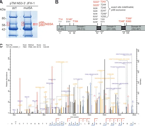
FIG 1Identification of NS5A phosphorylation sites. (A) Huh7-Lunet T7 cells were transfected with constructs encompassing NS3-5B JFH-1 with the NS5A
WT or harboring a triple-alanine mutation in the PFIS, abrogating PI4KA interaction (mut5A [39]). Twenty-four h later, cells were lysed, NS5A was immunoprecipitated, and p58 and p56 bands were cut out after SDS-PAGE and subjected to mass spectrometry analysis to identify phosphorylated peptides. (B) Schematic of the different domains of NS5A. The location of the identified phosphopeptides is indicated below. All numberings on top refer to amino acid positions within NS5A JFH-1 (GenBank accession numberKC164983.1). Residues shown in red are sites identified to be phosphorylated with high probability. Additional potential phosphoacceptor sites within each peptide that could not be identified as phosphorylated are shown in a smaller font size. Residues in italics of peptide V represent potential phosphoacceptor, where the exact site of phosphorylation could not be determined. Asterisks mark the residues already reported elsewhere: S146 (25), S222 (25, 41, 65), S225 (25), S232 (48), S235/238 (34), T249 (52), T348 (25), and T356 (40). (C) MSMS spectrum of peptide AT(ph)CTTHSNTYDVDMVDANLLM(ox)EGGVAQTEPESR with a mass of 3,723.5376 Da from NS3-3=JFH-1 detected in sample C1. This peptide was identified with a MaxQuant score of 65.19. Annotated fragments are color coded. y ions, red; b ions, blue; internal fragments, purple; a, b, and y ions and internal fragments with additional loss of ammonia, water or phosphoric acid, yellow. MaxQuant protein database software annotated the threonine in position two as most likely to be phosphorylated by the relation of the nonphosphorylated singly charged internal fragment TTHSNTYDVDM(ox)VDANL (m/z⫽1,793.76) and the phosphorylated internal fragment T(ph)CTTHSNTYD (m/z⫽1,261.42).
on November 6, 2019 by guest
http://jvi.asm.org/
[image:5.585.44.539.75.507.2]The phosphorylation pattern of the different mutants was comparable between cells
replicating a full-length viral genome (Fig. 3A) or expressing NS3-NS5B (Fig. 3B),
suggesting that transient expression of NS3-5B was indeed a feasible model to analyze
determinants of NS5A phosphorylation, as shown before (39). The p58/p56 ratio (Fig.
3C) validated this observation. Except for reduced p58 levels in the case of S146D,
which has been observed previously (25), no gross changes in the relative abundance
of the phosphoisoforms were observed for any other individual mutation. However, the
apparent molecular weight of p56 was clearly increased for the phosphomimetic
mutants at positions T242, T244, S247, and N-term, suggesting that phosphorylation at
these sites contributes to NS5A hyperphosphorylation. Furthermore, mutant N-termD
only gave rise to a single band of a size comparable to that of p58. These data were in
agreement with the observation that the phosphopeptide encompassing these
posi-tions was the only one exclusively found in p58. In addition, the Thr-rich cluster in LCS-I
was directly adjacent to S235 and S238, which have been found to be exclusively
phosphorylated in p58 as well (34). We therefore decided to focus our further studies
on the contribution of the Thr-rich cluster in peptide V to the hyperphosphorylation of
NS5A.
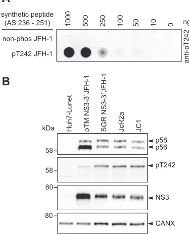
A phosphospecific antibody reveals phosphorylation of T242 exclusively in
p58.
To verify phosphorylation events in the Thr-rich cluster of peptide V, we aimed to
generate phosphospecific antibodies, which would further allow studying determinants
modulating phosphorylation. All HCV genotypes encode several potential
phosphoac-ceptor sites in this region, but only position T242 is fully conserved. In addition, there
was evidence by mass spectrometry indicating phosphorylation of this site (Fig. 1C),
and T242 was in the neighborhood of the bona fide phosphorylation site S238. We
therefore used a peptide encompassing amino acids 236 to 251 of JFH-1 NS5A, with a
single phosphorylation at position T242 to generate phosphospecific antibodies
tar-geting pT242. Indeed, after positive selection against the phosphorylated and negative
selection with the unphosphorylated peptide, only the phosphorylated peptide was
recognized with high specificity (Fig. 4A). These results overall indicated that the
pT242-specific antibodies indeed could be used to characterize phosphorylation events
at this site, with the limitation that proteins with multiple phosphorylations at
neigh-boring positions might be missed.
[image:6.585.41.549.83.190.2]We next analyzed the phosphorylation of T242 in cells either expressing NS3-5B or
replicating a NS3-3
=
JFH-1 subgenomic replicon (SGR JFH-1) or full-length viral
ge-nomes (JcR2a and JC1) (Fig. 4C). In all cases a clear band was detectable corresponding
to p58, demonstrating that T242 indeed was phosphorylated upon HCV replication and
expression of NS3-5B. However, pT242 seemed to be slightly more abundant in
replicating HCV than in the pTM expression model (Fig. 4B). This effect might be caused
by the higher expression level in the case of using pTM vectors, probably exceeding the
phosphorylation capacity of kinases involved in p58 synthesis. The restriction of the
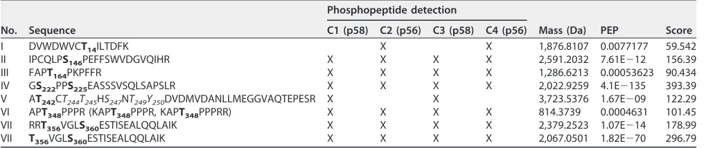
TABLE 1Phosphopeptides of wild-type (C1 and C2) and mutated (C3 and C4) HCV NS5A proteinsa
No. Sequence
Phosphopeptide detection
Mass (Da) PEP Score C1 (p58) C2 (p56) C3 (p58) C4 (p56)
I DVWDWVCT14ILTDFK X X 1,876.8107 0.0077177 59.542
II IPCQLPS146PEFFSWVDGVQIHR X X X X 2,591.2032 7.61E⫺12 156.39
III FAPT164PKPFFR X X X X 1,286.6213 0.00053623 90.434
IV GS222PPS225EASSSVSQLSAPSLR X X X X 2,022.9259 4.1E⫺135 393.39
V AT242CT244T245HS247NT249Y250DVDMVDANLLMEGGVAQTEPESR X X 3,723.5376 1.67E⫺09 122.29
VI APT348PPPR (KAPT348PPPR, KAPT348PPPRR) X X X X 814.3739 0.0004631 101.45
VII RRT356VGLS360ESTISEALQQLAIK X X X X 2,379.2523 1.07E⫺14 178.99
VII T356VGLS360ESTISEALQQLAIK X X X X 2,067.0501 1.82E⫺70 296.79
aHCV NS5A proteins were identified by Orbitrap MS analysis and MaxQuant data analysis (Fig. 1A). Predominant phosphorylation sites identified by microsequencing
are highlighted in boldface in the peptide sequences. Represented in italics are phosphoacceptors where the analysis could not clearly assign the phosphorylation to a specific site. Instances of phosphopeptides being detected in C1, C2, C3, and C4 are marked with an X. The mass of the phosphopeptides, the posterior error probability score (PEP;⬍0.02), and the individual peptide scores are presented for the most abundant MS scans. Note that all peptides identified contained only one phosphorylation event. Detection of multiply phosphorylated peptides would require previous enrichment, which was not done.
on November 6, 2019 by guest
http://jvi.asm.org/
2 3 4B 5A 5B 4A p7 E1 C E2 R-Luc ubi/2A C
JcR2a
Huh7-Lunet
72h
Huh7.5
72h
luc
luc
B
C
n=3
n=2
Replication
[RLU 72hpe / 4hpe]
Assembly & Release
[RLU 72hpi / 72hpe]
GD D Δ E1 E2 Δ JcR 2 a W
T A D A D A D A D A D A D A D A D A D A D A D A D A D A D 0.001 0.01 0.1 1 10 S 146 T 164 T 242 T 244 T 245 S 247 T 249 T 268 S 272 N-te rm (2 42-249) C-te rm (2 68 /
272) T348 T356 S360
GD D Δ E1 E2 Δ JcR 2 a W
T A D A D A D A D A D A D A D A D A D A D A D A D A D A D 0.1 1 10 100 S1 4 6 T1 6 4 T2 4 2 T2 4 4 T2 4 5 S2 4 7 T2 4 9 T2 6 8 S2 7 2 N-te rm (2 4 2 -2 4 9 ) C-te rm (268 / 272) T3 4 8 T3 5 6 S3 6 0
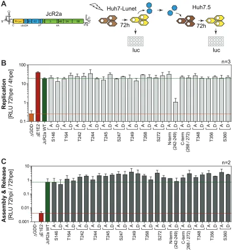
FIG 2Impact of phosphoablatant and -mimetic mutations at potential phosphoacceptor sites on replication and virus production. (A) Schematic
representation of the experimental procedure. Huh7-Lunet cells were transfected with WT and mutant full-length monocistronic JcR2a reporter virus genomes encoding renilla luciferase, and RNA replication efficiency was determined by luciferase measurement. Supernatants of transfected cells were transferred to Huh7.5 cells to assess production of infectious virus, again by quantification of luciferase activity 72 h postinfection (hpi). hpe, hours postelectroporation. (B and C) Analysis of phosphoacceptor sites harboring alterations of serine and threonine residues to alanine (to A) or aspartate (to D). Mutant “N-term” comprises all five serine and threonine mutations from T242 to T249. Mutant “C-term” comprises substitutions at both positions T268 and S272. (B) Replication is represented in relative light units (RLU) measured 72 h posttransfection and normalized to 4 h posttransfection. A replication-deficient JcR2a variant harboring a deletion in NS5B was used as a negative control for replication (ΔGDD), indicated by the orange line. A green line highlights the replication level of JcR2a WT. The data shown are the mean values with standard deviations (SD) from three independent experiments with two technical replicates each. (C) Supernatants of transfected cells described for panel B were used for reinfection of naive Huh7.5 cells to assess production of infectious virus. Huh7.5 cells were lysed 72 h postinfection and analyzed for luciferase activity. Virus production efficiency is expressed in luciferase activity RLU 72 h postinfection relative to 72 h posttransfection in order to normalize for defects in RNA replication. An assembly-deficient JcR2a variant harboring a deletion in E1E2 proteins was used as a negative control for reinfection (ΔE1E2), indicated by the red line. A green line indicates the virus production of JcR2a WT. The data shown are the mean values with standard deviations from two independent experiments with two technical replicates each.
on November 6, 2019 by guest
http://jvi.asm.org/
[image:7.585.40.500.73.566.2]signal to the p58 band was in line with the results from mass spectrometry, indicating
that the entire peptide V, comprising T242, is only phosphorylated in the p58 fraction
(Table 1). However, a minor reactivity of the antibodies to p56 was found in some
samples that could be due either to residual antibodies binding to the
nonphospho-rylated epitope or to a minor fraction of p56 phosphononphospho-rylated at T242.
Taking our findings together, we were able to generate antibodies specific to pT242
in NS5A, demonstrating that T242 was indeed phosphorylated upon HCV replication
only in the p58 isoform.
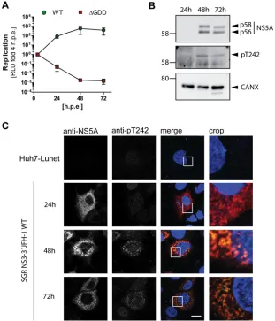
Subcellular localization of NS5A phosphorylated at T242.
After having validated
the genotype and phosphorylation specificity of pT242 detection by the antibody, we
next studied the kinetics of T242 phosphorylation and the subcellular localization of
NS5A phosphorylated at T242 (Fig. 5) upon transfection of a JFH-1 subgenomic reporter
replicon. Luciferase activity already peaked after 24 h, while NS5A became clearly
detectable only after 48 h (Fig. 5A and B). However, no clear kinetic difference was
observed in a Western blot comparing the pT242-specific antibody and the monoclonal
antibody 9E10, detecting both phosphoisoforms of NS5A, indicating that pT242
phos-phorylation was not delayed (Fig. 5B). In immunofluorescence analysis the pT242 signal
again was barely detectable at 24 h, even in cells with clear 9E10 signal, likely due to
the limited sensitivity and the slight background signal generated by this antibody.
JcR2a Lysates
58 kDa
58 kDa
pTM NS3-3´expression
A
B
C
n=2p58/p56
WT A D A D A D A D A D A D A D A D A D A D A D A D A D A D 0.0
0.5 1.0 1.5
JcR2a NS3-3' expression
n.d.
S
146
T1
6
4
T2
4
2
T2
4
4
T2
4
5
S
247
T2
4
9
T2
6
8
S2
7
2
N-te
rm
(242-249)
C-te
rm
(268 /
272) T3
4
8
T3
5
6
S
360
A D A D A D A D
T244 T245 S247 T249
A D A D A D T348 T356 S360
GD
D
Jc
R2
a W
T
A D A D A D
S146 T164 T242
A D A D A D A D N-term T268 S272 C-term
GD
D
JF
H-1 WT
A D A D A D
S146 T164 T242
A D A D A D A D
T244 T245 S247 T249
A D A D A D A D N-term T268 S272 C-term
A D A D A D T348 T356 S360
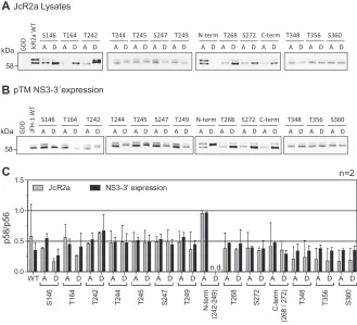
FIG 3Comparison of electrophoretic mobility of NS5A phosphoisoforms upon virus replication and
NS3-5B expression. Huh7-Lunet cells were electroporated with RNA encoding the full-length reporter virus JcR2a (A), or Huh7-Lunet T7 cells were transfected with pTM vectors expressing NS3-5B of isolate JFH-1 (B), either the WT or a mutant harboring the indicated phosphoablative alanine (A) or the phosphomimetic aspartic acid (D) mutations. Cells were lysed 72 h (A) or 24 h after transfection (B) and subjected to Western blotting using NS5A-specific antibody 9E10. The data shown are from one representative experiment (n⫽2). (C) Quantification of the p58/p56 ratios comparing JcR2a (gray bars) and pTM expression (black bars) based on the Western blots shown in panels A and B. n.d., not done, either due to replication defects (JcR2a) or due to the lack of separation between p56 and p58. The quantifications shown are from two experiments (n⫽2). Note for this and subsequent figures that the phosphoisoforms of NS5A of isolate JFH-1 have higher apparent molecular weights than 56 and 58 kDa but are still referred to as p56 and p58, respectively.
on November 6, 2019 by guest
http://jvi.asm.org/
[image:8.585.41.370.73.372.2]However, at 48 h and 72 h the pT242 signal was found in the same dot-like distribution
and at all sites with a strong total NS5A signal, suggesting that NS5A phosphorylated at
T242 has no particularly different localization compared to that of total NS5A (Fig. 5C). This
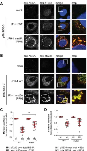
result was confirmed by a quantification of colocalization upon expression of NS3-5B in
Huh7-Lunet T7 cells (Fig. 6). Here we also included a replication-deficient NS5A mutant with
decreased PI4KA interaction, giving rise to increased p58 levels (PPH) (39). The pT242
signal widely and significantly colocalized with the total NS5A signal (Fig. 6A and C). In
contrast, not all NS5A colocalized with pT242, as expected for a p58-specific antibody.
For further control we used a pS235-specific antibody, which was previously shown to
be an efficient marker for hyperphosphorylated NS5A (Fig. 6B and D) (34), and obtained
a result similar to that for pT242. In the case of NS5A mutPPH, colocalization was even
more prominent, likely due to the higher signal intensity and to the more focused,
clustered perinuclear localization of the NS5A signal (Fig. 6A and B) (39).
In summary, NS5A phosphorylated at T242 is localized in a dot-like pattern
indis-tinguishable from total NS5A.
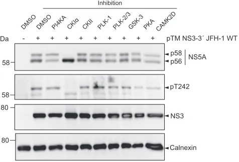
Impact of kinase inhibitors on T242 phosphorylation.
Several kinases have been
implicated in the regulation of NS5A phosphorylation. CKI
␣
appears to be the main
kinase involved in p58 formation, whereas CKII mainly contributes to basal
phosphor-ylation (26, 38). In addition, polo-like kinases have been shown to be involved in
hyperphosphorylation (35) and CAMK2
␥
and
␦
redundantly modulate S235
phosphor-ylation (28), whereas PI4KA is a negative regulator of p58 synthesis (39). To identify
kinases involved in phosphorylation of T242, we tested respective inhibitors regarding
their impact on p58 synthesis in general and on the pT242 signal in particular, both
relative to total NS5A (Fig. 7). Inhibition of CKI
␣
abrogated p58 synthesis completely,
H
u
h7-Lunet
SGR NS3-3´JFH-1 JcR
2
a
JC
1
p58 p56
pT242
NS3
CANX 58
kDa
58
80
80
pTM NS3-3`JFH-1
B
anti-pT242
non-phos JFH-1
pT242 JFH-1 synthetic peptide
(AS 236 - 251) 1000 500 250 100 50 10 0 ng
FIG 4T242 is exclusively phosphorylated in the hyperphosphorylated form of NS5A. (A) Different
concentrations of peptide either phosphorylated at position T242 or nonphosphorylated were titrated on PVDF membrane and incubated with purified, polyclonal anti-pT242 antibodies. (B) Huh7-Lunet T7 cells were either transfected with a pTM vector encoding JFH-1 NS3-3=or electroporated with in vitro transcripts encoding a bicistronic reporter replicon (SGR JFH-1), a full-length reporter virus genome (JcR2a), or an unmodified full-length virus genome (JC1). The cells were lysed 24 h posttransfection or 72 h postelectroporation, respectively, and analyzed by 7.5% SDS-PAGE/Western blotting using anti-NS5A (monoclonal 9E10)-, anti-anti-NS5A-pT242-, anti-NS3-, and anti-calnexin-specific antibodies. Shown is one representative experiment (n⫽2). Note the discrepancy between the 58-kDa molecular weight (MW) marker band (left) and the band referred to as p58/pT242 (right), which is due to a higher apparent MW of the NS5A phosphoisoforms in the case of JFH-1.
on November 6, 2019 by guest
http://jvi.asm.org/
[image:9.585.109.300.81.317.2]including a total loss of pT242 signal, suggesting that this kinase either directly
phosphorylates T242 or generates critical phosphorylation events essential for T242
phosphorylation. The PI4KA inhibitor increased the relative amount of total p58 and of
pT242. In contrast, inhibition of CKII, PLK-1, PLK-2/3, GSK-3, CAMK2D, and PKA had only
a minor impact on total p58 or pT242 (Fig. 7).
In conclusion, our data indicate that CKI
␣
is involved in phosphorylation at position
T242. In contrast, inhibition of other kinases previously linked to p58 synthesis did not
substantially reduce total p58 or pT242 levels.
Sequence determinants of T242 phosphorylation.
Several studies suggest that
p58 synthesis is based on saltatory phosphorylation events driven by CKI
␣
and further
involving priming phosphorylation by other kinases (25, 37). In particular, pS232, pS235,
and pS238 are very likely products of saltatory phosphorylation by CKI
␣
(34, 37, 48).
Phosphorylation of S232 in addition seems to be a main regulator of p58 synthesis,
FIG 5NS5A phosphorylated at T242 has no distinct subcellular localization compared to total NS5A.
Huh7-Lunet cells were transfected with in vitro transcripts encoding a JFH-1 reporter replicon (A to C) and analyzed for RNA replication (A), NS5A phosphorylation (B), and pT242 localization (C) at the indicated time points. (A) RNA replication of a WT replicon compared to a replication-deficient mutant (ΔGDD) is represented in relative light units (RLU) measured 24 h, 48 h, and 72 h posttransfection and shown as fold relative to values at 4 h posttransfection to normalize for transfection efficiency. (B) Transfected cells from panel A were lysed and analyzed by Western blotting for NS5A phosphorylation using anti-NS5A (monoclonal antibody 9E10)-, anti-NS5A-pT242-, and anti-calnexin (CAXN)-specific antibodies, as indicated on the right. (C) Cells were seeded on coverslips and fixed at the indicated time points postelectroporation. Total NS5A and NS5A phosphorylated at T242 were detected by immuno-fluorescence analysis using monoclonal antibody 9E10 (red) and anti-NS5A-pT242 (green), respectively. Nuclei were stained with DAPI. Scale bar, 10m. Shown is a representative experiment of two with comparable outcomes.
on November 6, 2019 by guest
http://jvi.asm.org/
[image:10.585.50.361.72.435.2]since phosphoablatant mutations at this site apparently abrogate
hyperphosphoryla-tion completely (19, 20). To understand the succession of phosphorylahyperphosphoryla-tion events
underlying phosphorylation at position T242, we mutated a number of potential
phosphoacceptor sites N and C terminally of this residue and analyzed the impact of
these mutations on the abundance of pT242 (Fig. 8 and 9). We included S146, since this
FIG 6Subcellular localization of WT NS5A compared to NS5A mutant with impaired PI4KA activation. (A
and B) Huh7-Lunet T7 cells were transfected with a pTM vector encoding NS3-5B of JFH-1 WT or mutPPH, seeded on coverslips, and fixed 24 h posttransfection. Total NS5A and NS5A phosphorylated at T242 (A) or S235 (B) was detected by immunofluorescence analysis using monoclonal antibody 9E10 (red) and anti-NS5A-pT242 or anti-NS5A-pS235 (both green), respectively. Nuclei were stained with DAPI. Scale bar, 10m. (C and D) Mander’s coefficient representing colocalization of pT242 with NS5A (M1) (C) or pS235 with NS5A (D) and vice versa (M2) was calculated for the pTM-transfected cells using Fiji. At least 10 cells per condition were analyzed.***,P⬍0.01 (homoscedastic, two-tailed t test).
on November 6, 2019 by guest
http://jvi.asm.org/
[image:11.585.61.352.67.572.2]site has been found to modulate NS5A hyperphosphorylation (25), as well as T164A,
which has been identified as a potential phosphorylation site in our study (Table 1).
Interestingly, none of the phosphoablatant mutations of serine residues N terminal of T242
abrogated T242 phosphorylation, although some of them strongly impaired overall p58
abundance (Fig. 8A and B). Even in the case of mutant S232A, which generated barely
detectable amounts of p58, the amount of pT242 phosphorylation relative to total NS5A
was not significantly reduced compared to that of the WT (Fig. 8B). In contrast, mutations
T242A and N-termA did not give rise to any pT242 signal, further proving the specificity of
the antibody, since neither mutant contains the specific phosphoacceptor site. In addition,
T244A and S247A strongly impaired T242 phosphorylation. While we cannot exclude at this
point that T244A interfered with pT242 recognition by the antibody, due to the close
proximity of both residues, this seems unlikely in the case of S247A, since T245A generated
a visible pT242 signal. Therefore, it seems likely that phosphorylation at S247 (and probably
T244) is a priming event crucial for T242 phosphorylation.
Taken together, these results so far suggested independence of T242
phosphory-lation from upstream phosphoryphosphory-lation in the Ser-rich cluster and vice versa. To further
validate this hypothesis, we analyzed the same samples with anti-pS235 (Fig. 8A).
Interestingly, p58 species phosphorylated at position S235 were found in all mutants,
even in the absence of detectable levels of total p58 (S232A), and in most cases
correlated with total p58 levels (34). This result indicated that T242 phosphorylation still
depends on phosphorylation events in the preceding Ser-rich cluster and might explain
the dependency of T242 phosphorylation on CKI
␣
. In contrast, lack of phosphorylation
of the Thr-rich cluster (N-termA) did not strongly impact the level of total p58 or pS235,
supporting the assumption that total p58 is mainly composed of phosphorylation
events in the Ser-rich cluster and that T242 phosphorylation only marks a minor species
of hyperphosphorylated NS5A.
We next assessed the impact of phosphomimetic mutations on T242
phosphoryla-tion (Fig. 9). Interestingly, S146D, which has been shown before to negatively regulate
p58 synthesis of JFH-1 NS5A (25), also reduced the relative amounts of pT242,
sug-gesting that phosphorylation at this site indeed is a master regulator of p58. All other
phosphomimetic mutations showed effects on total p58 synthesis very similar to those
of the phosphoablatant mutations: S232D again strongly reduced total p58 but not
pT242. T242D and N-termD did not react with the pT242-specific antibodies, likely due
to the loss of antigenicity. Surprisingly, T244D and S247D also negatively affected T242
phosphorylation, as did the phosphoablatant mutations. In case of pS235,
phosphor-DM SO
DM SO
PI4KACK Iα
CK II
PLK-1 PLK-2/3GSK-3 PKA
kDa - + + + + + + + +
Inhibition
pTM NS3-3´ JFH-1 WT +
CAMK2D
p58
p56 NS5A
58
pT242 58
80
NS3
Calnexin 80
FIG 7Impact of kinase inhibitors on phosphorylation of T242. Huh7-Lunet T7 cells were transfected with
a pTM vector encoding NS3-5B of JFH-1. Four hours after transfection the medium was replaced with new medium containing drugs targeting the indicated kinases that have been described to modulate NS5A phosphorylation. Cells were lysed 24 h after transfection and analyzed by Western blotting for NS5A phosphorylation using anti-NS5A (monoclonal antibody 9E10)-, anti-NS5A-pT242-, anti-NS3-, and anti-calnexin-specific antibodies as indicated. One representative of three experiments is shown.
on November 6, 2019 by guest
http://jvi.asm.org/
[image:12.585.89.327.73.235.2]ylation abundance was widely similar for the phosphomimetic (Fig. 9A) and ablatant
mutations (Fig. 8A). Altogether, the similarity of p58 phenotypes among
phosphoab-latant and phosphomimetic mutations indicated that replacement of the
phosphoac-ceptor site with a charged residue in fact did not rescue the priming function of a
phosphorylation event but rather interrupted the phosphorylation cascades up to this
point involved in p58 synthesis. This interpretation was confirmed by phosphatase
treatment of cleared cell lysates. While some of the aspartate mutations (S232D, S235D,
S238D, and T242D) induced an apparent decrease of the electrophoretic mobility of
NS5A p56, which has been interpreted before as a functional priming toward p58
synthesis (25), the same shift was still observed after treatment with lambda
phospha-tase (Fig. 9C). Overall these data demonstrated that a single mutation in this part of
LCS-I can strongly affect the electrophoretic mobility of NS5A. These results further
suggested that the increase in the apparent molecular weight of p56 in the case of
S232D, S235D, S238D, and T242D indeed was not due to further phosphorylation
events but based on the mutation itself. Interestingly, a similar shift in electrophoretic
mobility was found for phosphoablatant mutations of the same residues, S232A, S235A,
S238A, and T242A (Fig. 8A), indicating that the differences in the apparent molecular
weight are independent of the negative charge.
Taken together, the mutational analysis of phosphoacceptor sites N and C terminal
of T242 allowed several important conclusions. First, NS5A phosphorylated at pT242 is
FIG 8Impact of phosphoablative mutations of potential phosphoacceptor sites on T242
phosphoryla-tion. Huh7-Lunet T7 cells were transfected with pTM vectors expressing NS3-5B of isolate JFH-1, either WT or harboring the indicated phosphoablative alanine mutations. Mutant “N-termA” comprises all five serine and threonine mutations from T242 to T249. Mutant “C-termA” comprises both substitutions at positions T268 and S272. (A) Cells were lysed 24 h after transfection and analyzed by Western blotting using anti-NS5A (monoclonal antibody 9E10, upper)-, anti-NS5A-pT242-, anti-NS5A-pS235-, anti-NS3, and anti--actin (ACTB)-specific antibodies. (B) Ratio of p58 and pT242 to total NS5A. Intensities of the p58 bands, the pT242 bands, and the sum of p56 and p58 (total NS5A) as shown in panel A were quantified and used to calculate the indicated ratios. An additional staining of NS3 was performed subsequently on the same membranes previously used for detection of NS5A or pT242. These NS3 signals were used to obtain relative NS5A or pT242 levels for each membrane, which built the basis for pT242/NS5A levels. Bars represent mean values and SD from one representative experiment with three technical replicates. A second biological replicate was performed with a similar outcome (n⫽2). n.d., not detectable.***, P⬍0.01 (homoscedastic, two-tailed t test).
on November 6, 2019 by guest
http://jvi.asm.org/
[image:13.585.41.371.74.345.2]a minor subspecies of p58, since its abundance only marginally changed, even in cases
where total p58 levels were dramatically reduced (e.g., S229A and S232A). Second,
pT242 synthesis is regulated by C-terminal phosphorylation events, likely at position
S247 and potentially at T244. Third, phosphorylation of the p58 subspecies harboring
pS235/pS238, identified in previous studies (34), is regulated independently of pT242
and has only a minor impact on T242 phosphorylation. Fourth, some mutations of
phosphoacceptor sites in the LCS-I have a direct impact on the electrophoretic mobility
of NS5A p56, but the phosphomimetics most likely do not act as priming sites for
saltatory phosphorylation events. Overall, these data suggest that p58 is composed of
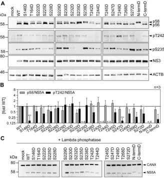
FIG 9Impact of phosphomimetic mutations of potential phosphoacceptor sites on T242
phosphoryla-tion. Huh7-Lunet T7 cells were transfected with pTM vectors expressing NS3-5B of isolate JFH-1, either WT or harboring the indicated phosphomimetic aspartic acid mutations. Mutant “N-termD” comprises all five serine and threonine mutations from T242 to T249. Mutant “C-termD” comprises both substitutions at positions T268 and S272. (A) Cells were lysed 24 h after transfection and analyzed by Western blotting using anti-NS5A (monoclonal antibody 9E10, upper)-, anti-NS5A-pT242-, anti-NS5A-pS235-, anti-NS3-, and anti--actin (ACTB)-specific antibodies. (B) Ratio of p58 and pT242 to total NS5A. Intensities of the p58 bands, the pT242 bands, and the sum of p56 and p58 (total NS5A) as shown in panel A were quantified and used to calculate the indicated ratios. An additional staining of NS3 was performed subsequently on the same membranes previously used for detection of NS5A or pT242. These NS3 signals were used to obtain relative NS5A or pT242 levels for each membrane, which built the basis for pT242/NS5A levels. Bars represent mean values and SD from one representative experiment with three technical replicates. A second biological replicate was performed with a similar outcome (n⫽2). n.d., not detectable.***,P⬍0.01 (homoscedastic, two-tailed t test). (C) Cells were lysed 24 h after transfection, and the cleared lysate was treated with lambda phosphatase. Phosphatase-treated lysates were analyzed by Western blotting using anti-NS5A (monoclonal antibody 9E10)- and anti-calnexin-specific antibodies (n⫽1). Note that dephosphorylation of NS5A by lambda phosphatase results in only one remaining band with an apparent MW similar to that of p56, but that different mutants show a consistently increased apparent molecular size.
on November 6, 2019 by guest
http://jvi.asm.org/
[image:14.585.42.370.71.427.2]differentially phosphorylated subsets of phosphoisoforms, with identical
electropho-retic mobility.
Assessment of phosphorylation events in the Thr-rich cluster for other HCV
genotypes.
Finally, we aimed to clarify whether T242 or other residues in the Thr-rich
cluster are phosphorylated in HCV genotypes other than 2a/JFH-1. We first assessed the
genotype specificity of the pT242 antibodies by expressing NS3-5B of gt1a, gt1b, gt2a,
gt3a, gt4a, and gt5a (isolates H77, Con1, JFH-1, S52, ED43, and SA1, respectively) in
Huh7 Lunet-T7 cells (Fig. 10A). A signal was obtained only for JFH-1/gt2a. To analyze
whether or not T242 was phosphorylated in genotypes other than 2a or whether the
pT242-specific antibodies were not able to bind to corresponding epitopes from other
genotypes due to high sequence divergence in this region (Fig. 10B), we synthesized
peptides for each genotype encompassing the region used for immunization, including
phosphorylation at T242. Indeed, the pT242 antibodies did not detect any genotype
other than 2a (Fig. 10C). We therefore adapted the peptide sequence in NS3-5B
expression constructs of all isolates to the JFH-1 sequence and analyzed T242
phos-phorylation again by Western blotting (Fig. 10D). We found an anti-pT242 positive
signal corresponding to p58 of the mutated ED43 (gt4a). This result provided the first
evidence that T242 also is phosphorylated in genotypes other than gt2a. However,
matching the surroundings of T242 to JFH-1 required 6 to 8 mutations (Fig. 10B), which
A P S L
K
A T C T
A N H D S P
D
A P S L
K
A T C T T
R H D S P
D
A P S L R A T C T T H S N T Y D
A P S L
K
A T C
Q
T H
R P H P
D
A P S L
K
A T C T
A P H D S P G
A P S L
K
A T C T T
Q G H H P
D
H77 Con1 JFH-1 S52 ED43 SA1
(56%)
(100%) (62%)
(62%) (50%) (62%)
236 242 251
mock H77
(1a)
Con1 (1b) JFH-1(2a) S52 (3a) ED43 (4a) SA1 (5a)
CANX pT242 NS5A 58
kDa
58
80
C
D
mock H77 WT mut. H77 Con1 WT Con1 K240R mut. Con1 JFH-1 WT S52 WT mut. S52 ED43 WT mut. ED43 SA1 WT mut. SA1
pT242 NS5A 58
58 kDa
CANX 80
anti-pT242
non-phos JFH-1
H77
Con1
JFH-1
S52
ED43
SA1
synthetic peptides (AS 236-251)
pT242
1000 500 100 50 10 0 ng
FIG 10Analysis of T242 phosphorylation in different HCV genotypes. (A) Huh7-Lunet T7 cells were transfected with pTM
vectors coding for the NS3-5B nonstructural proteins of the indicated HCV isolates. The cells were lysed 24 h after transfection and analyzed by 7.5% SDS-PAGE/Western blotting for total NS5A (monoclonal antibody 9E10), NS5A-pT242, and calnexin (CAXN). One representative experiment is shown (n⫽2). (B) Alignment of NS5A sequence from 236 to 251 (numbering according to NS5A JFH-1) from HCV isolates H77 (gt1a), Con1 (gt1b), JFH-1 (gt2a), S52 (gt3a), ED43 (gt4a), and SA1 (gt5a). Highlighted in boldface are the amino acids that are different from those of JFH-1. Numbers on the right represent the homology of the respective sequences to JFH-1. Note that the peptide sequence corresponding to JFH-1 was used to generate the pT242-specific antiserum. (C) Indicated amounts of peptides corresponding to the sequence of the indicated isolates and phosphorylated at position T242 were spotted on PVDF membrane and incubated with purified, polyclonal anti-pT242 antibodies. The nonphosphorylated peptide of JFH-1 served as a negative control for staining. (D) Huh7-Lunet T7 cells were transfected with pTM vectors coding for the NS3-5B nonstructural proteins of the indicated HCV isolates, either the WT or an NS5A mutated version. All mutated genotypes have amino acid sequence changed from their respective WT sequence to the sequence of JFH-1 in NS5A positions 236 to 251 (compare to panel B). In addition, a Con1 point mutant, K240R, was included to test if this change alone would allow antigen recognition. The experiments were performed twice with a comparable outcome.
on November 6, 2019 by guest
http://jvi.asm.org/
[image:15.585.42.423.74.345.2]could result in false-positive or -negative outcomes by interfering with potentially
varying phosphorylation cascades.
We chose an additional, unbiased approach and analyzed the immunoprecipitated
wild-type NS5A of isolates H77, Con1, S52, ED43, and SA1 by mass spectrometry (Fig.
11). We again used the NS3-5B expression model (Fig. 10A), since the WT variants of
these isolates are not replication competent and cell culture-adaptive mutations reduce
the abundance of p58 (21). Due to lower expression levels compared to those of our
previous experiment on JFH-1 (Fig. 1), we could not analyze p56 and p58 separately but
isolated the respective molecular weight range corresponding to both isoforms from a
gel (not shown) and subjected the contained tryptic peptides to MS/MS analysis (Fig.
11 and Table S2). We identified phosphorylated peptides in all cases carrying one or
two phosphate groups (Table S2). Interestingly, consistent phosphorylation for all
genotypes analyzed was found only within the Ser-rich cluster in LCS-I and at the N
terminus of domain III, albeit at various positions between 222 to 235 (Fig. 11B) and 364
to 399 (Table S2). In addition, we found phosphorylation sites with high probability in
domain I for gt1a at the same position as those for JFH-1 (146) (Table 1) (25) and for
gt3a (positions 87 and 117). Regarding the Thr-rich cluster, a double-phosphorylated
peptide was identified for gt4a with high probability of T242 phosphorylation (Fig. 11B
and Table S2). S249 was found phosphorylated in all genotypes, with a
phosphoac-Ser-rich cluster Thr-rich cluster
222 225 228 229 230 232 235 238 242 244 245 247 249
0.75 0.80 0.85 0.90 0.95
1.00 H77 Con1 S52 ED43 SA1
P
ho
sp
ho
(S
T
Y
) P
ro
ba
bilit
ie
s
B
A
AH
Domain I
LCS-IDomain II
LCS-IIDomain III
140 157 221 240 349 356
241 263 360 378
H77 (1a)
221 240
247 263 360 378
Con1 (1b)
272 294
272 294
79 108 221 240 360 378
113 122 263 277 379 404
272 288 385 404
S52 (3a)
224 240
ED43 (4a)
241 267
221 240 357 375
221 240
217 240 381 408
SA1 (5a)
FIG 11Phospho-mass spectrometry reveals additional phosphorylation events within the Thr-rich peptide. (A) Schematic of the different domains
of NS5A. The location of the identified phosphopeptides is indicated below for each isolate analyzed here, as given on the right. All numberings refer to amino acid positions within the respective genotype. (B) Phospho-(STY)-probability scores of different potential phosphoacceptor sites within the Ser-rich and Thr-rich cluster. Shown are only values above 0.75 that can be considered high-probability phosphorylation events at the respective site. The numbers below refer to all possible Ser/Thr phosphoacceptor sites existing in all analyzed genotypes. Note that phosphor-ylation at S249 was detected for all genotypes with a phosphoacceptor site at this position and that S52 (gt3a) and SA1 (gt5a) harbor a histidine (H) at position 249. The phospho-mass spectrometry was performed once.
on November 6, 2019 by guest
http://jvi.asm.org/
[image:16.585.45.494.83.419.2]cluster, carried a phosphate group in genotypes 1a, 1b, and 3a (Table S2). Only in the
case of gt5a/isolate SA1 did we find no indication of phosphorylation in the region
following the Ser-rich cluster, although nonphosphorylated peptides were detectable
(Table S2). In addition to these high-probability hits, several phosphorylated peptides
were found without clearly assignable phosphorylation sites, suggesting a mixed
population of peptides with variable phosphorylation events (Fig. 11A and Table S2). In
addition, we cannot exclude that many phosphorylation events remained undetected
in this analysis due to limited sensitivity, multiple and variable phosphorylation events
in one peptide, etc.
Taken together, our data suggest that the Thr-rich cluster is phosphorylated in the
case of JFH-1, and at least in genotypes 1a, 1b, and 4a, it is phosphorylated at different
positions. Combined evidence from genetics and mass spectrometry suggests that
T242 is phosphorylated in gt4a/ED43.
DISCUSSION
Our study aimed at a deeper understanding of NS5A phosphorylation, starting with
a phospho-mass spectrometry analysis of NS5A of isolate JFH-1 expressed in the
context of NS3-5B in Huh-7 cells, analyzing p56 and p58 separately. We thereby
identified seven phosphopeptides, with some of them having multiple potential
phos-phoacceptor sites. Overall, this result was well comparable with those of previous
studies, which already identified the peptides II, IV, V, VI, and VII (25, 33, 41).
The reason to use an expression system rather than a replication model in the first
place was to allow the analysis of replication-deficient mutants and inhibitors
interfer-ing with HCV replication. By comparinterfer-ing NS5A phosphorylation patterns from the
replicating full-length HCV and based on expression of NS3-5B, we observed overall
very similar results, as judged by p58/p56 ratios of numerous mutants. We only
observed a slight overrepresentation of the basal phosphorylated form in the
expres-sion system compared to replicating HCV, probably due to the higher expresexpres-sion level
of NS5A; therefore, limited expression of cellular kinases might restrict the amount of
p58 synthesis. These data overall confirmed that expression of NS3-5B indeed is an
excellent model to study NS5A phosphorylation, as already shown in previous studies
by us and others (21, 34, 39, 42, 48, 51, 53). In particular, we aimed to include an NS5A
mutant with strongly reduced PI4KA interaction, giving rise to enhanced p58 levels, to
understand the mechanism underlying the modulation of NS5A phosphorylation by
this lipid kinase. However, the pattern of phosphorylated peptides between the WT and
the NS5A mutant was comparable in mass spectrometry, suggesting that PI4KA does
not alter the overall phosphorylation pattern of NS5A but rather reduces the number
of hyperphosphorylated NS5A molecules by an unknown mechanism.
Interestingly, we identified for the first time a phosphopeptide restricted to the
hyperphosphorylated isoform of NS5A (peptide V), which was therefore the focus of our
study. This Thr-rich cluster in LCS-I was also found previously (25) and contains eight
potential phosphoacceptor sites, with one (T242) conserved throughout all genotypes.
A mutational analysis of all seven Ser/Thr residues individually revealed no impact on
RNA replication or particle production in cell culture, and a combination of only 5
phosphomimetic mutations in this region almost abrogated RNA replication (N-termD).
This result is in contrast to a very recent study demonstrating a strong reduction of RNA
replication for phosphomimetic mutations at positions T242 and T244, which was partly
restored by a triple mutation (54). The reason for these discrepant results is not clear so
far, but it might rely on the use of different Huh-7 cell populations with various
expression levels of cellular kinases regulating NS5A phosphorylation. Of note, both
studies agree on the lack of strong phenotypes for phosphoablative mutations,
includ-ing position T249 (52). Therefore, phosphorylation of individual residues within the
Thr-rich cluster in LCS-1 seems not to have an essential function in RNA replication or
virus production.
By generating phosphospecific antibodies against pT242, we were able to
on November 6, 2019 by guest
http://jvi.asm.org/
strate that this residue is indeed phosphorylated in a subspecies of NS5A molecules in
p58, thereby confirming the mass spectrometry data. T242 was phosphorylated upon
transient NS3-5B expression and virus replication, following the same kinetics as total
p58. The pT242-specific antibodies further allowed assessment of the determinants of
T242 phosphorylation. We used different inhibitors against the most prominent kinases
known to be involved in NS5A phosphorylation and monitored their impact on T242
phosphorylation. Only inhibition of CKI
␣
completely abrogated p58 and pT242
detec-tion, in line with previous studies (26, 37, 39, 49). This overall suggests that regulation
of phosphorylation at T242 is governed by the same mechanisms as total p58.
How-ever, it seems unlikely that T242 is directly phosphorylated by CKI
␣
, since the only
phosphoablative mutations found to abrogate pT242 phosphorylation, therefore being
suspect priming sites for CKI
␣
, are C terminal of this site (T244 and S247), whereas CKI
␣
requires an N-terminal priming site (pS-X-X-S) (55). Therefore, we cannot precisely
define the kinase responsible for T242 phosphorylation at this point.
Our data strongly indicate the existence of several independent subspecies of p58
with very different phosphorylation patterns. (i) Phosphoablative mutations at positions
S225, S229, and S232 almost entirely abrogate global p58 synthesis, as shown before
(19, 20, 25, 26), but have only a minor impact on T242 phosphorylation. Previous studies
furthermore, have shown that pS222, pS232, pS235, and pS238 are exclusively found in
p58 (25, 34, 48). Surprisingly, we found that even in the phophoablative mutants S225A,
S228A, and S229A, the p58-specific phosphosignal for S235 seems only slightly
af-fected, and even in S232A, lacking detectable levels of p58, small amounts of pS235
were detectable. This suggests that phosphorylation at position S235 occurs even if
global p58 levels are strongly impaired. (ii) In striking contrast, mutations T244A and
S247A abrogate T242 phosphorylation but hardly affect global p58 levels. Together,
these data suggest that pT242 defines a minor population of hyperphosphorylated
NS5A dependent on phosphorylation events in the N-terminal Ser-rich cluster, even
though we could not identify specific sites priming for downstream T242
phosphory-lation. These results challenge the concept of p58 as a homogenous NS5A isoform with
defined functions and rather suggest that p58 is a heterogeneous set of molecules that
are only defined by their homogenous apparent molecular weight. Individual
phos-phomimetic mutations at positions S235, S238, and T242 substantially increased the
apparent molecular weight of p56, resistant to phosphatase treatment, suggesting that
this increase was induced by the mutations itself, not by further phosphorylation. An
accumulation of five phosphomimetic mutations in the Thr-rich cluster even resulted in
a homogenous, phosphatase-resistant NS5A species indistinguishable from p58.
Alto-gether these data demonstrate that accumulation of mutations in LCS-I, particularly
adding negative charges, increases the apparent molecular weight of NS5A toward p58,
which can be achieved by a limited number of mutations at various sites in LCS-I.
However, these negative charges seem not to act as priming phosphorylation sites, as
suggested previously (25, 26), thereby challenging the use of these mutants in studies
addressing the function of distinctly phosphorylated NS5A subspecies. This observation
is supported by in vitro phosphorylation studies demonstrating that the
phosphomi-metic mutation S229E was unable to prime CKI
␣
activity compared to the pS229
peptide (37). Still, these phosphomimetic mutations demonstrate that a few changes in
this region suffice to generate p58. Therefore, p58 likely contains a heterogeneous
mixture of different phosphorylation patterns, either in the Ser-rich and/or in the
Thr-rich cluster of LCS-I. This model further explains the difficulties of identifying
distinct NS5A phosphorylation sites in mass spectrometry analyses, but why all these
various phosphoisoforms culminate in a distinct single p58 band in SDS-PAGE remains
puzzling.
What is the function of p58 in general and phosphorylation in the Thr-rich cluster of
LCS-I in particular? P58 has been regarded as the NS5A isoform inhibiting replication
and supporting assembly based on the fact that LCS-I is a hot spot for mutations
enhancing RNA replication of many HCV genotypes in cell culture but at the same time
abrogating or strongly reducing p58 synthesis (e.g., S225, S232, and S235) (19, 20,
on November 6, 2019 by guest
http://jvi.asm.org/
role of p58 in both virus production (26) and RNA replication (21).
Replication-enhancing mutations in LCS-I reduce p58 levels and abrogate activation of PI4KA,
which is an essential host factor of HCV RNA replication (51, 62–64) but too abundant
in Huh-7 cells (21). Therefore, the same mutations enhancing replication of many HCV
isolates by dampening PI4KA activity strongly decrease JFH-1 replication. JFH-1 also
requires PI4KA activation in Huh-7 cells, thereby reflecting more closely the WT
behavior of HCV in vivo. However, p232 and pS235, two of the phosphorylation events
required for PI4KA activation, are restricted to p58 (34). Even pS222, the major NS5A
phosphorylation identified in multiple studies (24, 25, 27, 65) and located at the N
terminus of the Ser-rich cluster in LCS-I, is mainly found in p58 (25). In addition,
mutations in the Ser-rich cluster have recently been shown to regulate multiple host
factor interactions required for replication and affect the membranous HCV replication
organelle (24, 46, 66). Taken together, these data strongly argue for essential functions
of distinctly hyperphosphorylated p58 subspecies of NS5A in HCV RNA replication.
We can only speculate on functions of phosphorylation events in the Thr-rich
cluster, since we found no impact of phosphoablative mutations on HCV replication
and virus production in cell culture. Previous studies have already shown that wide
parts of NS5A domain II (aa 246 to 304), encompassing the C-terminal half of the
Thr-rich cluster, can be deleted, with little impact on HCV replication or assembly (3).
However, this deletion mutant was strongly impaired in vivo due to increased
activa-tion of cytosolic pattern recogniactiva-tion receptors (17). In addiactiva-tion, the entire Thr-rich
cluster in LCS-I is part of the so-called interferon sensitivity-determining region (ISDR;
aa 237 to 276), which has been linked to the interferon response of HCV patients (67,
68). Therefore, it is tempting to speculate that phosphorylation events in the Thr-rich
cluster in LCS-I play a role in the interference with innate immune responses in vivo but
that these are not relevant for replication in cell culture due to the limited capacity of
Huh-7 cells to mount an innate immune response (17, 52). Our mass spectrometry data
on different HCV genotypes suggest at least a certain degree of conservation of
phosphorylation events in this region. T242 was found phosphorylated not only in gt2a
but also in gt4a, S249 in gt1a, gt1b (also found in reference 52), and gt4a, and S274 in
gt1a, gt1b and gt3a. These data, together with the complete conservation of T242,
suggest a functional relevance of phosphorylation events in the Thr-rich cluster.
In summary, our study identifies a novel minor subspecies of hyperphosphorylated
NS5A, marked by T242 phosphorylation. Our results challenge the concept of p58 being
a population of homogenously phosphorylated molecules but rather suggest a variety
of phosphoisoforms contained in p58 with identical electrophoretic mobility, likely
serving multiple functions. We thereby add a new level of complexity to NS5A
phos-phorylation and argue for a more differentiated view on p58.
MATERIALS AND METHODS
Cell lines.The Huh-7 ce