0022-538X/93/031357-08$02.00/0
Copyright© 1993, AmericanSocietyforMicrobiology
Mutations
in
Herpes Simplex Virus Type 1 Genes Encoding
VP5 and VP23
Abrogate
Capsid Formation
and
Cleavage of Replicated
DNA
PRASHANTDESAI, NEAL A. DELUCA, JOSEPH C. GLORIOSO, ANDSTANLEY PERSON* Department ofMolecular Genetics and Biochemistry, UniversityofPittsburgh,
Pittsburgh, Pennsylvania
15261Received 18September 1992/Accepted14December 1992
The herpessimplex virus type 1 capsid is composed of seven capsid proteins which are termed VP5, VP19c, VP21,VP22a, VP23, VP24, and VP26. Major capsid protein VP5 is encoded by the geneUL19. UL18, whose transcript is 3' coterminal with that of VP5, specifies capsid protein VP23. Vero cell lines have been isolated that are transformed with either theBglII N (UL19) or EcoRI G (UL16 to UL21) fragment of KOS. These cell lines,selected for the ability to support the replication of atemperature-sensitive VP5 mutant, were used to isolate VP5 and VP23 null mutants. The mutations in VP5 (K5AZ) and VP23 (K23Z) were generated by insertion ofthe lacZ gene at the beginning of the coding sequences of the genes. Both mutants failed to form plaques on the nonpermissive cell line, andtherefore, VP23, like VP5, is an essential gene product for virus replication. Both mutants expressed wild-type levels of infected-cell proteins upon infection of permissive and nonpermissive cell lines. However, the VP5 (150-kDa) and VP23 (33-kDa) polypeptides were absent in
lysates
prepared from K5AZ- and K23Z-infected Vero cells, respectively. No capsid structures were observed by electronmicroscopic
analysis
of thin sections ofK5AZ-and K23Z-infected Vero cells.Following sedimentation oflysates
from cellsinfectedby the mutants, capsid proteins were not observedinthefractions wherecapsidsnormally
sediment. The amounts of DNA replicated in the VP5 and VP23 mutant and inKOS-infected Vero cells were thesame asinpermissive cells. However, genomic ends were not evidentin Verocellsinfectedwith the mutants, suggesting that the DNA remains in concatemers and is not processed into unit length genomes.The herpes simplex virion is comprised of four distinct components,the core,capsid, tegument,and envelope. The electron-dense core contains the viral nucleic acid. An
icosahedral capsid structure encloses this core. An
amor-phous layer termed thetegumentsurrounds thecapsid, and the tegumentisenveloped byalipidmembrane inwhichare embedded the virus glycoproteins. The assembly of the
capsidisthereforeanessential step in virionmorphogenesis.
The herpes simplex virus type 1 (HSV-1) capsid is an icosahedral shell (42) which is composedof sevenproteins, VP5(150 kDa), VP19c (50 kDa), VP21 (43kDa),VP22a (40 kDa),VP23(33 kDa),VP24(24 kDa),and VP26(12 kDa)(9, 14, 16).There areapproximately 900 molecules of VP5 per
capsidforequineherpes virus1(24) andasimilarnumber for
HSV-1. Relative to900molecules ofVP5 percapsid, there
are approximately 1,400 of VP26, 900 of VP22a, 700 of VP19c, 400 of VP23, and 100 each of VP21 and VP24
(22-24).
Three types of capsids can be isolated from HSV-1-infected cells. Capsids are visualized as light-scattering bandsinsucrose gradientsandare designatedA, B, and C,
inorderofincreasingdistancesedimented (14). These differ inproteinandnucleic acidcompositionand intheireventual fate ininfected cells. A and Ccapsids aresimilar inprotein composition, but only C capsids contain viral DNA. B
capsidsdifferfrom A and Ccapsidsin that Bcapsidscontain an abundant VP22a protein inside the shell. VP22a is
inti-matelyinvolved in thepackaging of viral DNA(27, 33, 38).
VP22a may functionby formingascaffoldin theinnercapsid
*Corresponding author.
space,andacquisitionof DNAresults inconcomitant loss of VP22afrom thecapsidshell(4, 23). Conceptually,Bcapsids
may be the precursors to C capsids in the viral assembly
pathway,while Acapsidsmayresultfrom abortive attempts atDNApackaging.
The capsid shell is made up of 162 capsomeres (42) showingicosahedral symmetryoftriangulation class T=16. Forthisclass, 12 capsomeresarepentavalent(pentons)and the rest are hexavalent (hexons) (see, for example, refer-ences 4, 13, and 36). VP5, which represents 60% of the
capsidmass,is themajorcomponentof the hexons and may also form the penton structures (31, 40). The spatial
distri-bution of the other capsid proteins is largely unknown.
Hexamers areconnectedontheouter surfacebyY-shaped
structures, and these maybe composed of VP26, as
sug-gestedbyBakeretal. (4). Minor proteins(VP21andVP24)
mayaccountfor pentonsorspecialcapsidfunctions suchas
acquisition ofDNA
during
virusreplication
and release of DNAfollowing penetrationof thevirusintoaninfected cell. VP5extends from the outsidetothe inside of thecapsidand mustengageininteractions with itselftoform hexamers andwith the other majorproteins (VP19c, VP23, andVP26) to form thecompletecapsid (4, 13, 31,
36).
VP19c binds HSV-1 DNAand forms one or more covalent disulfide bondswith VP5 (6, 43). DNA is packaged within the centralregion
of thecapsid, occupyingthe entire spaceout tothebeginning
of thecapsidshell(5).
The functions and properties of the capsid
proteins
areunclear.Temperature-sensitive
(ts)
lesions in the genes that encode VP5 (29, 41)and VP19c(26)
result in the absence of mature capsids at thenonpermissive
temperature. UL26 encodesafamilyof relatedproteins,including VP22a,
which 1357on November 9, 2019 by guest
http://jvi.asm.org/
1358 DESAI ET AL.
areprocessed after translation (18-20, 27, 28). Temperature-sensitive mutants for UL26 gene products synthesize viral DNA and B capsids at the nonpermissive temperature but areunable to package this DNA to produce C capsids (33,
38).
The overall goal of our work is to construct null mutants for all of the capsid genes to identify the steps in the
assembly ofHSV-1 capsids and to characterize the role of eachproteininthis process. Null mutants are desirable since
theyallow comparisons to be made in the presence and in thecomplete absence of each protein. Complications due to
transdominant effects of mutant polypeptides are also avoided. Thecapsidproteins are expected to be essential for virus replication on the basis of thefinding that ts mutations have been mapped to genes that specify VP5, VP19c, and VP22a (26, 27, 41). Therefore, transformed cell lines that expressthesegenes in trans are required forpropagation of mutant viruses. In this report, we describe the isolation of transformed celllines that express VP5 and VP23, which are encoded by genes UL19 and UL18, respectively (10, 39). Furthermore, we have isolated and initiated the character-ization of null mutants for these genes.
MATERUILS AND METHODS
Cells and viruses. Human embryonic lung (HEL) cells were grown and maintained as described by Person et al.
(25). Vero cells and the E43 and G5 transformed cell lines were grown in Eagle's minimum essential medium supple-mented with 10% fetal calf serum (GIBCO-BRL) and
pas-saged as wereHELcells.Virus stocks of KOS (HSV-1)and of the VP5 (K5AZ) and VP23 (K23Z) null mutants were
preparedaspreviouslydescribed(25).The KOSisolate used was passaged twice from the P17 stock obtained from Priscilla A. Schaffer (Harvard University) in 1973. G5 was used as the permissive cell line for propagation of the null mutantsand gaveyields of approximately 200 PFU per cell. Plasmids. The EcoRI G (16.2-kb) and BglII N (5.7-kb)
fragmentsof KOS were cloned into theEcoRIsite of pUC9 and the BamHI site ofpUC19 and were designated pKEG and pKBgN, respectively (see Fig. 1). Plasmids are desig-natedas tothesourceof viral DNA(Kfor the KOS strain) andastotherestriction fragment, EG for the G fragment of restriction endonuclease EcoRI. A null mutation in VP5 was
generated first by partial digestion ofpKBgN with Ncol, which deleted 1.8 kb; this was followed by ligation with a
10-bpBglIIlinker(see Fig. 2A).ThelacZ gene, derived as a BamHI cassette from pSC8 (8), was then cloned into the
BglIIsite. Thiscassettedoes notcontain an initiation codon but does haveaTAAtermination codon. The construction of a VP23 null mutation was initiated by subcloning a 1.1-kb PvuI-StuIfragment intoSmaI-HincII-cut pUC19 after blunt ending thePvuIsite withT4 DNApolymerase (see Fig. 2B). This clone contains approximately 500 bp of the sequences both upstream and downstream of the VP23 initiation codon
(20). TheplasmidwascutwithNruI, which cuts in the11th
codon of VP23, and a 10-bp XhoI linker was inserted to createpKPSX.ThelacZ sequence from pSC8 was obtained as anXhoIcassetteandcloned into theXhoIsite ofpKPSX
such thatexpression of LacZ initiates at the VP23 ATG and the protein is synthesized fused in frame with the first 10 codons of VP23.
Antibodies. Monoclonal antibody LP12 directed to VP5 was a kind gift from Anthony C. Minson (University of
Cambridge). Polyclonal rabbit serum CP3 was prepared
against
VP23 isolatedby preparativesodiumdodecylsulfate-polyacrylamide gel electrophoresis (SDS-PAGE) of purified Bcapsids. This serum was prepared by East Acres Biolog-icals.Capsids werepurified by sedimentation ofinfected-cell lysates through 20 to50% sucrose gradients.
Construction of transformed Verocell lines. Theprocedure of DeLuca et al. (11) was followed for transformation of Vero cells.Subconfluent monolayers of Vero cells (2 x 10⁶) in 100-mm-diameter petri dishes were cotransfected with pSV2neo(1.5 ,ug) (35) and either pKBgN (5 ,ug) or pKEG (30
jig)
by usingthe calciumprecipitation procedure of Graham and Van der Eb (15). At 24 h after transfection, the cells were harvested and plated at a density of 4 x 10⁵/100-mm dish in medium containing 1 mg of G418(GIBCO-BRL) per ml. The medium was replenished every 3 days. G418-resistant colonies were harvested by using Perspex cloning chambersandtested for the ability to support the replication ofts1178 (41). Positive isolates were colony purified twice and then further characterized. Cell line E43 was trans-formed with the BglII N fragment, and G5 was transtrans-formed with theEcoRI-G sequence.Marker transfer of null mutations. Subconfluent monolay-ers of E43 or G5 cells in 60-mm dishes were cotransfected with 2
jig
of a linearizedplasmid and 10jig
ofKOS genomic DNA extracted from crude virionpreparations. The calcium precipitationtechnique of Graham and Van der Eb was used with the dimethyl sulfoxide enhancement modification of Stow and Wilkie (37). When foci were observed (48 h after transfection), the cell monolayers were harvested, freeze-thawed once, and sonicated and the titer of the total virus progeny was determined. To isolate viruses that expressed3-galactosidase,
plaques
wereallowed toform under meth-ylcellulose for 48 h after infection. This overlay was replaced with1% low-melting-point agarose in medium containing 300 ,ug of Bluo-gal (GIBCO-BRL) per ml. Plaques that stained blue were picked and further plaque purified three times by limiting dilution in 96-well trays.Southern blot hybridization. DNA sequences were de-tected after agarose electrophoresis as described by South-ern (34), by usingrandom-primer-labelled probes (12).
Sedimentation
analysis.
HEL cells(3 x 10⁷) were infected at a multiplicity of infection(MOI) of 10 PFU per cell. At6 h after infection, the monolayers were washed twice with tricine-buffered saline (25), methionine-free medium contain-ing 750 ,uCi of[35S]methionine
(NEN-DuPont) was added, and the mixture was incubated for a further 6 h. The cells were washed once with tricine-buffered saline, harvested, and pelleted at 3,500 xgfor 15 min at 4°C. The cell pellet wassuspended in capsid lysis buffer (500 mM NaCl, 20 mM Tris [pH 7.5], 1 mM EDTA [pH 8.0], 1% Triton X-100, 0.5 mM tosyl lysyl chloromethyl ketone) and left on ice for 5 min, and the nuclei were pelleted by centrifugation at 3,500 x g for 30 min at 4°C. The resulting nuclei were freeze-thawed three times and sonicated, and the DNA was sheared bybeing passed through a 26.5-gauge needle several times. The nuclear lysate was layered onto a 20 to 50% (wt/wt) sucrosegradient (in 300mM NaCl, 20 mMTris [pH 7.5], 1 mM EDTA [pH 8.0], 0.5 mM tosyl lysyl chloromethyl ketone) and centrifuged at 24,000 rpm for 100 min in a Beckman SW41 rotor at 4°C. Fractions were collected and precipitated with an equal volume of 16% trichloroacetic acid. The trichloroacetic acid-precipitated protein was resus-pended in sample buffer, and a fraction of this was analyzed bySDS-PAGE.Immunoprecipitation and SDS-PAGE. Radiolabelling of infected cells, immunoprecipitation of antigen, and
SDS-J. VIROL.
on November 9, 2019 by guest
http://jvi.asm.org/
E e B E
Ga
M
[image:3.612.60.317.68.245.2] [image:3.612.321.555.76.369.2] [image:3.612.60.300.652.716.2]T
T
...
ULI9 ULM UL21 UL16 UL17 UL18
30 35 40 45
I . . . I . . . . I
FIG. 1. TheEcoRI-G region of theKOS genome. The 16.2-kb EcoRI G fragment was cloned into pUC9. Restriction sites are shownin the topline for BglII(Bg) and EcoRI(E).Theremainder of the figure is from sequence analyses ofHSV-1 strain 17 (20, 21). ORFs and directions of translation are as indicated, as are tran-scripts (*-)and polyadenylation signals( t). Nucleotide numbers from theleft endof the strain17 genome aregivenatthebottomin
kilobases. The EcoRI G fragment specifies genes UL16 through UL21. UL18 and UL19 encode two of thecapsid proteins,VP23 and VP5,respectively. TheBglIINfragment of KOS specifies only the ULI9gene. Thetranscriptsweremapped byCosta etal.(10).
PAGE analyses were performed as described by Cai et al. (7).
RESULTS
Isolation of transformed cell lines expressing capsid genes. The capsid gene products are essential for virion
morpho-genesis,and therefore aprerequisite for the isolation of null mutants for these genes is construction of transformed cell lines that express these genes in trans. Vero cells were therefore cotransfected withpSV2neo(35)andplasmids that expressonly UL19 (pKBgN) orgenes UL16through UL21
(pKEG) (Fig.1)(21).Colonies thatwereresistanttothedrug G418 were harvested and tested for the ability to comple-mentthe lesion ints1178.This mutant is amemberof the G complementation group, and the lesion in it has been mapped to the VP5 locus (29, 41). Cell lines that tested
positiveforcomplementationwerecolony
purified
twice and then characterized further. Cell lines E43 and G5 were transformed with theBglII-N and EcoRI-G sequences,re-spectively. The cell lines were tested for the ability to support the growth of ts1178 at 34 and 39°C. E43 gave a
plaquingefficiencyforts1178(39/34°C) of62%, and G5 gave aplaquing
efficiency
of80% (Table 1). E43wasusedasthe permissive cell line for isolation ofa VP5 null mutant. G5 wasusedtoisolateaVP23 null mutantandtopropagate bothTABLE 1. Complementation ofts1178by transformed Vero cells
Platingefficiencyoftsll78aat:
Cell type
34'C(PFU/ml) 39°C(PFU/ml) 39/34-C (%)
Vero 2.1 x 10¹⁰ 6.5 x 10⁶ 0.031
E43 1.2 x 10¹⁰ 7.5 x 10⁹ 62
G5 2.5 x 10¹⁰ 2.0 x 10¹⁰ 80
aThe datashownareaverages oftwoexperiments.
A
| UL 19 ORF4122bp;
Bg N N
I I
N N Nr
N N
CCATGG C/AIGG
GAAGATC
NNr Bg I I I
N Ncol(partial cleavageandfill-in)
10bp BgIlllinkerligation
ULl9promoterand ...ATG GAA GAT CCC GTC...lac BamHtcassette
initiationcodon ...TACCT T CTAG GG CAG... frompSC8
ATGGAA GAT CCC GTC ...TAA
VP5 FUSIONCODONS lacZ
B
U118ORF -_lS N Nr Bg p x
Nrulcleavage 10bp Xhol linkerligation
UL18 promoter and ATG...ATCGC CC | AGGGCGAT.. cZXholcassette
tencodons TAC...'TAG CGG GAG OTI from pSC8
ATG...ATC GCC CTCGAGGGC...TAA
VP23codons 1 10
VP23 FUSION CODONS lacZ
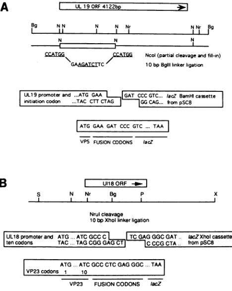
FIG. 2. Construction ofUL19 and UL18null mutationplasmids by insertion ofalacZcassette.(A) TheUL19ORF isdepictedatthe top in the left-to-right orientation. Immediatelyunderneath is the BglII N fragment of KOS thatwascloned into theBamHI site of pUC19. The five NcoIsites are shown(N).A 1.8-kb NcoIdeletion wasconstructedby apartialdigest.The UL19 start codon is within the most 5'NcoIsite.Followingafill-inreaction,a10-bpBglII (Bg) linkerwasaddedat thesiteof the deletion. Theresulting plasmid wasdigestedwithBglII,and theBamHIlacZcassettefrompSC8(8) wasintroduced intheorientation indicated. (B)TheUL18ORFis illustrated together with a map of restriction sites in this gene and thesurroundingsequences. A 1.1-kb PvuI(P)-to-StuI (S) fragment wascloned into theSmaI-HincII sites ofpUC19 aftertreatingthe PvuI end with T4 DNApolymerase. A10-bp XhoI (X) linkerwas
inserted into the NruI (Nr)siteof this plasmid. NruI cleaves this sequenceincodon 11 ofUL18. AnXhoI lacZcassettederived from pSC8wasthen cloned into thisXhoIsite.
mutants. Virus stocks of the VP5 null mutantprepared on E43 cellsgavelower titers andsmallerplaquesthan when G5 wasused. Chromosomal DNAextracted from G5was ana-lyzed by Southern blot hybridization using the EcoRI G fragment as aprobe. A 16-kbfragmentwas detected in G5 DNA, andthis waspresent at 10 to 20copies percell(data notshown).
Construction and isolation of VP5 and VP23mutants. The goal of these experimentswas toconstructmutations inthe VP5 and VP23 genes and transfer these into the KOS
genome by using E43 and G5 as permissive host cells. To constructthe VP5mutation,the5.7-kbBglIINfragmentwas
subcloned into theBamHIsite inpUC19
(Fig. 2). Figure
2A depicts the UL19 (VP5) open reading frame(ORF)
in the left-to-right orientation.BglII-Nof KOS contains five NcoIsites, and the most 5' site contains within it the ATG initiation codon of VP5(20).PartialNcoI
digestion
wasused tocleave at three of thesesites, andthis step resulted in aon November 9, 2019 by guest
http://jvi.asm.org/
1360 DESAI ET AL.
16.2 9.4 8.0
12
3
4
5
6
7
8
OMI.
6.9oft 5.7
4.3 _0
Om 3.3
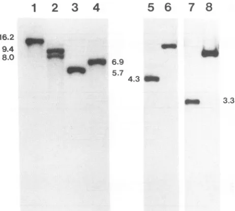
FIG. 3. Southern blot analysis ofVP5 andVP23 null mutants. DNAs(2 ,ug)extracted fromKOS(lanes1,3,5,and7)-infectedVero cells and K5AZ(lanes2 and4)-and K23Z(lanes6and8)-infected G5 cells were digested with restriction endonucleasesEcoRI (lanes 1 and2),BglII(lanes 3and4),StuI(lanes 5and6), and Sall (lanes7 and8). The resulting restriction fragmentswereseparatedon a1% agarosegel andtransferredtonitrocellulose. Filters werehybridized
to a 32P-labelled probe corresponding to BglII-N (lanes 1 to 4) sequences or the 1.1-kb PvuI-StuI (lanes 5 to 8) fragment. The numbersbeside thelanes indicate molecularsizesinkilobases.
deletion of 1.8 kb. The deletion was created so that the length of the resulting transcript would not be much greater than that ofwild-typeVP5.Inaddition,partial cleavagewas
employedbecause ofa concern that the sequence between the fourth and the fifth sites contains the VP23 promoter element. There is alsoapolyadenylationsignal at the end of the UL19 ORFwhichmaybe utilizedbyUL19and/orUL20. The 5' NcoI sitewas filled in, anda 10-bpBglIIlinkerwas added.Finally, thelacZgene, derivedas aBamHI cassette from pSC8 (8), was introduced into the BglII site in the
orientation shown. ThelacZ gene utilizes the VP5 promoter andinitiation codon. The LacZpolypeptideinthisconstruct starts at its ninth codon (GTG) and is preceded by three fusion codons. Translation termination occurs at the LacZ TAAcodon. The VP23 gene(UL18)(39)containsatits 10th and 11th codons anNruIsite whichwas usedto insert the lacZ gene (Fig. 2B). A 1.1-kb StuI-to-PvuI fragment was cloned into the
SmaI-HincId
sites ofpUC19.A 10-bpXhoI linkerwasintroducedinto the NruIsite,and thelacZgene, obtainedasanXhoIcassettefrompSC8,wascloned into this site such thatexpressionof this gene is drivenby the UL18 promoter and the LacZ polypeptide is synthesized as a fusion product containing the first 10 codons of VP23. Expression of LacZ was used to screen for mutant virusesbyvirtue of theirability to form blue plaques when overlaid withBluo-gal in cotransfection experiments.
Linearized plasmid DNA and intact KOS DNA were used tocotransfectE43 or G5 cells. Progenyvirus was harvested andassayed for theabilitytoformblueplaques in a Bluo-gal
overlay. Blue plaques were plaque purified three times before stocks were prepared. The VP5 and VP23 mutants were designated
K5A&Z
and K23Z, respectively. To confirm [image:4.612.64.295.75.283.2]the introduction of the lacZ gene into theseviruses, small
TABLE 2. Plating efficiency of viruses on transformed cells
Platingefficiency (PFU/ml)on: Virus
Vero E43 G5
KOS 2.6 x 10¹⁰ 1.9x 10¹⁰ 2.4 x 10¹⁰
K5AZ 7 x 10⁵ 6.7 x 10⁹ 1.03 x 10¹⁰
K23Z 3 x 10⁶ 2.9 x 10⁶ 1.51 x 10¹⁰
batches of viral DNAwereprepared from infected cells and analyzed by Southern blot hybridization. Results of this experiment are shown in Fig. 3. In the case of K5AZ, a deletion of 1.8 kb wascreated, followed by addition of the 3-kb lacZ fragment. Thisresulted in an overall insertion of 1.2 kb. Digestion of viral DNA withBglII resulted in the appearance of a band corresponding to the 5.7-kb BglII N fragment in KOS DNA (lane 3), and this increased in size to 6.9 kb in the K5AZdigest (lane4). The presence ofanEcoRI site in the lacZ gene resulted in cleavage of the EcoRI G fragment (16.2 kb) of KOS (lane 1) into 9.4- and 8-kb fragments in the mutant (lane 2). KOS DNA restricted with Stul andSall give risetobands thatwere4.3(lane 5)and 3.3 (lane 7) kb in size, respectively. These fragments both increased in sizeby 3 kb in the K23Z digest (lanes 6 and 8), owing to insertion of the lacZ gene.
Phenotypic
characterization of the mutants. The plating efficiency of the viruseswastested ondifferent cell lines to further confirm the genotype of thesemutants.Stocks of the mutantviruses prepared in G5 cells were plated on mono-layers of either Vero, E43, or G5 cells. The results of this assay are shown in Table 2. Asexpected, bothmutantsgave rise to plaqueson G5 monolayers, and onlyK5AZ formed plaques on E43 cells (transformed with only the BglII-Nsequence).The levels ofwild-typevirus in themutantstocks represent recombination between homologous DNA se-quences present in the mutant virus genomes and in the chromosome of thetransformed cellline. The percentages of
wild-type virus in K5AZ and K23Z stocks were 0.007 and
0.02%,respectively. Similar resultshave been obtained with otherindependentstocks of these viruses.
Thenextseriesofexperimentswascarriedout toconfirm the absence of either VP5orVP23in extractsfrom mutant-infected nonpermissive cells. Vero and G5 cells were in-fected withKOS, K5AZ,and K23Zat anMOI of 10PFU per cell andmetabolically labelled with [35S]methionine from 9 to 24hpostinfection. Totalinfected-cell polypeptideswere examined by SDS-PAGE (Fig. 4A). Results are shown for KOS-, K5AZ-, and K23Z-infected Vero(lanes 2 through4)
and G5 (lanes 6 through 8)cells, respectively. Both K5A&Z
(lane 3) and K23Z (lane 4) synthesized wild-type levels of
infected-cellproteins under nonpermissiveconditions. How-ever, the band corresponding to the 150-kDa VP5 protein was absent in K5AZ lysates and the 33-kDa band corre-spondingtoVP23wasabsent in K23Zlysates(arrowheads).
These proteins were present in lysates prepared from mu-tant-infected G5 cells (lanes7 and 8),albeit at lower levels. Apparently, the expression of VP5 and VP23 from the
integratedcopyofEcoRI-G in G5 cells is lower than expres-sion of these proteins from the wild-type virus. Expression
of VP23 in K5AZwasincreasedrelativetoits expressionin KOS(comparelanes 2 and3).The reason forthis is unclear. Thesameproteinlysateswere reactedwith eithera mono-clonalantibodytoVP5 (LP12)or apolyclonal rabbit serum raised against VP23 (CP3). Immunoprecipitates were ana-lyzed by SDS-PAGE, the results of which are shown in Fig.
J. VIROL.
on November 9, 2019 by guest
http://jvi.asm.org/
[image:4.612.316.556.89.153.2]A
-
_-_ _ _ _
,11 IIu
3m a 4E
1
2
3
4
5
6
7
8
B
[image:5.612.322.557.59.552.2]C
FIG. 4. Synthesis of viral polypeptides in K5AZ- and K23Z-infected cells. Vero (lanes1to4) and G5 (lanes5 to 8) cells were infected at anMOIof 10 PFU per cell with KOS (lanes 2 and 6), K5AZ (lanes 3 and7), or K23Z (lanes4and 8) or were mock infected (lanes1and5). Cellsweremetabolically labelled with [35S]methio-nine from 9to24 hafter infection. Lysates prepared from the cells weresubjected to SDS-12% PAGE (panel A) or precipitated with antibody LP12 (VP5) (panel B)or CP3 (VP23) (panel C), and the resulting immunoprecipitates were analyzed by SDS-12% PAGE.
4B and C. The results of the immunoprecipitation assay
further confirm these findings.
Ultrastructural
analysis
ofmutant-infected cells. TheVP5 and VP23 null mutants were examinedbyelectronmicros-copy to determine whether they form mature capsids or
accumulate visible capsid precursors under nonpermissive conditions. Mutantandparental viruseswereused to infect
permissiveandnonpermissive cells, andthe nuclei in tissue
sections were examined by electron microscopy. Samples
were fixed in glutaraldehyde at 12, 18, and 24 h after
infection. Resultsof the 12-h infection are showninFig. 5.
Capsidsweredetected inKOS-infectedVerocells(A)and in
G5cells infected with K5AZ
(B)
orK23Z(E).
Nocapsid-likestructure was observed in Vero cells infected with either
K5AZ (C) orK23Z (F). The nuclei of the mutant-infected
Vero cells appearedtobesimilar to those of mock-infected Vero cells
(D),
except for the condensation of chromatin material, a feature characteristic ofinfected cells. Most of the tissue slices showed capsids in preparations ofK5A&Z-and K23Z-infected G5 cellsorin KOS-infected Verocells.
However, no capsids were observed for approximately30
sections each of the mutant-infected Vero cells.
Sedimentation
analysis
ofmutants. The absence of maturecapsidsin Vero cells infected with K5AZ and K23Z does not
FIG. 5. Electron micrographs of thin sections of infected cells. Monolayers ofVerocellswereinfected with KOS(A),K5AZ(C), or K23Z(F)or weremock infected(D), and G5 cellswereinfected with K5AZ (B) or K23Z (E) at an MOI of 10 PFU per cell. At 12 h postinfection, cellswerefixed in2.5%glutaraldehyde in phosphate-buffered saline and embedded in Scipoxy 812 resin, and 50- to 70-nm-thick sectionsprepared for examination ina JEOL 100 CX electronmicroscope. Electron microscopic analysiswasperformed atthe StructuralBiology Center, University of Pittsburgh. Magni-ficationswere x27,750 for panelsA toD and x25,900 for panels E toF.Capsidsaremarkedwitharrowheads.
preclude the existence of capsid precursors which may accumulate in these cells. Therefore, nuclear extracts of mutant-infected cells were analyzed by sedimentation
through sucrose gradients. HEL cells were used as the
O'
I'Mon November 9, 2019 by guest
http://jvi.asm.org/
[image:5.612.78.287.64.370.2]1362 DESAI ET AL.
1 2 3 4 5 6 7 8 9 10 11 12 13 14
I~~*^ttei- * w * - - ~~~~VP5
.1. 9
- VP21
_-76' *<t - VP22
-VP22
-- VP24
- VP26
1
2
3
4
5
6
-
K
sof
i X -Q4&-q.
B
w-p a __ ..
FIG. 7. Southern blot analysis of replicated viral DNA. Mono-layers of Vero cellswereinfected with KOS(lane 2), K5AZ (lane3),
orK23Z(lane 5)or weremockinfected(lane1), and G5 cellswere infected withK5AZ (lane 4)orK23Z(lane 6)atanMOI of10 PFU percell. At 12 hpostinfection, total DNAwasextracted and 2.5,ug
wasdigested with BamHI. The restrictionfragmentswereseparated
on a1% agarosegeland transferredtonitrocellulose. The filterwas hybridized to a 32P-labelled probe corresponding to BamHI-K
sequences.Thejunction, K (5.9 kb), and terminal,Q(3.4 kb) and S (3.0 kb), fragmentsareindicated.
C
A
Agi4.o
FIG. 6. Sedimentation analysis of nuclear lysates from infected HEL cells. HELcellswere infected with KOS (A), K5AZ (B),or
K23Z (C)at anMOI of 10PFUpercell. Cellsweremetabolically labelled with[35S]methionine from 6to12 hpostinfection. Nuclear lysates were layered onto 20 to 50% sucrose gradients and sedi-mented at 24,000 rpm for 100 min in a Beckman SW41 rotor.
Fractionswerecollected, and proteinswereanalyzed by SDS-17%
PAGE. The direction of sedimentation is from left to right. The mobilities ofthesevencapsid proteinsaremarked. The positions of proteinstandards in orderofdecreasing molecularmass(200, 97, 68, 43, 29, 18, and14kDa)aremarked tothe left ofpanel A.
nonpermissive cellline. Cellmonolayerswereinfected with KOSorthemutantviruses andlabelled with[35S]methionine from 6to12 hpostinfection. Nuclear lysateswereprepared and layered onto 20 to 50% sucrose gradients. After sedi-mentation, fractions were collected and analyzed by
SDS-PAGE(Fig. 6). Fraction1,at the leftof Fig. 6, corresponds
tothetopof thecentrifuge tube. Two peaksof radioactivity wereobservedfor KOS-infectedcells (Fig. 6A), correspond-ingtothefaster-sedimenting C capsids (fraction 11) and the empty B capsids (fractions 6 through 8). Both B and C capsids contained VP5, VP19c, VP23, VP24, and VP26. VP22aand, perhaps,VP21weredetected only inBcapsids, asexpected. Webelieve that the double band of radioactiv-ity migratingat33 to37 kDa representsdifferent
phosphor-ylatedforms of VP23. VP23 in the virion has been shownto bephosphorylated (17).Thisprocessing activitymaybecell
type dependent, since VP23 in capsids isolated from the nuclei of Vero cellsmigratesas asingleband.Sedimentation analysis of the mutantlysates (Fig. 6B and C) revealed no
cosedimenting capsid proteinsin anyof the fractions. Pre-sumably, all of the capsid proteins were at the top of the gradient. Bands with mobilities corresponding to approxi-mately150 and 30 kDaappearedin allof the fractions forall of the virus-infected cells and thereforedonotcorrespondto
capsid proteins.
Viral DNAanalysis. Viral DNAanalysis experimentswere
carried out to determine the state ofviral DNA in
nonper-missive cells infected with the VP5 andVP23mutants.Both mutants failed to formmature capsids or capsid-like struc-tures upon infection of Vero cells. Nevertheless, the
pro-teins requiredforreplication, processing, and packaging of viral DNA are presumably expressed in both mutants. Southern blotanalysiswascarriedouttodetermine thelevel ofviral DNA replication and the processing of replicated DNA into unit length molecules in the absence of capsid structures. Vero and G5 cells were infected with KOS, K5AZ, and K23Z. Infected-cell DNA was extracted and analyzed bySouthern blothybridizationby usingthe BamHI Kjunction fragmentas aprobe (Fig. 7). InDNAs extracted fromKOS-infected Vero cells(lane 2) andG5 cells infected with K5AZ (lane 4) and K23Z (lane 6), the K junction fragmentand thetwoend, Q andS, fragmentsweredetected by hybridization, a result consistent with the presence of linearunit lengthgenomes.The presence ofmultiple bands ofQis duetotheheterogeneity ofasequencesatthatend of
the genome. Only the junction fragment was observed in
DNAextracted fromVero cells infected withK5AZ (lane 3) orK23Z(lane 5). Therefore, intheabsence of VP5orVP23 and, consequently, mature capsids, high-molecular-weight viral DNAisnotprocessedinto unit length molecules. The amountof thejunction fragment detected and, therefore, the extent of DNA replication are the same for mutant- and wild-type-infectedcells.
A
J. VIROL.
2a
on November 9, 2019 by guest
http://jvi.asm.org/
DISCUSSION
The ability toconstruct transformed cell lines that express agene in trans hasenabled the isolation of null mutations in essential genes of HSV-1. We have isolated Vero cell lines transformed with sequences that express VP5 alone (E43) or both VP5 and VP23 (G5). These cell lines were selected for by virtue of their ability to plaque ts1178, a ts mutant in VP5, at the nonpermissive temperature. Null mutants were gen-erated in the genes for VP5 (UL19) and VP23 (UL18) by insertion oflacZ sequences at codons 1 and 11, respectively. These were transferred into the KOS genome by homolo-gous recombinationby using transformed Vero cells as the permissive line. Both the VP5 (K5AZ) and VP23 (K23Z) mutants failed to replicate on Vero cells. Therefore, VP23, likeVP5, is an essential gene product. Only K5AZ replicated on the cell linetransformed with only the UL19 sequences. The absence of expression of the VP5 and VP23 polypep-tides in the respective mutants was confirmed by SDS-PAGE analysis of protein lysates and immunoprecipitation with antibodies specific for these proteins. Both mutants expressed wild-type levels of infected-cell polypeptides un-der nonpermissive conditions. Interestingly, expression of VP23 in K5AZ was increased relative to wild-type levels. The reasonfor this is unclear. The null mutants were unable to assemble capsids upon infection of nonpermissive cells, asjudged by electron microscopic analysis of thin sections. Byusingsedimentation analysis of nuclear extracts prepared from KOS-infected cells, B and C capsids were detected. However, foreither the VP5 or the VP23 mutant, no capsid structures were observed in the gradient at positions where wild-type capsids sediment.
The amount of DNA replication is normal in K5AZ- and K23Z-infected nonpermissive cells, but the DNA exists as concatemers that are not processed to genome length mole-cules. Normally during viral replication, concatemers are
cleavedinto unit length genomes, presumably at the time of
encapsidation. Itappears that if viral DNA is not packaged into capsids it is not cleaved (27, 32). In addition to null mutants for capsid genes that specify VP5 and VP23, ts mutations in VP19c and VP22a also result in accumulation of
concatemeric DNA (26, 27). Unlike the other three capsid
proteins, VP22a is found in empty but not in DNA-filled
capsids (33, 38). In addition to the capsid components, ts mutations that produce the same DNA-processing pheno-typehave beenidentified in five additional genes (1-3, 32). Therefore, capsid formation is necessary, but not sufficient, forcleavage ofDNAinto head-full lengths. While one of the noncapsid proteins may possess enzyme activity for
site-specific cleavage, the requirement of five gene products is
puzzling. Since DNAcleavage is linked to packaging, it is also surprising that one or more of these proteins have not beendetected ascapsid-binding proteins.
The HSV-1capsid is a complex structure composed of up tosevenproteins,each of which is important for assembly of the mature capsid, which is then incorporated into the virion. The steps thatoccurin the assembly of capsids have notbeenelucidated.Theisolation of mutations in each of the sevencapsidgeneproducts should enable one to identify the steps involved in capsid assembly and the role of each
protein in that pathway. ts lesions have been the primary sourceof mutations in the capsid genes, and these have been usedtocharacterize the roles of VP5, VP19c, and VP22a in capsidmorphogenesis. LesionsinVP22aresult insynthesis of viral DNA and emptycapsids; however, DNApackaging is defective in these mutants (27, 38). ts mutations in VP5
(41) and VP19c (26) result in lack of mature capsid forma-tion; nevertheless, small ring-like structures were observed by electron microscopic analysis of cells infected withts1178
(VP5) at the nonpermissive temperature (30). It is not clear whether this latter result is due to leakiness in the ts phenotype or a real precursor in the capsid assembly path-way. To overcome problems of leakiness in the ts pheno-type, null mutants need to be isolated so that the role of each gene can be defined in its complete absence. To this end, we have isolated null mutants for two important capsid proteins, VP5 and VP23. We have not detected any structure that resembles capsid precursors in infected cells. However, these studies require sedimentation parameters different
from those employed for complete capsids (20 to 50% sucrose gradients) and the experimental conditions, i.e., the salt concentration, may have to be varied to stabilize inter-actions between capsid molecules. The absence of large macromolecular structures in the sucrose gradients shown here does indicate thatVP5and VP23 play a major role in the assembly of capsids.
ACKNOWLEDGMENTS
This work was supported by Public Health Service grants from theNational Institutes ofHealth.
WethankBrendon Wahlberg forpreparation of figures.
REFERENCES
1. Addison, C., F. J. Rixon, J. W. Palfreyman, M. O'Hara, and V. G. Preston. 1984. Characterization of a herpes simplex virus type 1 mutant which has a temperature-sensitive defect in penetration of cells and assembly of capsids. Virology 138:246-259.
2. Addison, C., F. J. Rixon, and V. G. Preston. 1990. Herpes simplex virus type 1 UL28 gene is important for the formation of maturecapsids. J. Gen. Virol. 71:2377-2384.
3. Al-Kobaisi,M. F., F. J. Rixon,I.McDougall, andV. G. Preston. 1991. The herpes simplex virus UL33 gene product is required for the assembly of full capsids. Virology 180:380-388. 4. Baker, T. S., W. W. Newcomb, F. P. Booy, J. C. Brown, and
A. C. Steven. 1990. Three-dimensional structures of maturable and abortive capsids of equine herpesvirus 1 from cryoelectron microscopy. J. Virol. 64:563-573.
5. Booy, F. P., W. W. Newcomb, B.L. Trus, J. C. Brown, T. S. Baker, and A. C. Steven. 1991. Liquid-crystalline, phage-like packing of encapsidated DNA in herpes simplex virus. Cell 64:1007-1015.
6. Braun, D. K., W. Batterson, and B. Roizman. 1984. Identifica-tion and genetic mapping of a herpes simplex virus capsid protein that binds DNA. J. Virol. 50:645-648.
7. Cai, W., S. Person, C. DebRoy, and B. Gu. 1988. Functional regions and structural features of the gB glycoprotein of herpes simplex virus type 1: an analysis by linker insertion mutants. J. Mol. Biol. 201:575-588.
8. Chakrabarti, S., K. Brechling, and B. Moss. 1985. Vaccinia virus expression vector: co-expression of 1-galactosidase provides a visual screening of recombinant plaques. Mol. Cell. Biol. 5:3403-3409.
9. Cohen, G. H., M. Ponce de Leon, H. Diggleman, W. C. Lawrence, S. K. Vernon, and R. J. Eisenberg. 1980. Structural analysis of the capsid polypeptides of herpes simplex virus types 1 and 2. J. Virol. 34:521-531.
10. Costa, R. H., G. Cohen, R. Eisenberg, D. Long, and E. Wagner. 1984. Direct demonstration that the abundant 6-kilobase herpes simplex virus type 1 mRNA mapping between 0.23 and 0.27 map units encodes the major capsid protein VP5. J. Virol. 49:287-292.
11. DeLuca, N. A., A. M. McCarthy, and P. A. Schaffer. 1985. Isolation and characterization of deletion mutants of herpes simplex virus type 1 in the gene encoding immediate-early regulatory protein ICP4. J. Virol. 56:558-570.
on November 9, 2019 by guest
http://jvi.asm.org/
1364 DESAI ET AL.
12. Feinberg, A. P., and B. Vogelstein. 1984. A technique for radiolabellingDNArestrictionfragments tohighspecific activ-ity. Anal. Biochem. 137:6-13.
13. Furlong, D. 1978. Direct evidence for 6-fold symmetry of the herpesvirus hexon capsomers. Proc. Natl. Acad. Sci. USA 75:2764-2766.
14. Gibson, W., and B. Roizman.1972.Proteins specified by herpes simplex virus. VIII. Characterization and composition of mul-tiple capsid forms of subtypes1and2. J.Virol. 10:1044-1052. 15. Graham, F. L., and A. J. Van der Eb. 1973. A newtechniquefor
theassayofinfectivity of human adenovirus5 DNA. Virology 52:456-467.
16. Heilman, C. J., Jr., M. Zweig, J. R. Stephenson, and B. Hampar. 1979.Isolation ofanucleocapsidpolypeptide of herpes simplex virus types 1 and 2 possessing immunologically type-specific andcross-reactive determinants.J. Virol. 29:34-42.
17. Lemaster, S., and B. Roizman. 1980. Herpes simplex virus phosphoproteins. II. Characterization of the virion protein kinase andof thepolypeptides phosphorylated in the virion.J. Virol. 35:798-811.
18. Liu, F., and B. Roizman. 1991. The promoter, transcriptional unit, andcodingsequencesofherpessimplex virus1 family 35 proteinsarecontained within andin frame with theUL26open reading frame.J.Virol. 65:206-212.
19. Liu, F. Y., and B. Roizman. 1991. Theherpessimplex virus type 1 gene encoding a protease also contains within its coding domain the gene encoding the more abundant substrate. J. Virol. 65:5149-5156.
20. McGeoch, D. J., M. A. Dalrymple, A. J. Davison, A. Dolan, M. C. Frame, D. McNab, L. J.Perry,J. E. Scott, and P.Taylor. 1988. ThecompleteDNA sequenceof the long unique regionin the genome of herpes simplex virus type 1. J. Gen. Virol. 69:1531-1574.
21. McGeoch, D. J., S. K. Weller, and P. A. Schaffer. 1990. Herpes simplex virus,p. 1.115-1.120. In S. J. O'Brien (ed.), Genetic maps, Cold Spring Harbor Laboratory Press, Cold Spring Harbor,N.Y.
22. Newcomb, W. W.,and J. C. Brown. 1989. Useof Ar+plasma etching to localize structural proteins in thecapsid of herpes simplexvirus type 1. J.Virol. 63:4697-4702.
23. Newcomb, W. W., and J. C. Brown. 1991. Structure of the herpes simplex virus capsid: effects of extractionwithguanidine hydrochlorideandpartial reconstitution of extracted capsids.J. Virol.65:613-620.
24. Newcomb, W. W., J. C. Brown, F. P. Booy, and A. C. Steven. 1989.Nucleocapsidmassandcapsomerprotein stoichiometryin equine herpes virus type 1: a scanning transmission electron microscopic study.J. Virol. 63:3777-3783.
25. Person,S., R. W. Knowles, G. S. Read, S. C. Warner, and V. C. Bond. 1976. Kinetics of cell fusion induced by a syncytia [sic]-producingmutantofherpessimplex virustype 1. J.Virol. 17:183-190.
26. Pertuiset, B., M. Boccara, J. Cebrian, N. Berthelot, S. Chouster-man, F. Puvion-Dutilleul, J. Sisman, and P. Sheldrick 1989. Physical mapping and nucleotidesequenceofaherpes simplex virus type 1 gene required for capsid assembly. J. Virol. 63:2169-2179.
27. Preston, V.G., J. A. V. Coates, and F. J. Rixon. 1983. Identifi-cation and characterization of a herpes simplex virus gene product required for encapsidation of virus DNA. J. Virol.
45:1056-1064.
28. Preston, V.G., F. J. Rixon, I. M. McDougall, M.McGregor, and M. F. AlKobaisi. 1992.Processing of theherpessimplex virus assemblyprotein ICP35 nearitscarboxyterminal endrequires theproduct of thewhole of the UL26reading frame.Virology 186:87-98.
29. Schaffer, P. A., G. M. Aron, N. Biswal, and M. Benyesh-Melnick. 1973. Temperature-sensitive mutants ofherpes sim-plex virustype1: isolation,complementation and partial char-acterization. Virology52:57-71.
30. Schaffer, P. A., J. P. Brunschwig, R. M. McCombs, and M. Benyesh-Melnick.1974. Electronmicroscopic studies of temper-ature-sensitivemutantsofherpes simplex virustype 1.Virology 62:444-457.
31. Schrag, J. D., B. V. V. Prasad, F. J. Rixon, and W. Chiu. 1989. Three-dimensional structure of the HSV-1 nucleocapsid. Cell 56:651-660.
32. Sherman,G.,andS. L. Bachenheimer.1987.DNAprocessingin temperature-sensitive morphogenic mutants ofHSV-1. Virol-ogy158:427-430.
33. Sherman, G.,andS. L. Bachenheimer.1988.Characterization of intranuclear capsids made by ts morphogenic mutants of HSV-1.Virology 163:471-480.
34. Southern,E. M. 1975. Detectionofspecific sequences among DNAfragments separated by gel electrophoresis. J.Mol. Biol. 98:503-517.
35. Southern,P.J.,and P. Berg.1982.Transformation of
mamma-lian cellstoantibioticresistance withabacterialgeneunderthe controlof theSV40 earlyregionpromoter.J.Mol.Appl.Genet. 1:327-341.
36. Steven, A.C.,C. R. Roberts, J. Hay, M. E.Bisher,M.Pun, and B. L.Trus.1986.Hexavalent capsomersofherpessimplex virus type2: symmetry,shape,dimensions,andoligomericstatus.J. Virol. 57:578-584.
37. Stow, N., and N. M. Wilkie. 1976. Animprovedtechniquefor obtainingenhanced infectivity with herpessimplex virustype 1 DNA. J. Gen. Virol.33:447-458.
38. Rixon, F. J., A. M.Cross, C. Addison, and V.G.Preston.1988. The products of the herpes simplex virus type 1 gene UL26 which are involvedinDNApackaging arestrongly associated withempty but not with full capsids. J. Gen. Virol. 69:2879-2891.
39. Rixon, F. J., M. D. Davison, and A. J. Davison. 1990. Identifi-cation of the genes encoding two capsid proteins of herpes simplexvirus type 1bydirect aminoacidsequencing. J. Gen. Virol. 71:1211-1214.
40. Vernon,S.K.,M. PoncedeLeon, G.H.Cohen,R.J.Eisenberg, and B.A. Rubin. 1981.Morphologicalcomponentsof herpesvi-rus.III. Localization of herpessimplex virustype 1 nucleocap-sidpolypeptides by immune electron microscopy.J.Gen. Virol. 54:39-46.
41. Weller, S. K., E. P.Carmichael,D. P.Aschman, D. J.Goldstein, and P. A.Schaffer. 1987.Geneticandphenotypic characteriza-tion ofmutantsinfour essentialgenesthatmap tothe left half of HSV-1 ULDNA.Virology 161:198-210.
42. Wildy,P., W. C. Russell, and R. W. Horne. 1960. The morphol-ogyofherpes virus. Virology 12:204-222.
43. Zweig,M., C. J.Heilman, and B. Hampar. 1979. Identification ofdisulfide-linked protein complexes in the nucleocapsids of herpes simplex virustype2.Virology 94:442-450.
J.VIROL.