The Herpes Simplex Virus
1Gene
Encoding
a
Protease
Also
Contains
within Its
Coding Domain the Gene Encoding
the More
Abundant Substrate
FENYONG LIUAND BERNARD ROIZMAN*
TheMarjorie B. Kovler Viral Oncology Laboratories, The University of Chicago,
910 East58th
Street,
Chicago,
Illinois
60637Received25April 1991/Accepted 20 June 1991
The herpessimplexvirus 1openreading frames UL26 andUL26.5are3' coterminal.The larger,UL26open
reading frameencodesaprotein approximately 80,000 inapparentmolecular weight and containsthepromoter
and codingsequence of theUL26.5gene, whichspecifiesacapsidprotein designated infected cellprotein 35.
The largerproduct containsinits entirety the amino acidsequenceof the smaller protein. Wereportthat the
UL26 gene encodes a protease which catalyzes its owncleavage and thatof the more abundant product of
UL26.5. By inserting the coding sequence of an epitope to a cytomegalovirus monoclonal antibody and
homologs oftheimmunoglobulin Gbinding domain of staphylococcal protein A into the3' termini of the coding domainsofthetwoopenreadingframes, weidentified both products of the cleavage and determined that the
cleavage siteisapproximately 20amino acidsfromthe carboxyltermini of both proteins.
In this report, we describe the identification ofa herpes simplexvirus (HSV) protease and its substrate. This is the
firstherpesvirusprotease identifiedtodate. The remarkable properties of this protease are that (i) it catalyzes its own cleavage as well as that ofa more abundant, smaller
sub-strate and (ii) the gene encoding the smaller substrate, although transcribed and translated independently of the
pr&efase, is nevertheless entirelycontained within thegene encoding the latter. The relevant background and experi-mental design of this studymaybe summarizedasfollows.
Inthecourseof studiesontheproteins forming theherpes simplexvirus capsid, itwas noted thataprotein present in empty capsids lacking DNA, designated virion protein (VP22), wasreplaced in full capsids containing DNA byan apparently smaller, faster-migrating protein with similar
characteristics anddesignatedVP22A(8, 9).In thecourseof
enumeration of infected cellproteins, VP22and itsputative product, VP22A,weredesignated the infected cell protein 35 (ICP35) (3, 4). ICP35 has been extensively studied: it
con-sists ofafamily of highly heterogeneous proteins which form
numerous bands in both one-and two-dimensional
separa-tions. Pulse-chase experiments suggested that the major
translational products of the gene encoding these proteins
form bands designated ICP35c and -d, whereas the
post-translationally modified products form bandsICP35e and -f
(4). It hasrecentlybeensuggested thatICP35functions as a scaffolding protein (15).
Intheexpositionof the nucleotidesequenceof the HSV-1
genome, McGeoch et al. (14) assigned ICP35 to the open
reading frame UL26. The assignment was based on the observation that a temperature-sensitive mutation affecting
theprocessingofICP35andaccumulation ofmaturecapsids mapped in the UL26 open reading frame (18). However,
when studies on this gene were initiated (12), it became apparent that this open readingframe predicted to encode 635 amino acidswasmuch toobigtoencodeICP35 with an
apparent molecular weight of approximately 45,000. Our
* Correspondingauthor.
studies (12) showed thatUL26 consists oftwoopenreading frames, each encoded by its owntranscript (Fig. 1, lines 4 and5). Thus, UL26 encodes aprotein approximately 80,000
in molecular weight. In anticipation of the results to be
presentedin thisreport,we shall refertothis protein asthe protease, or Pr. The smaller reading frame, schematically
shown in Fig. 1, line 5, was designated UL26.5. The two
open reading frames share all of the amino acid sequences
encoded in UL26.5, and the promoter ofthe smaller open
reading frame is contained in the 5' coding and noncoding
domains ofUL26.
In this report, we show that the product of UL26 is a
protease. Indesigning these studies, we took advantage of
three observations: (i) a monoclonal antibody derived by
Lenore Pereira and analyzed jointly in our laboratories
reacts with the HSV-1 products of both UL26 and UL26.5
butnot with those of the HSV-2, (ii) UL26.5 is abundantly expressed in transfected cells, and (iii) HSV-1 strain F
[HSV-1(F)]istemperaturesensitive in the a4gene(10),and
atthenonpermissivetemperature itinducesagene
promot-ers (17) andexpressesprimarily agenes (1). We noted that
HSV-1(F) atthat temperature transactivates the UL26 and
UL26.5 open readingframes transfected into the samecells butnotitsownUL26orUL26.5openreadingframes(12).To
systematically analyze the site and attribute the cleavage
reactiontotheproductof theUL26openreading frame,we
used a movable epitope, that is, a sequence encoding 20 amino acidsspecifyingtheepitopeofamonoclonalantibody against a human cytomegalovirus (CMV) protein. This
epitope was inserted wherevernecessaryto trace the
prod-ucts of the reaction.
MATERIALS ANDMETHODS
Virus and cells. The properties of HSV-1(F) and
HSV-2(G),theprototypeHSV-1 and HSV-2strains,respectively,
used in thislaboratoryand the maintenance andpropagation
of the thymidinekinase-minus baby hamsterkidney (BHK)
cells have been describedpreviously (1, 6, 20).
Monoclonal antibodies. Monoclonal antibodies H725 and
5149
0022-538X/91/105149-08$02.00/0
CopyrightC 1991, American SocietyforMicrobiology
on November 10, 2019 by guest
http://jvi.asm.org/
El
~~~UL
ED
O I l_i_u
3 5
IB0
H
SMsflKA
L1 I 1 L- I I I I I2 122l
E IX B BaHBs SMsPI'
1 ISO 282138
l E
pRB4057E'-pRB4060 O
pRB4058
pRB4093
pRB4026g-pRB4092
ED
pRB4096
CD
pRB4102
pRB4080
pRB4140
pRB4184
pRB4185
pRB4103
pRB4090
pRB4186
pRB4188
pRB4213
pRB4214
pRB4215
ED
1000 1_ 2~138
X lIIBIIBIS l
X BBaHBs SMsPK
x
x
H
x
x
Ms 1 4
BS Ms
S
H Ms i
I I
H X-P
p. F
I *.
H p* F
H P*
Bs K
x K
- _
_~~~~
X
p
_p
_X P.
x
7p
x
x~~~~P.
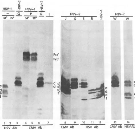
FIG. 1. SequencearrangementoftheHSV-1genome,p(
ofthe UL26 andUL26.5 openreadingframesand theirtran and structures ofthe test plasmids constructed for these s
Line1,schematicrepresentationof thesequencearrangemen
HSV-1genome.ULandUsrefer tothelonguniqueandshort
sequences flanked by terminal inverted repeats shown a
rectangles. Lines2and3, genomemap positions, nucleotid bersrelative totheapproximatetranscriptioninitiationsitec
indicatedbyletter Iat +1,andrestrictionendonucleasesite
HSV-1EcoNI-PstIDNAfragment.Alsoshownare theposit
thetranslationalterminationcodon(T)and thesinglepoly(A (A) which serve both the UL26and UL26.5RNAs. Lines 4 codingdomains oftheUL26and UL26.5openreadingframe rectangles).The numbersrefer tothepositionsofthetransc initiationsite,thetranslationinitiationandterminationcodoi
thepoly(A)signal for bothopen readingframesrelative to
tide +1 of UL26. Line 6, restriction endonuclease map drl
scale with reference to lines A through Z, which are sch
representations ofthe HSV-1 sequences containedin the F constructsusedinthestudiesdescribed inthisreport.Const
of the plasmids shown schematically in lines A throug] describedinMaterialsandMethods.Thesource ofthe a4pr4
(openrectangle)shown inplasmids B, J, L, P, W,X,and Z
U-a CH28-2 to ICP35 and CMV
glycoprotein B, respectively,
wereobtained from Lenore Pereira and have been described0~
previously
(3,
4,12).
0.35~5 Construction of plasmids. Construction of plasmids I g ; (pRB4026), A (pRB4057), C (pRB4058), B
(pRB4060),
E (pRB4093), L(pRB4096),
N(pRB4102),
and P(pRB4080)
PS has been described previously (12). Two short double-stranded DNA fragments were made by synthesizing com-plementary strands of each in an Applied Biosystems 380B DNA synthesizer and annealing the two strands.Sequence
_j A (5'-AGGGACAGAAGCCCAACCTGCTAGACCGACTG
PS
CGACACCGCAAAAACGGGTACCGACAC-3')
encodes Xj theepitope of monoclonalantibody CH28-2(12,16).Plasmid PS Q(pRB4140)
was derivedby inserting oligonucleotide
A-j
Ps with its complement into theunique PmlI site ofplasmidA._j Sequence C (5'-TCGACGTTGACACGGCCCGCGCCGCC
Ps
GATTTGTTCGTCTCTCAGATGATGGGGGCCCGCC
pr ACGTGTGA-3') encodes the authenticUL26sequence from thePmlI sitetothe translation termination site but with the
J addition ofa new PmiI site between the
carboxyl-terminal
Ps amino acid and thestop
codon of theUL26
open reading_j frame. In addition, the sequence GTG atthe authenticPmlI Ps site has been changed to GTC. Insertion of sequence Cinto Ps the
unique
PmlI
site of theUL26
openreading
frame in the PsJ properorientation resulted in the creation ofanewPmlI site
Ps between the carboxyl-terminal amino acid and the stop
J codon of UL26. The original authentic PmlI site was
de-Ps stroyed without changing the UL26 amino acid sequence
J since the codons GTG and GTC encode the same amino
s acid.
Therefore,
theneteffectof theinsertion ofoligonucle-Ps otide C with its
complement
totheUL26
openreading
framewas that UL26 had two additional amino acids between its authentic carboxyl-terminal amino acid and its stop codon which were encoded by the new created PmlI recognition sequence. Plasmid R (pRB4184) was derived by
inserting
sequence C into thePmlI site ofplasmid E(pRB4093), and
Ps plasmid S (pRB4185) was derived by inserting sequence A -* into the PmlI site of plasmid R. Plasmids T, U, V, and W
)s
(pRB4103, pRB4090, pRB4186, and pRB4188)were derived from plasmids B and S. Plasmids X, Y, and Z(pRB4213,
Ps
pRB4214, and pRB4215) were constructed by inserting in___X frame into theUL26 open reading frame at the site between
Ps the3'end of the CMV sequence and the stop codon eithera -j sequence encoding 256 amino acids comprising five
ho-Ps
mologs of immunoglobulin G (IgG) binding domains of ositions staphylococcal protein A or a sequence encoding 129 amino vscripts, acids and comprising two such domains. These sequencesstudies. were theBclI-HincII andHindlIl-HinclI fragments, respec-itofthe
tively,
of theprotein
A gene fusion vectorpRIT5(Pharma-unique
cia, Piscataway,
N.J.). Thevectorforplasmids T, U, V, and Ls open Y was derived from pGEM3Zf(+) (Promega, Madison,lf UL2m
Wis.); these plasmids could be used as templates for in vitro ofthe transcription by T7 or SP6 RNA polymerase. The vectors fortions of all other
plasmids
were derived frompUC18.
All insertion signal sites ofsequencesA and C into theplasmidsweresequencedand 5, toverifythat the CMVepitopeandtheamino acidsequence s(filled encoded by sequence C were inserted in frame with the
:ription UL26 open reading frame.
ns,
and
nucleo-awn to
iematic
)lasmid
ruction h Z is
omoter
7wasa
BamHIZ DNA fragment (17) inserted in proper transcriptional
orientation.The CMVepitopeis shownasafilled oval.
Oligonucle-otideC withitscomplement is shownas afilled hexagon,andthe
new created PmI site is marked (P*).Restriction endonuclease
sites: B, BamHI; Ba, Ball; Bs, BstEII; E, EcoNI; H, HpaI; K, KpnI;Ms,MstII; P, PmII;Ps,PstI; S, Sall;X, XcmI.
1
2
3
4
5
6
A
B
C
E
J
L
N
p
R
S
T
U
V
w
x
y
z
on November 10, 2019 by guest
http://jvi.asm.org/
[image:2.612.65.307.69.527.2]In vitro transcription and translation. Plasmid DNA tem-plates (5 ,ug) were prepared andtranscribedin thepresence of capped analog GppG (New England BioLabs, Beverly, Mass.) with SP6or T7 RNA polymerase as recommendedby Promega; 1
,ug
of either synthetic RNAs or brome mosaic virus RNA (supplied by Promega) was translated in a50-pI
reaction mixture containing nuclease-treated rabbit reticulo-cyte lysate and
[35S]methionine
(New England Nuclear, Boston, Mass.) by using a kit from Promega. After incuba-tion of the translaincuba-tion reacincuba-tion mixture at 37°C fordifferent time intervals as stated in Results, the translation reaction was terminated by the addition of either disruption buffer (0.05 M Tris [pH 7.0], 8.5% [vol/vol] sucrose,5%[vol/vol]2-,B-mercaptoethanol,
2% [vol/vol] sodium dodecyl sulfate [SDS]) or cycloheximide (Sigma, St. Louis, Mo.) to a final concentration of 100,ug/ml.
In some experiments,the trans-lation mixture was diluted 10-fold in phosphate-buffered saline containing cycloheximide at a final concentration of 100,ug/ml.
In all cases, the contents of thereactionmixtures were solubilized in disruption buffer and boiled for 1 min before electrophoretic separationin denaturinggels.Transfections and superinfection ofcells transfected with plasmid DNAs. Transfections were done as described by Kristie and Roizman (11). In most experiments, the trans-fected cells were exposed 18 to 20 h posttransfectionto 10 PFU of HSV-1(F) orHSV-2(G)percellas stated inResults. After 2 h of exposureof cells tovirus at 10°C, theinoculum was replaced with Dulbecco's modified Eagle's medium supplemented with 10% fetal bovine serum and the cells were incubated at 34, 37, or 39°C for 20 h as stated in Results. In the experiments which did not involve viral infection, the cells were harvested 40 to 42 h posttransfec-tion. The harvestedcells werewashedonce with phosphate-buffered saline, pelleted bycentrifugation at4,000 rpm for 5 min in a Sorvall SS34 rotor spun in a Dupont centrifuge, suspended in the disruption buffer, sonicated for 20 s in ice, and boiled for 1 min before electrophoretic separation in denaturing gels.
Electrophoretic separationandstainingof ICPs with mono-clonal antibody. The denatured, solubilized polypeptides from cell lysatesorin vitrotranslation were separated on 9.5 or 12%(vol/vol)SDS-polyacrylamide gels cross-linked with N,N'-diallyltartardiamide (4, 8, 9). The separatedpolypeptides from BHKcells were transferred electrically to nitrocellu-lose membranes andreactedin anenzyme-linked immunoas-say onlywith anti-mouse IgG conjugated with horseradish peroxidase(Amersham, ArlingtonHeights,Ill.)orwiththis anti-mouseIgG inadditiontothemonoclonalantibodyH725 against HSV-1 ICP35 or CH28-2 against the CMV epitope, as previously described (4, 12). The gels containing the separated polypeptides translated from the reticulocyte ly-sate were driedand exposed to Kodak X-Omatfilm.
RESULTS
Experimental design. We used two different monoclonal antibodies to trace the synthesis and processing ofICP35. Thefirstwas amonoclonal antibody specific forICP35. The epitope for thisantibody mapped at or near the 5' terminus of thecoding domain of the UL26.5 openreadingframe. The secondtracer was amonoclonalantibody to aCMV
epitope
described previously (12). An oligonucleotide sequence
en-coding 21 amino acids described in Materials and Methods was inserted into the coding domains of the UL26 and UL26.5 atsites shown in Fig. 1. Plasmids A throughPwere
described previously (12). All others aredescribed in
Mate-Mr.
-3
BMV U T
110
97 4_
11
PraICP35 _c
d
35
I z v
FIG. 2.
Autoradiographic
image
of[35S]methionine-labeled
poly-peptidestranslated inanuclease-treated rabbit
reticulocyte
lysate
andelectrophoretically
separated
in a9.5%denaturing
polyacryl-amide gel. Lanes: 1, translation
products
of brome mosaic virustemplatesprovidedwith the kit
(Promega)
andtranscribedaccording
to the manufacturer's
suggestions;
2, translationproduct
of theUL26openreadingframe in
plasmid
U; 3,translationproduct
oftheUL26.5 open reading frame in
plasmid
T. Molecularweights
areindicated in thousandsontheleft.
rials and Methods. These
plasmids
wereused todetermine the site andrequirement
for theprocessing
ofICP35
by
cleavage.
In vitro translation of
UL26
andUL26.5
openreading
frames. Braun et al.
(4)
reported
thatICP35
proteins
areprocessed
posttranslationally
into at least sixspecies
(ICP35a
to-4)differing
inelectrophoretic
mobility. Recently
wedemonstratedthat
ICP35
is encodedby UL26.5
(12).
Toidentify
theunprocessed
species
ofICP35,
both theUL26.5
and
UL26
openreading
frames werecloned intopGEM3Z-f(+)
toderiveplasmids
TandU,
respectively
(Fig.
1).
RNAscorresponding
to the mRNAs ofUL26
andUL26.5
were transcribedby
SP6
RNApolymerase
and translated in nuclease-treated rabbitreticulocyte lysates.
The results(Fig.
2)
indicated thatUL26
andUL26.5
specify proteins
each of whichforms double bandswithapparent
molecularweights
of80,000
(Pra)
and45,000
(ICP35d
and-c),
respectively.
Thetwo
species
ofUL26.5 (ICP35)
protein synthesized
in vitrowere foundto
comigrate
withICP35c
and -dsynthesized
in vivo inHSV-1(F)-infected
cells(data
notshown).
The
unprocessed
forms ofUL26.5
ICP35c and-d
can beprocessed
into ICP35eand -f. Earlierexperiments suggested
that
ICP35c
and-dwerethe precursors ofICP35e
and -f(4,
18).
Totestthishypothesis,
BHKcellsweretransfected withplasmid
Econtaining
theUL26.5
gene(Fig.
1)
andsuperin-fected with
HSV-1(F)
at39°C.
As note in theintroduction,
this virus is
temperature
sensitive and at39°C
does notexpress its own
UL26
andUL26.5
openreading
frames(12).
The results
(Fig.
3)
show thefollowing.
(i)
Asexpected,
theICP35
gene resident in the viral genomewasexpressed
at34°C
(lane 1)
butnot at39°C
(lane
2),
asevidencedby
the presence andabsence,
respectively,
of the
ICP35
bandsreactive with monoclonalantibody
H725to
ICP35.
on November 10, 2019 by guest
http://jvi.asm.org/
[image:3.612.395.480.77.287.2]340 390 340 390
- - A E - C C
A
340D° 390 340
CO < _ u,. LL
_ _ + + + + +
z -z z z z z z z
B
1iP9OF L+B(p9) HS-1
B L
12pg 48 24 12 6 3 o 10lpg
_.`-. g:
dff
1 2 3 4 5 6 7
FIG. 3. Photograph of electrophoretically separated polypep-tides fromcellstransfected withplasmidconstructs, superinfected withHSV-1(F) at either 34°C(340)or39°C(390),electrophoretically separatedinpolyacrylamidegels,electrically transferredtoa nitro-cellulose sheet, reacted with monoclonal antibody H725toHSV-1 ICP35, and stained withgoat anti-mouse IgG antibody coupled to peroxidase. Experimental details are described in Materials and Methods. Letters above the lanes identify the plasmid constructs withwhich the cells weretransfected.Adash indicates that the cells were infectedbut not transfected. Letters at the sidesrefertothe different species of ICP35 asdesignated byBraun etal. (4).
(ii) ICP35c and -d were the only two species of ICP35 expressed from the UL26.5 openreadingframe inplasmidE at 39°C (lane 4), whereas at least ICP35c to -f could be detected in lysates of productively infectedcellsmaintained
at34°C (lane 1).
We conclude that (i) ICP35c and -dare the unprocessed forms oftheICP35proteins, (ii) they canbe processedinto ICP35e and-f, and(iii)theprocessing requiresatrans-acting factor since processing did not occur in the absence of HSV-1(F) lategeneexpression.
Localization of the DNA sequences in the viral genome required for theprocessing of ICP35c and -d into ICP35e and
-f. BHK cells were transfected with a series of plasmids containingdifferent lengths of HSV-1 DNA sequences each containing an intact ICP35 gene and superinfected with HSV-1(F) at 39°C. Figure 3 shows that BHK cells trans-fected with plasmid A containing the intact UL26 gene (Fig. 1) generated ICP35e and -f in addition to ICP35c and -d (lane 3), whereas cells transfected with plasmid C in which the promoterregion of the UL26 gene was deleted and only the coding sequenceofUL26.5 was included (Fig. 1) generated only the unprocessed ICP35c and -d (lane 7). These results suggested that the gene product of UL26 was required for the processingof ICP35c and -d into ICP35e and -f.
UL26canactintrans to process ICP35c and -d into ICP35e
and -f. To determine whether UL26 acts in trans or in cis, BHKcells were transfected with plasmid N as the substrate for processing and with a series of plasmids containing deletions in the UL26 open reading frame, infected with HSV-1(F), and maintained at 39°C. The results (Fig. 4A) showed the following.
(i) ICP35c and -d did not autocatalyze their processing into ICP35eand-f,inasmuchas thelysates of cells cotransfected
I
2 3 4 5 6 7 8 9 10 11 12 13 14 150 16 17 18
HSV Ab CMVAb
FIG. 4. Photograph of electrophoretically separated polypep-tidesfromcellstransfectedwithplasmidsandeither mock infected orsuperinfected with HSV-1(F) (HSV-1)at34°C (340; lanes 1 and 10),39°C(390;lanes 2to9),or37°C (lanes 11to18),
electrophoret-ically separated inpolyacrylamidegels, electrically transferredto a nitrocellulose sheet, reacted with monoclonalantibodyH725 (HSV Ab) orCH28-2 (CMV Ab), and stained withgoat anti-mouse IgG antibody coupledtoperoxidase.Lettersabovethe lanesidentifythe plasmid constructs with which the cellswere transfected. A dash indicatesthat thecellswereinfected butnottransfected. Lettersat thesides refertoICP35bandsasdesignated byBraunetal.(4).c', d', e',andf'bandsidentify proteins with decreased electrophoretic mobility relativetothecorresponding authenticproteins duetothe insertion oftheCMVepitope.
withplasmidsN and E(Fig. 1) did not containICP35e and-f reactive with the CMV monoclonalantibody (lane 8).
(ii) ICP35c and -d were not processed in BHK cells
cotransfected with plasmids N and C or I (lanes 7 and 6). Plasmids C and I contain deletions in the promoter region and at the polyadenylation site of the UL26 open reading frame, respectively (Fig. 1).
(iii) ICP35c and -d were processed into ICP35e and -fin
BHK cells cotransfected with plasmids N and A or B.
Plasmids A and B contain the intact UL26 promoter and
openreadingframe and theUL26 coding sequencedrivenby the a4 promoter, respectively (lanes 5 and 4). The ox-trans-ducingfactor inHSV-1(F) inducesthe a4promoterto ahigh level (2, 17) at 39°C. The high level of expression of UL26 may explain the presence of the processed forms of ICP35 (formseandf)inlysates of cells cotransfectedwithplasmids N and B(lane 4).
These results indicate that UL26 encodes a protein
in-volvedintheprocessing of ICP35c and -d into ICP35e and -f. UL26 iscompetent and the only viral protein required for
the processing of ICP35c and -d into ICP35e and -f. To determine whetherUL26 is the only viral protein required for thisprocessingandtoexclude the possibility that viral genes expressed bytheHSV-1(F)genomeat39°C contribute to the catalysis of ICP35, BHK cells were cotransfected with a constant amount of plasmid L and different amounts of
plasmid B as the genes encoding the substrate and the enzymefor theprocessing, respectively. Inplasmid L(Fig.
a
d
a,
b'
. ift C.
.iz:..-.:.
d'
lw'r
.,fe:on November 10, 2019 by guest
http://jvi.asm.org/
[image:4.612.322.557.79.303.2] [image:4.612.124.238.80.277.2]1), the UL26.5 open reading frame was regulated by thea4 promoter and the CMV epitope was inserted at the MstII restriction endonuclease site, whereas plasmid B contained the intact UL26 open reading frame driven by the same promoter. Since the a4 promoter is a strong eukaryotic promoter constitutively expressed in transfected cells (11, 17), expression of the UL26.5 and UL26 proteins in cells transfected with plasmids L and B did not require superin-fection with HSV-1(F). The results (Figure 4B) were as follows.
(i) In the absence of viralinfection, ICP35c and -d were the only two species expressed in cellstransfected with plasmid L (lane 17). The epitopically marked ICP35 expressed by plasmid L was fully processed in cells superinfected with HSV-1(F) at the permissive temperature (lane 18). As ex-pected, plasmidB did notproduce products reactive with the anti-CMV antibody (lane 11).
(ii) In the presence of plasmid B containing UL26, the epitopically marked ICP35c and -d expressed by plasmid L were processed intoICP35e and -f. At lowconcentrations of plasmid B, the extent ofaccumulation of ICP35e and -f was directly proportional to the amount ofUL26 plasmid DNA cotransfected with plasmid L into BHK cells (lanes 12 to 16). The decrease in the amounts ofICP35e and -fobserved in the presence of thehighestamounts of plasmid B may reflect competition between the two plasmids or reduced yield as a result of thetoxicity caused by the high amounts of DNA.
We conclude from these studies that theproduct ofUL26 is the only viral factor both competent and sufficient to process ICP35c and -d into ICP35e and -f.
Processing of ICP35c and -d to ICP35e and -f involves carboxyl-terminal proteolytic cleavage. Inasmuch as ICP35e
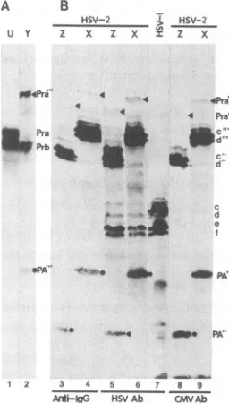
and -fspecified by plasmid N comigrated in denaturing gels with ICP35c and -d produced in HSV-1-infected cells (Fig. 4A, lanes 1 and 5), it may be deduced that the portion of ICP35 cleaved during processing is roughly equivalent to the size of the CMV amino acid sequence inserted into plasmid N. To determine whether ICP35 processing involves car-boxyl-terminal proteolytic cleavage, BHK cells were trans-fected with plasmids J, Q, R, S, and W and superinfected with HSV-2. Plasmid Q contained the CMV epitope (se-quence A) inserted in the PmlI site of UL26.5, whereas in plasmids S and W the insert was at the carboxyl-terminal amino acid (Fig. 1). Analyses of the electrophoretically separated, electrically transferred polypeptides with the anti-HSV-1 (H725) and anti-CMV (CH28-2) monoclonal an-tibodies revealed the following (Fig. 5).
(i) Cells transfected with plasmid J in which the CMV epitope was inserted at the MstII site 122 amino acids upstream from the UL26 stopcodon made both the precur-sors ICP35c and -d and the processed products ICP35e and -f, which reacted with the CMV antibody (lane 8). The decrease in the electrophoretic mobility of ICP35c to -f relative to the wild-type proteinscorresponds to the increase in the molecular weight due to the insertion of the CMV epitope.
(ii)Only ICP35c and -d were made in cells transfected with plasmid Q (lanes 3 and 6). In this plasmid, the CMVepitope was inserted into the
PmIl
siteofUL26, which is 21 amino acidsupstream from theUL26.5stopcodon. Identification of theICP35c and -d forms was based on the observation that they comigrated with the corresponding forms specified by plasmid L, which expressed only ICP35c and -d intrans-fected cells (Fig. 4B, lane 17).
(iii) Insertion of sequence C into plasmid R at the PmlI restrictionendonuclease site destroyed this site and created
1 cs
HSV-1~HSV-U:C _S-I __
- 0 P 0 L
34° 3-9°- 73-934-3
-HSV-2 >
J S S R x
HSV-2
w w
Pra' _ ~~~~~Prb'
cl d d, ' d
d e dtc
e..
1 2 3 4 5 6 7 8 9 10 11 12 13 14
HSV Ab CMV Ab CMV Ab HSV Ab CMVAb HSVAb
FIG. 5. Photograph ofpolypeptides from cells transfected with
plasmids andeithermockinfected or superinfected withHSV-1(F) (HSV-1) orHSV-2(G) (HSV-2) at 340C (340; lanes 1 and 4), 39°C
(390;lanes 2 and5),or 370C(lanes3 and 6 to 14),electrophoretically separatedindenaturing polyacrylamide gels, electrically transferred to anitrocellulose sheet, reacted with monoclonal antibody H725
(HSV Ab)or CH28-2(CMV Ab),and stained withgoat anti-mouse
IgG antibody coupledto peroxidase. Letters above the lanes
iden-tifythe plasmidconstructs withwhichthecells weretransfected. A dashindicatesthatthe cells were infected butnottransfected.
a new PmIl cleavage site between the
carboxyl-terminal
aminoacid and the stopcodonofUL26without changingthe aminoacid sequence of either UL26 or UL26.5. ICP35c to -f detected with monoclonal antibody H725 comigrated with theauthenticproteins (lane 11), indicatingthat the insertion of sequence C had no effect on ICP35 expression and processing.
(iv) In plasmids S and W, the CMV epitope was inserted intothenewPmlI siteof plasmidRatthecarboxyl terminus of UL26.5. Cells transfected with these plasmids
accumu-latedICP35c to-freactivewith HSV-1 monoclonal antibody H725(lanes 10 and 14), butonly ICP35c and -d reacted with the CMV monoclonal antibody CH28-2 (lanes 9 and 13). The significantfinding isthat whereas ICP35c and -d of plasmid S comigrated with the corresponding forms ofplasmid J, i.e., they were 21 amino acids longer than wild type, ICP35e and -f comigrated with the wild-type ICP35e and -f,
indicating
that the inserted amino acid sequence encoding the CMV epitope was removed (lanes 9 and 10). The products speci-fiedbyplasmidWbehaved in thesamemanner(lanes 13 and 14). ICP35e and -f specified by plasmid W were more
abundant than those specified byplasmids S and R, possibly because in plasmid W the entire UL26 open reading frame was reconstituted and more of the protein product was
expressed and made available toprocessICP35.
We conclude that the cleavage of the precursor ICP35 protein is approximately 20 amino acids from the carboxyl-terminal codon and that insertion of the CMV epitope 21 amino acids from the terminus interfered with the cleavage whereas insertion of the epitope at the carboxyl terminus enabled the cleavage to take effect.
on November 10, 2019 by guest
http://jvi.asm.org/
[image:5.612.321.557.83.309.2]DILUTIONV:10)
CYCLO-CHASE CYCLO-CHASE PULSE CYCLO-CHASE PULSE
360 90 30 360 90 30 10 360 90 30 10 360 90 30 10 360 90 30 10
4Pra' 4 4 4pra"
4
4 Pra'
Pra W ...
N Prb
d:
Pra' 't
Pra _. W_1
Prb
2 3 4 5 6 7 8 9 10 11 2 13 14 15 % 17 18 19
FIG. 6. Autoradiographic image of[35S]methionine-labeled poly-peptides encoded bythe UL26 openreading frame
electrophoreti-cally separatedinadenaturingpolyacrylamide gel. The UL26open reading frame contained in plasmids U and V (Fig. 1) was
tran-scribed in vitro and translatedinnuclease-treated rabbitreticulocyte
lysates. The lanes shown represent portions removed from the translation mixture at 10, 30, 90, and 360 min after initiation of translation. For samples shown in lanes 4 to 7 and 12 to 15, cycloheximide (CYCLO)wasaddedtothetranslation mixtureat10 minafter initiation of translation toinhibit furthertranslation. For thesamples inlanes 1to 3, the translation mixtureat 10 minafter initiation of translation was diluted 10-fold in phosphate-buffered salinecontaining cycloheximide (100p.g/ml).
Autoprocessing of UL26involvescarboxyl-terminal
proteo-lytic cleavage. In the preceding sections, we have demon-strated that UL26 is theonly viral factorresponsiblefor the carboxyl-terminal proteolytic processing of ICP35. Re-cently, wealsodemonstrated that UL26 and ICP35 share the
samecarboxyl-terminal amino acidsequence (12). The pos-sibilitythat UL26 cleaves itselfemerged from the observa-tion that BHK cells transfectedwith plasmidP(Fig. 1) and superinfected with HSV-1(F) ateither 34or39°C expressed
adoublet band of UL26(Fig.5, lanes4and5) which reacted with monoclonal antibody CH28-2. This observation sug-gested the possibility that UL26 catalyzes its own cleavage since HSV-1(F)expressesprimarily a genesat 39°C.
Additional evidence that UL26 can catalyze its own cleav-age emerged from in vitro studies. RNAs transcribed from plasmid U or V (Fig. 1) in vitro by SP6 or T7 RNA polymerasewere translated in nuclease-treated rabbit retic-ulocyte lysatein the presence of
[35S]methionine.
Analyses oftheelectrophoretically separated products ofthe transla-tion reaction were asfollows (Fig. 6).(i)Incubation of the translation products of plasmid U in the presenceof cycloheximideresultedin a gradual accumu-lation of the cleavageproduct (Prb)of the UL26 protein. The amount of accumulated cleavage product was proportional
tothedurationofthe incubation (lanes 12 to 15).(ii)Identical results were obtained with the translation products of plas-mid V (lanes 4 to 7). The significance of this experiment
stemsfrom the presence of the CMV epitope at the carboxyl terminus of UL26. As expected, the translation product Pra of UL26 made from plasmid V migrated more slowly than the authentic protein derived from plasmid U. However, the processed form Prb of UL26 synthesized from plasmid V comigrated with that of the authentic protein from plasmid U, indicatingthat UL26 autoprocessing involves carboxyl-terminal proteolytic cleavage.
The cleavages of ICP35c, ICP35d, and Pra are sequence specific and atthe same site. The results of the experiments
*PA" w_ _.ob
PA:"
_4 PA
1 2 3 4 5 6 7 8 9
AMniHgG VSV Ab CMVAb
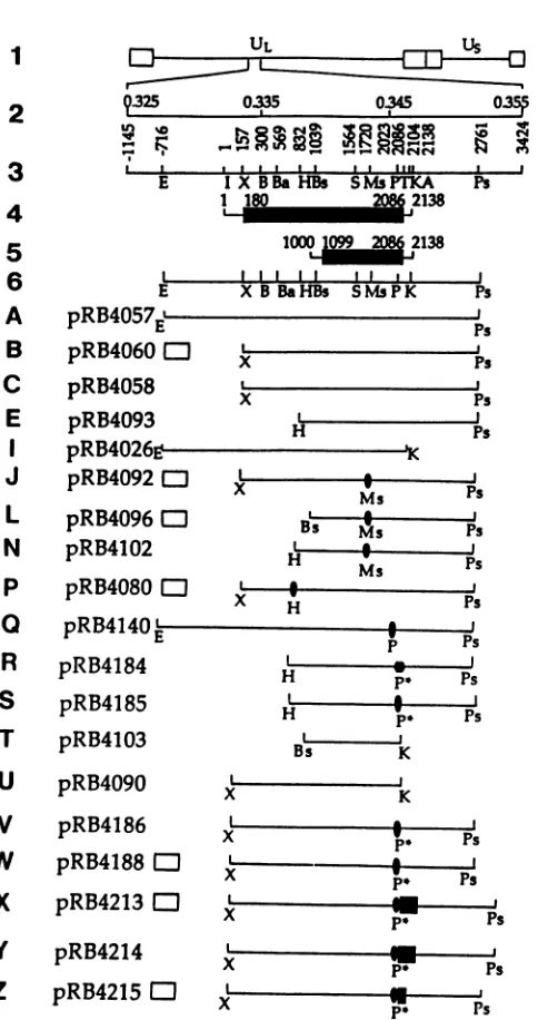
FIG. 7. Autoradiographic images (A) and photograph (B) of
polypeptideseithersynthesizedin vitro fromsequencesencodedin
plasmid U or Y or contained in lysates of cells transfected with
plasmid XorZ andsuperinfectedwithHSV-1(F) (HSV-1)or
HSV-2(G) (HSV-2). The in vitro-synthesized polypeptides and those
containedin cell lysateswere electrophoretically separatedin the
samedenaturing12%polyacrylamide gel, electricallytransferred to
anitrocellulosesheet,and reacted withgoat anti-mouseIgG conju-gatedwithhorseradishperoxidase(Anti-IgG) onlyorwith this
anti-IgG antibodyinadditiontomonoclonalantibodyH725(HSV Ab)or CH28-2(CMV Ab).Adash indicates that the cellswereinfected but
not transfected. The polypeptides shownin panel A were labeled
with [35S]methionine. Banddesignationsare asfollows:letterscto
f withoutprimes identify authenticICP35 products ofthe UL26.5
openreading frame;Pra and Prbarethetranslation-processedforms
ofthe proteaseproductsof theUL26openreading frame;the double
andtriple primesindicate that the protein also contains the CMV
epitope and the sequence encoding two IgG and five IgG binding domains, respectively, of staphylococcal protein A; PA" and PA"'
arethecarboxyl-terminal productsof thecleavageof the ICP35c and
-d andPraproteins containinginserts of theCMVepitopeandIgG bindingdomains.
presentedin the precedingsectionpredictedthat the
cleav-age and processing of the products of UL26 and UL26.5
occurred at a site approximately 20 amino acids from the
carboxyl terminus of the proteins. To demonstrate that the
processing of theseproteins occurs atthepredicted site, it
wasnecessarytodemonstrate bothproductsof thecleavage
reaction on the same gel. To visualize both products, we
inserted into the codingsequenceatthepredicted carboxyl
terminus both theepitopefor the CMVmonoclonalantibody
and the sequences encoding the IgG binding domains of
staphylococcal protein A. Plasmids Z and X were
con-structedby insertingin frame the sequences coding for 129
and 256 amino acids comprising two and five IgG binding
domains ofprotein A, respectively,betweenthe 3' terminus
of the CMV epitope and the stop codon ofUL26 (Fig. 1).
Two experiments were done. In the first, the HSV-1 open
reading frames in plasmids U and Y were transcribed and
translatedfor 6 h. Theproteinstranslated in vitrowerethen electrophoretically separated in a denaturing gel (Fig. 7A).
In thesecondexperiment,BHKcells weretransfected with
A B
HSV-2 :. HSV-2 U Y Z X Z X I Z X
d e
on November 10, 2019 by guest
http://jvi.asm.org/
[image:6.612.66.298.67.225.2] [image:6.612.376.504.85.310.2]plasmid Z or X and then superinfected with HSV-2(G). The cell lysates were electrophoretically separated in the same gel as that used for the separation of the in vitro-translated protein, electrically transferred to a nitrocellulosesheet, and reacted with antibody to CMV or HSV or with anti-IgG antibody that would bind to IgG binding domains of protein A (Fig. 7B). The results were as follows.
(i) Autocatalytic processing of the in vitro-transcribed/ translated HSV-1 sequences in plasmid U yielded, as ex-pected, the protein bands designated Pra and Prb. Similar autocatalytic processing of the products ofthe Y plasmid yielded three bands. The first band migrated slower than the authentic precursor Pra band, as would be expectedfrom the presence of the additional 256 amino acids of protein A and the 21 amino acids constituting the CMV epitope. The second band comigrated with the Prb band and is therefore the product of the autocatalytic cleavage ofthe translation product. The third band comigrated with the bands de-scribed below, which reacted with the CMVantibodyas well as with the anti-IgG antibody. Cycloheximide chase experi-ments after a shortpulseindicated thatthe first band is the precursor of the other two bands (data not shown).
(ii) The expected translation products of plasmid X were ICP35c and -d and Pra. It could be expected that the translation products would react with the CMV, HSV, and anti-IgG antibodies. The predicted translation products of plasmid Z should be similar except that because of the smaller inserts of the protein A sequences, these proteins should migrate correspondingly fasterthanthose of plasmid X. This was in fact the case (Fig. 7; comparelanes 3,5, and 8withlanes4, 6, and 9). It could also bepredicted that if the cleavage of theICP35c and -d occurs asexpected 20amino acids from the carboxyl terminus of the authentic protein, then the amino-terminal products of the cleavage reaction should comigrate with the authentic ICP35 and react only with the HSV-1 monoclonal antibody. This was in fact the case: ICP35e and -fproduced by plasmids Zand X (lanes 5 and 6)comigrated with theauthentic ICP35e and -f(lane 7) and were detectable solely by the HSV-1-specific monoclo-nal antibody. Conversely, it could be expected that the carboxyl-terminal products of the cleavage reaction should migrate in accordance with their size and should react with bothanti-IgG and CMVantibodies. As shown in Fig. 7B, the bands reactive with theanti-IgGantibodyfromlysates of cell transfected with plasmid X migrated slower than the corre-sponding Z bands. However, since all of the carboxyl-terminal cleavage products contained the IgG binding do-mains of protein A, all of the protein products would be expected to react with IgG irrespective of specificity of the immunoglobulin (e.g., lanes 3 and 4).
Inasmuchas we detected both products, the results indi-cate that ICP35 (forms c and d) and Pra, the products of UL26.5 and UL26,respectively, are both posttranslationally processed by cleavage. Since the two proteins share amino acid sequences for the entire length of ICP35c and -d and since the products of the cleavage of the two proteins comigrate, the two proteins are cleaved at identical sites. Finally, the translational products of both open reading frames in vitro resolve into double bands. The double bands areparticularly noticeable inthe case of ICP35 (forms c and d). In all of the experiments done to date, including those shown in Fig. 7, thecarboxyl-terminal product of the cleav-age formed a single band. Thisobservationis consistent with the hypothesis that the differences in the proteins which form the doublets are at the amino rather than carboxyl termini of the proteins.
DISCUSSION
The keyfindingpresented in this report is that theproduct of the UL26 open reading frame is both necessary and the soleviral protein that suffices to effect its own cleavage and that ofthe product of the UL26.5 openreading frame. We have previouslyidentified the product ofUL26.5 as ICP35. In thisreport, we have designated theproduct of the UL26as
Pra. Incell-free systems, Pracleaved itself toPrb, suggest-ing that the translation product, Pra, can function as a
protease.Prb, the product ofautocatalytic cleavage ofPra, isapproximately 20 aminoacids smaller. Experimentsarein progress to determine whether it too exhibits proteolytic activity.
The distinguishing features of this protease are that (i) it catalyzesits own cleavage,(ii) the more abundant substrate onwhich it acts isencodedbyasequenceentirelycontained withinthegene encoding the protease,and (iii) the protease and thesubstrate share amino acid sequences.
Thesubstrate of this protease,ICP35,hasbeenpreviously identified as a proteinwhich changes structure in the transi-tion from empty tofull capsids(8, 9). It has been
suggested
that ICP35functions as ascaffoldingproteinin theassembly of the capsid (15). Homologs ofICP35 have been detected in other herpesviruses; indeed, it has been reported
recently
that the CMV equivalent of the ICP35 protein is cleaved at
thecarboxyl terminus (7, 19,21), but the CMV protease has not been identified as yet.
Theprotease reported in this studyis the first identified for HSV-1. We have already noted that the CMV
protein
corresponding toICP35 is cleaved at its
carboxyl
terminus,
and therefore it is likely thatthis virusalso encodes its own
protease. Theexistence ofahomologousopenreadingframe in the varicella-zoster virus genome (5, 14) suggests that the ICP35 equivalent and the corresponding protease are
con-served among the various herpesviruses. Gibson et al.
(7)
noted the presence of the conserved amino acid sequence -Val-Asn-Ala-Ser-near thecarboxylterminusoftheproteins predicted by the homologous open
reading
frames in the HSV-1 and human and simian CMV genomes. The serinein this sequence is 25amino acids fromthecarboxyl
terminus,
approximately at or near the site of the cleavage predicted from the results shown inFig. 5. Thefindingsthat the amino acid sequence of ICP35is entirely containedin the
carboxyl
terminus ofPrand that ICP35 does not show demonstrable proteolytic activity lead us to predict that the
proteolytic
activity exhibited byPris expressed by the amino-terminal domainof the protein.
Preliminary
studies (13) indicate that deletion of sequencesencoding
approximately
150 amino acids at thecarboxylterminusdoes notaffect theproteolytic
activity of the mutatedproduct ofUL26. Itis of interest that in the varicella-zoster virus genome (5), the open
reading
frame corresponding to UL26 of HSV-1 exhibit greater homology inamino acidsequenceatthe amino terminus than at the carboxyl terminus. The observation that the temper-ature-sensitive mutation of Prestonet al.
(18)
maps atthe 5' terminus of the UL26 open reading frame and that at thenonpermissive temperature ICP35c and -dare not
processed
further is congruent withbut does notprove our
prediction.
Theonlyfunction of the protease identified todate is that it cleaves itself andICP35. Because of its
overlap
with the more abundant ICP35, it is conceivable that it assembles with and is dedicated to theprocessing
ofthisprotein.
Given the size and the number of openreading
frames encoded in the HSV-1genome, and should the protease identified in this study be indeed dedicated to thecleavage
of itself and ofon November 10, 2019 by guest
http://jvi.asm.org/
ICP35, it would not be too surprising if additional viral
proteases were to be detected. Further studies will deter-mine whether this is the case. Because processing of the ICP35 is an essential step in the assembly of the capsid, the protease appears to be a suitable target of antiviral drug research.
Inthis study, we made use of two monoclonalantibodies, one to a stationary epitope encoded by both UL26 and UL26.5openreading frames andonereactive withamovable epitope. The latter was an indispensable tool in identification of theproducts of the two open reading frames, in determi-nation of the function of the proteins, and in mapping of the cleavage site. Without the movable epitope, we would have had to rely solely onradioactive tracers or make antibodies to oligopeptides corresponding to various domains of the genes. The movable epitope offers instant antibody to the product of any open reading frame and, when used in the context described in this and preceding studies, can enor-mously facilitateidentificationof the function of theproduct of the gene into which it has been inserted.
ACKNOWLEDGMENTS
We thank Lenore Pereira for the invaluable gift of themonoclonal antibodiesand RichardRoller for advice.
These studieswereaided by Public Health Service grants from the National Cancer Institute (CA47451)and the National Institute for Allergy and Infectious Diseases (AI124009 andA11588-11).
REFERENCES
1. Arsenakis, M., J. Hubenthal-Voss, G. Campadelli-Fiume, L. Pereira,and B.Roizman.1986. Construction andproperties ofa cell line constitutively expressing the herpes simplex virus glycoprotein Bdependentonfunctionala4protein synthesis.J. Virol.60:674-682.
2. Batterson, W., and B. Roizman. 1983. Characterization ofthe herpes simplex virion-associated factor responsible for the induction ofotgenes. J. Virol.46:371-377.
3. Braun, D. K., L. Pereira, B. Norrild, and B. Roizman. 1983. Application of denatured, electrophoretically separated, and immobilized lysates of herpes simplex virus-infected cells for thedetection of monoclonal antibodies and for studies of the properties of viralproteins. J. Virol. 46:103-112.
4. Braun, D. K., B.Roizman, and L. Pereira. 1984. Characteriza-tion ofpost-translational products of herpes simplex virus gene 35proteins bindingtothesurfaces of full capsidsbut not empty capsids.J.Virol. 49:142-153.
5. Davison, A. J., and J. E. Scott. 1986. The complete DNA sequenceofvaricella-zostervirus. J. Gen. Virol. 67:1759-1816. 6. Ejercito,P.M., E. D.Kieff, and B. Roizman. 1968. Characteri-zation of herpes simplexvirus strains differing in their effect on
socialbehavior ofinfected cells. J.Gen. Virol.2:357-364. 7. Gibson, W., A. I. Marcy, J. C. Comolli, and J. Lee. 1990.
Identificationofprecursor tocytomegaloviruscapsid assembly protein and evidence that processing results in loss of its carboxyl-terminal end.J. Virol. 64:1241-1249.
8. Gibson,W., and B. Roizman.1972. Proteinsspecified by herpes simplexvirus. VIII. Characterization andcomposition of mul-tiplecapsid formsof subtypes 1and2. J. Virol. 10:1044-1052. 9. Gibson, W., and B. Roizman.1974. Proteinspecified by herpes
simplexvirus. Staining and radiolabelingproperties ofBcapsids
andvirionproteinsinpolyacrylamide gels.J.Virol.13:155-165. 10. Knipe, D. M., W. T. Ruyechan, B. Roizman, and I. W. Halli-burton.1978. Moleculargenetics of herpes simplex virus: dem-onstration ofregions ofobligatory and nonobligatory identity
within diploid regions of thegenomebysequencereplacement andinsertion. Proc. Nat.Acad. Sci. USA 75:3896-3900. 11. Kristie, T.M.,and B. Roizman. 1984. Separationofsequences
defining basalexpressing from thoseconferringa gene recogni-tion within theregulatory domains of herpes simplex virus1 a
genes. Proc. Natl.Acad. Sci. USA 81:4065-4069.
12. Liu, F., and B. Roizman. 1991. The promoter, transcriptional unit,andcodingsequenceofherpessimplex family35proteins arecontained within and in frame with the UL26openreading frame.J. Virol.65:206-212.
13. Liu, F., and B. Roizman. Unpublisheddata.
14. McGeoch, D. J., M. A. Dalrymple, A. J. Davison, A. Dolan, M. C. Frame, D. McNab, L. J. Perry, J. E.Scott,and P.Taylor. 1988. ThecompleteDNAsequenceof thelong uniqueregionin the genome of herpes simplex virus type 1. J. Gen. Virol. 69:1531-1574.
15. Newcomb, W. W., and J. C. Brown. 1991. Structure ofthe herpessimplex viruscapsid: effects of extraction withguanidine hydrochloride and partial reconstitution of extracted capsids.J. Virol. 65:613-620.
16. Pereira, L., and A.Sears. Unpublished data.
17. Post, L.E., S. Mackem, and B. Roizman. 1981.Theregulation of agenes ofherpes simplex virus: expression of chimericgenes produced by fusion ofthymidine kinase witha gene promoters. Cell24:555-565.
18. Preston, V.G., J. A. V.Coates,and F.J. Rixon.1983. Identifi-cation and characterization of a herpes simplex virus gene product required for encapsidation of virus DNA. J. Virol. 45:1056-1064.
19. Robson, L., and W. Gibson. 1989. Primate cytomegalovirus assemblyprotein:genomelocalization and nucleotidesequence. J. Virol.63:669-676.
20. Roizman, B., and P. G. Spear. 1968. Preparation of herpes simplex virus of high titer.J. Virol.2:83-84.
21. Schenk, P., A. S. Woods, and W. Gibson. 1991. The 45-kilodaltonprotein of cytomegalovirus(Colburn) B-capsids isan amino-terminal extension form of theassembly protein.J.Virol. 65:1525-1529.
![FIG. 2.peptidesandtoamidetemplatesindicatedUL26UL26.5 the Autoradiographic image of [35S]methionine-labeled poly- translated in a nuclease-treated rabbit reticulocyte lysate electrophoretically separated in a 9.5% denaturing polyacryl- gel](https://thumb-us.123doks.com/thumbv2/123dok_us/1312758.84649/3.612.395.480.77.287/peptidesandtoamidetemplatesindicatedul-autoradiographic-methionine-translated-reticulocyte-electrophoretically-denaturing-polyacryl.webp)